Embed Size (px)
Citation preview
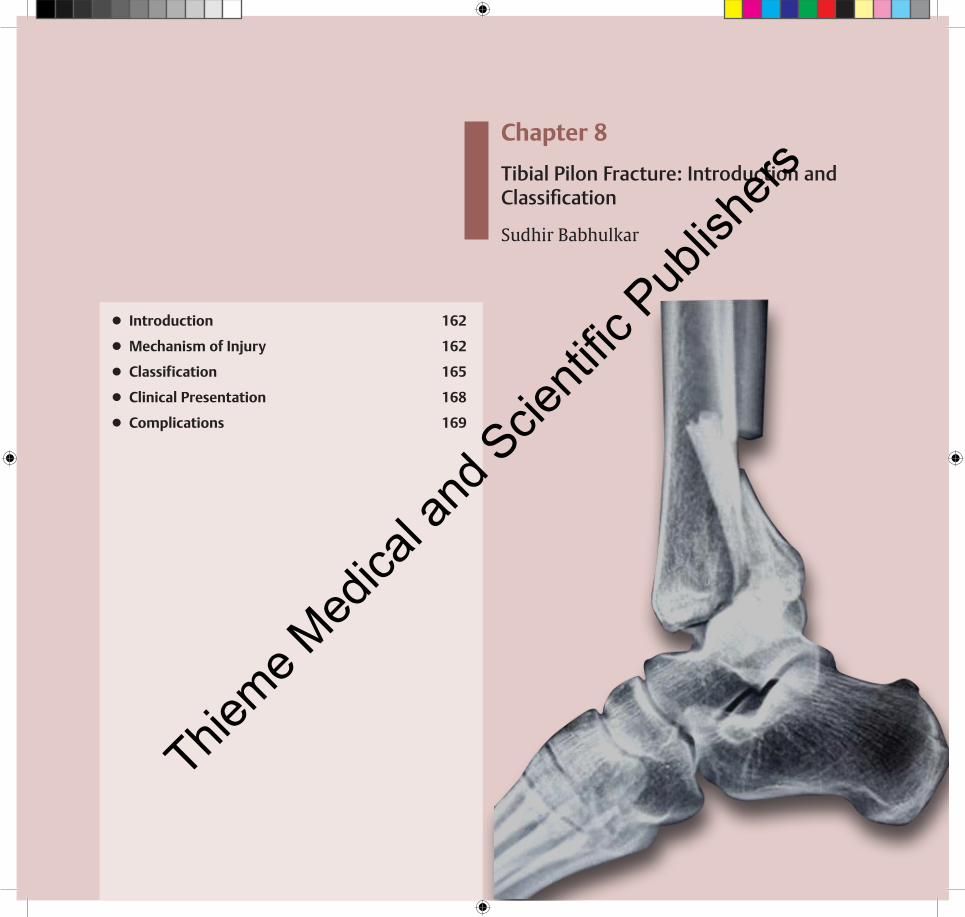
Chapter 8
Tibial Pilon Fracture: Introduction and Classification
Sudhir Babhulkar
● Introduction 162
● Mechanism of Injury 162
● Classification 165
● Clinical Presentation 168
● Complications 169
Thieme M
edica
l and
Scie
ntific
Pub
lishe
rs

162
Tibial Pilon Fractures
8 Tibial Pilon Fracture: Introduction and Classification
fractures. Pilon fractures account for 7 to 10% of all tibia fractures. Most pilon fractures are a result of high-energy mechanisms. Thus, concomitant injuries are common and should be ruled out. The injury is commonly seen in males, around 30 to 40 years of age.
█Mechanism of Injury
Fracture pattern is dictated by the position of the foot and talus at time of impact (Fig. 8.1):
1. Plantarflexion injury: posterior lip fragment2. Neutral ankle: anterior and posterior fragments3. Dorsiflexion injury: anterior lip fragment
Axial compression: This results from fall from a height. The force is axially directed through the talus into the tibial plafond, causing impaction of the articular surface. It may be associated with significant comminution. If the fibula remains intact, the ankle is forced into a varus position with impaction of the medial plafond. Plantar flexion or dorsiflexion of the ankle at the time of injury results in primarily posterior or anterior plafond injury, respectively (Fig. 8.2).
Shear: This is commonly seen after skiing accident. The mechanism is primarily torsion combined with a varus or valgus stress. It produces two or more large fragments and minimal articular comminution. There is usually an asso-ciated fibula fracture, which is usually transverse or short oblique (Fig. 8.3).
Combined injury: The mechanism of injury is combined compression and shear patterns demonstrating components of both compression and shear. The vector of these two forces determines the fracture pattern (Fig. 8.4).
█ Introduction
The term “pilon” was first used by Destot in 1911, likening the pilon to a pestle.1 The terms tibial plafond fracture, pilon fracture, and distal tibial explosion fracture all have been used to describe intra-articular fractures of the distal tibia. These terms encompass a spectrum of skeletal injury ranging from fractures caused by low-energy rotational forces to fractures caused by high-energy axial compression forces arising from motor vehicle accidents or falls from a height. High-energy fractures frequently are associated with open wounds or severe, closed, soft-tissue trauma. The fracture may have sig-nificant metaphyseal or articular comminution or diaphyseal extension.2,3
Patients are typically present with variable gross deform-ity of the involved distal leg. Assessment of neurovascular status and evaluation of any associated injuries are essential. Swelling is often massive and rapid and necessitates serial neurovascular examinations as well as assessment of skin integrity, necrosis, and fracture blisters. Soft-tissue injury including edema, contusion, and blisters is associated with pilon fractures and needs special consideration. Meticulous assessment of soft-tissue damage is made. Significant damage occurs to the thin soft-tissue envelope surrounding the distal tibia as the forces of impact are dissipated. Outcomes after tibial plafond fractures are variable but typically poor. Patients frequently have pain, impaired ankle function, and decreased general health status.
Classification of these fractures is important in determin-ing their prognosis and choosing the optimal treatment. The fibula is fractured in 85% of these patients, and the degree of talar injury varies. All fractures of the tibia involving the distal articular surface should be classified as pilon fractures, except for medial or lateral malleolar fractures and trimalleolar Thie
me Med
ical a
nd S
cienti
fic P
ublis
hers

Tibial Pilon Fracture: Introduction and Classification
163
Tibial Pilon Fractures
A B C
Fig. 8.1 Different mechanism of injury of the lower end of the tibia: (A) plantar flexion injury, (B) neutral ankle injury, and (C) dorsiflexion injury.
A B C
Fig. 8.2 X-ray showing axial compression force causing tibial pilon fracture, stabilized by open reduction internal fixation.Thieme M
edica
l and
Scie
ntific
Pub
lishe
rs

164
Tibial Pilon Fractures
A
Fig. 8.3 X-ray showing shear type of force causing tibial pilon fracture stabilized by open reduction internal fixation.
Fig. 8.4 X-ray showing combination of axial compression and shear forces causing pilon fracture. CT scan showing comminuted pilon fracture following combination injury. (A and B) X-ray AP and lateral of Tibia-fibula with ankle joint showing comminuted pilon fracture. (C and D) CT scan of the same patient showing badly comminuted pilon fracture with marked displacement of articular fragments, nicely seen in 3D reconstruction.
A
B C D
B
Thieme M
edica
l and
Scie
ntific
Pub
lishe
rs

Tibial Pilon Fracture: Introduction and Classification
165
█ Classification
The fracture classification systems are a tool for communi-cation and information, relative to treatment decisions and prognosis. Reliability must be obtained with regard to con-sistent reproducibility, thereby into readily classifying various
fracture patterns. Classification systems have some utility for management of individual patients and can serve as a frame-work for decision making.
An earlier commonly used classification system was pro-posed by Rüedi and Allgöwer in 1969, which classifies plafond fractures into three categories (Fig. 8.5I–III; Fig. 8.6A–C).4
Fig. 8.5 Rüedi-Allgöwer classification of distal tibia fracture.
Type I Type II Type IIIThieme M
edica
l and
Scie
ntific
Pub
lishe
rs

166
Tibial Pilon Fractures
Subsequently, Orthopedic Trauma Association (OTA) in 1991 described the classification systems that cover more accurately the wide range of distal tibial articular fractures. The Association for Osteosynthesis (AO) or OTA classification system provides a comprehensive description of distal tibial fractures (Fig. 8.7).5
Type A: Fractures are extra-articular distal tibial fractures, which are subdivided into groups A1, A2, and A3, based on the amount of metaphyseal comminution.
Type B: Fractures are partial articular fractures in which a portion of the articular surface remains in continuity with the shaft; these are subdivided into groups B1, B2, and B3, based on the amount of articular impaction and comminution.
Type C: Fractures are complete metaphyseal fractures with articular involvement; these are subdivided into groups C1, C2, and C3, based on the extent of metaphyseal and articular comminution.
High-energy tibial pilon fractures generally are of Type C. Because of the complexity of tibial pilon fractures, there is statistically significant interobserver and intraobserver vari-ability when these injuries are classified with the use of the AO/OTA system.6–8
While considering tibial pilon fractures, it is essential to consider the associated soft-tissue injury. Gustilo and Anderson have developed the classification of open fractures, but the Tscherne classification considers all essential factors and guides the proper timely treatment. Gustilo and Anderson developed their bony classification on the basis of analysis of open fractures.9 The Tscherne classification is mainly based on soft-tissue injuries and is grouped into four categories according to severity of injury and whether it is a closed fracture or an open wound.10,11
Fig. 8.6 (A) Type I: Fractures are nondisplaced cleavage fractures that involve the joint surface. (B) Type II: Fractures have cleavage-type fracture lines with displacement of the articular surface, but minimal comminution. (C) Type III: Fractures are associated with metaphyseal and articular comminution.
A
B
C
Thieme M
edica
l and
Scie
ntific
Pub
lishe
rs

Tibial Pilon Fracture: Introduction and Classification
167
Fig. 8.7 AO/OTA classification of distal tibia fracture.
A1 B1 C1
A2 B2 C2
A3 B3 C3
Thieme M
edica
l and
Scie
ntific
Pub
lishe
rs

168
Tibial Pilon Fractures
█ �Tscherne�Classification�of�Open�Soft-Tissue�Injuries
In the Tscherne classification, soft-tissue injuries are grouped into four categories according to severity. The fracture is labeled as open or closed by an “O” or a “C.”
1. Open fracture grade I (Fr. O1): The skin is lacerated by a bone fragment from the inside. There is no or mini-mal contusion of the skin, and these simple fractures are the result of indirect trauma (Types A1 and A2 fractures according to the AO classification).
2. Open fracture grade II (Fr. O2): There is a skin lacera-tion with a circumferential skin or soft-tissue contu-sion and moderate contamination. All open fractures resulting from direct trauma (AO classification Type A3, Type B, and Type C) are included in this group.
3. Open fracture grade III (Fr. O3): There is extensive soft-tissue damage, often with an additional major vessel and/or nerve injury. Every open fracture that is accompanied by ischemia and severe bone comminu-tion belongs in this group. Farming accidents, high- velocity gunshot wounds, and compartment syndrome are included because of their high risk of infection.
4. Open fracture grade IV (Fr. O4): These are subtotal and total amputations. Subtotal amputations are defined by the Replantation Committee of the International Society for Reconstructive Surgery as a “separation of all important anatomical structures, especially the major vessels, with total ischemia.” The remaining soft-tissue bridge may not exceed one-fourth of the circumference of the limb.}} Cases requiring revascularization can be classified
as grade III or IV open.
█ Tscherne�Classification�of�Closed�Fractures
5. Closed fracture grade 0 (Fr. C0): There is no or minor soft-tissue injury with a simple fracture from indirect trauma. A typical example is the spiral fracture of the tibia in a skiing injury.
6. Closed fracture grade I (Fr. C1): There is superficial abrasion or skin contusion, simple or medium severe fracture types. A typical injury is the pronation– external rotation fracture dislocation of the ankle joint. The soft-tissue damage occurs through fragment pressure at the medial malleolus.
7. Closed fracture grade II (Fr. C2): There are deep con-taminated abrasions and localized skin or muscle con-tusions resulting from direct trauma. The imminent compartment syndrome also belongs to this group. The injury results in transverse or complex fracture patterns. A typical example is the segmental fracture of the tibia from a direct blow by a car fender.
8. Closed fracture grade III (Fr. C3): There is extensive skin contusion, destruction of muscle, or subcutaneous tissue avulsion (closed degloving). Manifest compart-ment syndrome and vascular injuries are included. The fracture types are complex.
█ Clinical Presentation
█ Plain�RadiographyAnkle films in anteroposterior (AP), lateral, and mortise views. delineate articular incongruity and fragmentation. Tibial films are necessary to fully evaluate the metaphyseal and dia-physeal extents. Proximal injuries may easily be overlooked.
Thieme M
edica
l and
Scie
ntific
Pub
lishe
rs

Tibial Pilon Fracture: Introduction and Classification
169
have been associated with a high rate of complications. Even when accurate reduction is obtained, predictably excellent outcomes are not always achieved, and less than anatomic reduction can lead to satisfactory outcomes.
█ Early�Postoperative�ProblemsSkin necrosis, superficial and deep infection, and loss of fixa-tion are complications with fracture healing. Delayed union or nonunion of the metaphyseal–diaphyseal junction varus or valgus malunion of the distal part of the tibia is also observed. Nonanatomical reduction or postoperative loss of reduction of the articular surface, soft-tissue slough, necrosis, and hema-toma results from initial trauma as well as improper handling of soft tissues. Avoid excessive stripping and avoid skin clo-sure under tension. Secondary closure, skin grafts, or muscle flaps may be required for adequate closure. Prevalence of postoperative skin and wound problems decreased substan-tially with use of the technique of indirect reduction with external fixation and reconstruction of the articular surface with small plates or screws, or both.
Traction X-ray, traction, and ligamentotaxis often pull the dis-placed fragments back into position, allowing for a better defi-nition and understanding of the fracture pattern. Computed tomography (CT) is used as an adjunct to plain films, com-monly after applying external fixator. CT shows details often not readily available on most plain films. It acts as a guide to the articular injury for fracture orientation, fragment location, and amount of comminution or impaction (Figs. 8.4 and 8.8). It aids in surgical decision making.
█ Complications
The treatment of tibial plafond fractures requires careful man-agement of the soft-tissue envelope, reconstruction of the articular surface, and stable fixation with minimal additional damage. Hence, proper planning and assessment by classifi-cation is a suitable predictor and has prognostic importance for the final outcome for this intra-articular fracture. Pilon fractures, especially those caused by high-energy trauma,
A B Fig. 8.8 (A) X-ray showing comminuted distal tibia fracture;
(B) Axial CT scan shows three classical articular components of distal tibia articular fractures.Thie
me Med
ical a
nd S
cienti
fic P
ublis
hers

170
Tibial Pilon Fractures
References1. Destot E. Traumatismes du pied et
rayons x malleoles, astragale, calcaneum, avant–pied. Paris: Masson; 1911:1–10
2. Kellam JF, Waddell JP. Fractures of the distal tibial metaphysis with intra-articular extension—the distal tibial explosion fracture. J Trauma 1979;19(8):593–601
3. Thordarson DB. Complications after treatment of tibial pilon fractures: prevention and management strategies. J Am Acad Orthop Surg 2000;8(4):253–265
4. Rüedi TP, Allgöwer M. The operative treatment of intra-articular fractures of the lower end of the tibia. Clin Orthop Relat Res 1979; (138):105–110
5. Muller ME, Allgower M, Schneider R, et al. Manual of Internal Fixation. 3rd ed. New York: Springer-Verlag; 1991
6. Swiontkowski MF, Sands AK, Agel J, Diab M, Schwappach JR, Kreder HJ. Interobserver variation in the AO/OTA fracture classification system for pilon fractures: is there a problem? J Orthop Trauma 1997;11(7):467–470
7. Dirschl DR, Adams GL. A critical assessment of factors influencing reliability in the classification of fractures, using fractures of the tibial plafond as a model. J Orthop Trauma 1997;11(7):471–476
8. Martin JS, Marsh JL, Bonar SK, DeCoster TA, Found EM, Brandser EA. Assessment of the AO/ASIF fracture classification for the distal tibia. J Orthop Trauma 1997;11(7):477–483
9. Gustilo RB, Anderson JT. Prevention of infection in the treatment of one thousand and twenty-five open fractures of long bones: retrospective and prospective analyses. J Bone Joint Surg Am 1976;58(4):453–458
10. Tscherne H, Oestern HJ. A new classification of soft-tissue damage in open and closed fractures (author’s transl) [in German]. Unfallheilkunde 1982;85(3):111–115
11. Ruedi TP, Buckley RE, Moran CG. AO Principles of Fracture Management, Second Expanded Edition. Thieme and AO Publishing; 2007
Thieme M
edica
l and
Scie
ntific
Pub
lishe
rs