Embed Size (px)
Citation preview

The Isolation, Identification and Characterization of
Endophytes of Sw m virgatum L.), itchgrass (Panicu
a Bioenergy Crop
Francois Gagne‐Bourque
Master’s of science
Department of Plant Science
Faculty of Agricu mental Sciences ltural and Environ
McGill University
Montr nada eal, Quebec, Ca
October 2011
A thesis t of the submitted to McGill University in partial fulfillmen
requirements for the deg ee of Master’s of science r
©Copyright 2011 All rights reserved.

ABSTRACT
It has been established that perennial grasses harbour different types of
endophytic bacteria and fungi. Switchgrass (Panicum vergatum L.) is
identified as a model perennial energy crop. This study was conducted to
explore fungal and bacterial endophyte communities inhabiting switchgrass
cultivars of Quebec. The primary focus of this study was to isolate the
endophytes, and provide taxonomic identifications based on comparative
analysis of ITS rDNA gene sequences. A total of 145 endophytes isolates were
recovered (52 bacteria and 93 fungi) from whole plant samples collected at
early vegetative, and full reproductive stages. Five and nine different taxa of
bacteria and fungi were identified, respectively. We evaluated the
antagonistic activity of some endophytes against several fungal pathogens
and selected candidate endophytes for future introduction into commercial
switchgrass cultivars for biomass enhancement. We demonstrate the vertical
transmission ability of some endophyte from one switchgrass generation to
the next using species‐specific primers. Artificial inoculation of young
switchgrass seedlings with selected bacterial endophytes hold promise as a
ethod of reinfection switchgrass seedlings. m
i

Résumé
Le panic érigé (Panicum vergatum L.) est reconnu comme une des
plantes modèles pour la production de biomasse végétale. Il est connu que
la plupart des plantes vasculaires étudiées à ce jour sont colonisées par des
champignons et bactéries endophyte. Cette étude avait pour but d’explorer
les communautés d’endophytes présentes dans différents cultivars de panic
érigé au Québec, pour ensuite isoler les endophytes et effectuer leur
identification taxonomique en comparant leur séquence ITS rADN. Nous
avons obtenu un total de 145 isolats (52 bactéries et 93 champignons)
enant de feuille de plante au stade végétative et au stade reproductive. v
Une fois les isolats identifiés, nous avons obtenu cinq différents
groupes taxonomiques pour les bactéries et neuf pour les champignons.
Nous avons évalué le potentiel antifongique des différents endophytes
bactériens, avec pour objectif d’identifier les candidats potentiels à la ré‐
inoculation de cultivar de panic érigé commercial afin augmenter leur
production de biomasse. À l’aide de séquences d’amorces spécifiques, nous
avons pu démontrer la transmission verticale des endophytes.
ii

ACKNOWLEDGMENTS I would like to thank my supervisor Dr. Suha Jabaji for all her financial and
moral support and patience during my M.Sc program. She taught me the
scientific thought process and helped me edit countless times different
versions of my thesis. I would also like to thank my advisory committee, Dr.
Philippe Seguin, Department of Plant Science, McGill University and Mr.
Roger Samson, R.E.A.P.‐Canada, Resource Efficient Agricultural Production
for their advice and assistance during my studies. Thanks also go to my
fellow labmates Dr. Konstantinos Aliferis, Mamta Rani, Rony Chamoun and
Tanya Copley for their support, assistance and patience. I would like to
demonstrate my gratitude to Rony Chamoun for mentoring me all along my
project. My parents Anne Gagné and Jean‐Louis Bourque for teaching me the
work ethics required to accomplish such work. Thanks to my good friends,
David, Louis‐Philippe, Matthew, Rosemarie and Raphaelle who kept me sane
by helping me clearing my head at night. I also would like to thank R.E.A.P.‐
Canada for providing me with different switchgrass cultivars and helping me
in setting up the field experimental trials. A special thanks to Ferme Caron
for contributing space to set‐up my field trials. I wish to acknowledge the
funding agency Le Ministere d’agriculture, pecherie et alimentation du
Quebec (MAPAQ) for making this work possible. Also I would like to
acknowledge the support of the FQRNT Regroupement Stratégique Center,
S.È.V.E. for contributing towards travel cost for attending scientific
conferences.
iii

Table of Contents
Abstract………………………………………………..………………...................
Résumé……………………
i
………………………………………………………….
Acknowledgemen
ii
ts……………………………………………………………
Table of Cont
iii
ents.........................................................................................
List of Tables…
iv
…………………………………………………………………… vi
vList of Figures……….......………………………………………………………..
1.0 In
ii
troduction……………………………………………………………….... 1
1.1 Problem definition………………………………………………... 1
1.2 Rational for research…………………………………………...... 2
1.3 Objective…………………………………………………………….....
1.4 Hypotheses…
3
…………………………………………………………
2.0 Li
3
terature review……………………………………………………...….
2.1 Switchgrass…………………………………………
5
………………...
2.1.1 Switchgrass as Biofuels feedstock…………………
5
5
2.1.2 Switchgrass Cultivars used in NorthEastern
States Québec and Ontario…………………………………..
2.2 En tes of grasses……………………………
7
dophy ………………
Balansiaceous fungal endophytes ........
8
2.2.1. ............... 10
2.2.2 NonBalansiaceous fungal endophytes............... 11
2.3 Bacterial endophytes…………………………………………..…
2.4 Beneficial roles of endophytes……
12
…………………………..
2.4.1 Effects on Plant
14
Physiology…………………………..
2.4.2 Photosynthesis ……
15
…………………………………...…
2.4.2 Drought resistance…………………………………...…
14
17
iv

2.4.3 Resistance to herbivory…………………………….… 18
2.4.4 Reduction in disease occurrence……………..……
.
19
22.5 Secondary metabolites……………………………………….…
2.6 Common technique for observation, isolation and
characterization of endophyte……………………………………..
0
22
2.6.1. Histological staining and tissue observation...
2.6.2 Surface sterilization....................................................
22
23
2.6.3 Maintenance and culture of isolated
endophytes…………………………………………………………
2.6.4 Detection …………………………………………………..
24
25
2.6.5 Identification using morphological
parameters……………………………………………………...…
2.6.6 Identificatio
25
n using molecular markers..............
3.0 M
25
aterials and Methods………………………………………………… 27
3.1 Cultivars of switchgrass ………………………………………. 27
3.2 Field sites and sample selection…………………………….. 27
3.3 Sampling and plant sample processing ………………….. 28
3.4 Efficacy of sterilization………………………………………….. 30
3.5 Isolation and maintenance of endophytes.......................
.......
30
33.6 Identification of fungal and bacterial isolates.........
3.7 Species‐specific primer design and phylogenetic
..
1
analysis.................................................................................................
3.8 Detection of fungal and bacterial endophytes using
33
PCR methods‐ plants and seeds...................................................
33
33.9 Antagonistic study by dual culture method....................
3.10 Inoculation of switchgrass seedling with bacterial
4
v

isolate and confirmation of the re‐isolated bacterial.........
4.0 Re
35
sults……………………………………………………………………...….. 37
34.1 Efficacy of sterilization………………………………………
4.2 Survey of Switchgrass endophytes and molecular
..
…. 7
identification......................................................................................
4.3 Detection of fungal and bacterial endophytes using
...............
37
PCR methods‐ plants and seeds....................................
4.4 Antagonistic property of endophytes and re‐
inoculatio
40
n of switchgrass with endophytes.........................
5.0 Discussion……………
41
………………………………………………………. 42
6.0 Tables and figures……………………………………………………….. 49
7.0 Conclusion……………………………………………………...…………….
8.0 References……………………………………….........................................
62
64
vi

List of Tables
Table 4.1. List of isolated endophytes, specific and universal
primers designed and used in polymerase chain reaction
(PCR) assa …... ys……………………………………………………………………
Table 4.2. Molecular identification of bacterial and fungal
endophytes from Panicum virgatum L. based on blastN
queries in ……….
94
NCBI……………………………………………………………
Table 4.3. Dual culture antagonistic experiment using
different fungi……………………………………………………………………
25
54
vii

viii
List of Figures
Figure 4.1. Distribution of fungal and bacterial endophytes
across the leaf tissues.……………………….. different cultivars and
Figure 4.2. Antagonistic test of Pantoea ananatis and Bacillus
subtilis aga ………...
55
inst Rhizoctonia solani………………………………
Figure 4.3. Maximum likelihood phylogenic tree of all
putative en ..
65
dophytic bacteria……………………………………………….
Figure 4.4. Example of PCR detection of fungal and bacterial
endophytes detection in different switchgrass cultivars using
species‐spe ……………………….
75
cific primers………………………………
Figure 4.5. PCR detection of Bacillus subtilis, Microbacterium
testaceum and example of vertical transmission of the fungal
ndophyte Epicoccum nigrum………………………………………………
95
e
60

Chapter I Introduction
1.1 Problem Definition
The need to find sustainable alternatives to replace increasingly expensive fossil
fuels and to reduce greenhouse gas (GHG) emissions has peaked interest in biofuels around
the world. Biofuel produced from renewable biological resources such as plant biomass and
treated municipal and industrial wastes, is envisaged as being part of the solution. For
example, the Québec Energy Strategy for 2006‐2015 was established in order to reduce the
consumption of fossil fuels and promote renewable fuels. In particular, the use of annual
grains such as corn for biofuel production is discouraged in Québec and priority is given to
energy crops. Perennial energy crops more effectively capture and store solar energy as
biomass through photosynthesis, increase the overall energy produced per hectar, and are
productive on marginal farmland. In 1985, the USA began a 5‐year program to develop
herbaceous energy crops. Switchgrass (Panicum vergatum L.) was identified as a model
herbaceous energy crop species (Samson, 1991). Switchgrass had a number of positive
attributes especially being a productive long‐lived perennial crop with high resource use
efficiency and good adaptability to marginal soils (McLaughlin and Kszos, 2005). The first
scientific studies in Canada on switchgrass were subsequently conducted by Resource
Efficient Agricultural Production (REAP)‐Canada in 1991. These studies proved that
switchgrass is well adapted to Québec and Ontario environment and productive in Quebec
1

and Ontario (Samson, 1997), and that it has an excellent energy balance, low cost of
production and an ability to increase landscape carbon sequestration and an ability to
increase landscape carbon (Donner and Kucharik, 2008). Because of the above‐mentioned
attributes, more efforts focused on switchgrass development as an energy crop could
benefit Quebec’s agriculture, and represent a new source of income for Quebec farmers.
Already, 116 Quebec farms dedicate 816 hectars to switchgrass production, a 50% increase
compared to what was planted in 2008 (REAP‐Canada, unpublished). The development of a
national capacity to utilize perennial herbaceous crops, including the native prairie plant
switchgrass, as biofuels could benefit Quebec’s agriculture by optimizing productivity on
marginal farmland or degraded lands and by providing a new source of income for Quebec
armers. f
1.2 Rational for research
New approaches are required to assess and improve the genetics and cultural
management of the crop for Quebec’s marginal farming areas. For example, perennial
grasses are the hosts of fungal and bacterial endophytes that systemically colonize the
roots or the above ground portions of grasses and their seeds (Ryan et all., 2008; Schulz and
Boyle, 2005). The close link between endophyte fitness and its host grass is presumed to
align the interests of both partners towards a mutually beneficial cooperation. The
endophytes gain shelter, nutrition, and dissemination via host propagules, and can
2
contribute an array of host fitness enhancements including imporvement of vigor, leading
to superior agronomic qualities, greater drought tolerance, and increased resistance to
herbivory and also against pathogen. (Clay, 1990; Lodewyckx et al., 2002; Schulz et al.,
1993; Schulz et al., 2002). However, the importance and use of endophytes in switchgrass
remains poorly understood.
1.3 Hypotheses
The ob jectives are supported by three hypotheses:
I. Endogenous endophytes of switchgrass can be successfully isolated on synthetic
microbiological media.
II. Endogenous endophytes can be detected in plantae using polymerase chain reaction
(PCR) method.
III. Switchgrass endophytes are transmitted from maternal plant to offspring via seeds.
1.4 Objectives
The objectives were:
Objective 1: To study the distribution and frequency of fungal and bacterial endophytes
in switchgrass cultivars established in field sites across in southwestern
region of Quebec, followed by their isolation and characterization using
accurate and sensitive detection methods.
3

Objective 2: To establish that endophytes are inherited by host offspring in a vertical
transmission mode via seeds.
bjective 3: To study the biological activity of selective isolated endophytes. O
4

Chapter II Literature Review
2.1 Switchgrass
2.1.1 Switchgrass as biofuels feedstock
Switchgrass (Panicum vergatum L.) is a perennial warm‐season (C4) grass native to
the grassland of North America (Lemus et al., 2002). Switchgrass has a wide native
geographic range and has evolved into two ecotypes: Lowland ecotypes are tetraploid, are
characterized as vigorous, tall, thick‐stemmed and is adapted to wet conditions. The upland
ecotypes are hexaploid or octaploid (Lemus et al., 2002), are characterized as short,
rhizomatous and thin‐stemmed, and are adapted to drier conditions. Both ectotypes of
switchgrass offer a significant opportunity to improve agricultural sustainability as they
decrease erosion and improve water quality, when compared to row crops. Highly eroded
and will benefit the most from the perennial nature of switchgrass. l
The selection of switchgrass as a biofuel feedstock is based on several significant
attributes, including its efficiency in mitigating greenhouse gases (GHG) emissions by
sequestering large amounts of carbon in its extensive root system (Donner and Kucharik,
2008), its high biomass mainly having a high concentration of lignin and cellulose, and low
5

amounts of water, nitrogen and ash (Lemus et al., 2002; Samson, 1997; Samson, 1991).
Additionally, it has high productivity across a wide geographical range, superior suitability
for marginal quantity land, low water and nutrient requirements and positive
environmental attributes. Because of these attributes, research projects across North
America and Europe were initiated to pin point important new information on the yield
potential of this species as a biofuel crop. Research focused on evaluation of the most
promising existing varieties in regional field trials, cultural treatments to enhance the
establishment and growth of stands, breeding for superior yield, identification of
physiological markers for assessing and promoting improved growth and development, and
the development of tissue culture techniques for biotechnological improvement (Berdahl et
al., 2005; Casler and Boe, 2003; Cassida et al., 2005; Elbersen et al., 2001b; Lee and Boe,
2005; Madakadze et al., 1999; Muir et al., 2001; Sanderson et al., 1999; Vogel et al., 2002).
These tasks combined the interrelated needs of realizing maximum near‐term
requirements in productivity potential through cultural improvement and the longer‐term
equirements for enhancing and protecting yield capacity. r
In the USA, development in switchgrass is mostly aimed for cellulosic ethanol
production (Bouton 2008) in order to displace fossil fuels, thereby, reducing expenditure
on imported fuels. While in Quebec and Ontario, the focus has been more on agri‐fiber
pellet that is used in commercial boilers such as in greenhouse industries (Samson and
6

Stamler, 2007). It is estimated that these two provinces could produce 14 millions tons of
switchgrass biomass if 20 % of crop land and 40% of forage land would be converted to
switchgrass production (Samson and Stamler, 2007). This technology has the potential to
produce 770–890% more net energy gain/ha than growing grain corn for ethanol.
However, it remains substantially less efficient than direct combustion of energy grasses or
corn silage biogass as means to produce energy from farmland.
2.1.2 Switchgrass Cultivars used in North-Eastern States, Québec and Ontario
There are currently 20 different switchgrass cultivars available on the market. A
great deal of effort is focused presently on breeding and creating new lines (Lemus et al.,
2002). The most widely used cultivars in North‐Eastern America and recommended by
experts is Cave‐in‐Rock (Lemus et al., 2002). Others such as Sunburst, Shelter and
Forestburg are also suitable cultivars for production in northern locations (Samson, 2007).
Warm‐season grasses such as switchgrass are increasingly being cultivated in North
America for summer forage and biomass production. The cooler temperatures and shorter
growing seasons typical of Canadian production areas, are major limiting factors to warm‐
season grass production in these areas. Assessment of the morphological development and
relationship of growing degree‐days (GDD) to plant morphology and tiller characteristics
were evaluated in nine cultivars of switchgrass. Cave‐in‐rock switchgrass, a cultivar
7

originating from Southern Illinois (Elbersen et al., 2001a) showed increases in tiller number
and had the highest ground cover ratings after a three‐ year trial conducted in
southwestern regions of Quebec, thereby demonstrating that it is well adapted to Quebec
climate and soil type (Madakadze et al., 1999). The cultivar Blue Jacket is an upland
ecotype evolved from Sunburst, a cultivar originated in South Nebraska, and its main
advantage over other cultivars is its superior seedling vigor due to heavy seed set. This
cultivar is well adapted to sandy soils and low rainfall than Cave‐in‐Rock (Boe and Ross,
1998). Tecumseh switchgrass, an upland ecotype evolved from Summer which originated
from South Dakota, well adapted to heavy soils, and requires a lot of precipitation (Elbersen
et al., 2001a; Samsons, 2011). Finally, the cultivar Sand Lover is a population of switchgrass
derived from cultivar NU 94‐2. It is an upland ecotype. This cultivar originated in Oklaoma
and is well adapted to sandy soils and dry environments (Taliaferro, 2002).
2.2 Endophytes of grasses
For the purpose of this study, the term endophytes will only refer to bacteria or
fungi that establish a mutualism or commensalism interaction with the aerial parts of
grasses. Endophytes are present in the spaces that run parallel to the long main axis of
leaves and stem (Clay, 1990). These organisms have the ability to absorb freely available
nutrients found in the intercellular spaces or obtain their nutrients from the surrounding
cells (Schulz and Boyle, 2005; Schulz et al., 1999).
8

Endophytes are microorganisms that inhabit healthy plant tissues during at least
one stage of a grass life cycle, and do not cause any apparent symptoms of disease or
negative effects. Presently, multiple studies have shown that several plant species are hosts
to a great deal of endophytic biodiversity. According to Schulz and Boyle (2005), some
endophytic species can occur in unique ecological niches and may be sources for a variety
of bioactive metabolites that may have great potential for pharmacological applications.
Many fungal endophytes are seed‐borne and migrate in the seed during seed
germination. On plants that propagate vegetatively, the endophytes use the reproductive
tissues as vector for the infection in new plants. In order to colonize non‐infected plants,
the fungi gain access to host cells using infective specialized structures such as appressoria
and haustoria that develop intracellularly. They can also penetrate directly through the cell
wall or enter through the stomata and the substomatal chambers (Schulz and Boyle, 2005)
Grasses and fungi have a long history of symbiosis ranging in a continuum from
mutualisms to antagonisms (Schulz and Boyle, 2005). This continuum is particularly
evident among symbioses involving the fungal genus Epichloë. In the more mutualistic
symbiota, the Epichloë endophytes are vertically transmitted via host seeds, and in the
more antagonistic symbiota they spread and suppress host seed set (Schardl et al., 2004).
Generally, the endophytes gain shelter, nutrition, and dissemination via host propagules,
9

and can contribute to host fitness enhancements including protection against insect and
vertebrate herbivores, enhancements against drought tolerance and nutrient status, and
improved growth particularly of the roots. Recent advances in endophytes’ molecular
biology promise to shed light on the mechanisms of the symbioses and host benefits
(Malinowski and Belesky, 2000; Pavlo et al., 2011). For example, Pavlo and coworkers
(2010) applied enzymatic activity and Real‐Time PCR assays, to shed light on some
important plant mechanisms (e.g., ISR and SAR) for protection against pathogen and are up‐
regulated by the presence of endophyte (see section 3.5). Fungal endophytes can be
classified into three different ecological groups: the mycorrhizal fungi, the balansiaceous
and the non‐balansiacceous taxa. Mycorrhizal associations will not be covered, as they are
not the subjects of this research.
2.2.1 Balansiaceous fungal endophytes
The balansiaceous or ‘grass endophytes’ is a group of closely related fungi with
specific ecological requirements and adaptations that are different from other fungal
endophytes. The balansiaceous endophytes Epichloë and Balansia, (Anamorphs:
Neotyphodium and Ephelis, respectively) belong to the Ascomycetes. They are the most
studied above ground endophytic interactions because of their ecological and economical
impact. They grow systemically and intercellularly within all above ground plant organs of
grasses, rushes and sedges (Schulz and Boyle, 2005). They are dependent on nutrients
10

present in the apoplast for growth. A signal communicating system between the host and
the plant insures a perfect balance between the host and the endophyte virulence (Schulz
and Boyle, 2005) with benefits to both partners: the endophyte gains access to nutrients
and protection from abiotic stress, and the host becomes more resistant to insect or
pathogen attack due to the presence of alkaloids or the production of chitinase enzymes
(Schulz and Boyle, 2005). Many fungal endophytes are seed‐borne and migrate in the seed
uring seed germination processes. d
2.2.2 Non-Balansiaceous fungal endophytes
In contrast to balansiaceous endophytes, the non‐balansiaceous endophytes are a
diverse group of fungi both from the taxonomic and life‐history strategy point of view.
These fungi are Ascomycetes and can belong to any of the following genera: Acremonium,
Alternaria, Cladosporium, Conithyrium, Epicoccum, Fusarium, Geniculosporium, Phoma,
Pleospora. Colletotrichum, Guignardia, Phyllosticta, Pestalotiopsi, and Lophodermium. They
are found in all the organs and have been shown to be present in all sampled plants (Schulz
and Boyle, 2005). Many of these fungi produce an array of secondary metabolites (for
review Schultz et al. 2002) with varied biological activity (refer to section 4; Bashyal et al.,
2006; Suryanarayanan et al., 2009b).
The colonization of non‐balansiaceous endophytes can be localized or systemic.
They can grow intercellularly or intracellularly and form different types of interactions
11
varying from non‐aggressive, mutualistic to pathogenic. The non‐balansiaceous endophyte
composition population of a plant varies according to the tissues (root, stem, leaf, flower).
The fungi are not obligate host specific; they have a certain level of adaptation to different
hosts. While others are more specific and can only be found in specific organs of specific
plant (Schulz and Boyle, 2005).
2.3 Bacterial endophytes
Endophytic bacteria have been found in virtually every plant studied, where they
colonize the internal of their host plant and can form different relationships including
symbiotic, commensalistic, mutualistic and trophobiotic (Ryan et al., 2008). Most
endophytes appear to originate from the rhizosphere or the phyllosphere, however some
may be transmitted via seeds. They are able to colonize the internal tissue of the plant
showing no external sign of infection or negative effect on their host (Schulz and Boyle,
2005). They are often isolated from surface‐sterilized tissues or extracted from internal
plant parts. Both gram‐positive and gram‐negative bacterial endophytes have been isolated
from several tissue types in numerous plant species. However, several different endophytic
bacteria may reside within a single plant (Kobayashi and Palumbo, 2000). These
endophytes either remain localized at their entry points or spread to other parts of the
plant (Hallmann et al., 1997).
12

Bacterial endophytes cover a significant range of Gram‐positive and Gram‐negative
bacteria (Lodewyckx et al., 2002). Most bacterial endophytes belong to Cellulomonas,
Clavibacter, Curtobacterium, Bacillus, Sphingomonas, Pantoea, Pseudomonas and
Microbacterium genera (Kang et al., 2007; Ryan et al., 2008; Thomas et al., 2007). They will
colonize the host without external signs of infection or negative effects on the host
(Rosenblueth and Martinez‐Romero, 2006). Bacteria from the same genera can colonize
both the roots and the aerial parts depending on the specificity of the host and the bacteria
(Lodewyckx et al., 2002). Many bacterial endophytes are seed borne. On plants that
propagate vegetatively, the endophyte can use the reproductive tissues as vector for
infecting the new plant. In order to colonize non‐infected plant, bacterial endophytes
secrete cellulolytic, pectinolytic, cell wall degrading (endogluconase and polygalacturonase)
enzymes. These enzymes are useful to penetrate the host (Rosenblueth and Martinez‐
Romero, 2006) .
As with their fungal counterparts, bacterial endophytes also produce secondary
metabolites exhibiting antifungal and antibacterial properties. For example, fengycin,
bacillomycin D, Zwittermicin A, Iturin A, Surfactin are peptides with strong antimicrobial
activities and are produced by Bacillus spp (Athukorala et al., 2009; Ramarathnam et al., 2007).
13

Only few plants have ever been completely studied relative to their endophytic
biology. Consequently, the opportunity to find new and beneficial endophytic
icroorganisms among the diversity of plants indifferent ecosystems is considerable. m
2.4 Beneficial roles of endophytes
Generally, endophytic bacteria and fungi can promote plant growth and yield and
can act as biocontrol agents. Endophytes can also be beneficial to their host by producing a
range of natural products that could be harnessed for potential use in medicine, agriculture
or industry (Ryan et al., 2008; Schulz and Boyle, 2005; Schulz et al., 2002; Schulz et al.,
1999). In addition, it has been shown that they have the potential to remove soil
contaminants by enhancing phytoremediation and may play a role in soil fertility through
phosphate solubilization and nitrogen fixation (Ryan et al., 2008). There is increasing
interest in developing the potential biotechnological applications of endophytes for
improving phytoremediation and the sustainable production of nonfood crops for biomass
and biofuel production.
2.4.1 Effects on Plant Physiology
Endophytes are known to have many effects on their hosts. Infected plants may
produce more inflorescences and seeds than uninfected plants. This can be explained by a
14

greater vegetative vigor of infected plants (Clay, 1990). Seeds of some grass species (e.g.,
tall fescue and perennial ryegrass) germinate more rapidly and grow faster when infected
by an endophyte. Thus, resulting in faster growth of the infected seedling. Fungal
endophyte‐infected seeds contain higher concentrations of alkaloids, which may explain
this faster germination/growth rate. The alkaloids concentration makes it less likely for the
seed to be eaten by vertebrates and invertebrates plant and seed feeders (Clay, 1988).
Grasses infected by endophytes have tillers that are more profuse and spread horizontally
via stolons and rhizomes (Clay, 1990). Enhanced growth rate of infected plants was also
emonstrated with perennial ryegrass and purple nutsedge (Latch and Christensen, 1985). d
2.4.2 Photosynthesis
Endophytes influence the source and sink mechanism of the plant. Experiments
conducted on photosynthate metabolism reveal that endophytic fungi were constantly
transforming plant sucrose into sugar alcohols that are unavailable to the plant to
metabolize (Smith et al., 1985). If the level of photosynthetic activity of the plant is high,
the plant metabolism in charge of processing the outcome of the photosynthesis (sugar)
cannot work fast enough. Thus, there is a sucrose build up, which in turn will trigger a plant
feedback inhibition mechanism, leading to reduction in photosynthetic activity. Therefore,
entophytic fungi prevent the feedback inhibition of photosynthetic rates, allowing higher
photosynthetic rates and subsequently increase the average growth of the plant. In the case
15

of root‐borne endophytes, siderophores are produced that can increase the solubility of
utrient in the rhizosphere (Rosenblueth and Martinez‐Romero, 2006). n
2.4.2 Drought resistance
Drought stress usually induces a series of adaptations in the plant, in order for it to
survive. These adaptations include mechanisms of drought avoidance, tolerance, and
ecovery from drought. Endophytic presence can impact theses different adaptations. r
Drought avoidance is the series of mechanisms by which the plant attempts to
maintain an efficient water supply to above ground organs or conserving water during
periods of soil water deficit. A good way to improve and maximize water uptake is by
developing a deeper and denser root system. Perennial ryegrass, tall fescue, meadow
fescue infected by fungal endophytes showed an increase in root dry matter, root hair
length, and decrease root diameter (Malinowski and Belesky, 2000; Thomas et al., 2007).
These traits increase root surface, leading to increase in water and minerals absorption.
For example, Papaya infected by the bacterial endophytes Pantoea ananatis, Bacillus subtilis
and Microbacterium esteraromaticum showed an increase in root development (Kang et al.,
2007; Thomas et al., 2007). The superior root system is a simple way to maximize the
uptake of available water in the soil, thus improving the drought capacity of the plant.
16

Another drought avoidance technique is to reduce transpiration. The stomata of Tall
fescue and meadow fescue endophyte‐infected plant close faster under water stress
conditions than non‐infected plants (Malinowski et al., 1999; Turner, 1986). The internal
plant hormones balance has to be modified in order to influence the stress behaviour of the
plant. Of interest is the presence of auxin, a major plant hormone that is involved in the
drought resistance signalling pathway (Chaves et al., 2009). Clay (1990) found that auxin
can be produced by the endophyte Balansia epichloe. Water content of tiller bases in
endophyte‐infected plants are maintained at higher levels than those in non‐infected plants
during drought conditions. This may be due to a greater accumulation of solutes in
endophyte‐infected tissues.
Drought tolerance is series of adaptations that enable the plant to withstand water
deficits. A good way to cope with drought is to have carbohydrate reserves used up during
stress periods. An important part of drought tolerance adaptation is to be able to maintain
normal osmotic pressure. Accumulation of solutes in tissue helps maintain the turgor
pressure, which in turn facilitates physiological and biochemical processes. Endophyte‐
infected plants produce a number of these solutes; water‐soluble sugars, mannitol and
arabitols, proline and loline (Malinowski and Belesky, 2000). Endophyte‐ infected plants
17

help maintaining cell wall elasticity in the same way as they do maintain osmotic pressure,
which further help the biological processes.
The combination of drought avoidance and tolerance mechanisms determines the
natural potential of a plant to withstand drought. Endophytes can play a key role on many
of the different strategy used, thus increasing the drought tolerance of plant.
2.4.3 Resistance to herbivory
Endophytes are known to increase resistance to herbivory (Clay, 1990). Several
findings showed that endophyte‐infected plant materials reduce feeding and egg laying
intensity of aphids, large milkweed bugs, and fall armyworms (Clay, 1988). The endophyte‐
infected plant reduces the survival, growth and developmental rates of feeding insects.
These effects, are due to the presence of secondary metabolites, with insecticidal property,
produced in plant tissues by the endophytes (Johnson et al., 1985; Suryanarayanan et al.,
2009a; Suryanarayanan et al., 2009b). These secondary metabolites are often alkaloids in
nature. To name a few: the Ergots alkaloids like clavines, lysergic acid, ergopeptides are the
cause of fescues toxicosis in grazing lifestock (Malinowski and Belesky, 2000). Peramine is
another alkaloid that is found on endophyte‐infected grasses; it has insect‐feeding
deterrent properties, but in this cases does not impact mammalian herbivory (Malinowski
and Belesky, 2000). Endophyte–infected grasses have been reported to be also more
resistant to soil‐born nematodes, with the resistance probably attributable to alkaloids
18

present in roots or secretion of phenolic‐like compounds into the rhizosphere (Malinowski
nd Belesky, 2000). a
2.4.4 Reduction in disease occurrence
Numerous reports have shown that endophytic microorganisms can have the
capacity to control plant pathogens (Krishnamurthy and Gnanamanickam, 1997; Rodriguez
Estrada et al., 2011), insects (Azevedo et al., 2000) and nematodes (Hallmann et al., 1997).
In some cases, they can also accelerate seedling emergence, promote plant establishment
under adverse conditions (Chanway, 1997) and enhance plant growth (Bent and Chanway,
998). 1
It is believed that certain endophytic bacteria trigger a phenomenon known as
induced systemic resistance (ISR), which is phenotypically similar to systemic‐acquired
resistance (SAR) (van Loon et al., 1998). ISR is effective against different types of pathogens
but differs from SAR in that the inducing bacterium does not cause visible symptoms on the
host plant (van Loon et al., 1998). Bacterial endophytes and their role in ISR have been
reviewed recently by Kloepper & Ryu (2006). Pavlo et al. (2010) demonstrated that the
bacterial endophyte of potato, Pseudomonas spp. increased resistance toward the pathogen
Pectobacterium atrosepticum by priming the host’s defense responses. With the help of
enzymatic activity assay and Real‐Time PCR, they discovered that the plant antioxidant
19

system that produces superoxide dismutase (SOD), catalase, guaiacol peroxidase (GPOX)
and ascorbate peroxidase (APX) was moderately activated as well as both the ISR and the
SAR pathway by the presence of endophyte. Reduced nematode populations were
associated with endophyte infection in tall fescue, both in pot culture and in field trials
(Pedersen et al., 1988; West et al., 1988). Possible mechanisms by which endophytes could
inhibit plant pathogens include competition for resources (Sturz et al., 2000), induction of
generalized defense responses, and the production of antimicrobial compounds
(Athukorala et al., 2009; Ramarathnam et al., 2007).
2.5 Secondary metabolites
Endophytes have been shown to prevent disease development through endophyte‐
mediated de novo synthesis of novel compounds and antifungal metabolites. Investigation
of the biodiversity of endophytic strains for novel metabolites may identity new drugs for
ffective treatment of diseases in humans, plants and animals (Strobel et al., 2004) e
Most of the effects of fungal endophytes on their hosts can be associated with the
production of secondary metabolites, more specifically alkaloids, that are generally host‐
specific and can be extracted from endophyte cultures outside the host (Clay, 1990; Schulz
and Boyle, 2005; Suryanarayanan et al., 2009a; Suryanarayanan et al., 2009b). Alkaloids are
defined as naturally occurring molecules containing nitrogen atoms (Clay, 1990). It has
20

been shown that greater resistance to pathogens and predators is usually associated with
antimicrobial metabolites (Schulz and Boyle, 2005). These alkaloids become part of the
plant tissue and are in part responsible for the poisonings of grazer animals and insect
herbivore. Endophyte‐infected grasses contain a series of alkaloids not found in uninfected
grasses.
Generally, metabolites produced by endophytes can be isolated from pure culture of
endophytes. The balansiaceous endophytes produce diverse array of secondary metabolite,
including peramine and lolines and the anti‐vertebrate alkaloids lolitrem B and ergovaline
(Schulz and Boyle, 2005) that are harmful for mammals. While the non‐balansiaceous
endophytes are known to produce a diversity of metabolites that are known to have
herbicidal (e.g., brefeldin A produced by Aspergillus clavatus), anti‐bacterial (e.g.,
pyrrocidine A and B produced by Acremonium zeae), anti‐viral (e.g., mellein produced by
Penicillium janzcewskii), anti‐fungal (e.g., pyrrocidine A and B produced by Acremonium
zeae), and anti‐cancer ( e.g., vincristine produced by Fusarium oxysporum) properties
(Bashyal et al., 2006; Suryanarayanan et al., 2009b), as well as growth promoting with
phytohormone properties (Schulz et al., 2002). Thus, the fact that endophytes can produce
secondary metabolites with biocidal properties make them attractive to the pharmaceutical
industry (Schulz et al., 2002).
21

As with fungal endophytes, a number of low molecular weight compounds active at
low concentrations against plant pathogenic fungi, animals and humans have been isolated
from bacterial endophytes. For example, the peptides, Fengycin, Bacillomycin D,
Zwittermicin A, Iturin A, Surfactin exhibit a strong antimicrobial activity produced by
Bacillus species. An exhaustive list of antimicrobial compounds produced by different
bacterial endophytes are described in Ryan et al. 2008.
2.6 Common techniques for observation, isolation and characterization of endophyte
In order to assess the viability of endophytes, some methods exist for detection,
identification and isolation of endophytes from plant tissues.
2.6.1. Histological staining and tissue observation.
This technique involves observing the endophyte structures within the plant tissues
using light microscopy. It is not the ideal method for the detection of bacterial endophytes.
The recent application of green fluorescent protein (GFP) for tagging bacterial or fungal
endophyte is a more precise way to visualize the presence of the endophyte in situ. With
the help of specific vectors, it is possible to introduce the GFP gene into other organisms.
This gene produces a protein when exposed to blue light produces a bright green
fluorescent which can be visulaized using fluorescence microscopy to detect the presence of
endophyte inside plant tissue. Vectors such as pHRGFPT, pHRGFPGU have been used to
22

transform endophytic bacteria (Ramos et al., 2007; Rouws et al., 2010). Fungal endophytes
can also be transformed using specific pPd‐EGF vector (Mukherjee et al., 2010).
2.6.2 Surface sterilization
The host tissue is subjected to surface sterilization techniques followed by isolation
of the endophytes on specific synthetic growth medium. The most common procedure
employs a surfactant such as ethanol, followed by a sterilizing agent, such as sodium
hypochlorite (Schulz and Boyle, 2005). The efficiency of sterilization is confirmed by the
imprint technique. The technique consists of imprinting the plant tissue before and after
sterilization on different growth media such as Malt Peptone Yeast Agar, Malt Extract Agar
(MEA) (Arnold et al., 2000), Potato Dextrose Agar (PDA) (Latch and Christensen, 1985). All
these media support the growth of fungal endophytes and are often amended with
antibiotics in order to prevent bacterial endophytes growth (Schulz et al., 1999). For
bacterial endophytes, a wide range of media are used for growth; Nutrient Agar (NA),
Viande‐Levure (Pavlo et al., 2011) or Rice extract Modified Rennie (RMR) are just some
examples (Miyamoto et al., 2004). Once successfully isolated, endophytes are then sub‐
cultured, purified and maintained under controlled conditions for future applications.
2.6.3 Maintenance and culture of isolated endophytes
23

Fungal endophytes growing from the leaf section after sterilization are sub‐cultured
in order to obtain a pure culture, using the same media that they have been isolated on
(MEA and PDA amended with antibiotics). The cultures are then incubated at 24° C. Once
the fungus covers approximately ¾ of the plate, 5mm diameter punch holes are removed
from the edge of the colony which represents the younger part of the fungal growth.
Twenty plugs are place in a 2 ml sterile screw‐cap tube, and covered with a sterile solution
of Glycerol at a concentration of 25% (Kitamoto et al., 2002). The tubes are then frozen in
liquid nitrogen and stored at ‐80°.
Bacterial endophytes require to pass through a series of 4 single colony isolations to
ensure their purity. Pure bacterial colonies are grown on LB (1.0% tryptone (Difco, Texas),
0.5% Yeast Extract (Difco, Texas), 1.0% and NaCl for 18 hours. An aliquot of 750 μl of
bacterial solution are then pipetted into a 2 ml sterile screw‐cap tube and mixed with
sterile glycerol to obtain a 25% final glycerol solution. The tubes are then frozen in liquid
nitrogen and stored at ‐80°C (Costa and Ferreira, 1991).
2.6.4 Detection
Fungal and bacterial endophytes in surface sterilized tissues can be detected by
polymerase chain reaction methods (PCR) using the universal primers ITS 1 and ITS 4
encoding the ribosomal DNA space genes in fungi (Schulz and Boyle, 2005), or the 16sRNA
24

and ITS 16 (Miyamoto et al., 2004) encoding a spacer section in the ribosomal DNA of
bacteria.
2.6.5 Identification using morphological parameters
In order to identify the endophytes, different techniques can be used. For fungi,
condiogenesis and spore morphology are both acceptable methods of classification (Arnold
et al., 2000; Schulz and Boyle, 2005). For the non‐sporulating fungi, the morpho‐species
method is used. This method is based on the observation of growth and morphological
characteristic (Arnold et al., 2000). Identification of bacterial endophytes can be
accomplished by morphological characteristics such as color, form, texture opacity as well
as gram staining (Zinniel et al., 2002).
2.6.6 Identification using molecular markers
Molecular techniques such as the polymerase chain reaction (PCR) has proved
powerful in detecting DNA of symbionts or endophytes that can not be cultured and
separated from their co‐symbionts (Haddad et al., 1995). Ribosomal DNA is now widely
employed for estimating the phylogenies of various organisms followed by sequencing of
the amplified product and homology comparison using Genbank or other database for
dentification (Arnold et al., 2000; Chiang et al., 2001). i
25

The primer sequences of the internal transcribed spacer region of the nuclear
ribosomal DNA (Sessitsch et al., 2004) is widely used for resolving phylogenetic
relationships at the species or generic levels (White et al., 1990). Employing phylogenetic
analysis is a common method for fungal and bacterial endophyte identification (Chiang et
al., 2001; Larran et al., 2002; Mendes et al., 2007; Pavlo et al., 2011; Sun et al., 2008).
26

Chapter III Materials and Methods
3.1 Cultivars of Switchgrass
The cultivar, Cave-in-Rock originated from Southern Illinois and is the most
recommended and widely used cultivar in North‐Eastern America (Lemus et al., 2002).
Cave‐in‐Rock evolved in a humid climate, for this reason it is well adapted to wet
environment. Three other cultivars were also screened for the presence endophytes. Blue
Jacket, an upland ecotype evolved from Sunburst and originated from South Nebraska (Boe
and Ross, 1998; Samsons, 2011), Tecumseh, another upland ecotype that evolved from
Summer and originated from South Dakota (Elbersen et al., 2001; Samsons, 2011), and Sand
lover, an upland ecotype evolved cultivar NU 94‐2 which originated from South Dakota
(Samsons, 2011; Taliaferro, 2002). These three cultivars are well adapted to dry growth
ondition. c
3.2 Field sites and sample selection
Two field sites in Valleyfield, Qc, Canada were selected for switchgrass (Panicum
virgatum L.) collection. Field site 1 (45’ 16’ 29’’ N and 74° 4’2’’ W) was seeded in 1995 with
cultivar Cave‐in‐Rock and received no fertilizer amendments until 2006 after which, only
27

50 kg N/ha was annually applied. Field site 2 (45’ 16’ 23’’ N and 74°0’59’’ W) was seeded in
2006 with cultivars Sand lover, Tecumseh and Blue Jacket and received no fertilizers in
2006 and 2007, but they received 50 kg /ha of N in 2008 and 2009. Both field sites were
annually mowed in the fall and the material was baled in the spring.
Seeds from switchgrass plants showing the best agronomical traits from the 4
cultivars were collected on October 28, 2009 from both field sites, placed in envelopes and
stored in the dark at room temperature. A portion of the seeds was reserved for DNA
extraction, while the remaining seeds (912 seeds/cultivar) were planted on March 18th
2010 in 38‐cells trays (Plant products Co. Ltd) containing 50 Pro‐Mix HP /50 Pro‐mix BX of
potting mixture from of Agro mix® (Plant products Co. Ltd) , grown in a greenhouse at
temperatures of 21°/19° day/night and under spring light conditions, and watered 3 times
a week. Plant height was recorded after two months of growth. The tallest 20 plants of each
cultivar were selected, transplanted into larger pots (10‐cm diameter) containing the same
substrate and after one month, they were transferred into field site 2 in Valleyfield on June
11th 2010. Plots were designed as a grid pattern (8.25 m2), in which there is 50 cm between
plants on each axis
3.3 Sampling and plant sample processing
Plants were collected over two growing seasons in 2010 and 2011. At each sampling
date (September 2010 and October 2010) four tillers per cultivar originated from seed
28

were grown and collected from different parts of the field and GPS coordinates were
recorded and used for subsequent sampling. Additionally, tillers of 25 Cave‐in‐Rock
switchgrass plants were collected in October 2011 from the oldest and established
switchgrass field (Site 1). Leaves of eight (4 leaves per sampling date) asymptomatic plants
(i.e., from seed grown switchgrass plants) of each cultivar were randomly sampled at two
defined growth stages: late vegetative stage (i.e., the leaf at the upper node) and at full
reproductive stage (i.e., the flag leaf) in the months of September and October 2010,
respectively. A total of 25 flag leaves of Cave‐in Rock from the established field were
sampled. All samples were processed for bacterial and fungal endophytes. One leaf of each
plant/cultivar/stage was sampled and immediately stored in individual Ziploc® bags,
ransported to the lab and processed within 48 hours. t
Leaves were surface sterilized by step‐wise washing in 99% ethanol for 1 min,
rinsed in sterile water for 1 min, then immersed in a 5% solution of sodium hypochlorite
for 5 min, followed by a rinse in sterilized water for 1 min then immersed in 99% ethanol
for one min, and followed by three rinses (one min. each) in sterile distilled water (Schulz et
al., 1993). The grass leaf was then separated into leaf sheath and leaf blade using a sterile
blade, and each were cut into several 1‐cm section pieces. Sections from each tissue were
plated onto Potato Dextrose Agar (PDA) and Malt Extract Agar (MEA) (Difco, Texas) at pH of
5.6 and 6.0 respectively amended with 60 mg l‐1 penicillin G + 80mg l‐1 streptomycin
29

sulphate + 50 mg l‐1 chloromphenicol or onto Nutrient Agar (NA) (BBL, New‐York), and
incubated at 24°C in the dark for 4‐6 weeks. The remaining leaf sections were transferred
into small tubes, immersed in liquid nitrogen and kept in a ‐800 C freezer for genomic DNA
xtraction. e
Seeds (1 gram) of the four cultivars were also subjected for surface sterilization
method according Sauer and Burroughs (1986). Briefly, seeds were soaked in 5% solution
of sodium hypochlorite for 20 min. with continuous stirring using an electromagnet. The
seeds were subjected to three rinses of one min each in sterile distilled. Seeds were ground
with liquid nitrogen and stored at ‐80°C for DNA extraction.
3.4 Efficacy of sterilization
The efficiency of surface sterilization procedure was ascertained following the
imprint method of Schulz et al. (1993). In each Petri dish, 3 segments/tissue prior and after
surface sterilization were imprinted for 5‐10 seconds by carefully pressing the leaf sections
onto antibiotic amended PDA, MEA and NA. The dishes were sealed with parafilm and
incubated at 24°C ± 2 C for 4‐6 weeks in dark. If there were microbes appearing on the
imprinted culture plate after sterilization, the tissues were discarded.
3.5 Isolation and maintenance of endophytes
30

Cultures were examined regularly for emerging fungal and bacterial colonies.
Emerging fungal colonies were passed through two rounds of sub‐culturing on the same
media in which they were isolated prior to long‐term storage method according to
Kitamoto et al. (2002). Briefly, 20 plugs (5 mm in diameter) were taken from 7 day‐old
fungal cultures, placed into screw‐capped 2 ml tubes, covered with 25% solution of sterile
glycerol, immersed immediately in liquid nitrogen and stored at ‐80°C. Emerging bacterial
colonies were also passed through 4 rounds of single colony isolation by streaking them on
NA culture medium to ensure purity of the organism prior to long term storage according to
the method of Costa and Ferreira (1991). Briefly, single colonies were grown on LB (1.0%
tryptone; 0.5% Yeast Extract, and 1.0% NaCl) for 18 hours. An aliquot of 750 μl of bacterial
solution was placed in 2 ml sterile screw‐caped tube and mixed with 25% glycerol solution.
The tubes were flash frozen in liquid nitrogen then stored at ‐80°C
3.6 Identification of fungal and bacterial isolates
Sporulating fungi were identified based on colony morphology, conidiosphore and
conidia morphology (Ellis, 1971). Isolated endophyte that failed to sporulate were broadly
grouped by their macro‐morphological characteristics. For bacterial endophytes, those
were grouped on the basis of phenotypic characteristics, e.g., colony color and morphology,
gram reaction staining (Steinbach and Shetty, 2001) and their antagonistic activity against
selected fungi. All fungal groups were further refined and identified into taxa by DNA
31

cloning and sequencing of ITS regions. Bacterial strains that showed promising antagonistic
activity against fungi were further grouped into their taxa based on ITS cloning and
equencing. s
For further characterization of endophytes, 1x10⁹ of bacterial cells were harvested
for genomic DNA extraction of test bacterial strains grown in LB broth for 18 hours with
QIAGEN DNeasy® Blood & Tissues kit. Genomic DNA (100 mg) of test fungi grown on PDA
covered with a cellophane membrane was extracted with QIAGEN DNeasy® Plant Mini kit
and following the manufacturer’s recommendations. PCR amplification of the 16S rDNA
gene for bacterial endophytes and of the ITS (ITS1, 5.8S, ITS2) coding sequence of fungal
rDNA was performed with their respective universal primer set (Table 1), and was PCR
amplified by using 20 ng genomic bacterial or fungal DNA. The PCR primers were used to
sequence the purified PCR products. Briefly, 3 μl of the putative PCR products were cloned
using the TOPO® TA‐cloning Kit (Invitrogen, Carlsbad, CA) following the manufacturer’s
protocol. Plasmid DNA was purified using the PureLink™ Quick Plasmid Miniprep Kit
(Invitrogen) and sent for sequencing at Genome Quebec (Montreal, QC). Gene sequences
were manually inspected and edited into contigs using DNA sequence assembly using CAP3
Sequence Assembly Program. Sequences were then subjected to Blastn searches against
NCBI database. The top 5 hits, with the lowest e‐value, were used to assign identity. The
nucleotide sequences were deposited to GenBank public database (Table 1)
32

3.7 Species-specific primer design and phylogenetic analysis
The most similar sequences of endophytic fungi and bacteria were further aligned
using ClustalW software in SDSC Biology Workbench (Subramaniam, 1998) and the non‐
conserved regions were used to design specific primers for each endophytic fungus and
bacterium. Specific primers were synthesized by Integrated DNA Technologies Inc
(Coralville, Iowa USA), and were tested against all fungi and bacteria to insure specificity.
To construct phylogenetic trees for bacterial endophytes, the nucleotide sequence of
each bacterial endophyte was aligned with sequences of selected known strains of bacteria
using ClustalW software, and the trees for bacterial endophytes were built using MEGA 5.05
software (Tamura et al., 2011). Maximum likelihood method was employed to infer the tree
topology. The reliability of the trees was tested by bootstrapping 1,000 replicates
generated with a random seed.
3.8 Detection of fungal and bacterial endophytes using PCR methods- plants and seeds
Sterilized switchgrass plant tissues (i.e., sheaths and blades of leaf stages and seeds)
were reduced to powder under liquid nitrogen using a mortar and pestle and subjected to
DNA extraction extracted using the Dneasy Plant Mini kit 50 (GIAGEN®, Ontario). The
33

presence of endophytes within switchgrass tissues was confirmed by PCR using the
GeneAmp® PCR System 9700 (Applied Biosystem, California). Each amplification mixture
contained 2.5μl of 10x PCR buffer (Fermantas ®, Ontario, Ontario), 2.5 μl of dNTP (2mM), 2
μl of MgCl2 (15mM), 1.5 μl of each primer (2mM), 0.5 U of Taq polymerase (Fermantas®,
Ontario), and 8μl of template DNA (5ng/μl) in a total volume of 25μl. All PCR reactions
were run under the following conditions: one cycle of initial denaturation at 94°C for 10
min., followed by 35 cycles of denaturation at 95°C for 45 s, annealing for 30 s at specific
temperatures (Table 1), and extension at 72°C for 45 s. The program ended with an
additional 7 min and additional extension at 72°C followed by a cool down to 4°C. All
primer sets were run with a positive control and a negative control containing no template
DNA. All samples were run on a 1% agarose (Applied Biological Materials, Inc., B.C., Cat #
GO60‐2) gel electrophoresis and visualized using Gel Logic 200 Imaging system® from
Mendel under U.V. light.
3.9 Antagonistic study by dual culture method
Twenty‐one bacterial endophyte isolates were screened for antifungal activity by
dual culture method against plant pathogenic fungi and biological control agents that were
obtained from established fungal data banks and from collaborators (Table 3). Bacterial
endophytes and test fungi were grown on PDA culture plates and incubated at 24°C. A 5mm
diameter mycelial plug taken from the edge of actively‐growing test fungus was placed in
34

the middle of the culture plate containing 15 ml of PDA. A 10μl aliquot of LB broth
containing bacterial of suspension with a concentration of 105 CFU ml−1 was aseptically
deposited on both sides at 2.5 cm from test fungus. Simultaneously, culture plates
inoculated with test fungi and the endophytes served as control. All plates were allowed to
grow at 24°C in the dark for 5 days. Three replicates were used for each test fungus. The
inhibitory effect on fungal growth was evaluated by development of an inhibition zone on
either side of the test fungus and compared with control (fungus alone).
3.10 Inoculation of switchgrass seedling with Bacterial endophyte isolate and
confirmation of the re-isolated bacteria.
To test whether bacterial endophytes were able to colonize switchgrass, bacterial
isolates were introduced into seedling grown aseptically under controlled conditions.
Magenta® GA‐7 Plant Culture Boxes 3 x 3 x 4" (Magenta, Chicago III) containing 100 g of
sand and vermiculite (50/50% v/v) were autoclaved for 1 h every 24 h for a period of 72 h.
Twenty surface sterilized switchgrass seeds were seeded in each Magenta box and grown in
a growth chamber at 22°C under a 12‐h/12‐h of light/dark cycle. Each box received 5 ml of
sterile‐distilled water at seeding. Bacterial endophytes were grown in LB broth for 24
hours to the mid‐log phase, pelleted by centrifugation, washed and suspended in sterile
distilled water. After two weeks, plants were thinned down to 15 seedlings, which received
35

5 ml of water containing 105 CFU/ml of bacteria. Switchgrass seedlings receiving
autoclaved distilled water served as control. Putative endophytes Bacillus subtilis and
Microbacterium testacum were tested alone and in combination. Plants were incubated
further for another 2 weeks. Four‐week‐old seedlings were dipped in a solution of 70%
ethanol, rinsed in autoclaved‐distilled water, separated into roots and shoots, and subjected
to DNA extraction using QIAGEN DNeasy® Plant Mini kit. Using specific primers designed
for each endophyte, the presence of endophytes was assessed in roots and shoots of
inoculated and non‐inoculated switchgrass. The experiment was replicated 8 times and
each replicate contained 15 plants.
36
Chapter IV Results
4.1 Efficacy of sterilization
The surface sterilization protocol was a critical prerequisite for isolating plant
endophtyic bacteria and fungi. This study proved that the surface sterilization protocol
combined with the imprint technique was effective in removing epiphytic organisms and
that the bacterial and fungal isolated strains can be considered to be true endophytic
organisms.
4.2 Survey of Switchgrass endophytes and molecular identification
Over the course of this study (2010 and 2011), 594 switchgrass leaf segments were
incubated and the total number of culturable endophytes was 145 among which 93 strains
were fungi and 52 strains were bacteria (Figures 1A‐B). The majority of the fungal (91%)
and bacterial (73%) endophytes were recovered from the switchgrass cultivar Cave‐in‐
Rock. Of those, 80 fungal and 26 bacterial isolates originated from old stands of Cave in
37

Rock, followed by those recovered from Blue Jacket, while the remaining endophytes were
distributed equally between Tecumseh and Sand Lover (Figures 4.1A‐4.1B). Interestingly,
irrespective of the cultivar or leaf type (i.e., vegetative or reproductive), 75 out of 93 fungal
endophytes originated from switchgrass blades, while bacterial endophytes were almost
qually distributed between the sheaths and the blades (Figures 4.1C‐1D). e
The fungal isolates were grouped into 9 morphogenic groups and were identified
into different taxa based on cloning and sequencing of amplified fragments (300‐700 bp) of
the ITS region (Table 4.1). All isolates showed a good homology ranging from 93 to 100%
with other known sequences (Table 4.2) and partial sequence data for the 18s rDNA have
been deposited in the GenBank (NCBI) nucleotide sequences data base library. Data for
endophytic strains have been deposited under the following accession numbers (JN689341‐
JN689349). The fungi (Table 4.2) were identified as Chaetomium globosum (Sordiales)
Epicoccum nigrum (Dothideales), Emericella spp. (Eurotiales), Ascochyta sp.
(Sphaeropsidales), Penicillium resedanum (Eurotiales), Alternaria alternata (Pleosporales),
Aspergillus versicolor (Eurotiales), Cladosporium tassiana (Mucorales), and Syncephalastrum
racemosum (Mucorales). All of which are known as common fungal endophytes. C.
globosum, E. nigrum and Ascochyta sp. were the most widely isolated strains (data not
shown).
38

Twenty‐two of the bacterial isolates tested gram‐positive and 30 were gram‐
negative. A total of twenty‐one strains of bacterial endophytes were randomly selected out
of 52 isolated strains and tested for their antagonistic activities against selected pathogenic
fungi in dual culture assays (Figure 4.2). Strains showing antagonistic activity were
subsequently identified by cloning and sequencing the amplified fragment (1505 bp) using
the ITS sequence data. Also, two additional strains that did not exhibit any antagonistic
activity were also sequenced. Partial sequence data for the 16s rDNA gene have been
deposited in the GenBank (NCBI) nucleotide sequences data base library. Data for
endophytic strains have been deposited under the following accession numbers (JN689336‐
JN689340). All were grouped into 5 different taxa that shared high homology of 98‐99%
with other known sequences (Table 4.2). Bacterial endophytes were identified as
Microbacterium testaceum (Gram‐positive; Actinomycetales), Curtobacterium
flaccumfaciens (Gram‐positive; Actinomycetales), Pseudomonas fluorescens (Gram‐negative;
Pseudomonadales), Bacillus subtilis (Gram‐positive; Bacillales) and Panteoa ananatis
(Gram‐negative; Enterobacteriales), with the latter as the most frequently isolated
endophyte (data not shown).
Phylogeny analysis based on a maximum likelihood with a bootstrap analysis
repeated 1000 times was performed on the identified using the bacterial 16S rRNA gene
sequences. The analysis revealed a good homology. For example, the homology value
39

between the isolate Panteoa ananatis JN689340 and Pantoea ananatis GQ383910 from
Genbank was 99%. The rooted maximum likelihood tree showed good bootstrap values at
the nodes many of them above 70%, which demonstrate the most probable branching of the
tree. Each of the isolated endophyte clustered with a single group of species, which indicate
the good identification of the organism.
4.3 Detection of fungal and bacterial endophytes using PCR methods- plants and seeds
The presence of the identified endophytes in various tissues (leaves and seeds) of
field‐grown switchgrass cultivars was confirmed by PCR assays using species‐specific
designed primer for each one of the identified endophytes. The presence of endophytes
varied with cultivars and tissue types. The bacterial endophytes, B. subtilis and C.
flaccumfactiens, and the fungal endophytes, E. nigrum, Ascochyta sp., S. racemosum and P.
resedanum were detected in tissues of switchgrass grown from seeds collected in 2009
(Figure 4.4). Others were detected in one switchgrass cultivar only such as, the bacterial
endophytes M. testacum and the fungal endophytes C. globosum, A. versicolor were found in
Cave‐in Rock cultivar. The fungal endophyte Emericella sp. was found in Tecumseh and
Blue Jacket. The species‐specific primer sets failed to detect the bacterial endophytes:
Pseudomonas fluorescens, Panteoa ananatis and fungal endophytes A. alternata and
Cladosporium tassiana in field switchgrass although they successfully amplified the
endophyets when grown in pure culture (Data not shown). Interestingly, vertical
40

transmission of the following endophytes via seeds; B. subtilis, E. nigrum, Ascochyta sp., P.
resedanum, and S. racemosum was confirmed in switchgrasss grown in 2010 and originated
from seeds collected in 2009 (Figure 4.5D).
4.4 Antagonistic property of endophytes and re- inoculation of switchgrass with
endophytes
Six out of 21 bacterial strains were antagonistic against all different test fungi.
Antagonism towards the test fungi was recorded as inhibition zone developing during dual
culture and ranging from 1 mm to greater than 3 mm (Figures 4.2A and B; Table 4.3).
Bacillus subtilis (strains B26 and B32) and P. fluorescens (strain B25) were effective against
all test fungi, followed by P. ananatis (strains B45, B46 and B47) that was antagonistic
gainst selected test fungi. a
The presence of endophytes was successfully detected in roots and shoots of four-week-
old switchgrass inoculated singly (Figures 4.5A and 4.5B) or in combination with Bacillus
subtilis and Microbacterium testacum (Figures 4.5C). Absence of endophytes was confirmed in
non-inoculated switchgrass seedlings (Figures 4.5A and 4.5B).
41

42

Table 4.1. List of isolated endophytes, specific and universal primers designed and used in polymerase chain reaction (PCR) assays
Targeted organism Forward and reverse primer sequences (5’ to 3’)
Primer Tm(°C)
PCR product size
Genebank accession number for targeted gene
Reference
Bacteria
Microbacterium testaceum CCCTATCGCATGGTGGCGAGTGTCCAAAGAGTTG
60 836 JN689336 1
Curtobacterium flaccuxmfaciens
CTGGCCGCATGGTCTACACCGACCACAAGGGGGC
60 778 JN689337 1
Pseudomonas fluorescens TGCATTCAAAACTGACTGAATCACACCGTGGTAACCG
55 850 JN689338 2
Bacillus subtilis CAAGTGCCGTTCAAATAGCTCTAGGATTGTCAGAGG
60 650 JN689339 1
Pantoea ananatis GTCTGATAGAAAGATAAAGACCGGTGGATGCCCTGGCA
57 450
JN689340
3
Fungi
Chaetomium globosum TATGGAGGTGGATGCGAATACC AAACGCGTCAAAGGTTCCAA
60 301 JN689341 4
Epicoccum nigrum GCTGGCAATGGTGTTGGCAAAGGGCAATGCAAGGAAGACC
65 355 JN689342 1
43
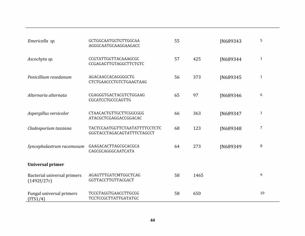
Emericella sp. GCTGGCAATGGTGTTGGCAAAGGGCAATGCAAGGAAGACC
55 JN689343 5
Ascochyta sp. CCGTATTGGTTACAAAGCGCCCGAGAGTTGTAGGCTTCTGTC
57 425 JN689344 1
Penicillium resedanum AGACAACCACAGGGGCTGCTCTGAACCCTGTCTGAAGTAAG
56 373 JN689345 1
Alternaria alternata CGAGGGTGACTACGTCTGGAAGCGCATCCTGCCCAGTTG
65 97
JN689346 6
Aspergillus versicolor CTAACACTGTTGCTTCGGCGGGATACGCTCGAGGACCGGACAC
66 363 JN689347 1
Cladosporium tassiana TACTCCAATGGTTCTAATATTTTCCTCTCGGGTACCTAGACAGTATTTCTAGCCT
68 123 JN689348 7
Syncephalastrum racemosum GAAGACACTTAGCGCACGCACAGCGCAGGGCAATCATA
64 273
JN689349
8
Universal primer
Bacterial universal primers (1492f/27r)
AGAGTTTGATCMTGGCTCAG GGTTACCTTGTTACGACT
58 1465 9
Fungal universal primers (ITS1/4)
TCCGTAGGTGAACCTTGCGGTCCTCCGCTTATTGATATGC
58 650 10
44

45
Plant universal primer CATTACAAATGCGATGCTCTTCTACCGATTTCGCCATATC
55 300‐700 11
1. Present study; 2. Scarpellini, Franzetti et al. 2004; 3. Walcott, Gitaitis et al. 2002; 4. Hynes, Chaudhry et al.
2006; 5. Matsuzawa, Tanaka et al. 2010; 6. Pavon, Gonzalez et al. 2010; 7. Qing‐Yin Zeng, Sven‐Olof Westermark
et al. 2006; 8. I. Nyilasi and K. Krizsa ́n 2008; 9. Frank, Reich et al. 2008; 10. White et all, 1990; 11. Taberlet,
Gielly et al. 1991

Table 4.2. Molecular identification of bacterial and fungal endophytes from Panicum virgatum L. based on blastN queries in NCBI Taxon Switchgrass
Cultivar Genbanck accession No.
Closest blast match (Genbanck accession No.) Query/reference ITS length (Similarity %)
BACTERIA
Curtobacterium flaccumfaciens
Cave‐in‐Rock JN689336 Curtobacterium flaccumfaciens (AM410688) 1502/1504 (99)
Pseudomonas fluorescens
Cave‐in‐Rock JN689337 Pseudomonas fluorescens (DQ439976) 1476/1500 (98)
Microbacterium testacum
Cave‐in‐Rock JN689338 Microbacterium testacum (EU714365) 1455/1466 (99)
Bacillus subtilis Cave‐in‐Rock JN689339 Bacillus subtilis (HQ727971) 1502/1503 (99)
Pantoea ananatis Cave‐in‐Rock JN689340
Pantoea ananatis (GQ383910) 1483/1490 (99)
FUNGI
46
Chaetomium globosum
Cave‐in‐Rock JN689341 Chaetomium globosum (HQ529775) 575/576 (99)
Epicoccum nigrum Cave‐in‐Rock and Tecumseh
JN689342 Epicoccum nigrum (FN868456) 576/576 (100)
Emericella sp. Cave‐in‐Rock JN689343 Emericella sp. (AB249015) 581/581 (100)
Ascochyta sp. Cave‐in‐Rock JN689344 Ascochyta hordei (HQ882800) 479/490 (98)
Penicillium resedanum
Cave‐in‐Rock JN689445 Penicillium resedanum (AF033398) 576/579 (99)
Alternaria alternata Cave‐in‐Rock JN689446 Alternaria alternata (JN618076) 423/423 (100)

47
Aspergillus versicolor
Cave‐in‐Rock JN689447 Aspergillus versicolor (AY373880) 568/569 (99)
Cladosporium tassiana
Tecumseh JN689348 Cladosporium cladosporioides (HQ380766) 548/548 (99)
Syncephalastrum acemosum r
Cave‐in‐Rock JN689349 Syncephalastrum racemosum (HQ2855713) 303/326 (93)

Table 4.3. Dual culture antagonistic experiment using different fungi
Test fungus
No inhibition; +, zone of inhib ion 1 3 mm ; ++, ne o nhib on o ore
th
it to zo f i iti f m
an 3 mm $ numbers in bracket represent the strain designation
1. G. Lazarovits, Agriculture, Agri‐Food Canada, London, Ontario, Canada; 2.
S. Neate, DPI, Queensland, Australia; 3. M.A. Cubeta, CIFR, North Carolina
State University, NC, USA; 4. American Type Culture Collection (ATTC),
Manassas, VA20108, USA.
B4 B8 B25 B26 B32 B45 B46 B47$
Fusarium solani1
‐ ‐ ++ ++ ++ ‐ ‐ ‐
B inucleate rhizoctonia 17072 ‐ ‐ +
+
++
‐
+
+
+
+
+ Rhizoctonia solani3
‐ ‐ ++ ++ ++ + + +
Trichoderma virens4
‐ ‐ + + + ‐
‐
‐
Stachybotrytis elegens4
‐ ‐ + + + ++ ++ ++
Phytophtora infestans1
‐
‐
‐
‐
+
+
+
+
+
+
+
+
+
+
+
+ Verticillium albo atrum1
48

Fig.4.1. Distribution of fungal and bacterial endophytes across the different
cultivars and leaf tissues. (A) Fungal isolates across the different cultivars (B)
Bacterial isolates across the different cultivar (C) Fungal endophytes in
different leaf tissues (D) Bacterial endophytes in the different leaf tissues.
49

Fig.4.2. Antagonistic test of (A) Pantoea ananatis and (B) Bacillus subtilis
against Rhizoctonia solani. (A1) R. solani ; (A2 and A3) R. solani and P.
ananatis; (B1) Rhizoctonia solani alone; (B2 and B3) Rhizoctonia solani
and Bacillus subtilis.
50

51

Fig.4.3. Maximum likelihood phylogenic tree of all putative endophytic
bacteria. The tree based analysis of partial 16S rDNA sequences of bacterial
isolates from switchgrass. Numbers above each node indicate percentage of
confidence levels generated from 1000 bootstrap trees and the GeneBank
Accession numbers precede the species names.
52

Fig. 4. Example of PCR detection of fungal (A) and bacterial (B) endophytes
detection in different switchgrass cultivars using species‐specific primers.
(A) Ascochyta sp. and (B) Bacillus subtilis. 100 bp DNA ladder lanes 1, lanes 2
Pure fungal genomic DNA of (A) Ascochyta or (B) Bacillus subtilis lanes 2, No
template lanes 3, PCR on template isolated from leaves of Tecumseh lanes 4‐
7, Cave‐in‐Rock lanes 8‐11, Blue Jacket lanes 12‐15, Sand Lover lanes 16‐19,
PCR on template isolated from seeds of Tecumseh, Cave‐in‐Rock and Blue
Jacket, respectively lanes 20‐21.
53

Fig. 5. PCR detection of Bacillus subtilis (A), Microbacterium testaceum (B),
the combination of both (C) recovered from colonized switchgrass seedlings.
(D) example of vertical transmission of the fungal endophyte Epicoccum
nigrum. (A) 100 bp DNA ladder lane 1, Pure DNA of B. subtilis lane 2, No
template lane 3, DNA of non‐inoculated switchgrass seedlings lanes 4‐6, DNA
of re‐inoculated switchgrass seedling with B. subtilis lane 7, DNA from roots
of colonized switchgrass lane 8, DNA from leaves and stems of colonized
switchgrass lane 9; (B) 100 bp DNA ladder lane 1, DNA of non‐inoculated
switchgrass seedlings lane 2‐5, DNA from roots of colonized M. testaceum
switchgrass seedling lanes 6‐7, DNA from stems and leaves of colonized M.
testaceum switchgrass lanes 8‐9, Pure genomic DNA of M. testaceum lane 10,
No template lane 11; (C) 100 bp DNA ladder lane 1, Pure genomic DNA of B.
subtilis lane 2, No template lane 3, DNA from roots of colonized switchgrass
with B. subtilis and M. testaceum and tested with B. subtilis primers lane 4,
DNA from shoots of colonized switchgrass with B. subtilis and M. testaceum
switchgrass and tested with B. subtilis primers lane 5, pure genomic DNA of
54

B. subtilis lane 6, No template lane 7, DNA from roots of colonized
switchgrass with B. subtilis and M. testaceum switchgrass and tested with M.
testaceum primers lane 8, DNA from shoots of colonized switchgrass with B.
subtilis and M. testaceum and tested with M. testaceum primers lane 9; (D)
100 bp DNA ladder lane 1, Pure genomic DNA of E. nigrum lane 2, No
template lane 3, DNA of flag leaf collection from cultivar Cave‐in‐Rock line 4,
collected seeds from cultivar Cave‐in‐Rock line 5, flag leaf from Cave‐in‐Rock
grown from seed lane 6.
55

Chapter V Discussion
Endophytic fungi and bacteria are ubiquitous in nature, infecting
virtually all plants in both natural and in agronomic ecosystems (Hyde and
Soytong, 2008; Ryan et al., 2008; Schulz and Boyle, 2005). Some of these
likely have no beneficial effects on host fitness and may be either latent
pathogens or saprophytes that remain inactive until environmental cues
trigger a developmental shift for the fungus or bacteria to continue its
lifecycle. Others have been repeatedly isolated and described as endophyte
and showed to be beneficial in some way to the plant. The endophytes
isolated over the course of this work were obtained using a traditional
method, in which only endophytes that can grow on microbiological media
were accounted for which may create a negative bias towards endophytes
that are slow growing or that can’t grow on biological media. In order to
assess the full range of endophyte future work should incorporate techniques
like DGGE or T‐RLFP (Bridge and Newsham, 2009; Duong et al., 2006). As the
over arching aim of this study is to identify endophyte species that could be
introduced into elite switchgrass cultivars for maximizing their utility as a
bionergy crop, we focused on these endophytes that could be grown on pure
culture and by using different media with different sources of carbon and pH,
e believe to have countered as many as possible. w
56

Our research goals were to survey switchgrass grown in Quebec for
the presence of endophytic bacteria and fungi, and to determine their
taxonomic position. Strategically, in order to cover a maximum genetic
diversity, we sampled leaves from old and well‐established stand as well as
from recently established fields of four switchgrass cultivars having different
genetic background. In this study, we demonstrated that a wide range of
fungal and bacterial species from 11 different taxa orders exist as endophytes
in switchgrass cultivars. Similarly, other researchers have reported on the
isolation of indigenous bacterial endophytes from leaf tissue (Zinniel et al.,
2002) and fungal endophytes from leaf and root tissues (Ghimire et al., 2011)
of switchgrass that naturally inhabits prairies. To the best of our knowledge,
our study is the first to describe both indigenous fungal and bacterial
endophytes from elite switchgrass cultivars produced through breeding.
There appears to be significant variation in the types of indigenous fungi and
bacteria from naturally occurring and breeding lines of switchgrass. The
only common fungal endophyte recovered from prairie switchgrass (Ghimire
et al., 2011) and from the switchgrass breeding lines (our study) was A.
alternata and Ascochyta sp. (anamorph Didymella). In the case of bacterial
endophytes, Curtobacterium and Microbacterium species were common as
they were recovered endophytes from prairie switcthgrass (Zinniel et al.,
2002) and from the cultivars used in this study. Several factors may explain
these differences, including cultivar type, plant age and tissue type and
seasonality (Clay, 1984; Ghimire et al., 2011)
57
Some of the fungal genera to which our switchgrass isolates have been
assigned are known to display a variety of interactions with plants. For
instance, Alternaria alternata has been described a frequently recovered
endophyte (Guo et al., 2004) as well as a plant pathogen causing post‐harvest
diseases of fruits and vegetables (Tian et al., 2011). Another fungal genus,
Cladosporium exhibits interactions with plants ranging from facultative
biotrophs to endophytes and epiphytes (Wirsel et al., 2001) and has been
reported along with Aspergillus versicolor to display anti‐micorbial activity
against several human pathogens (Sette et al., 2006). We are currently
investigating these isolates in details to establish their physiological
capacities.
Over the course of this study, endophytes were isolated from 594
switchgrass leaf segments with the majority (85%) of bacterial and fungal
endophytes originating from an old stand of Cave‐in‐Rock cultivar that was
established sixteen years ago. This is not surprising as the disparity in the
number of isolates per cultivar may be the result of plant age. As time of
exposure to endophyte inoculum increases, plants seem to accumulate an
increasing number of endophytes in their tissues. Because of this, older plant
parts may harbor more and diverse populations of fungal and bacterial
endophytes than younger ones (Arnold and Herre, 2003; Clay, 1984).
Another plausible explanation for increased incidence of recovered
endophytes from Cave‐in‐Rock may be related to the environmental
58

conditions favoring the incidence of endophytes. It has been shown that
humid and hot conditions are likely more conducive for increased incidence
of endophytes than arid and cold conditions (Arnold and Lutzoni, 2007;
Sánchez Márquez et al., 2010). Among the switchgrass cultivars used in this
study, cultivar Cave‐in‐Rock was developed in humid areas of southern
Illinois, USA (Elbersen et al., 2001), and probably evolved to promote higher
endophyte incidence as the plant under such such conditions have boundless
access with fungal endophytes and pathogens.
The findings of our study show that the distribution of fungal, but not
bacterial endophytes, in different leaf parts was substantially unequal. We
were able to isolate significantly more fungal strains from the leaf blade
compared to the leaf sheath. We are not able to explain this observation,
however recent studies have shown that the extent of leaf colonization by
fungal endophytes including that of leaf blades varies among host grasses
and probably reflects the stage of development of the primordial leaf when
the hyphae become established (Christensen and Voise, 2009). Also, leaf
colonization by fungal endohoytes is related to the timing of entry of the
hyphae into developing leaves (Christensen et al., 2000). The ligular zone
found just below the leaf blade lacks continuous intercellular spaces and acts
as a physical barrier preventing the entry of hyphae into leaf blades (Hinton
and Bacon, 1985). Although in our study, changes in endophyte distribution
over time was not addressed, it is likely that leaf blade colonization of
59

switchgrass by fungal endophytes occurred before a certain stage in the
development of the ligular zone. Another plausible explanation is the
difference in the nature of the tissues composing the leaf sheath and the leaf
blade might explain the difference in the number of isolate. The leaf sheath
has a much greater content of lignified tissues than the leaf blade
(Rangasamy et al., 2009). Lignified tissues are harder to penetrate for fungi
and provide a natural plant protection, thus making it less hospitable for the
endophytes (Raven et al., 2005).
The dominant taxa recovered from switchgrass reflect an unequal
distribution of isolate richness among species. This kind of abundance has
also been observed in other grasses (Sánchez Márquez et al., 2010; Wirsel et
al., 2001). It is not yet clear whether this abundance inequality is method
related, that is selection of certain taxa by the cultivation method. Several
dominant taxa of Switchgrass endophytes also have been identified in other
grasses and plants of other families implying that these species are host‐
generalists. Examples of these taxa are Epicoccum, Chaetomium, Ascochyta
and Panteoa (Hashizume et al., 2010; Mano et al., 2007; Neubert et al., 2006;
ánchez Márquez et al., 2010). S
Our results demonstrated that the five different recovered taxa of
bacteria from switchgrass are also associated with grasses and plants as
endophytes (Lodewyckx et al., 2002; Zinniel et al., 2002). The beneficial
60

effects of bacterial endophytes on their host plants appear to occur through
similar mechanisms described for plant‐growth promoting rhizobacteria
(Lodewyckx et al., 2002). Plant growth‐promoting bacteria can affect plant
growth directly or indirectly. The direct promotion of plant growth by PGPR
or endophytes such as Microbacterium testaceum and Panteoa ananatis
(Thomas et al., 2007; Zinniel et al., 2002) for the most part, entails providing
the plant with a compound that is synthesized by the bacterium and the
uptake of certain nutrients from the environment. The indirect promotion of
plant growth occurs when PGPR or endophytes (e.g., Pseudomonas
fluorescens, Bacillus subtilis, Curtobacterium flaccumfaciens, and Panteoa
species) decrease or prevent the deleterious effects of one or more
phytopathogenic organisms via release of antimicrobial metabolites (Bacon
t al., 2001; Landa et al., 2004; Mano et al., 2007; Thomas et al., 2007). e
Our ITS‐sequence database for switchgrass‐associated fungi was not
only the basis for molecular taxonomy but also served as a source for
designing species‐specific primers for monitoring individual fungal species
within DNA isolated from leaves and seeds of switchgrass cultivars. We have
applied this approach for tracking individual endophytes by PCR analysis in
seeds and of mixed endophytes reintroduced into switchgrass seedlings.
Seed‐borne endophytes can provide improved vigor to switchgrass, which
could be most important when establishing new switchgrass stands. In our
study, the endophytes B. subtilis, E. nigrum, P. resedanum, S. racemosum were
61

62
frequently detected in switchgrass seeds as well as plants grown in 2010 that
originated from seeds collected in 2009. These results demonstrate that the
endophytes are carried by seeds as a viable propagule and can be vertically
transmitted to the next generation of the host. The same type of transmission
is found in common reed, corn, coffee and some native grasses (Bacon et al.,
001; Ernst et al., 2003; Vega et al., 2006) 2
Endophytic bacteria inside a plant might either become localized at
the point of entry or spread throughout the plant (Hallmann et al., 1997).
These microorganisms reside within cells, in the intercellular space or in the
vascular system (Hallmann et al., 1997) . In our colonization experiments, B.
subtilis and M. testaceum were recovered from sterilized roots, and leaves
two weeks after inoculation of switchgrass seedlings, indicating successful
internal colonization within the roots and the leaves. These results indicate
that the endophytes in switchgrass seedlings moved from the roots and
travelled upward to the stem and leaves. Our results concur with previous
findings, which also demonstrate the ability of B. subtilis endophytic strain to
enter the roots and migrate in the upper stems and leaves of eggplants (Lin et
l., 2009). a

Chapter VI Conclusion
In summary, we recovered 9 different endophytic fungi and 5
different endophytic bacteria from four switchgrass cultivars. These
endophytes were identified using molecular techniques. Our findings are in
agreement with other studies that the recovered identified endophyte are
known endophytes in other plants, including perennial grasses.
The in‐vitro dual‐culture method using endophytic bacteria,
demonstrated their bioactivity potential against plant pathogenic fungi. Our
results corroborate the finding of several studies which show the
antagonistic activity of bacterial endophytes. Thus, switchgrass endophytes
represent a potential source of symbionts that can play an important role in
switchgrass production.
The species‐specific primers that we have developed can now be
applied to systematically screen samples from various natural habitats,
locations, and seasons in order to reveal putative correlations between
switchgrass health and the switchgrass‐associated endophytes discovered in
this study.
The successful colonization of switchgrass with selected endophytes,
either applied singly or in combination suggests that they can be utilized in
63

future applications such as delivery of degradative enzymes or antimicrobial
metabolites for controlling diseases.
64

Chapter VII References Arnold A.E., Herre E.A. (2003) Canopy cover and leaf age affect colonization
by tropical fungal endophytes: Ecological pattern and process in
Theobroma cacao (Malvaceae). Mycologia 95:388‐398.
Arnold A.E., Lutzoni F. (2007) Diversity and host range of foliar fungal
endophytes: Are tropical leaves biodiversity hotspots? Ecology 88:541‐
549.
Arnold A.E., Maynard Z., Gilbert G.S., Coley P.D., Kursar T.A. (2000) Are
tropical fungal endophytes hyperdiverse? Ecology Letters 3:267‐274.
Athukorala S.N.P., Fernando W.G.D., Rashid K.Y. (2009) Identification of
antifungal antibiotics of Bacillus species isolated from different
microhabitats using polymerase chain reaction and MALDI‐TOF mass
n u l spectrometry. Canadia Jo rna of Microbiology 55:1021‐1032.
Azevedo J.L., Maccheroni Jr. W., Pereira J.O., de Araújo W.L. (2000)
Endophytic microorganisms: a review on insect control and recent
advances on tropical plants. Electronic Journal of Biotechnology 3:15‐
16.
Bacon C.W., Yates I.E., Hinton D.M., Meredith F. (2001) Biological control of
Fusarium moniliforme in maize. Environmental Health Perspectives
109:325‐332.
Bashyal B.P., McLaughlin S.P., Gunatilaka A.A.L. (2006) Zinagrandinolides A‐C,
cytotoxic delta‐elemanolide‐type sesquiterpene lactones from Zinnia
grandiflora. Journal of Natural Products 69:1820‐1822.
65

Bent E., Chanway C.P. (1998) The growth‐promoting effects of a bacterial
endophyte on lodgepole pine are partially inhibited by the presence of
other rhizobacteria. Canadian Journal of Microbiology 44:980‐988.
Berdahl J.D., Frank A.B., Krupinsky J.M., Carr P.M., Hanson J.D., Johnson H.A.
(2005) Biomass yield, phenology, and survival of diverse switchgrass
cultivars and experimental strains in western North Dakota. Agronomy
Journal 97:549‐555.
Boe A., Ross J.G. (1998) Registration of 'Sunburst' switchgrass. Crop Science
38:540‐540.
Bouton J. (2008) Improvement of Switchgrass as a Bioenergy Crop. Genetic
Improvement of Bioenergy Crops:309‐345.
Bridge P.D., Newsham K.K. (2009) Soil fungal community composition at
Mars Oasis, a southern maritime Antarctic site, assessed by PCR
amplification and cloning. Fungal Ecology 2:66‐74.
Casler M.D., Boe A.R. (2003) Cultivar X environment interactions in
switchgrass. Crop Science 43:2226‐2233.
Cassida K.A., Muir J.P., Hussey M.A., Read J.C., Venuto B.C., Ocumpaugh W.R.
(2005) Biofuel Component Concentrations and Yields of Switchgrass in
South Central U.S. Environments. Crop Science 45:682‐692.
Chanway C.P. (1997) Inoculation of tree roots with plant growth promoting
soil bacteria: An emerging technology for reforestation. Forest Science
43:99‐112.
66

Chaves M.M., Flexas J., Pinheiro C. (2009) Photosynthesis under drought and
salt stress: regulation mechanisms from whole plant to cell. Annals of
Botany 103:551‐560.
Chiang Y.C., Chou C.H., Lee P.R., Chiang T.Y. (2001) Detection of leaf‐
associated fungi based on PCR and nucleotide sequence of the
ribosomal internal transcribed spacer (ITS) in Miscanthus. Botanical
Bulletin of Academia Sinica 42:39‐44.
Christensen M.J., Voise C.R. (2009) Tall Fescue – Endophyte Symbiosi, in: D. B.
H. In H.A. Fribourg, and C.P. West (Ed.), Tall fescue for the twenty‐first
centur, Agronomy Monograph, Southampton. pp. 251‐27.
Christensen M.J., Simpson W.R., Al Samarra T. (2000) Infection of tall fescue
and perennial ryegrass plants by combinations of different
Neotyphodium endophytes. Mycological Research 104:974‐978. DOI:
d nuoi: ll.
Clay K. (1984) The effect of the fungus AtkinsonellaHypoxylon
(Clavicipitaceae) on the reproductive‐system and demography of the
grass DanthoniaSpicata. New Phytologist 98:165‐175.
Clay K. (1988) Fungal endophytes of grasses – A defensive mutualism
between plants and fungi. Ecology 69:10‐16.
Clay K. (1990) Fungal Endophytes of grasses. Annual Review of Ecology and
Systematics 21:275‐297.
Costa C.P., Ferreira M.C. (1991) Presevation of Microorganisms‐ A Review
Revista De Microbiologia 22:263‐268.
67

Donner S., Kucharik C. (2008) Corn‐based ethanol production compromises
goal of reducing nitrogen export by the Mississippi River. Proceedings
of the National Academy of Sciences 105(11):4513‐4518.
Duong L.M., Jeewon R., Lumyong S., Hyde K.D. (2006) DGGE coupled with
ribosomal DNA gene phylogenies reveal uncharacterized fungal
phylotypes. Fungal Diversity 23:121‐138.
Elbersen H.W., Christian D.G., Bassem N.E., Bacher W., Sauerbeck G.,
Alexopoulou E., Sharma N., Piscioneri I., Visser P.d., Berg D.v.d. (2001)
Switchgrass Variety Choice In Europe. Aspects Appl. Bio 65:21–28.
Ellis M.B. (1971) Denatiaceous hyphomycetes. Comowealth Mycological
Institute Kew 608; 1‐25.
Ernst M., Mendgen K.W., Wirsel S.G.R. (2003) Endophytic fungal mutualists:
seed‐borne Stagonospora Spp. enhance reed biomass production in
0‐587. axenic microcosms. Molecular Plant‐Microbe Interactions 16:58
Ghimire S.R., Craven K.D. (2011) Enhancement of Switchgrass (Panicum
virgatum L.) Biomass Production under Drought Conditions by the
Ectomycorrhizal Fungus Sebacina vermifera. Applied and
Environmental Microbiology 77:7063‐7067.
Ghimire S.R., Charlton N.D., Bell J.D., Krishnamurthy Y.L., Craven K.D. (2011)
Biodiversity of fungal endophyte communities inhabiting switchgrass
(Panicum virgatum L.) growing in the native tallgrass prairie of
northern Oklahoma. Fungal Diversity 47:19‐27.
68
Guo L.D., Xu L., Zheng W.H., Hyde K.D. (2004) Genetic variation of Alternaria
alternata, an endophytic fungus isolated from Pinus tabulaeformis as
determined by random amplified microsatellites (RAMS). Fungal
ty . Diversi 16:53‐65
Haddad A., Camacho F., Durand P., Cary S.C. (1995) Phylogenetic
characterization of the epibiotic bacteria associated with the
hydrothermal vent Polychaete Alvinellapompejana. Applied and
Environmental Microbiology 61:1679‐1687.
Hallmann J., Quadt‐Hallmann A., Mahaffee W.F., Kloepper J.W. (1997)
Bacterial endophytes in agricultural crops. Canadian Journal of
Microbiology 43:895‐914.
Hashizume Y., Fukuda K., Sahashi N. (2010) Effects of summer temperature
on fungal endophyte assemblages in Japanese beech (Fagus crenata)
leaves in pure beech stands. Botany 88:266‐274.
Hinton D.M., Bacon C.W. (1985) The distribution and ultrastructure of the
hendop yte of toxic tall fescue. Canadian Journal of Botany 63:36‐42.
Hyde K.D., Soytong K. (2008) The fungal endophyte dilemma. Fungal
Diversity 33:163‐173.
Johnson M.C., Dahlman D.L., Siegel M.R., Bush L.P., Latch G.C.M., Potter D.A.,
Varney D.R. (1985) Insect feeding deterrents in Endophyte‐Infected
Tall Fescue Applied and Environmental Microbiology 49:568‐571.
69

Kang S.H., Cho H.S., Cheong H., Ryu C.M., Kim J.F., Park S.H. (2007) Two
bacterial entophytes eliciting both plant growth promotion and plant
defense on pepper (Capsicum annuum L.). Journal of Microbiology and
Biotechnology 17:96‐103.
Kitamoto Y., Suzuki A., Shimada S., Yamanaka K. (2002) A new method for the
preservation of fungus stock cultures by deep‐freezing. Mycoscience
43:0143‐0149.
Kloepper J.W., Ryu C.‐M. (2006) Bacterial Endophytes as Elicitors of Induced
Systemic Resistance 9:33‐55.
Microbial Root Endophytes, in: B. J. E. Schulz, et al. (Eds.), Springer Berlin
Heidelberg. pp. 33‐52.
Kobayashi D.Y., Palumbo J.D. (2000) Bacterial endophytes and their effects on
plants and uses in agriculture: Dekker (Ed.), Microbial endophytes,
Marcel Dekker, New‐York . 199‐243
Krishnamurthy K., Gnanamanickam S.S. (1997) Biological control of sheath
blight of rice: Induction of systemic resistance in rice by plant‐
3 . associated Pseudomonas spp. Current Science 72: 31‐334
Landa B.B., Navas‐Cortes J.A., Jimenez‐Diaz R.M. (2004) Influence of
temperature on plant‐rhizobacteria interactions related to biocontrol
potential for suppression of fusarium wilt of chickpea. Plant Pathology
53:341‐352.
70

Larran S., Perello A., Simon M.R., Moreno V. (2002) Isolation and analysis of
endophytic microorganisms in wheat (Triticum aestivum L.) leaves.
World Journal of Microbiology & Biotechnology 18:683‐686.
Latch G.C.M., Christensen M.J. (1985) Artifical infection of grasses with
endophytes Annals of Applied Biology 107:17‐24.
Lee D.K., Boe A. (2005) Biomass production of switchgrass in central South
Dakota. Crop Science 45:2583‐2590.
Lemus R., Brummer E.C., Moore K.J., Molstad N.E., Burras C.L., Barker M.F.
(2002) Biomass yield and quality of 20 switchgrass populations in
southern Iowa, USA. Biomass and Bioenergy 23:433‐442.
Lin L., Qiao Y.S., Ju Z.Y., Ma C.W., Liu Y.H., Zhou Y.J., Dong H.S. (2009) Isolation
and characterization of endophytic Bacillius subtilis Jaas ed1 antagonist
of eggplant Verticillium wilt. Bioscience Biotechnology and
Biochemistry 73:1489‐1493.
Lodewyckx C., Vangronsveld J., Porteous F., Moore E.R.B., Taghavi S., Mezgeay
M., van der Lelie D. (2002) Endophytic bacteria and their potential
applications. Critical Reviews in Plant Sciences 21:583‐606.
Madakadze I.C., Stewart K., Peterson P.R., Coulman B.E., Smith D.L. (1999)
Switchgrass biomass and chemical composition for biofuel in eastern
Canada. Agronomy Journal 91:696‐701.
Malinowski D.P., Belesky D.P. (2000) Adaptations of endophyte‐infected cool‐
season grasses to environmental stresses: Mechanisms of drought and
mineral stress tolerance. Crop Science 40:923‐940.
71

Mano H., Tanaka F., Nakamura C., Kaga H., Morisaki H. (2007) Culturable
endophytic bacterial flora of the maturing leaves and roots of rice
plants (Oryza sativa) cultivated in a paddy field. Microbes and
nEnviro ments 22:175‐185.
Mendes R., Pizzirani‐Kleiner A.A., Araujo W.L., Raaijmakers J.M. (2007)
Diversity of cultivated endophytic bacteria from sugarcane: Genetic and
biochemical characterization of Burkholderia cepacia complex isolates.
Applied and Environmental Microbiology 73:7259‐7267.
Miyamoto T., Kawahara M., Minamisawa K. (2004) Novel endophytic
nitrogen‐fixing clostridia from the grass Miscanthus sinensis as revealed
by terminal restriction fragment length polymorphism analysis.
Applied and Environmental Microbiology 70:6580‐6586.
Muir J.P., Sanderson M.A., Ocumpaugh W.R., Jones R.M., Reed R.L. (2001)
Biomass production of 'Alamo' switchgrass in response to nitrogen,
phosphorus, and row spacing. Agronomy Journal 93:896‐901.
Mukherjee S., Dawe A.L., Creamer R. (2010) Development of a transformation
system in the swainsonine producing, slow growing endophytic fungus,
Undifilum oxytropis. Journal of Microbiological Methods 81:160‐165.
DOI: 10.1016/j.mimet.2010.02.015.
Neubert K., Mendgen K., Brinkmann H., Wirsel S.G.R. (2006) Only a few fungal
species dominate highly diverse mycofloras associated with the
common reed. Applied and Environmental Microbiology 72:1118‐1128.
72

Pavlo A., Leonid O., Iryna Z., Natalia K., Maria P.A. (2011) Endophytic bacteria
enhancing growth and disease resistance of potato (Solanum tuberosum
L.). Biological Control 56:43‐49.
Pedersen J.F., Rodriguezkabana R., Shelby R.A. (1988) Ryegrass cultivars and
endophyte in Tall Fescue affect nematodes in grass and succeding
soybean. Agronomy Journal 80:811‐814.
Ramarathnam R., Bo S., Chen Y., Fernando W.G.D., Gao X., de Kievit T. (2007)
Molecular and biochemical detection of fengycin‐ and bacillomycin D‐
producing Bacillus spp., antagonistic to fungal pathogens of canola and
h n o o 5 ‐w eat. Ca adian J urnal f Microbiology 3:901 911.
Ramos H.J.O., Souza E.M., Soares‐Ramos J.R.L., Pedrosa F.O. (2007)
Determination of bean nodule occupancy by Rhizobium tropici using the
double gfp and gusA genetic markers constitutively expressed from a
new broad‐host‐range vector. World Journal of Microbiology &
Biotechnology 23:713‐717.
Rangasamy M., Rathinasabapathi B., McAuslane H.J., Cherry R.H., Nagata R.T.
(2009) Role of leaf sheath lignification and anatomy in resistance
against southern Chinch bug (Hemiptera: Blissidae) in St.
Augustinegrass. Journal of Economic Entomology 102:432‐439.
Raven P.H., F. E.R., Eichhorn S.E. (2005) Biology of Plants. 7 ed. Sara Tenney,
New‐York. 686
73
Rodriguez Estrada A.E., Hegeman A., Corby Kistler H., May G. (2011) In vitro
interactions between Fusarium verticillioides and Ustilago maydis
through real‐time PCR and metabolic profiling. Fungal genetics and
biology : FG & B 48:874‐85.
Rosenblueth M., Martinez‐Romero E. (2006) Bacterial endophytes and their
interactions with hosts. Molecular Plant‐Microbe Interactions 19:827‐
837.
Rouws L.F.M., Meneses C., Guedes H.V., Vidal M.S., Baldani J.I., Schwab S.
(2010) Monitoring the colonization of sugarcane and rice plants by the
endophytic diazotrophic bacterium Gluconacetobacter diazotrophicus
marked with gfp and gusA reporter genes. Letters in Applied
Microbiology 51:325‐330.
Ryan R.P., Germaine K., Franks A., Ryan D.J., Dowling D.N. (2008) Bacterial
endophytes: recent developments and applications. Fems Microbiology
Letters 278:1‐9.
Samson R. (1991) Switchgrass: A living solar battery for the prairies.
Sustainable Farming:4‐8
Samson R. (1997) Technology evaluation and development of short rotation
forestry and switchgrass for energy production, in: N. R. Canada (Ed.),
Ottawa. pp. 218.
Samson R., Stamler S.B. (2007) The emerging Agro‐Pellet industry in Canada
in: REAP‐Canada (Ed.), REAP‐Canada Librairy, REAP‐Canada, Ste‐Anne‐
de‐Bellevue.
74

Sánchez Márquez S., Bills G., Domínguez Acuña L., Zabalgogeazcoa I. (2010)
Endophytic mycobiota of leaves and roots of the grass Holcus lanatus.
Fungal Diversity 41:115‐123.
Sanchez‐Azofeifa A., Oki Y., Wilson Fernandes G., Ball R., Gamon J. (2011)
Relationships between endophyte diversity and leaf optical properties.
07/s00468‐011‐059Trees ‐ Structure and Function:1‐9. DOI: 10.10 1‐5.
Sanderson, A. M., Read, C. J., Read, L. R. (1999) Harvest management of
switchgrass for biomass feedstock and forage production American
Society of Agronomy 91: 5‐10.
Sauer D.B., Burroughs R. (1986) Disinfection of seed surfaces with Sodium‐
Hypochlorite. Phytopathology 76:745‐749.
Schardl C.L., Leuchtmann A., Spiering M.J. (2004) Symbioses of grasses with
seedborne fungal endophytes. Annual Review of Plant Biology 55:315‐
340.
Schulz B., Boyle C. (2005) The endophytic continuum. Mycological Research
109:661‐686.
Schulz B., Wanke U., Draeger S., Aust H.J. (1993) Endophytes from herbaceous
plants and shrubs ‐ Effectiveness of surface sterilization methods
Mycological Research 97:1447‐1450.
Schulz B., Rommert A.K., Dammann U., Aust H.J., Strack D. (1999) The
endophyte‐host interaction: a balanced antagonism? Mycological
Research 103:1275‐1283.
75

Schulz B., Boyle C., Draeger S., Rommert A.K., Krohn K. (2002) Endophytic
fungi: a source of novel biologically active secondary metabolites.
Mycological Research 106:996‐1004.
Sessitsch A., Reiter B., Berg G. (2004) Endophytic bacterial communities of
field‐grown potato plants and their plant‐growth‐promoting and
antagonistic abilities. Canadian Journal of Microbiology 50:239‐249.
Sette L.D., Passarini M.R.Z., Delarmelina C., Salati F., Duarte M.C.T. (2006)
Molecular characterization and antimicrobial activity of endophytic
fungi from coffee plants. World Journal of Microbiology &
8 . Biotechnology 22:11 5‐1195
Smith K.T., Bacon C.W., Luttrell E.S. (1985) Reciprocal translocation of
carbohydrates between host and fungus in Bahiagrasss infected with
Myciogenospora atramentosa. Phytopathology 75:407‐411.
Steinbach W.J., Shetty A. (2001) Use of the diagnostic bacteriology laboratory:
a practical review for the clinician. Postgraduate Medical Journal
8 77:14 ‐156.
Strobel G., Daisy B., Castillo U., Harper J. (2004) Natural products from
endophytic microorganisms. Journal of Natural Products 67:257‐268.
Sturz A.V., Christie B.R., Nowak J. (2000) Bacterial endophytes: Potential role
in developing sustainable systems of crop production. Critical Reviews
in Plant Sciences 19:1‐30.
Subramaniam S. (1998) The biology Workbench‐‐a seamless database and
analysis environment for the biologist. Protein 32;1‐2
76

Sun L., Qiu F., Zhang X., Dai X., Dong X., Song W. (2008) Endophytic bacterial
diversity in rice (Oryza sativa L.) roots estimated by 16S rDNA
sequence analysis. Microbial Ecology 55:415‐424.
Suryanarayanan T.S., Thirumalai E., Prakash C.P., Rajulu M.B.G.,
Thirunavukkarasu N. (2009a) Fungi from two forests of southern India:
a comparative study of endophytes, phellophytes, and leaf litter fungi.
Canadian Journal of Microbiology 55:419‐426.
Suryanarayanan T.S., Thirunavukkarasu N., Govindarajulu M.B., Sasse F.,
Jansen R., Murali T.S. (2009b) Fungal endophytes and bioprospecting.
Fungal Biology Reviews 23:9‐19.
Taliaferro C.M. (2002) Breeding and selection of new Switchgrass varieties
for increased biomass production, in: U. S. D. o. E. O. o. Biomass (Ed.),
Tennessee.
Tamura K., Peterson D., Peterson N., Stecher G., Nei M., Kumat S. (2011)
MEGA5: Molecular Evolutionary Genetics Analysis using Maximum
Likelihood, Evolutionary Distance, and Maximum Parsimony Methods.
Melecular Biology and Evolution. Doi:10.1093/molbev/msr121.
Thomas P., Kumari S., Swarna G.K., Gowda T.K.S. (2007) Papaya shoot tip
associated endophytic bacteria isolated from in vitro cultures and host‐
endophyte interaction in vitro and in vivo. Canadian Journal of
Microbiology 53:380‐390.
77

Tian J., Ban X.Q., Zeng H., Huang B., He J.S., Wang Y.W. (2011) In vitro and in
vivo activity of essential oil from dill (Anethum graveolens L.) against
fungal spoilage of cherry tomatoes. Food Control 22:1992‐1999.
Turner N.C. (1986) Adaptation to water deficites ‐ A changins perspective
Australian Journal of Plant Physiology 13:175‐190.
Van Loon L.C., Bakker P., Pieterse C.M.J. (1998) Systemic resistance induced
b z c u ty rhi osphere ba teria. Ann al Review of Phy opathology 36:453‐483.
Vega F.E., Posada F., Peterson S.W., Gianfagna T.J., Chaves F. (2006)
Penicillium species endophytic in coffee plants and Ochratoxin A
Production. Mycologia 98:31‐42.
Vogel K.P., Brejda J.J., Walters D.T., Buxton D.R. (2002) Switchgrass biomass
production in the Midwest USA: Harvest and nitrogen management
A imer can Society of Agronomy 93: 413‐420
West C.P., Izekor E., Oosterhuis D.M., Robbins R.T. (1988) The effect of
Acremonium‐Coenophialum on the growth and nematode infestation in
i 6Tall Fescue. Plant and So l 112:3‐ .
White I.R., Backhouse D. (2007) Comparison of fungal endophyte
communities in the invasive panicoid grass Hyparrhenia hirta and the
native grass Bothriochloa macra. Australian Journal of Botany 55:178‐
185.
78

79
White T.J., Bruns T., Lee S., Taylor J.W. (1990) Amplification and direct
sequencing of fungal ribosomal RNA genes for phylogenetics, in: M. A.
Innis, et al. (Eds.), PCR Protocols: A Guide to Methods and Applications,
Academic Press Inc., New York. pp. 315‐322.
Wirsel S.G.R., Leibinger W., Ernst M., Mendgen K. (2001) Genetic diversity of
fungi closely associated with common reed. New Phytologist 149:589‐
598.
Zinniel D.K., Lambrecht P., Harris N.B., Feng Z., Kuczmarski D., Higley P.,
Ishimaru C.A., Arunakumari A., Barletta R.G., Vidaver A.K. (2002)
Isolation and characterization of endophytic colonizing bacteria from
agronomic crops and prairie plants. Applied and Environmental
Microbiology 68:2198‐2208.