Embed Size (px)
Citation preview

MATTES
1.133
Protective Caps availble according to diameter of nailSchutzkappen für Nägel passend zum Durchmesser des Nagels
15198 ø 1.5 mm15200 ø 2.0 mm15202 ø 2.5 mm15204 ø 3.0 mm15206 ø 3.5 mm15208 ø 4.0 mm15210 ø 4.5 mm15212 ø 5.0 mm
Principle / Prinzip:
2 Gliding Nails produce a 3-point support in an elastic and stable system.2 Gleitnägel bilden eine 3-Punkt-Unterstützung für ein elastisches und stabiles System.
Intramedullary Gliding Nails for childrenGleitnägel für Kinder
Stainless Steel DIN ISO 5832-115250 1.5 x 150 mm15251 1.5 x 400 mm15252 2.0 x 200 mm15253 2.0 x 400 mm15254 2.5 x 250 mm15255 2.5 x 400 mm15256 3.0 x 300 mm15257 3.0 x 400 mm15258 3.5 x 350 mm15259 3.5 x 400 mm
Titanium DIN ISO 5835-3Titanium 6-4 Vanadium
ELI Alloy ForgingsColour Coding
15263 2.0 x 450 mm Green15264 2.5 x 450 mm Pink15265 3.0 x 450 mm Gold15266 3.5 x 450 mm Blue15267 4.0 x 450 mm Violet15268 4.5 x 450 mm Gray15269 5.0 x 450 mm Silver (natural)

MATTES
1.134
Trocar with slotted sleeve for implantation of medullary gliding nails
After opening of the medullary canal with trocar, it happens quite often, that the point of entry cannot be found. By using the new trocar, the slotted sleeve remains in situ and avoids unnecessary searching or preparating of soft tissue. Gliding nails with bent tip can easily be introduced through the slot of the sleeve.
Pfriem mit geschlitzter Hülse zur Implantation intramedullärer Schienen
Häufig ensteht nach Eröffnung der Markhöhle mit einem Pfriem das Problem, die Eintrittsstelle wieder zu fin-den. Bei Verwendung des neuen Pfriems verbleibt die geschlitzte Hülse am vorgebohrten Loch und erspart unnötiges Suchen oder Freipräparieren der Weichteile. Durch die Schlitzung der Gewebeschutzhülse können Schienen mit gebogener Spitze leicht eingeführt werden.
15310 Trocar for ø 3,5 - 5,0 mm Nails15312 Trocar for ø 1,5 - 3,5 mm Nails
Opening of medullary can-nal with trocar and sleeve
Eröffnen der Markhöhlemit Pfriem und Hülse
Removal of trocar. Sleeveis held in position
Entfernen des Pfriems - dieHülse wird in der Positiongehalten
Introduction of nail throughslot of sleeve
Einführen der Schienedurch die geschlitzte Hülse
MATTES
1.135
Centering Sleeves for diam.Zentrierhülse für ø
15320 1,5 - 2,0 mm15322 2,5 - 3,0 mm15324 3,5 - 5,0 mm
Centering SleeveZentrierhülse
Impactor headSchlagkopf
HandleHaltegriff
Fixation ScrewFeststellschraube
15316 Impactor Einschlaginstrument
The new Impactor for „Intramed Gliding Nails“ serves for quick intro-duction avoiding tiresome manipulating during fixation of implants. Owing to the lateral handle the danger of insuries to the surgeon in case of slipping-through of nails is practically impossible. A new clamping system serves for fixation of nails in the impactor without applying much power.
Der neue Impactor für Intramed-Schienen ermöglicht das schnelle Einbringen von intramedullären Schienen ohne lästiges Hantieren bei der Fixation der Implantate. Durch den seitlichen Griff wird die Verletzungsgefahr für den Chirurgen, beim Durchrutschen der Schienen ausgeschlossen. Die Schienenfixation im Einschlagin-strument ist durch eine neuartige Verklemmung ohne grossen Kraftaufwand möglich.
Impactor for all diameters of Gliding NailsEinschlaginstrument für alle Gleitnägel-Durchmesser

MATTES
1.136
151010 Sterilization Container (recommended)
15001 Tray (empty)
Listing: M 050000 Implant Set for Intramedullary Gliding Nails (Steel and Titanium Gliding Nail Set)
Implant Set for Intramedullary Gliding Nails

MATTES
1.137
Listing: M 050000 Implant Set for Intramedullary Gliding Nails (Steel and Titanium Gliding Nail Set)
15001 Tray (empty)
Steel Intramedullary Gliding Nail Implants ( Stainless Steel DIN ISO 5832-1 )
15250 Dia. 1,5 mm x Length 150 mm 4 Pcs.15251 Dia. 1,5 mm x Length 400 mm 4 Pcs.15252 Dia. 2,0 mm x Length 200 mm 4 Pcs.15253 Dia. 2,0 mm x Length 400 mm 4 Pcs.15254 Dia. 2,5 mm x Length 250 mm 4 Pcs.15255 Dia. 2,5 mm x Length 400 mm 4 Pcs.15256 Dia. 3,0 mm x Length 300 mm 4 Pcs.15257 Dia. 3,0 mm x Length 400 mm 4 Pcs.15258 Dia. 3,5 mm x Length 350 mm 4 Pcs.15259 Dia. 3,5 mm x Length 400 mm 4 Pcs.
Titanium Intramedullary Gliding Nail Implants ( Titanium DIN ISO 5832-3 )
15263 Dia. 2,0 mm x Length 450 mm Colour Coding Green 4 Pcs.15264 Dia. 2,5 mm x Length 450 mm Colour Coding Pink 4 Pcs.15265 Dia. 3,0 mm x Length 450 mm Colour Coding Gold 4 Pcs.15266 Dia. 3,5 mm x Length 450 mm Colour Coding Blue 4 Pcs.15267 Dia. 4,0 mm x Length 450 mm Colour Coding Violet 4 Pcs.15268 Dia. 4,5 mm x Length 450 mm Colour Coding Gray 4 Pcs.15269 Dia. 5,0 mm x Length 450 mm Colour Coding Silver 4 Pcs.
Implant Set for Intramedullary Gliding Nails
MATTES
1.138
Intramedullary Gliding Nails
Diaphiseal fractures of long bones in children an adolescents in the growing age are mostly treated conservatively with plaster of Paris and external splintage. The borderline is there, where retention obviously can not be maintained. Quite often additional anaesthesias were necessary due to post-reduction. In spite of the often long therapeutical process the final result was not satisfactory neither for the child, nor for the parents and therapists.The treatment with plates and screws or with an external fixateur were reduced to exceptions.Reasons for it:- during growth 75% the periosteal healing is predominant- with plates and screws a rigid anatomical reduction is given but followed by a long time of immobilization,
extensive scar and reoperation due to metal removal.- The treatment with external fixator for open fractures is advantageous and offers with good rigidity the
possibility of early dynamization. However the axial positioning can not always be achieved. In addition regularely pin-care visits are necessary and the child always is confrontated with the osteosyntheses material.
DemandInspite of the „healing potential“ in childlike bone the treatment of bone in the growing skeleton the axial anatomical alignment must be looked at with priority.Is there a reduction necessary then the fracture may not remain in an axial deviation or in a wrong rotational position. Independent from age the aim must be to achieve the optimum position.
Intramedullary osteosyntheses offers an alternative with- little additional traumatization- sufficiant axial stability- optimum healing due to stimulized micro movements- low complication rate- early mobilization- exellent cosmetical result- outpatient metal removement after 10 to 12 weeks and- is indicated for patients, in which the conserative therapy has already started, but post reduction is
predictable.
The use of about 10.000 intramedullary gliding nails in Germany has led to expand the indications also to adults. This alternative to the treatment with intramedullary gliding nails results in:- less invasive treatment- shorter OR-time- cost saving procedure- less X- Ray exposure for patient and therapist- outpatient metal removal after 6 to 9 months.

MATTES
1.139
The rigidity of the elastic intramedullary gliding nail is adequat to the rigidity of the bone. Incoming forces (F1), which lead to material breackage in rigid implants, are transformed and bend the implant. The elasticity of the implant and its ability of reversible deformation reset the intramedullary gliding nail into its origin position due to reset forces (R).
Is the intramedullary gliding nail (pict. 2) pre-bent (F2), arises a new „Zero Point“, that means that the reset forces reset the gliding nail in its „new“ point of origin.
Pict. 1
Pict. 2
MATTES
1.140
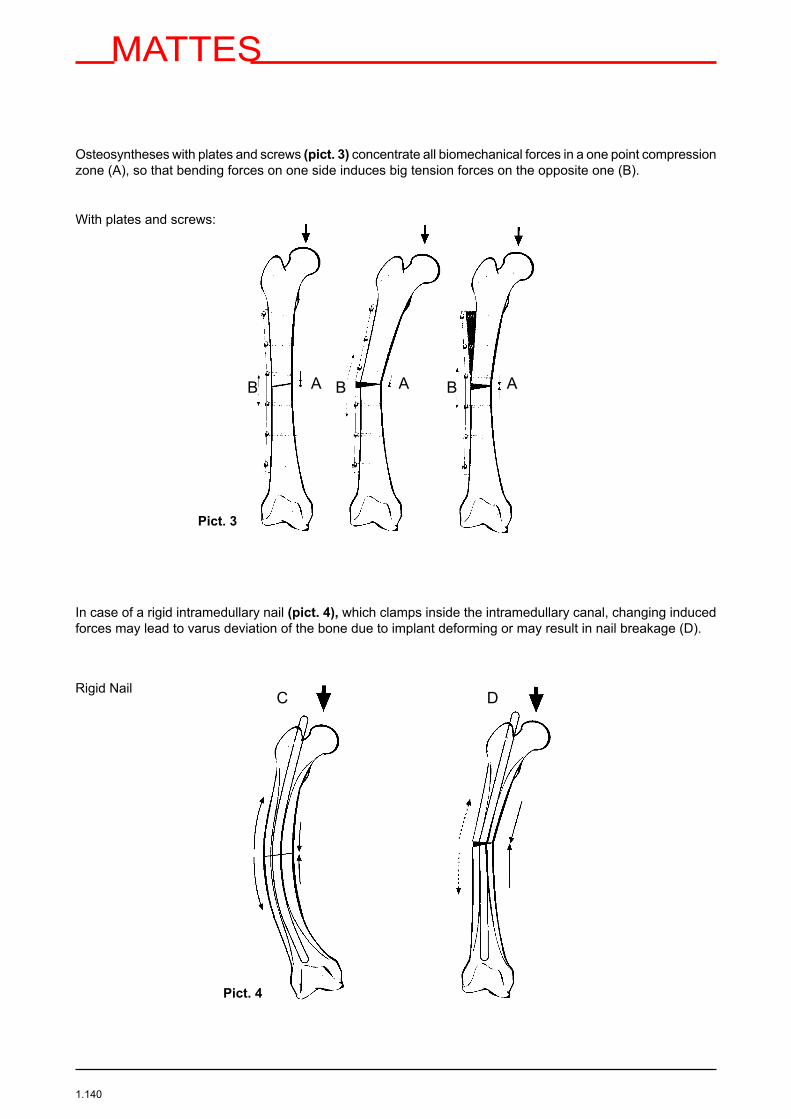
Osteosyntheses with plates and screws (pict. 3) concentrate all biomechanical forces in a one point compression zone (A), so that bending forces on one side induces big tension forces on the opposite one (B).
With plates and screws:
In case of a rigid intramedullary nail (pict. 4), which clamps inside the intramedullary canal, changing induced forces may lead to varus deviation of the bone due to implant deforming or may result in nail breakage (D).
Rigid Nail
Pict. 3
C D
Pict. 4
B A A AB B
MATTES
1.141
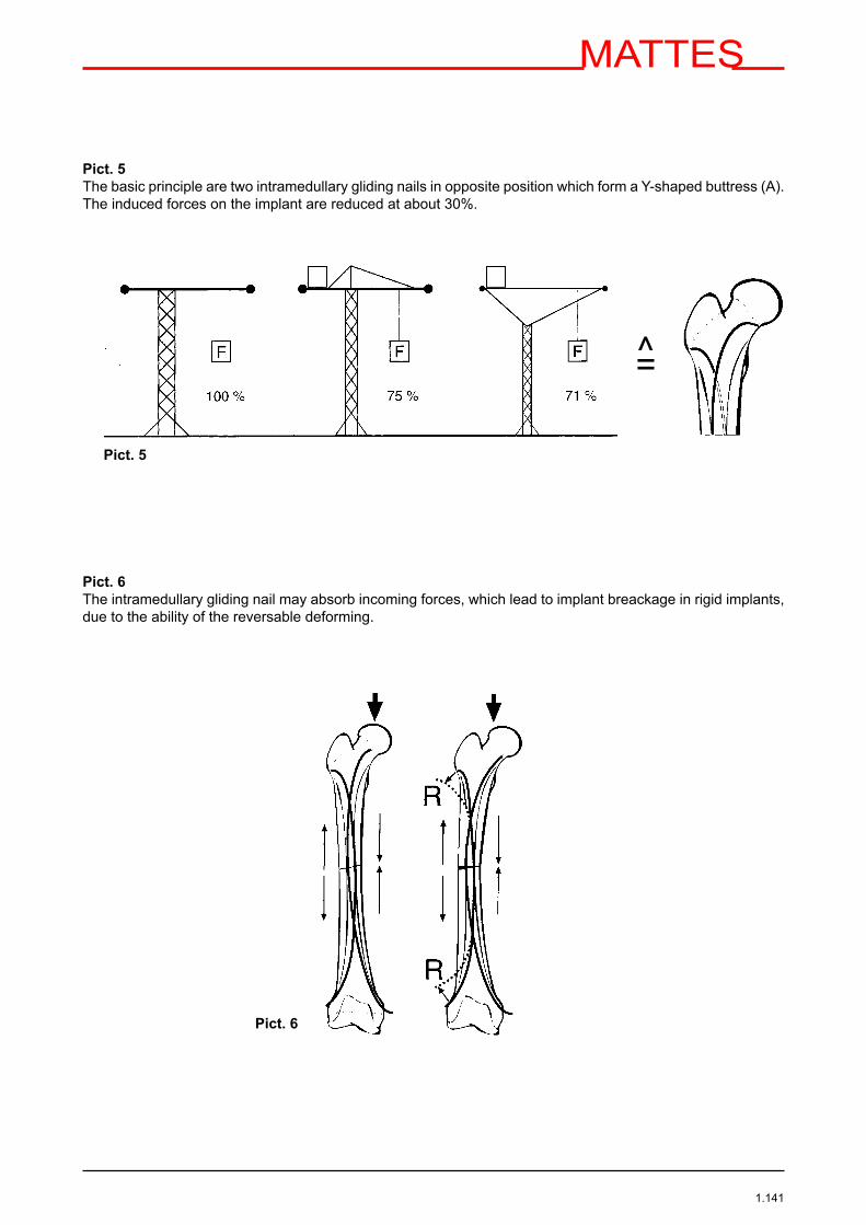
Pict. 5The basic principle are two intramedullary gliding nails in opposite position which form a Y-shaped buttress (A). The induced forces on the implant are reduced at about 30%.
Pict. 6The intramedullary gliding nail may absorb incoming forces, which lead to implant breackage in rigid implants, due to the ability of the reversable deforming.
=̂
Pict. 6
Pict. 5
MATTES
1.142
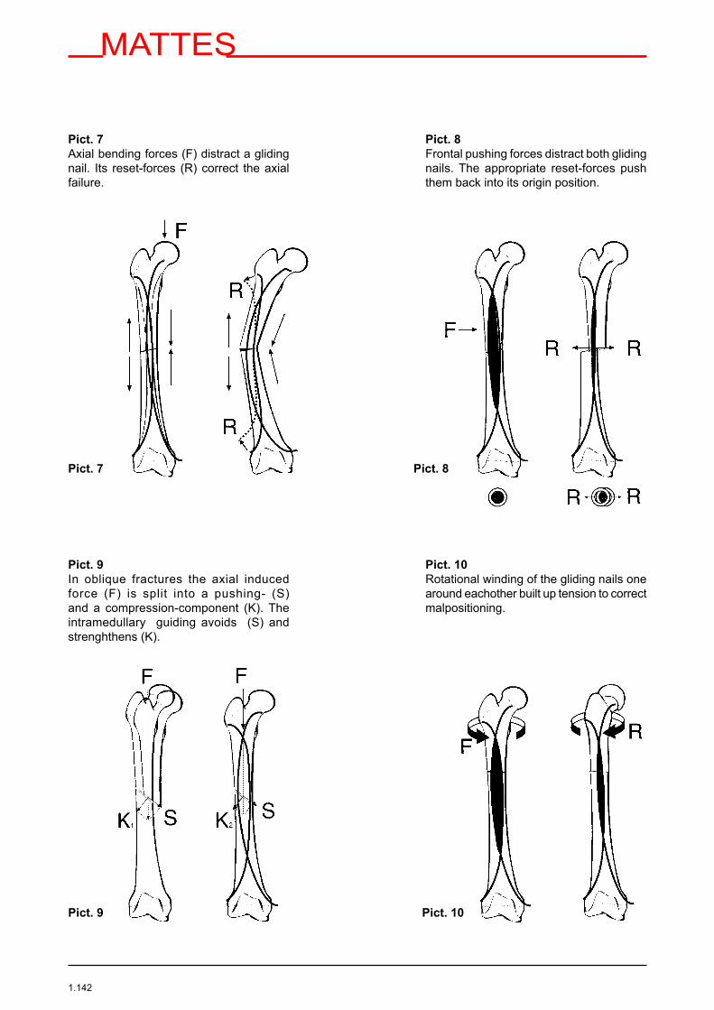
Pict. 7Axial bending forces (F) distract a gliding nail. Its reset-forces (R) correct the axial failure.
Pict. 8Frontal pushing forces distract both gliding nails. The appropriate reset-forces push them back into its origin position.
Pict. 9In oblique fractures the axial induced force (F) is split into a pushing- (S) and a compression-component (K). The intramedullary guiding avoids (S) and strenghthens (K).
Pict. 10Rotational winding of the gliding nails one around eachother built up tension to correct malpositioning.
Pict. 7 Pict. 8
Pict. 9 Pict. 10

MATTES
1.143
Pict. 11Sagittal pushing forces reduce the contact- area within the intramedullary canal and provoque, due to induced tension, the reset of the fragments. Are there excentric sagittal forces it leads to ante- or recurvation which will be corrected by the elasticity of the implants.
Pict. 12Due to the fact that the oval between the two gliding nails cannot be enlarged, axial compression leads to re-inforcement pressure on the gliding nails against the endost.
Technique:Determination of the thickness size of the gliding nail
Medullary canal diameter in mid-shaft Gliding nail size = —————————————————————— 3
2 x Medullary canal diameter in mid-shaft Forearm = ———————————————————————- 3
Pict. 11 Pict. 12
MATTES
1.144
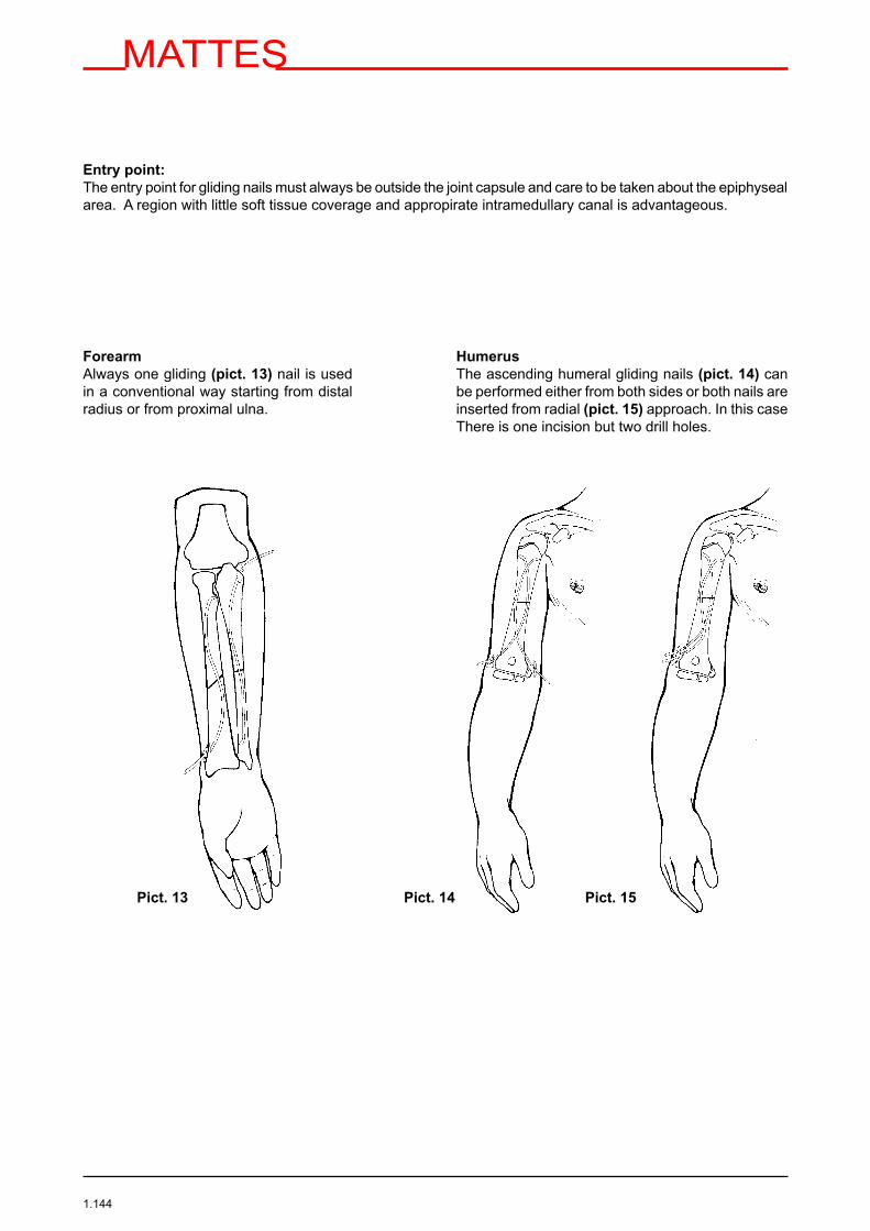
Entry point:The entry point for gliding nails must always be outside the joint capsule and care to be taken about the epiphyseal area. A region with little soft tissue coverage and appropirate intramedullary canal is advantageous.
Forearm Always one gliding (pict. 13) nail is used in a conventional way starting from distal radius or from proximal ulna.
Humerus The ascending humeral gliding nails (pict. 14) can be performed either from both sides or both nails are inserted from radial (pict. 15) approach. In this case There is one incision but two drill holes.
Pict. 14 Pict. 15Pict. 13
MATTES
1.145
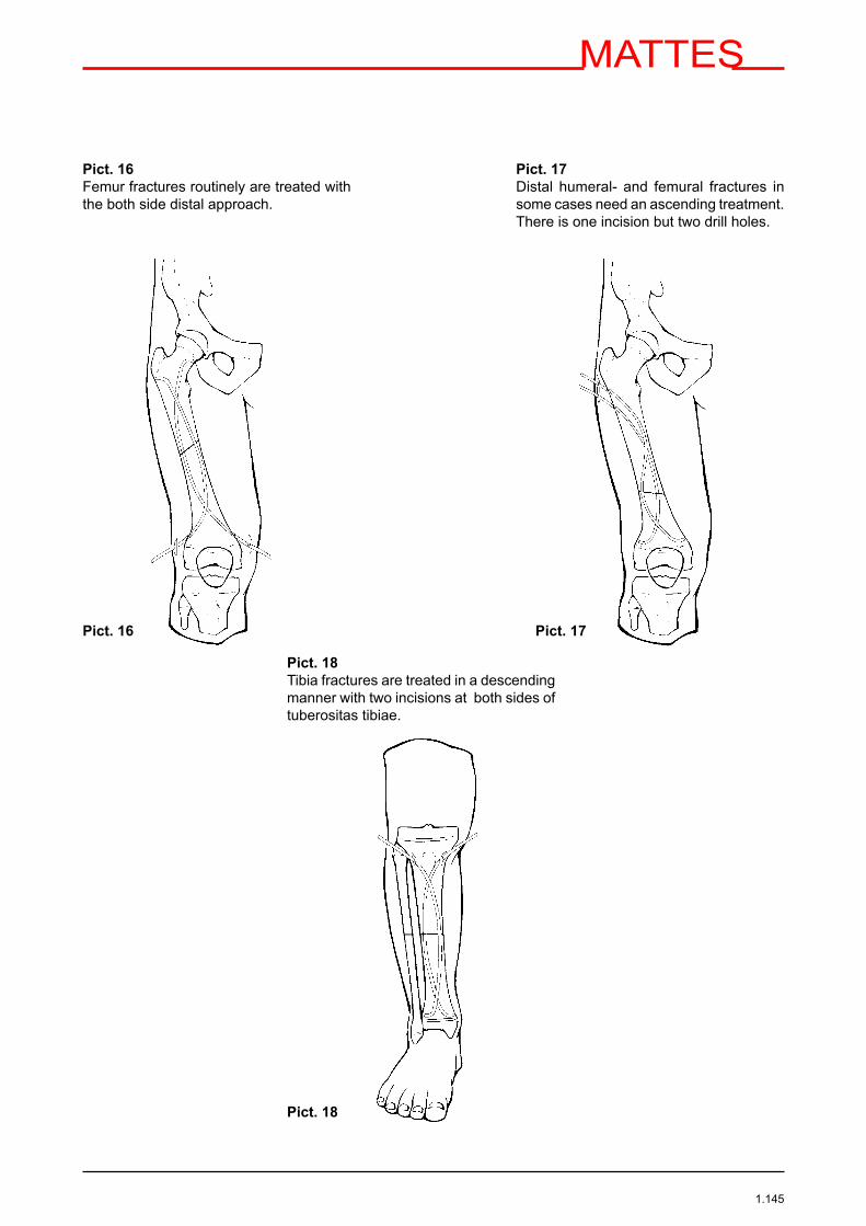
Pict. 16Femur fractures routinely are treated with the both side distal approach.
Pict. 17Distal humeral- and femural fractures in some cases need an ascending treatment. There is one incision but two drill holes.
Pict. 18Tibia fractures are treated in a descending manner with two incisions at both sides of tuberositas tibiae.
Pict. 16 Pict. 17
Pict. 18

MATTES
1.146
OP- TechniqueSkin incision is made from the planned entrance point in epiphyseal direction (pict. 19). A 2 to 3 cm long incision faciliates the following steps.
The entrance point is marked with the trokar which has been put into the sleeve to protect soft tissue. The marking is done in a 90° angulation (pict. 20) to the bone surface. The bone opening can alternatively done either with the trocar or with a drill (pict. 21).
The size of the trocar or drill must be at least 0,5 mm bigger in diameter than the indicated intramedullary nail. The bone perforation is performed in angulation less than 45° (pict. 22). After removal of the trocar or drill the tissue protection sleeve is held in position (pict. 22a). The gliding nail in then introduced through it. The slot of the sleeve (pict. 23) faciliates the introduction of nails with bent tips.
Pict. 19
Pict. 20 Pict. 21
Pict. 22 Pict. 23Pict. 22a
<45°

MATTES
1.147
Implantation of intramedullary gliding nailIs pre-bending of the gliding nail necessary? It is then indicated when the gliding nail in the bone entrance fragment shall early reach the opposite cortical bone, e.g. in those cases when the fragment is relatively short or if the implant sticks on the opposite coritical bone and cannot be moved further on into the medullary canal. Primarily that implant is introduced which leads to the highest reduction effect of the fracture. The gliding nail is introduced through the tissue sleeve into the intramedullary canal. The gliding nail then must be turned in the way that the tip shows in direction to the medullary canal and not to the cortex.
Pict. 24With the T-handle with Jacobs Chuck the gliding nail is shortly fixed and introduced step by step. This faciliates the introduction and avoids unintended bending of the implant.
Pict. 25Is there little fragment contact at the fracture side, the tip of the gliding nail can be aligned while twisting the gliding nail.
Tip:Often it is useful, that prior to the transfer of the first gliding nail from one side of the fracture to the other one that the second gliding nail has been inserted and being pushed onto the borderline of the fracture. There are now two starting points given which can be used for reduction in detail.
Pict. 24 Pict. 25

MATTES
1.148
Pict. 26To introduce the second gliding nail in the opposite fragment easily, the first gliding nail can be twisted to correct fragment position.
Pict. 27The implant is fixed in the metaphyseal spongiosa of the counter fragment with some soft impacts with the hammer.
Pict. 28Slight misalignments in axial position can be corrected by restricted twisting manoevers of the gliding nail.
Tip:A possible distraction at the fracture line which may occur due to the implantation can be corrected through axial compression. Afterwards the implants are impacted.Finally the gliding nail will be cut subcutaniously with the wire cutter. A protective cap is put onto the sharp end of the gliding nail to prevent soft tissue irritation and nail perforation.
Pict. 26
Pict. 27 Pict. 28

MATTES
1.149
Pict. 29In oblique fractures that gliding nail is easier to implant which nail tip is 90° to the fracture area.
Pict. 30In spiral fractures primarily the implantation at the side with the long coritical bone is recommended.
Implant removalImplant removal can be done in outpatient treatment. The end of the gliding nail is exposed in the conventional way. The protection cap is removed with a forceps (pict. 31). The implant is grasped with an adjustable extraction forceps (pict. 32), with a longitudinal groove to cover the implant.
Pict. 29 Pict. 30
Pict. 31 Abb. 32

MATTES
1.150
Pict.35Twisted implants one around eachother avoid the adequate tension fixation and the necessary elasticity.
Pict. 34Too short or too thin used implants are not able to fulfil the demands of the elastic- stable treatment with intramedullary gliding nails.
Pict. 33The assymetrical implantation leads to two tension bows and therefore can induce malposition.
Implantation Problems
Pict. 33 Pict. 34
Pict. 35
MATTES
1.151
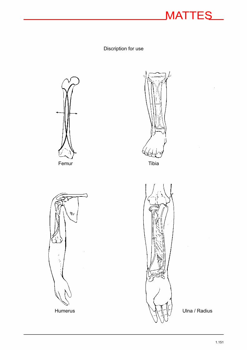
Discription for use
Femur Tibia
Humerus Ulna / Radius

MATTES
1.152