Embed Size (px)
Citation preview

マイクロプレート凝集反応を用いたブルセラカニスのイヌにおける血清疫学
誌名誌名 The journal of veterinary medical science
ISSNISSN 09167250
著者著者
木村, 昌伸今岡, 浩一鈴木, 道雄ほか2名,
巻/号巻/号 70巻7号
掲載ページ掲載ページ p. 707-709
発行年月発行年月 2008年7月
農林水産省 農林水産技術会議事務局筑波産学連携支援センターTsukuba Business-Academia Cooperation Support Center, Agriculture, Forestry and Fisheries Research CouncilSecretariat
NOTE Public Health
Evaluation of a Microplate Agglutination Test (MA T) for Serological Diagnosis of
Canine Brucellosis
Masanobu KlMURA1), Koichi 1MAOKA1h, Michio SUZUKl1), Tsun巴oKAM1YAMA1) and Akio YAMADA1)
J)Department of Veterinary Science, Nat附 wllnst山 teof Infectious D山 ases,/-23-1Toyama, Shinjuku-ku, Tokyo 162-8640, Japan
(Received 12 November 2007/Accepted 19 February 2008)
ABSTRA汀.A microplate agglutination test (MAT) was compared with the tube agglutinin test (TAT), a standard test for the diagnosis of 8rucella canis, in terms of the sensitivity and specificity. The results showed that MAT was more sensitive, simpler to perform and easier to read the results than TAT. On top of出創出eMA T a1lows us to handle a larger number of samples at once. Using this method we conducted sero-surveillance of the prevalence of 8. canis in dogs kept in an Animal Shelter located in Kanagawa Prefecture. Twelve of 485 (2.5%) showed seropositive against 8. canis. These results indicate that B. canis infection in dogs is still occurring in Japan. 阻 YWORDS・8rucellacanis, canine bruc巴llosis,microplate agglutination t巴st
Brucellosis, one of th巴 majorzoonoses worldwide, is caused by a bacteria belonging to the genus Brucella [4] Among many species of the genus Brucella, B. melitensis, B.αbゅrtus,B. suis and B. canis are known to result in human brucellosis. Although Brucella spp. with smooth-type lipopolysaccharides (LPS), such as B. melitensis, B. abortus and B. suis, are known to infect several domestic animals, such as cows, sheep, goats and pigs, B. canis, one of Bru-cella spp. with rough LPS, infects a limit巴dhost range, such as dogs and wild canidae. B. canis infection in dogs is usu-ally asymptomatic but can sometimes cause contagious abortion, epididymitis, testicular atrophy and infertility [3]. Most canine infections occur by di陀 ctcontact with lochia at the tIme of abortion or vaginal discharges in infected female dogs. Semen and urine from infected male dogs have also been implicated as sources of infection [7]. Drug therapy for B.. canis infection requires an appropriate regimen of antibiotic combination, but relapse may ensue, because B.
canis often persists within macrophages or other type of cells [3]. Humans are ra陀 Iyinfected with B. canis. Most human infections are asymptomatic; however, several c1ini-cal symptoms, which are milder than those observed with other Brucella spp. , are sometimes noticed [12].
1n Japan, B. canis infection was first reported in a breed-ing colony of beagles in 1972 [20]. Several epidemiological studies of canine brucellosis in Japan were conducted in th巴1970s and 1980s [10,11,15-18], but th巴rehave only been a few reports since then. ln 2003 and 2006, canine brucellosis emerged as outbreaks in large br田:dingcolonies, suggesting that B. canis infection is still enzootic in Japan. To assess the possible risk of B. canis on human, detβrmination of the prevalence of B. canis in the dog population in Japan seemed helpful.
Although tube agglutination test (T AT) is the most widely used laboratory test for the detection of B. canis anti-
*Co限 ESPONDENCETO: IMAoKA, K., D巴partmentof Veterinary Sci-ence, National Institute of Infectious Diseases, 1-23ー1Toyama, Shinjuku-ku, Tokyo 162-8640, Japan. 巴-mail:[email protected]
J. Vet. Med. Sci. 70(7)・707-709,2008
bodies in both humans and canines, it is time-consuming and cumbersome in terms of performance and measurement of results [2]. On the other hand, microplate agglutination test (MAT) described for B. canis [5] and B. abortus [1, 6] appear巴dadvantageous, because a larger number of samples can be processed simultaneously by this method. ln the present study, we attempted to evaluate wh巴therthe use of MAT with safranine-stained bacterial cells as antigens could serve as a substitute for T AT to conduct sero-epidemi-ologic investigations of canine brucellosis in Japan.
TAT was carried out by placing 0.5 ml of 2-fold serially diluted sera and an equal volume of B. canis antigen solu-tion (OD600= 1) purchased from the Kitasato 1nstitute (Tokyo, Japan) in glass tubes. After incubation at 500C for 24 hr, the agglutination titer was determined and exprl巴ssedas a reciprocal of final serum dilutions, which gave rise to agglutination as observed in the 50% control tube. Titers of 160 or higher were considered positive. Anti-B. canis anti-
body was prepared in our laboratory by immunizing a rabbit with inactivated B. canis whole antigen and was included as
a reference. MAT was performed as follows. First, serum samples, 2-
fold serially diluted in phosphate-buffered saline, were pre-pared in a 96-well U-bottom microplate. Then, an equal volume (25μl) of B. canis antigen solution (Kitasato 1nsti-tute), which is same as used in TAT, containing 0.005% saf-ranine solution (2% of Favor G@, Nissui Pharmaceutical Co., Tokyo, Japan) was added to each well. The sealed plates were mixed gently for 20 sec and incubated at 500C for 24 hr in a humid atmosphere. The titers were expressed as a reciprocal of the highest dilution of sera showing agglu-tination. Safranine-stained antigens made it possible to judge the results more easily and objectively. An agglutina-tion titer greater than 160 was considered positive.
We have experienced an outbreak of B. canis infection in 2003 [8]. Sera obtained from dogs involved in the outbr巴akwere examined for the presence of anti-Brucella antibody by T A T. Fifty-one of 110 sera tested positive for antibody against B. canis. These sera were subjected to MAT for
708 ルf.KI恥1URAET AL.
xl0240 y-o.32487+ 1∞48x
R̂ 2=O.894
1 • n
u
n
u
““
2
x
×
(H〈冨)HS戸
3 • 3. 3 •
x160
<x160 Eミヨ
<x160 x160 x2560 xl0240 x640
Titer (TAT)
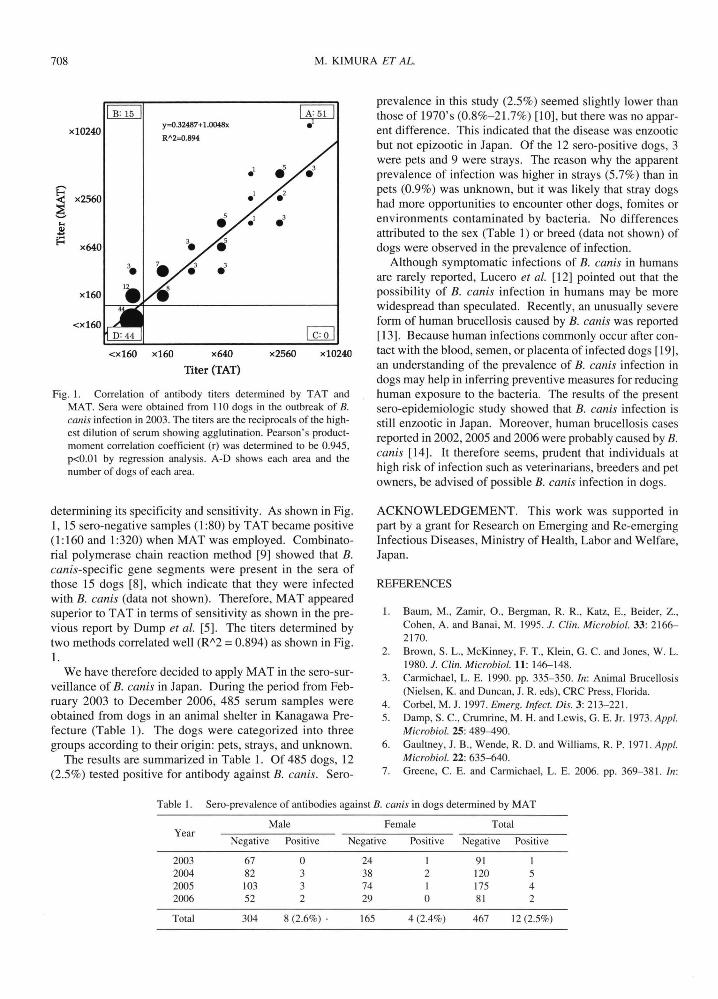
Fig. 1. Correlation of antibody titers determined by TAT and MAT. Sera were obtained from 110 dogs in th巴 outbre紘 ofB.
canis infection in 2003. The titers are出巴 reciprocalsof the high-est dilution of serum showing agglutination. Pearson's product-moment correlation coefficient (r) was d巴terminedto be 0.945, p<O.O 1 by regression analysis. A-D shows each area and the number of dogs of each area.
determining its specificity and sensitivity. As shown in Fig.
1,15 sero-negative samples (1:80) by TAT became positive
(1:160 and 1:320) when MAT was employed. Combinato-
rial polymerase chain reaction method [9] showed that B
canis-specific gene segments were present in the sera of
those 15 dogs [8], which indicate that they were infected with B. canis (data not shown). Therefore, MAT appeared
superior to T AT in terms of sensitivity as shown in th巴pre-
vious report by Dump et al. [5]. The titers d巴terminedby
two methods correlated well (R八2= 0.894) as shown in Fig.
We have therefore decided to apply MAT in the sero-sur-
veillance of B. canis in Japan. During the period from Feb-
ruary 2003 to December 2006, 485 serum samples were
obtained from dogs in an animal shelter in Kanagawa Pre-fecture (Table 1). The dogs were categorized into three
groups according to their origin: pets, strays, and unknown. The results are summarized in Tabl巴1.Of 485 dogs, 12
(2.5%) tested positive for antibody against B. canis. Sero-
prevalenc巴 tn出isstudy (2.5%) seemed slightly lower than
those of 1970's (0.8%-21.7%) [10], but there was no appar-
ent differenc巴. This indicat巴dthat the disease was enzootic
but not epizootic in Japan. Of the 12 sero-positive dogs, 3
were pets and 9 were strays. The reason why the apparent
prevalence of infection was higher in strays (5.7%) than in
pets (0.9%) was unknown, but it was likely that stray dogs
had more opportunities to encounter oth巴rdogs, fomites or environments contaminated by bacteria. No differences
attributed to the sex (Table 1) or breed (data not shown) of
dogs were observed in th巴prevalenceof infection.
Although symptomatic infections of B. canis in humans
are rarely report巴d,Lucero et al. [12] pointed out that the
possibility of B. canis infection in humans may be more
widespread than speculated. Recent1y, an unusually severe form of human brucellosis caused by B. canis was r巴ported
[13]. Because human infections commonly occur after con-
tact with the blood, semen, or placenta of infected dogs [19], an understanding of th巴preval巴nceof B. canis infection in
dogs may help in inferring preventive measures for reducing
human exposure to th巴bacteria.The results of the present
sero叩 iderniologicstudy showed that B. canis infection is
still enzootic in Japan. Moreover, human brucellosis cases
reported in 2002, 2005 and 2006 were probably caused by B. canis [14]. It therefore s巴巴ms,prudent that individuals at
high risk of infection such as veterinarians, breeders and pet owners, be advised of possible B. canis infection in dogs.
ACKNOWLEDGEMENT. This work was supported in
part by a grant for Research on Emerging and Re-emerging
Infectious Diseases, Ministry ofHealth, Labor and Welfare,
Japan.
REFERENCES
1. Baum, M., Zamir, 0., Bergman, R. R., Katz, E., Beider, Z.,
Coh巴n,A. and Banai, M. 1995. J. Clin. Microbiol. 33: 2166-2170.
2. Brown, S. L., McKinney, F. T., K1ein, G. C. and Jones, W. L. 1980. J. Clin. Microbiol. 11: 146ー148.
3. Carmichael, L. E. 1990. pp. 335-350. ln: Animal Brucellosis (Ni巴Isen,K. and Duncan, J. R. eds), CRC Press, Florida.
4. Corbel, M. J. 1997. Emerg. lnfect. Dis. 3: 213-221. 5. Damp, S. C., Crumrin巴, M.H. and Lewis, G. E. Jr. 1973. Appl
Microbiol. 25: 489-490. 6. Gaultney, J. B., Wende, R. D. and Williams, R. P. 1971. Appl
Microbiol. 22: 635-640 7. Greene, C. E. and Carmichael, L. E. 2006. pp. 369-381. ln:
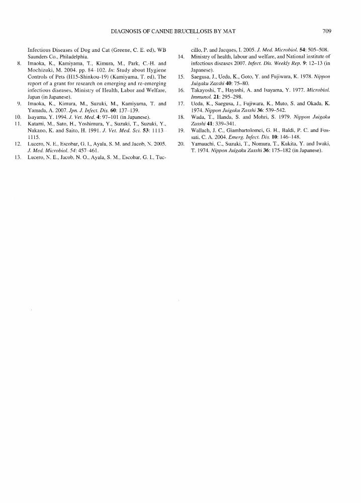
Table 1. Sero-pr巴valenceof antibodies against B. canis in dogs determined by MAT
Male F巴male Total Year
Negative Positive Negative Positiv巴 Negative Positive
2003 67 。 24 91 2004 82 3 38 2 120 5 2005 103 3 74 175 4 2006 52 2 29 。 81 2
Total 304 8 (2.6%)・ 165 4(2.4%) 467 12 (2.5%)
DIAONOSIS OF CANINE BRUCELLOSIS BY MAT 709
Infectious Diseases of Dog and Cat (Oreene, C. E. ed), WB Saund巴rsCo., Philad巴Iphia
8. Imaoka, K., Kamiyama, T., Kimura, M., Park, C.-H. and Mochizuki, M. 2004. pp. 84-102. In: Study about Hygiene Controls of Pets (H 15-Shinkou-19) (Kamiyama, T. ed), The
report of a grant for research on emerging and re-emerging
infectious diseases, Ministry of Health, Labor and Welfare,
Japan (in Japanese)
9. Imaoka, K., Kimura, M., Suzuki, M., Kamiyama, T. and
Yamada, A. 2007. Jpn. J. Infect. Dis. 60: 137ー13910. Isayama, Y. 1994. J. Vet. Med. 4: 97ー101(in Japanes巴).
11. Katami, M., Sato, H., Yoshimura, Y., Suzuki, T., Suzuki, Y.,
Nakano, K. and Saito, H. 1991. J. Vet. Med. Sci. 53: 1113 1115.
12. Lucero, N. E., Escobar, 0.1., Ayala, S. M. and Jacob, N. 2005
J. Med. Microbiol. 54: 457-461.
13. Lucero, N. E., Jacob, N. 0., Ayala, S. M., Escobar, O. 1., Tuc-
cillo, P. and Jacques, 1. 2005. J. Med凡1icrobiol.54: 505-508.
14. Ministry of health, labour and welfar巴,and National institute of
infectious diseases 2007. Infect. Dis. Weekly Rep. 9: 12-13 (in Japanese)
15. Saegusa, J., Ueda, K., Ooto, Y. and Fujiwara, K. 1978. Nippon
Juigaku Zasshi 40: 75-80. 16. Takayoshi, T., Hayashi, A. and Isayama, Y. 1977. Microbiol.
Immunol. 21: 295-298.
17. U巴da,K., Saegusa, J., Fujiwara, K., Muto, S. and Okada, K.
1974. N,伊'ponJuigaku Zasshi 36: 539-542 18. Wada, T., Handa, S. and Mohri, S. 1979. Nippon Juigaku
Zasshi41: 339-341 19. Wallach, J. C., Oiambartolomei, O. H., Baldi, P. C. and Fos-
sati, C. A. 2004. Emerg. Infect. Dis. 10: 146-148. 20. Yamauchi, c., Suzuki, T., Nomura, T., Kukita, Y. and Iwaki,
T.1974.NipponJuigakuZasshi36: 175-182 (in Japane則。