Embed Size (px)
Citation preview
同舟共济 守望生命
Optical Coherence Tomography (OCT) for the Coronary Artery Disease Patients with Moderate
Stenosis
上海市同济医院 蒋金法
同舟共济 守望生命
INTRODUCTION
Cardiovascular disease (CVD) causes almost half of all deaths in the developed world and 25% of deaths in the developing world. Coronary artery disease (CAD) is the leading cause of cardiovascular mortality worldwide, with more than 4.5 million deaths occurring in the developing world each year.
By the year 2030, cardiovascular disease will cause an estimated 23.6 million deaths worldwide, becoming the predominant cause of death in the world, surpassing infectious diseases and death caused by cancer.
同舟共济 守望生命
INTRODUCTION
Cardiologists now know that the “vulnerable plaque” is the atherosclerotic lesion defined as the precursor to coronary plaque rupture, hence the proximate cause of acute coronary syndromes (ACS).
Acute coronary syndromes often result from rupture of a modestly stenotic vulnerable plaque,not visible by x-ray angiography.
同舟共济 守望生命
INTRODUCTION
The most common mechanism, occurring in approximately 60-75% of cases is the rupture of a mild to moderately sized plaque in a coronary artery with a necrotic core and thin, structurally weak intimal wall, referred to as a thin-capped fibroatheroma (TCFA).
The presence of a plaque with an echolucent central core and a thin fibrous capsule, the so-called “vulnerable plaque,”can also be identified by IVUS. Whether these lesions should be treated by percutaneous coronary intervention (PCI)versus medical therapy has not been elucidated by clinical trials
同舟共济 守望生命
INTRODUCTION
(OCT) has emerged as one of the most promising investigation modality thanks to its ability to provide unique information about the plaque composition, the thickness of the fibrous cap, the presence of macrophages and tissue collagen composition.
coronary angiography may no longer be the true gold standard for detecting coronary stenosis.Coronary angiography only displays the opacified “silhouette”.
同舟共济 守望生命
INTRODUCTION
An intermediate coronary lesion on angiography is defined as a luminal narrowing with a diameter stenosis 40% but 70%.
All measures of noninvasive assessment of myocardial ischemia are compared with the presence on angiography of a focal stenosis 50% diameter. This cutoff is based on animal
studies and human clinical correlations that demonstrate functional significance (the induction of ischemia) with the anatomic presence of a 50% diameter stenosis .
同舟共济 守望生命
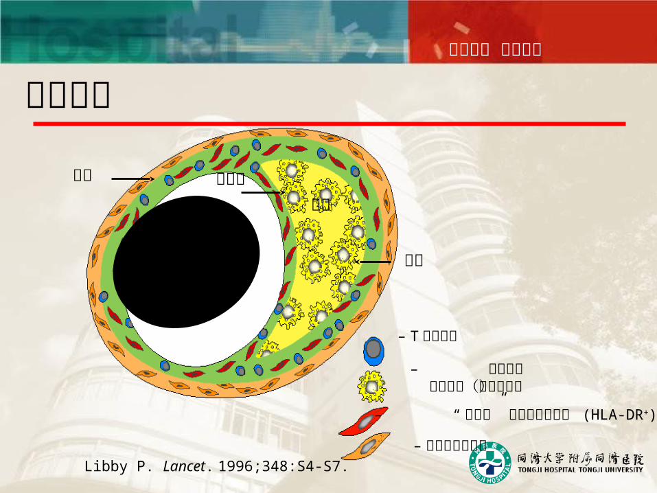
Libby P. Lancet. 1996;348:S4-S7.
中层
– T 淋巴细胞
– 巨噬细胞泡沫细胞(组织因子)
– “激活的” 内膜平滑肌细胞 (HLA-DR+)
– 正常平滑肌细胞
纤维帽内膜脂核
管腔
易损斑块
同舟共济 守望生命
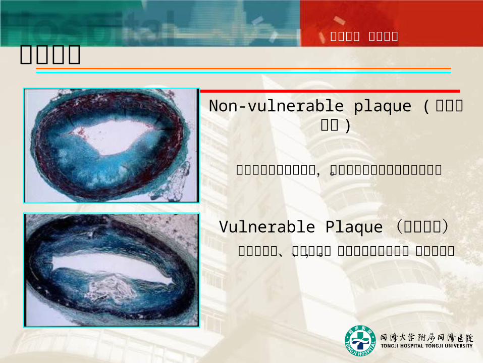
Non-vulnerable plaque ( 非易损斑块 )
纤维组织部分阻塞血流,但不易引起血凝块及心脏事件。
Vulnerable Plaque (易损斑块)富含脂质核、纤维帽薄、边缘炎症反应明
显,易于破裂。
易损斑块
同舟共济 守望生命
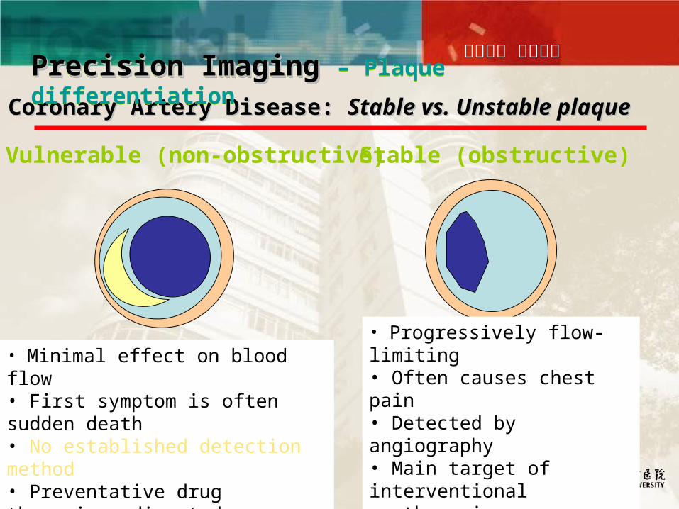
Coronary Artery Disease: Coronary Artery Disease: Stable vs. Unstable plaqueStable vs. Unstable plaque
Stable (obstructive)Vulnerable (non-obstructive)
• Progressively flow-limiting• Often causes chest pain• Detected by angiography• Main target of interventional therapies (angioplasty, stents)
• Minimal effect on blood flow • First symptom is often sudden death • No established detection method• Preventative drug therapies; directed therapies still unproven
Precision ImagingPrecision Imaging – Plaque differentiationPrecision ImagingPrecision Imaging – Plaque differentiation
同舟共济 守望生命
同舟共济 守望生命
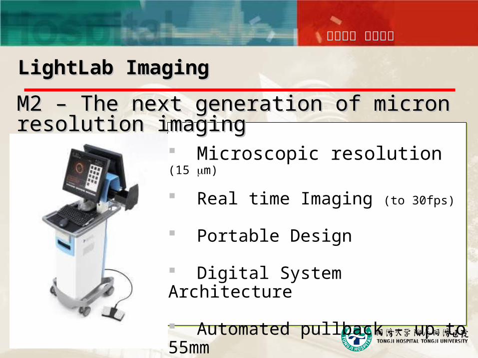
Microscopic resolution (15 m)
Real time Imaging (to 30fps)
Portable Design
Digital System Architecture
Automated pullback – up to 55mm
LightLab ImagingLightLab Imaging
M2 – The next generation of micron resolution M2 – The next generation of micron resolution imagingimaging
同舟共济 守望生命
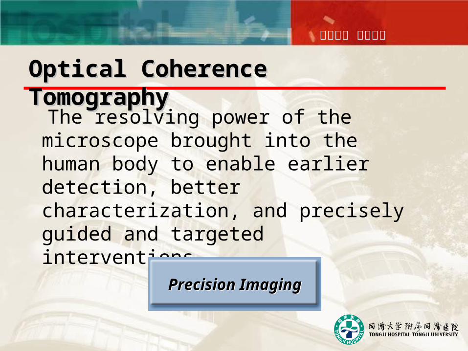
The resolving power of the microscope brought into the human body to enable earlier detection, better characterization, and precisely guided and targeted interventions.
Precision ImagingPrecision Imaging
Optical Coherence TomographyOptical Coherence Tomography
同舟共济 守望生命
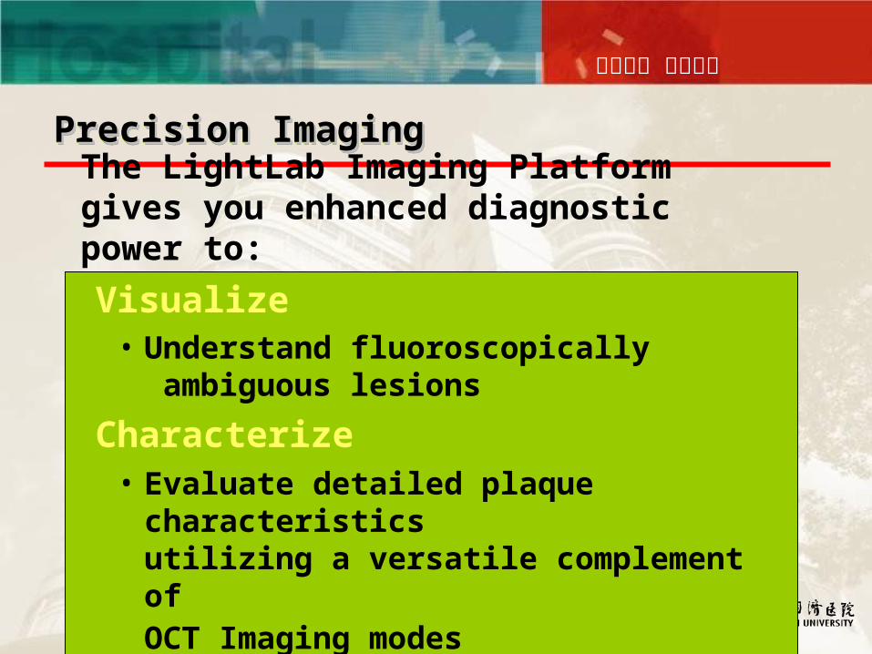
Visualize• Understand fluoroscopically
ambiguous lesions
Characterize• Evaluate detailed plaque characteristics
utilizing a versatile complement ofOCT Imaging modes
The LightLab Imaging Platform gives you enhanced diagnostic power to:
Precision ImagingPrecision ImagingPrecision ImagingPrecision Imaging
同舟共济 守望生命
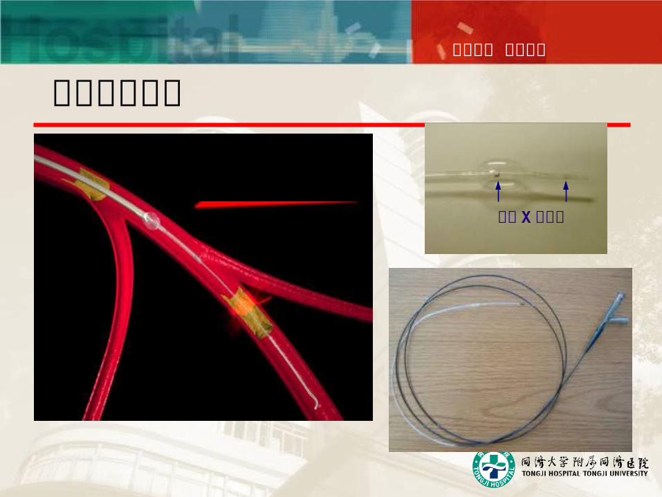
阻断球囊导管
低压力、大直径阻断球囊
不透 X 线标记
同舟共济 守望生命
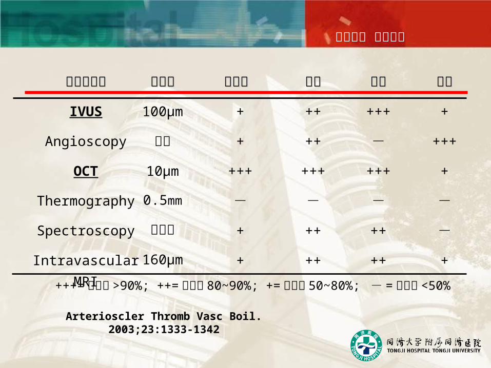
影像学技术影像学技术
IVUS
Angioscopy
OCT
Thermography
Spectroscopy
Intravascular MRI
分辨率分辨率
100μm
不知
10μm
0.5mm
不适用
160μm
纤维帽 纤维帽
+
+
+++
-
+
+
脂核脂核
++
++
+++
-
++
++
钙化钙化
+++
-
+++
-
++
++
血栓血栓
+
+++
+
-
-
+
+++= 灵敏度 >90%; ++= 灵敏度 80~90%; += 灵敏度 50~80%; - = 灵敏度 <50%
Arterioscler Thromb Vasc Boil. 2003;23:1333-1342
同舟共济 守望生命
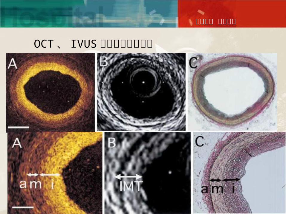
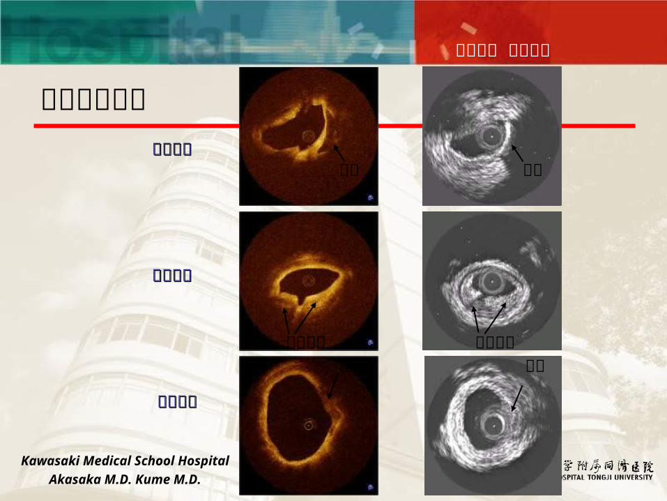
OCT 、 IVUS 与病理解剖的对比
同舟共济 守望生命
Kawasaki Medical School Hospital
Akasaka M.D. Kume M.D.
钙化斑块钙化斑块
纤维斑块纤维斑块
脂质斑块脂质斑块
各种斑块形态
钙化 钙化
纤维斑块 纤维斑块脂池 脂池
同舟共济 守望生命
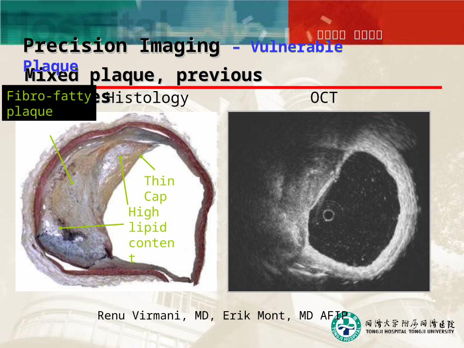
Histology OCTMixed plaque, previous rupturesMixed plaque, previous ruptures
High lipidcontent
Fibro-fattyplaque
Thin Cap
Renu Virmani, MD, Erik Mont, MD AFIP
Precision ImagingPrecision Imaging – Vulnerable PlaquePrecision ImagingPrecision Imaging – Vulnerable Plaque
同舟共济 守望生命
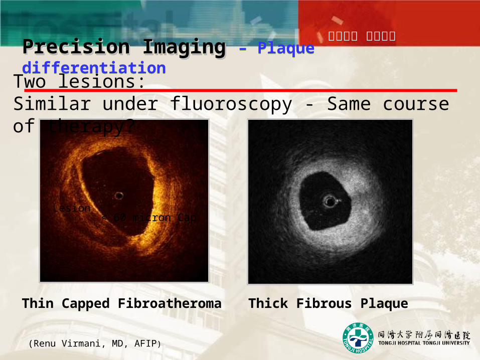
Thick Fibrous PlaqueThin Capped Fibroatheroma
(Renu Virmani, MD, AFIP)
Two lesions: Similar under fluoroscopy - Same course of therapy?
Precision ImagingPrecision Imaging – Plaque differentiationPrecision ImagingPrecision Imaging – Plaque differentiation
< 60 micron CapLesion
同舟共济 守望生命
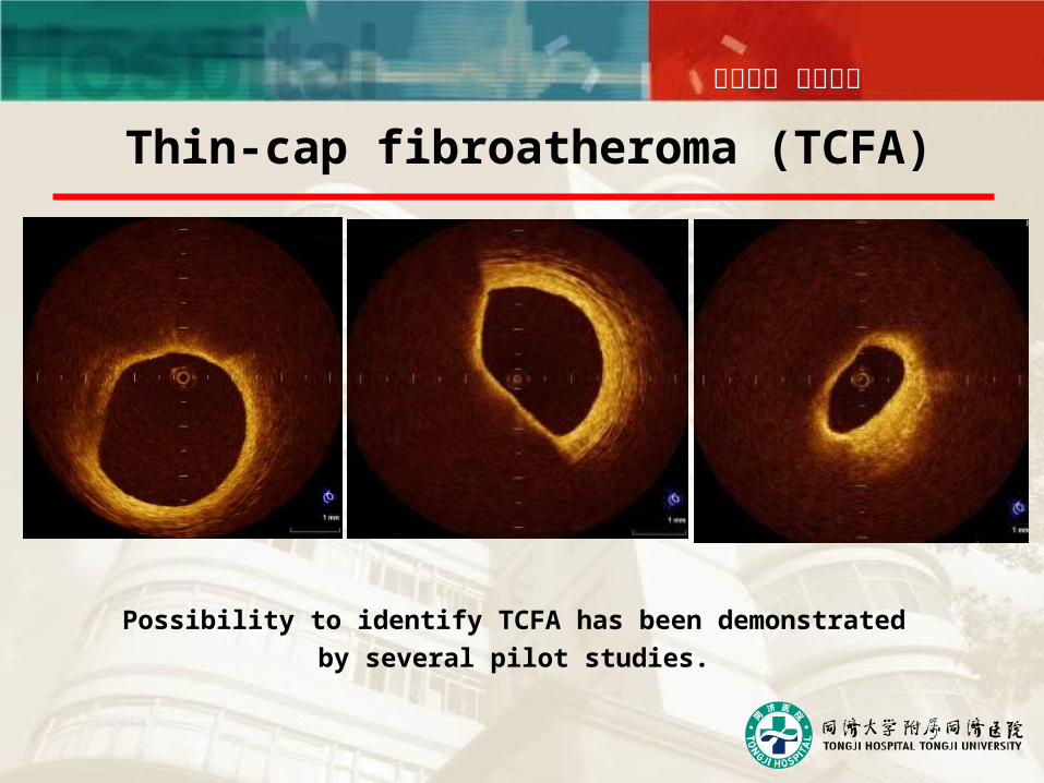
Thin-cap fibroatheroma (TCFA)
Possibility to identify TCFA has been demonstrated
by several pilot studies.
同舟共济 守望生命
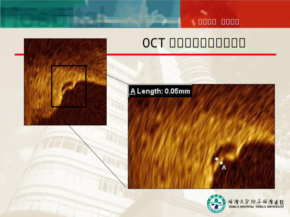
OCT 能精确测量纤维帽厚度OCT 能精确测量纤维帽厚度
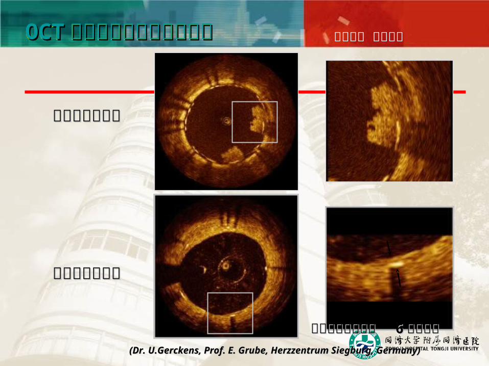
同舟共济 守望生命OCTOCT 能帮助进行支架术后随访能帮助进行支架术后随访OCTOCT 能帮助进行支架术后随访能帮助进行支架术后随访
~0.07mm
((Dr. U.Gerckens, Prof. E. Grube, Herzzentrum Siegburg, Germany)Dr. U.Gerckens, Prof. E. Grube, Herzzentrum Siegburg, Germany)
晚期支架内血栓晚期支架内血栓
支架内内膜增生支架内内膜增生
雷帕霉素药物支架 雷帕霉素药物支架 66 个月随访个月随访
同舟共济 守望生命
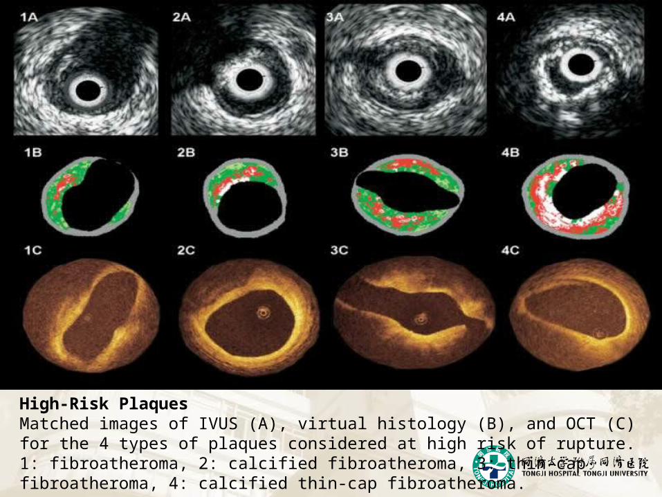
High-Risk PlaquesMatched images of IVUS (A), virtual histology (B), and OCT (C) for the 4 types of plaques considered at high risk of rupture.1: fibroatheroma, 2: calcified fibroatheroma, 3: thin-cap fibroatheroma, 4: calcified thin-cap fibroatheroma.
Precision ImagingPrecision ImagingPrecision ImagingPrecision Imaging
同舟共济 守望生命
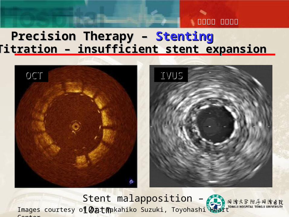
Titration – insufficient stent expansionTitration – insufficient stent expansion Precision Therapy – Precision Therapy – StentingStenting
Stent malapposition – 10atm
OCTOCT IVUSIVUS
Images courtesy of Dr. Takahiko Suzuki, Toyohashi Heart Center
同舟共济 守望生命
同舟共济 守望生命
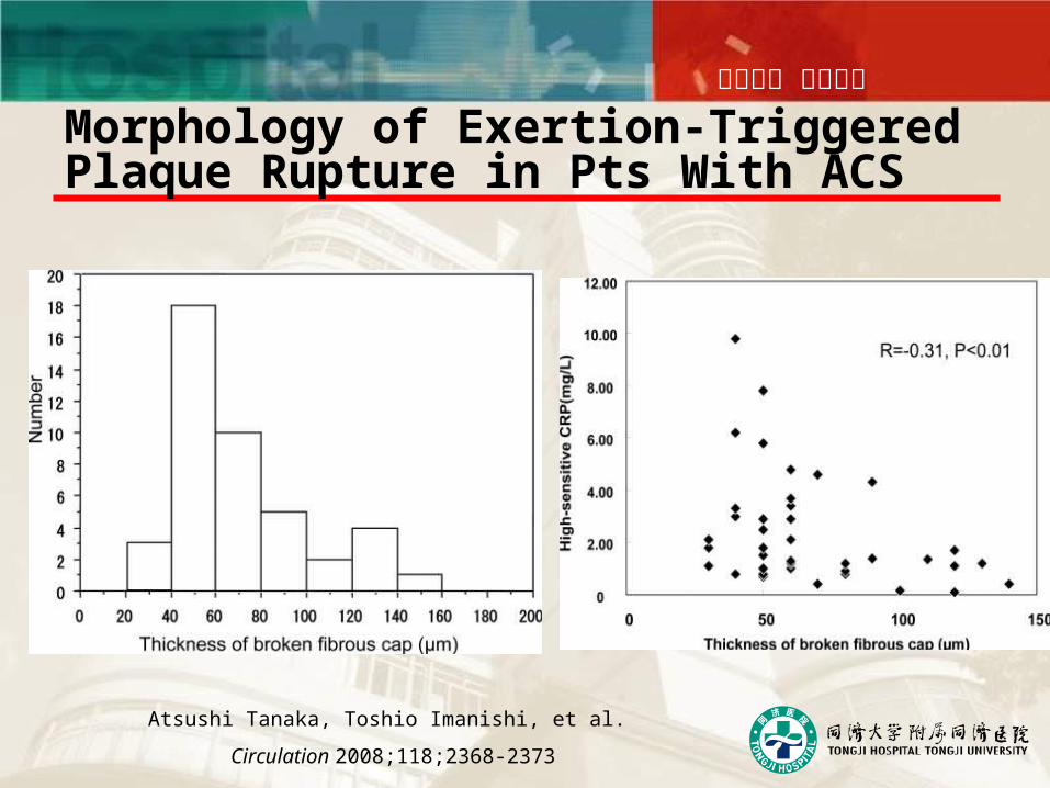
Morphology of Exertion-Triggered Plaque Rupture in Pts With ACS
Atsushi Tanaka, Toshio Imanishi, et al.
Circulation 2008;118;2368-2373
同舟共济 守望生命
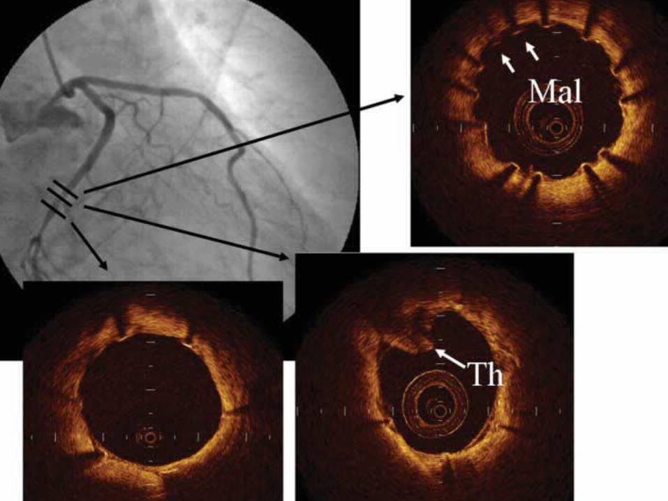
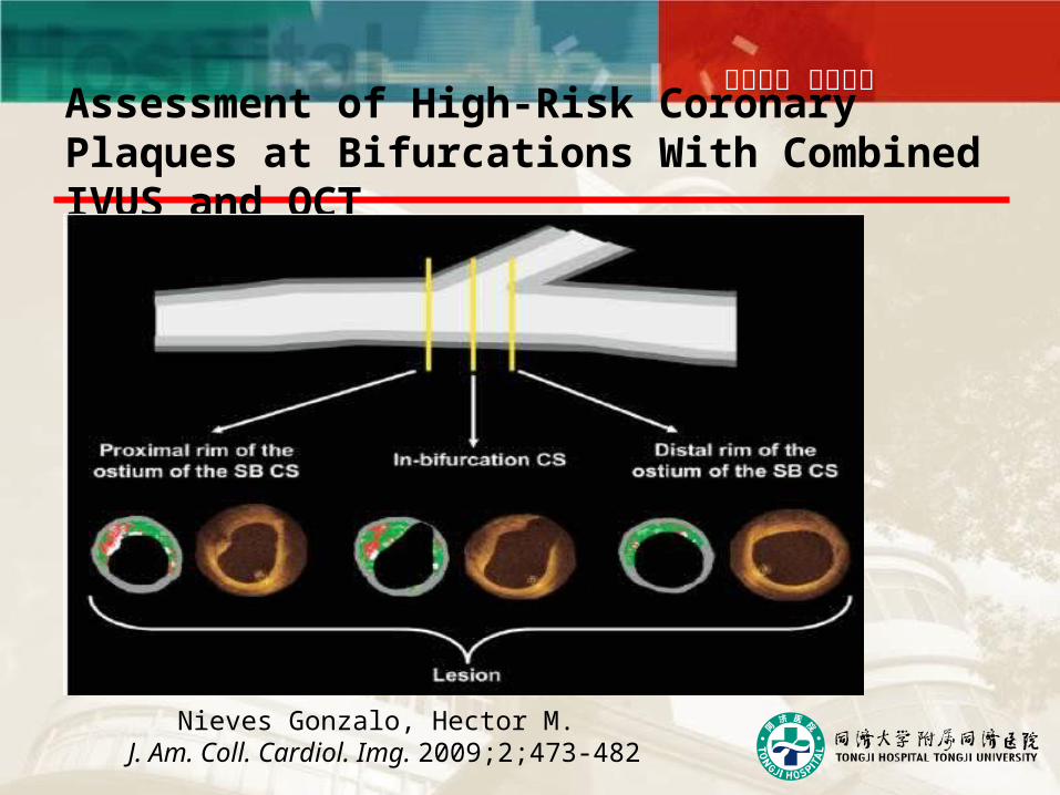
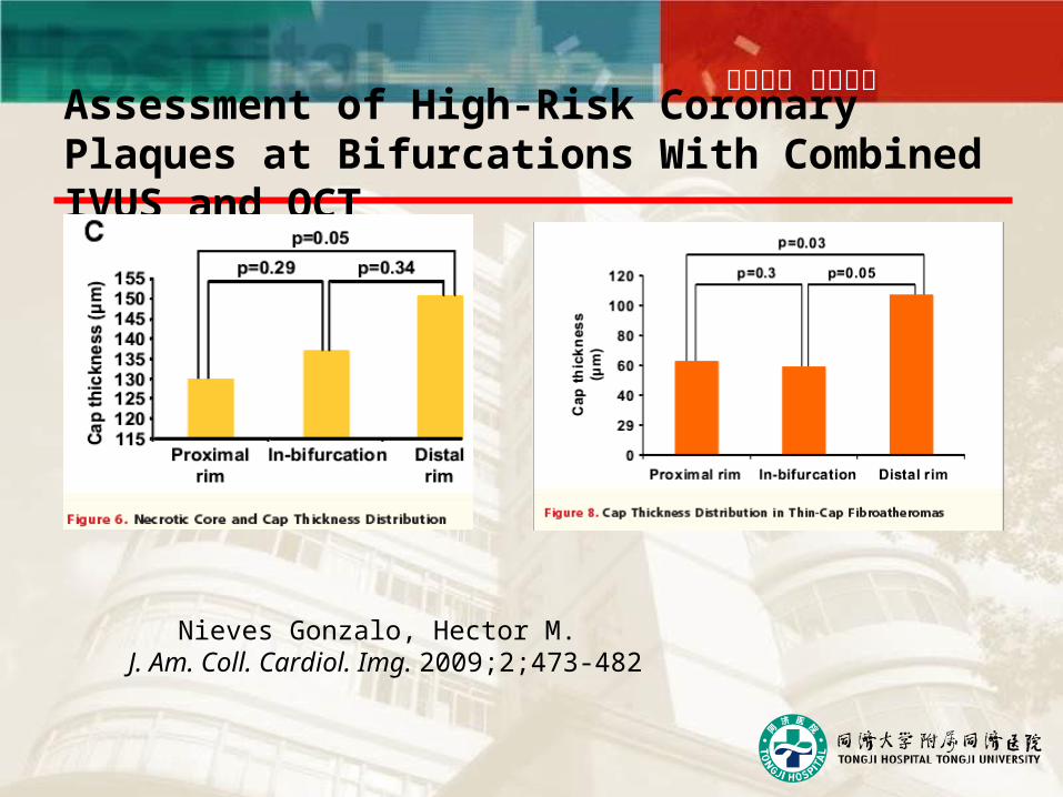
Assessment of High-Risk Coronary Plaques at Bifurcations With Combined IVUS and OCT
Nieves Gonzalo, Hector M. J. Am. Coll. Cardiol. Img. 2009;2;473-482
同舟共济 守望生命
Assessment of High-Risk Coronary Plaques at Bifurcations With Combined IVUS and OCT
Nieves Gonzalo, Hector M. J. Am. Coll. Cardiol. Img. 2009;2;473-482
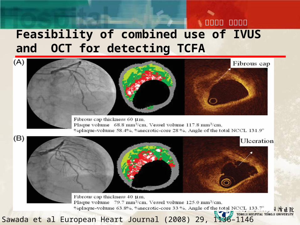
同舟共济 守望生命Feasibility of combined use of IVUS and OCT for detecting TCFA
T. Sawada et al European Heart Journal (2008) 29, 1136–1146
同舟共济 守望生命
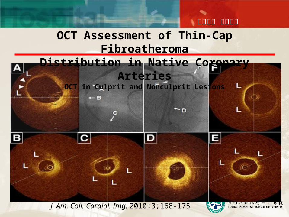
OCT Assessment of Thin-Cap FibroatheromaDistribution in Native Coronary Arteries
OCT in Culprit and Nonculprit Lesions
J. Am. Coll. Cardiol. Img. 2010;3;168-175
同舟共济 守望生命
同舟共济 守望生命
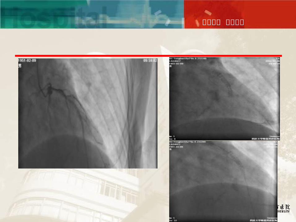
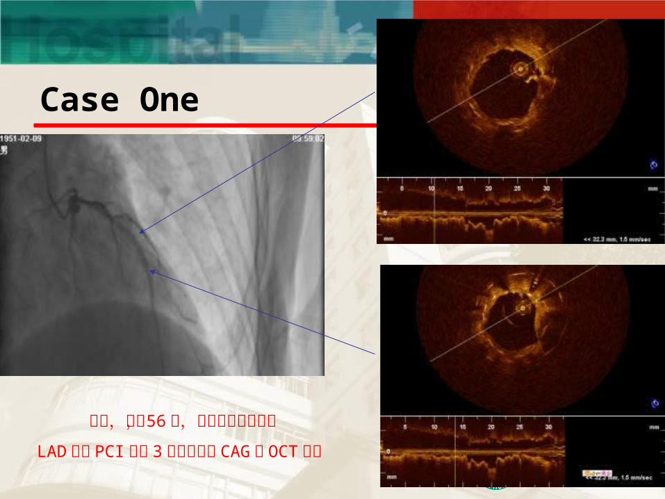
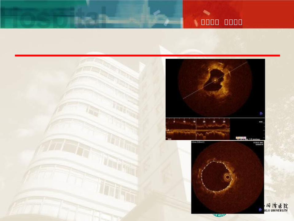
Case One
患者,男, 56 岁,前壁心肌梗死再梗
LAD 再次 PCI 术后 3 个月后复查 CAG 及 OCT检查
同舟共济 守望生命
同舟共济 守望生命
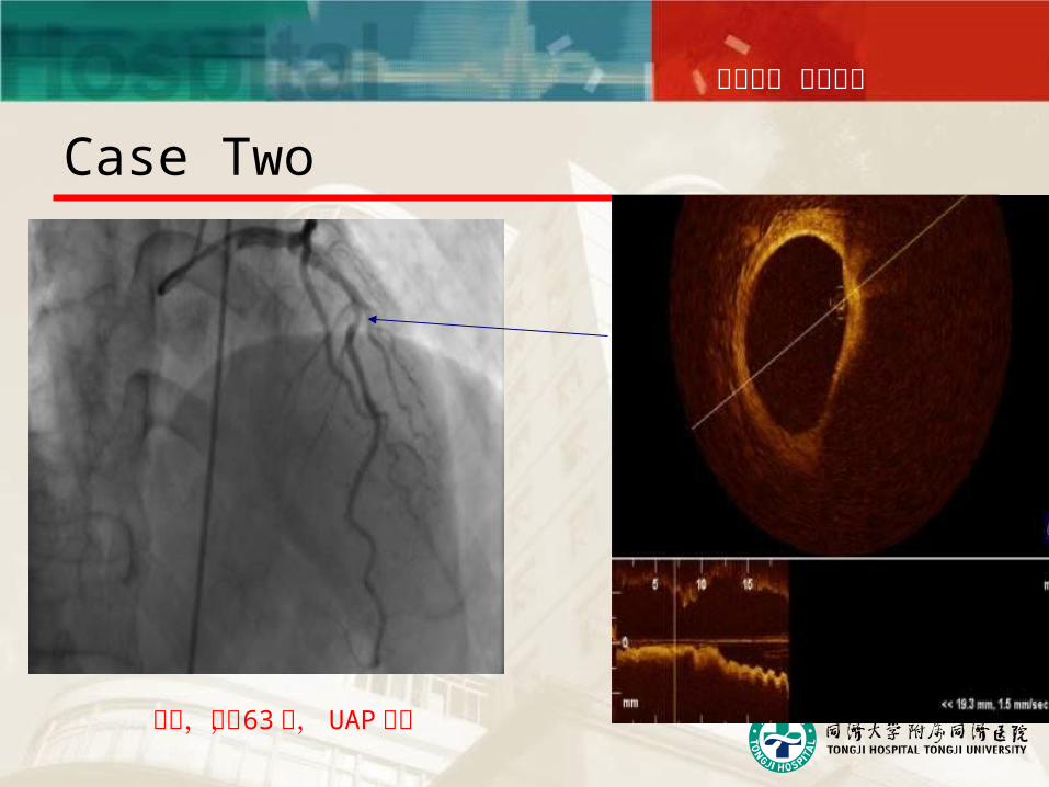
Case Two
患者,男, 63 岁, UAP 入院
同舟共济 守望生命
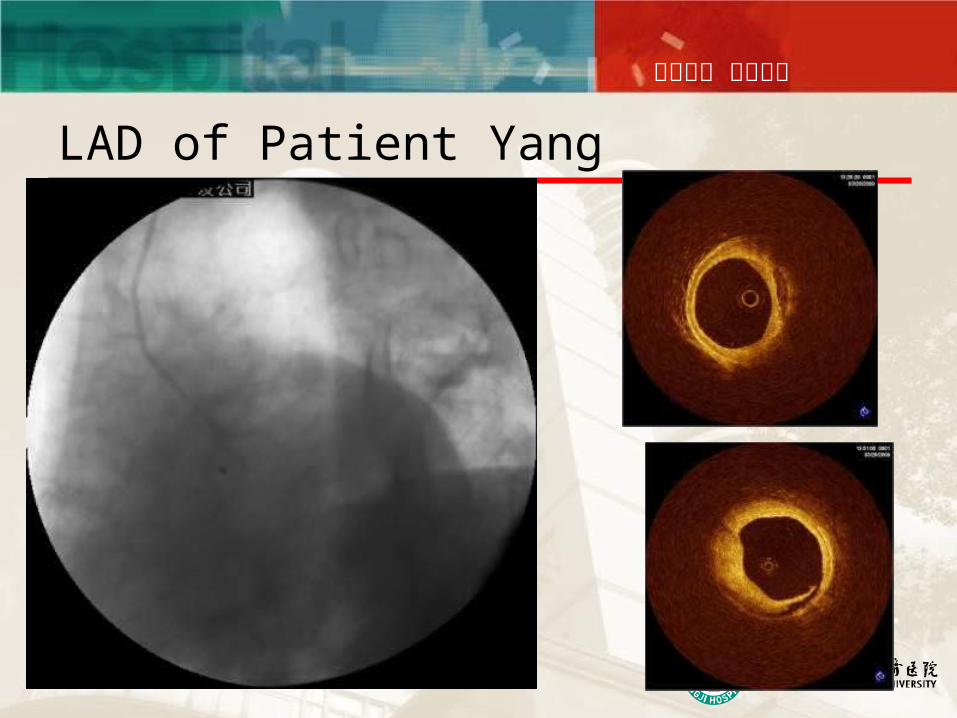
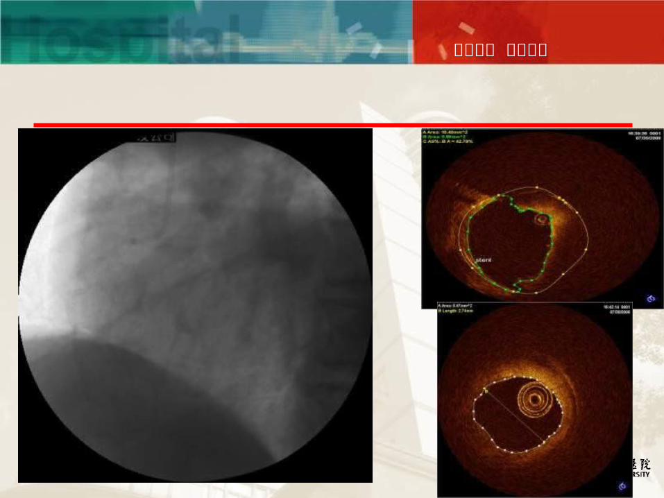
LAD of Patient Yang
同舟共济 守望生命
同舟共济 守望生命
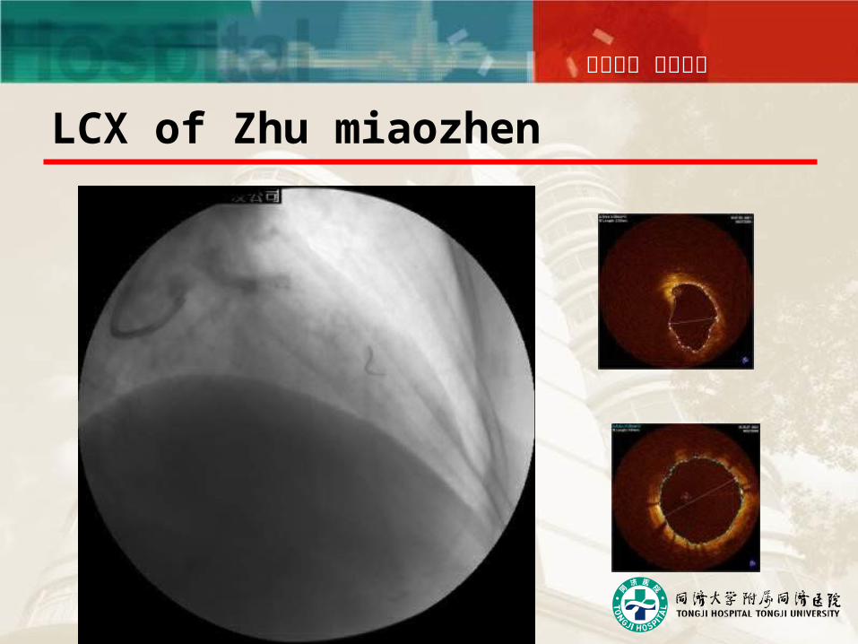
LCX of Zhu miaozhen
同舟共济 守望生命
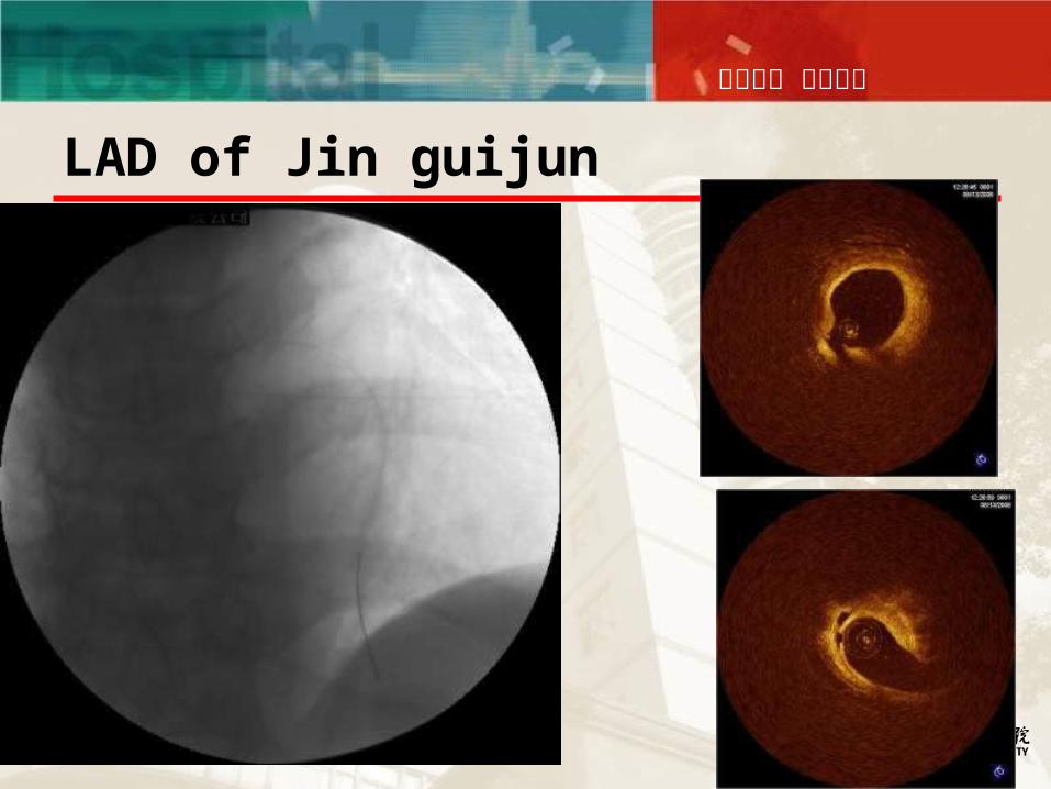
LAD of Jin guijun
同舟共济 守望生命
CONCLUSION
a powerful new tool to assist in diagnosis and treatment of coronary artery disease
Visualization of Neointimal Growth in Drug Eluting Stents
Visualization of Thin-Capped Lesions and Ruptured Plaques
In-Vivo 15μm Imaging allows for development of new areas of invesigation

同舟共济 守望生命