Embed Size (px)
Citation preview

Biological Procedures Online Vol. 5 No. 1 May 1, 2003 www.biologicalprocedures.com
© 2003. Biological Procedures Online. Published in Biological Procedures Online under license from the author(s). Copying,printing, redistribution and storage permitted.
Biol. Proced. Online 2003;5(1): 123-135.
Shotgun Phage Display - Selection for Bacterial Receptins or other Exported Proteins Karin Jacobsson1*, Anna Rosander1, Joakim Bjerketorp1 and Lars Frykberg1 1Department of Microbiology, Swedish University of Agricultural Sciences, UPPSALA, Sweden. *To whom correspondence should be addressed: To whom correspondence should be addressed: Department of Microbiology, Box 7025, SE-750 07 UPPSALA, Sweden. Tel: +46-(0)18-67 32 99; Fax: +46-(0)18-67 33 92; Email: [email protected] Submitted: February 11, 2003; Revised: March 31, 2003; Accepted: April 8, 2003; Published: May 1, 2003 Indexing terms: Peptide Library, Staphylococcus aureus.
ABSTRACT Shotgun phage display cloning involves construction of libraries from randomly fragmented bacterial chromosomal DNA, cloned genes, or eukaryotic cDNAs, into a phagemid vector. The library obtained consists of phages expressing polypeptides corresponding to all genes encoded by the organism, or overlapping peptides derived from the cloned gene. From such a library, polypeptides with affinity for another molecule can be isolated by affinity selection, panning. The technique can be used to identify bacterial receptins and identification of their minimal binding domain, and but also to identify epitopes recognised by antibodies. In addition, after modification of the phagemid vector, the technique has also been used to identify bacterial extracytoplasmic proteins.
INTRODUCTION Phage display The phage display technique, first described by G. Smith in 1985, employs Escherichia coli filamentous phage such as M13, fd and f1, to express proteins or peptides in fusion to one of the coat proteins (1). These phages infect the host by attaching to F-pili, and progeny phage are subsequently secreted without killing the host. All five structural proteins that constitute the phage coat are inserted into the E. coli inner membrane before incorporation into the phage particle (2). All of these have been used for display of foreign peptides or proteins on the phage surface, though most commonly the minor coat protein (pIII) present in 3 to 5 copies, or the major coat protein (pVIII) present in approximately 3000 copies per phage particle, is used. Synthetic or native DNA can be cloned into the gene encoding the coat protein in phage genome, or in
a phagemid. However, cloning into the genome may result in technical problems, e.g. phage infectivity may be impaired if large inserts are present in all copies of pIII, or phage instability if large proteins are expressed in fusion to pVIII. To avoid this, phagemids are used which results in hybrid phage with a mix of wild type and fusion coat protein. Hybrid phage can also be obtained by using modified phage genomes containing two copies of the gene encoding the coat protein, one providing the wild type protein, and one engineered to allow insertion of foreign DNA (3). This eliminates the need for helper phage. When a fusion protein is expressed at the phage surface, phage displaying a certain binding affinity can be isolated from a majority of other phage by an affinity selection procedure known as panning. This procedure involves immobilisation of a ligand in a microwell or on a bead, addition of the phage library, removal of unbound phage and, after elution, infection of E. coli with the eluted phage. After this enrichment for a certain binding affinity, the sequence of the inserted, foreign DNA can be determined and the interaction with the ligand analysed further. The affinity selection replaces the traditional screening with a labelled ligand, or antibody, and enables the analysis of a much larger number of clones in a short time. In a hybrid system, display in fusion to pIII commonly results in less than one copy of the fusion protein per phage particle. For pVIII display, multivalent display is achieved and the number of fusion protein varies, and are believed to depend on a number of different factors, such as the size, the folding and amino acid composition of the foreign protein. Monovalent display may be of advantage when for example a peptide library is selected against a ligand and the strongest interact is sought. Multivalent display may result in selection for low affinity interactions. When panning a gene fragment library, or
Jacobsson et al.
Biological Procedures Online Vol. 5 No. 1 May 1, 2003 www.biologicalprocedures.com
124
a library made from genomic DNA, true interactions, i.e. naturally occurring interactions are sought, and then multivalent display should result in a more efficient selection. However, multivalent display may increase the risk of selecting for weak background interactions. In our hands, panning libraries made from genomic DNA in a gene III-based vector never gave more than 10-20% positive clones, while gene VIII-based display resulted in almost 100% positive clones (4, 5). Shotgun phage display cloning We have applied the phage display technology in our work to identify and characterise staphylococcal receptins. Receptins are microbial proteins, secreted from or attached to the cell, that interact with host components in serum or the extracellular matrix (6) and, are as such, putative vaccine components. Staphylococcus aureus has been reported to interact with many
different ligands and for several of these interactions, the receptin has been identified and the corresponding gene cloned. For example, four different fibrinogen-binding (7-10), two IgG-binding (11, 12), two fibronectin-binding (13, 14), a bone sialoprotein-binding (15) and a collagen-binding protein (16) have been thoroughly characterised. In addition, two different broad-spectrum recognition receptins that bind several different host proteins have been identified, Map (also known as Eap) and Emp (17, 18). In shotgun phage display, randomly fragmented chromosomal DNA is cloned into a phagemid vector. After transformation into E. coli and infection with helper phage, this results in a library consisting of phages together expressing parts of all proteins encoded by the bacterial genome. By panning against an immobilised ligand, or a mixture of ligands, the gene encoding the corresponding receptin can be identified. The procedure is schematically outlined in Figure 1.
Fig. 1: Schematic outline of library construction and panning procedure.
MATERIALS AND METHODS Materials E. coli strain TG1 (SupE thi∆(lac-proAB) F´[traD36 proAB+ LacIq lacZ∆M15]) are grown in liquid or on solid Luria-Bertani (LB)-medium, when required supplied with 50 µg/ml ampicillin (amp). Helper phage R408 are from Promega. MaxiSorp microwell plates (Nunc) are used for immobilisation of ligands for panning. Anti-E-tag antibodies and horse radish-peroxidase labelled anti-mouse antibodies are from Amersham Biosciences.
Library construction Libraries are constructed by ligation of DNA fragments into a phagemid vector. A library representing an entire bacterial genome normally has to be quite large which requires good quality vector DNA and DNA fragments. The vectors we use for making phage display libraries employs the blunt-end restriction site SnaBI for insertion of foreign DNA fragments. The reason for using a blunt-end site is to keep manipulation of the DNA fragments at a minimum, since no addition of linkers or restriction enzyme cleavage of the fragments are required before ligation into the vector.
Jacobsson et al.
Biological Procedures Online Vol. 5 No. 1 May 1, 2003 www.biologicalprocedures.com
126
The chromosomal DNA is fragmented by sonication to the desired size and made blunt-ended with T4-DNA polymerase (Protocol 1). DNA can also be fragmented by DNase treatment or shearing with a gauge needle (19). Since restriction enzyme cleavage is not random, this should be avoided. Sonication has the advantage that a sample can be sonicated for a few seconds, the fragment size analysed by gel electrophoresis, and if the fragments are regarded too long, the sonication can easily be repeated. Thus, fragments of the desired length can easily be obtained. The size desired depends on the intended use of the library. If the main purpose is to map a binding domain, small fragments should be used, while for identification of genes encoding binding proteins, larger fragments are to prefer. Sonication of chromosomal DNA results in a broad range of the DNA fragment size. For identification of genes, we usually aim for fragments in the range of 0.5-3 kbp, which are used without any further size fractionation. This procedure results in a library containing phages displaying polypeptides of varying sizes and thus, from such a library, also proteins containing large binding domains can be isolated. We do not know if there is an upper limit to the size of the protein that can be displayed in fusion to pVIII, but we have isolated several different clones that encode more than 500 amino acids. However, most likely factors other than just the size, such as folding and net charge, may also affect the display at the phage surface. The vector is digested with SnaBI and thereafter 5´-phosphate groups are removed by treatment with calf intestine alkaline phosphatase, to prevent self-ligation of the vector in subsequent ligations. Blunt-end ligations can be troublesome but with the modern ligation kits, large libraries can rather easily be obtained also when fragments with blunt ends are ligated directly into the vector. However, it is also possible to add adaptors to the fragments, and ligate into a vector digested with an enzyme that generates overhangs compatible with the adaptors. In any case, it is important to first ensure that both the vector and the chromosomal fragments ligate well and that the vector cannot recirculate without insert. A large library also requires good electrocompetent E. coli cells with transformation frequencies of 109-1010 transformants per µg plasmid DNA. These frequencies can be obtained by following Protocol 2, but several attempts may be required since the transformation frequency can vary a great deal between batches. Before making the library, a number of test-ligations should be performed to ensure that optimal conditions are used, i.e. that the ratio of vector to DNA fragments gives a high number of clones with an inserted DNA fragment. Thus, several ligations with varying amounts of vector and DNA fragments, respectively, should be made and electrotransformed into E. coli. The fraction of clones containing an insert is then investigated by colony PCR on the obtained transformants. The number of clones obtained from the test ligations are used to estimate how many ligations that are required to obtain the desired size of the library. Usually, the upscaling results in
slightly lower number of clones than could be expected from the tests. The number of clones required to cover a genome can be calculated with the equation: N= ln(1-P)/ln(1-a/b) where N= number of clones required P= probability that a certain fragment is present a= average size of the DNA fragments b= total size of the genome. However, it is important to remember that with this type of library, only one clone in 18 will contain an insert that is in the correct orientation and in the same reading frame as the vector sequences. In addition, in a successful panning experiment, a number of clones with overlapping inserts are desired. Also, it is conceivable that not all variations of a protein are displayed equally well. Thus, the size of the library should exceed the minimum number of clones required several times. When we make a library from S. aureus (2.8 x 10⁶ bp) using fragments of an average size of 1 kbp, we aim at a library of more than 107
clones. Panning procedure (Protocol 3) Usually the ligand is coated onto a microwell at high pH after which any remaining binding sites are blocked with PBS containing 0.05% Tween20. Sometimes, an additional proteineous blocking agent may be required, but then the blocking protein should also be added to library prior to the panning to avoid selection for any phage with affinity for the blocking agent. In the first panning, relatively few phage can be expected to bind the ligand. Therefore, this panning should be allowed to proceed for 4 h at room temperature (RT) or over night at 4°C, while for repannings one hour at RT is sufficient. Panning can also be made in solution using a biotinylated ligand, so called biopanning (20). Here the biotinylated ligand is added to the library, and the mixture is transferred to a streptavidin coated well to capture phage that have reacted with the ligand. This requires that the ligand can be biotinylated and that this does not interfere with the interaction with the corresponding bacterial protein. On the other hand, it is conceivable that in some cases, immobilisation of the ligand directly to the microwell may cause a sterical hindrance that interferes with binding of recombinant phage, which can be avoided by biopanning. To elute bound phage, our standard protocol is to use a citrate buffer (pH 2) for 5 minutes after which the solution is transferred to an Eppendorf tube containing Tris-buffer to ensure an immediate neutralisation of the low pH. The eluted phage should directly be used to infect E. coli TG1 cells, and thereafter the mixture spred on LB/amp-plates. To make sure that single colonies are obtained, varying amounts of the eluate

Jacobsson et al.
Biological Procedures Online Vol. 5 No. 1 May 1, 2003 www.biologicalprocedures.com
127
should be used. The total number of colonies will correspond to the number of bound and eluted phage. These colonies can be screened with a labelled ligand (or for expression of the E-tag, see below). Alternatively, phagemid DNA can be prepared and the sequence of the inserted DNA determined. The phage stock for repanning can either be made in solution, or in soft agar. We prefer the soft agar method since, in our hands, it gives the most reproducible results, and phage stocks with high titres. Another advantage is that differences in growth rate will not have the same impact on the total amount of bacteria produced as when bacteria are growing in solution. Therefore, slow-growing clones will not be completely outcompeted when grown on a plate, and the phage produced by such clones will not be lost to the same extent as when grown in solution. Thus, after panning, the entire eluate is also used to infect E. coli TG1 cells and the mixture spread LB/amp-plates. The day after, resistant colonies are resuspended in a small volume of LB-broth, infected with helper phage, mixed with softagar and poured onto an LB/amp-plate. After overnight growth, the soft agar is collected, mixed with LB-broth and the phage eluted by diffusion into the soluble phase. For details, see Protocol 3. In the second panning, a positive result is usually manifested by a 50 to 1000-fold increase in eluted phage compared the first panning (pure ligand). However, an increase in the eluted phages of approximately 10 times between the first and second panning is occasionally also seen in experiments from which no true binding phage can be identified. Thus, until clones with overlapping inserts are found and the binding confirmed by other means, the result should be viewed with an open mind. Screening for E-tag expression (Protocol 4) In the pG8SAET-vector (Figure 2), the E-tag is in frame with gene VIII, but out of frame with the signal sequence and thus, no expression can be detected. However, insertion of DNA fragments of random length should theoretically restore the reading frame in about one clone in eighteen. Empirically, in a library made in this vector usually 1-2 % of the clones will express the E-tag. After a successful panning, the frequency of clones with an open reading frame from the signal sequence into gene VIII should increase, and subsequently, the frequency of E-tag positive clones should increase. Thus, the frequency of E-tag positive clones is a measure of putative correct clones and is a useful tool for following specific enrichment of clones after panning.
Fig. 2: The pG8SAET-phagemid: The pG8SAET phagemid is a gene VIII-based vector with a universal screening tag, i.e., independently of the ligand used in the panning, putative positive clones can be detected in a colony screening assay by screening for expression of the E-tag. The signal sequence and the E-tag are not in the same reading frame. Instead, insertion of a foreign DNA into either of the cloning sites, NcoI or SnaBI, are required for expression of gene VIII-E-tag-fusion protein. When expressed in a non-supressor strain, protein can be produced without fusion to protein VIII, and recombinant protein purified on a commercially available affinity-column with anti E-tag antibodies (Amersham Biosciences). The complete sequence is available from Genebank AF130864. Primers for sequencing of the inserted DNA: Forward: 5´- TAT CTG GTG GCG TAA CAC CTG CT -3´ Reverse: 5´- GAT CGT CAC CCT CGG ATC CCT AGG -3´ When a library has been made, the frequency of E-tag positive clones should be determined. This is done by picking 150-300 clones, which are transferred to a nitrocellulose filter that is placed on an LB/amp-plate where the colonies are allowed to grow over night. The colonies are lysed in chloroform vapours and the filter is washed, blocked and screened for binding of anti-E-tag antibodies (Protocol 4). Knowledge of the frequency of E-tag positive clones in the original library is useful for comparison with frequencies in panning experiments. In addition, it is also a control of the quality of the library. If the frequency is very low or very high, it is likely that something has gone wrong in the construction. A low frequency may indicate a low frequency of clones with an insert or alternatively, a high frequency of clones with multiple inserts. A high frequency of E-tag clones could indicate a phagemid contamination from an earlier experiment. In a panning experiment, 150 clones are picked after each round of panning and the frequency of E-tag positive clones is determined. These data are especially useful in cases where the panning does not result in a clear-cut enrichment. It can tell whether it is worth continuing the experiment with further pannings, whether clones should be sequenced and in that case, what clones to sequence. The E-tag expression is a useful tool for following the enrichment and finding the clones to be sequenced. However, it is just an indicator and should be interpreted with caution. Sometimes the information can be misleading, see sections Different elution conditions and Pitfalls.

Jacobsson et al.
Biological Procedures Online Vol. 5 No. 1 May 1, 2003 www.biologicalprocedures.com
128
RESULTS AND DISCUSSION
Panning against a purified ligand When panning against a novel ligand, our standard protocol includes making two or three consecutive pannings. Usually a specific binding is manifested by a huge increase in the number of eluted phages from the first to the second panning, as exemplified in Table 1 which shows the results after panning an S. aureus library, made in pG8SAET, against von Willebrand factor (21). However, occasionally this increase is seen only after the third panning. In libraries made in the pG8SAET-vector, an increase in the number of correct clones can also be followed by screening for expression of the E-tag. In the original library, the frequency of E-tag positive clones normally is 1-2 %, and after a successful cycle of pannings, this figure usually increases to 30-100 % (Table 1). A more direct way to find correct clones is to screen the colonies with a labelled ligand. However, this is a more laborious procedure since the ligand has to be labelled. In addition, labelling of the ligand can affect the binding properties and render the molecule unable to interact with the receptin. If no screening system is available, colonies can just be picked at random after the second panning and the DNA sequence of the inserts determined. A true selection should be manifested by clones containing different inserts derived from the same gene (overlapping inserts). If only identical inserts are found, it is important to establish the true nature of the selection, before too much energy is invested in the clone. In our experience, identical clones usually suggest that selection has been for some property other than specific binding to the ligand, such as replication, packaging into the phage particle etc. We have just once identified a true interaction after finding only identical inserts. However, in that case a large binding domain was
required (over 300 aa) and apparently, only one such clone existed in the original library since when a new library was made, overlapping clones were found (22). Table 1: Results obtained after panning of an S. aureus library against von Willebrand factor
Panning Phage eluted
(cfu/ml)
E-tag-positive
(%)
Number of sequenced
clones
Number of correct clones
1st 2 × 10⁴ 8
2nd 5 × 10⁷ 70
3rd 2 × 10⁸ 90 32 26
Pure phage stocks and binding specificity When a number of clones encoding putative binding polypeptides have been found, it is important to determine the specificity of the binding. This can easily be done by making phage stocks from one or several candidate clones, and panning these stocks against the ligand. One simple and useful test is to pan the stock against several different ligands coated onto wells at the same concentration. A specific binding is usually manifested by phage binding in a 1000-fold higher number to the original ligand than to unrelated ligands. For example, when a stock made from a von Willebrand factor (vWF)-binding clone was panned against several different ligands, the number of eluted phage was 1000-10000 fold higher after panning against vWF than against any unrelated ligand (Table 2, from Ref. 21).
Table 2: Panning of a stock made from a vWF-binding clone against different proteins
Fig. 3: Inhibition of the binding between vWF and vWbp using antibodies against vWbp
Ligand Phage eluted (cfu/ml)
vWF 3 × 10⁷
Fibronectin 4 × 10⁴
Fibrinogen 5 × 10³
Vitronectin 1 × 10³
IgG 1 × 10³
HSA 1 × 10³
Casein 2 × 10³
Uncoated well 3 × 10³
0
20
40
60
80
100
120
140
160
0 10 20 30 40 50
Antibody concentration (µg/ml)
Elu
ted
phag
e (k
cfu/
ml)

Jacobsson et al.
Biological Procedures Online Vol. 5 No. 1 May 1, 2003 www.biologicalprocedures.com
128
It is also important to determine whether the interaction can be inhibited specifically. This can be done using a single clone phage stock in several different ways, using either the ligand, antibodies against the ligand, the identified binding polypeptide itself, or antibodies against the polypeptide as inhibitors. Shown in Figure 3 (from Ref. 21) are the results from an experiment where we used antibodies against the von Willebrand factor-binding protein vWbp in S. aureus to inhibit the binding of phages to immobilised vWF. The interaction between vWF and vWbp can be inhibited using either the receptin, or antibodies directed against the binding domain in this protein, as inhibitor. Antibodies against the ligand may not be directed against the part that is recognised by the receptin and thus, may not inhibit the interaction. The ligand itself is often an active inhibitor of the interaction. However, the binding polypeptide is displayed in several copies at the phage surface and may bind the ligand at several different sites, and subsequently, the interaction may not easily be inhibited by free ligand even if added in excess. Mapping of binding domains One advantage with phage display, compared to traditional gene cloning, is that not only is the gene of interest identified, but if several clones are sequenced this usually gives a good picture of the localisation and size of the binding domain (12, 21, 22). To improve the mapping of the binding domain, libraries can be made from small DNA fragments derived from a cloned gene. For example, when a library made from the gene encoding the streptococcal receptin Mag, was panned against serum albumin, the binding domain was mapped to 42 amino acids (23). This domain is one amino acid longer in the N-terminal, and three amino acids shorter in the C-terminal end, than the region conserved between albumin-binding proteins in Gram-positive cocci. Using gene fragment libraries made from cDNA, we have also made two attempts to map the domains in human proteins to which bacterial receptins bind. In one case, a gene III-based library made from cDNA encoding human fibronectin was panned against a fibronectin-binding protein in S. aureus. However, no enrichment was obtained and no interacting phages were isolated. Still, the library was functional, and after panning against polyclonal antibodies directed against fibronectin, both enrichment and overlapping clones was obtained. Similarly, a gene VIII-based library made from cDNA encoding human von Willebrand factor did not generate clones that interacted with vWbp, while panning against vWF antibodies resulted in a huge increase in binding clones and gave a good overview of the domains against which the antibodies mainly were directed (data unpublished). These examples show the usefulness of phage display to map interactions, but show also a limitation of the technique; protein
folding and protein modification. Both fibronectin and vWF have a complex folding, containing several repetitive domains as well as different types of domains, and in addition, both proteins are heavily modified mainly by glycolysations. Obviously, these complex proteins cannot be completely mimicked when expressed in E. coli and displayed at the phage surface. Thus, if the receptin in question interacts with a domain in the native protein that is not correctly displayed at the phage surface, no binding will be achieved. However, this is a problem of less importance when panning against polyclonal antibodies since these usually are directed both against linear, and discontinuous epitopes, of which at least linear epitopes should be recognised also if the polypeptide is incorrectly folded at the phage surface. Panning against complex mixtures of ligands One attractive application is panning against complex mixtures of ligands, e.g. mammalian serum coated onto wells. This procedure can reveal interactions between the bacterium and the host without any prior knowledge of either interacting partner. When an S. aureus library was panned against newborn calf serum, a strong increase in the number of eluted phage and in the number of tag-positive clones was seen after the second and third panning (24). However, after the third panning almost exclusively IgG-binding clones were obtained. This enrichment is probably a reflection of the high IgG concentration in serum (5-10 mg/ml) and the existence of two IgG-binding proteins in S. aureus. Mixing the phage with IgG before addition to the well inhibited enrichment for IgG-binding clones and instead fibronectin- and β2-glycoprotein I (β2-GPI)-binding phage was obtained. Both these proteins are found at relatively high concentrations in serum, about 0.3 mg/ml. Although it is likely that the efficiency of binding to the wells varies between different proteins, it is conceivable that the proteins coated onto the well-surface in general reflects the concentration found in serum, i.e. that proteins present at a high concentration will be present in higher amounts in the well than a low-concentration protein. Thus, enrichment of phage displaying protein interacting with minor components in the serum may be concealed by phage that binds to major components, as in the example above. This may be overcome by inhibition with specific components e.g. IgG. However, we have found that addition of fibronectin did not inhibit enrichment of phage displaying fibronectin-binding proteins. Thus, some interactions may be more difficult to inhibit than others. We do not know what concentration of a ligand that is required for isolation of binding phage in this type of panning experiment, but if the protein coated onto the well cannot bind more phage than the background of bound and eluted phage, the binding will be difficult to detect.

Jacobsson et al.
Biological Procedures Online Vol. 5 No. 1 May 1, 2003 www.biologicalprocedures.com
129
Different elution conditions S. aureus encodes many different receptins, many of which have been found in different panning experiments. However, we have failed to isolate some receptins e.g. the multi-binding proteins Map/Eap and Emp (17,18). This could have several explanations. One may be that the elution condition has not been right. Therefore, in a panning against human serum, several different elution conditions were tested (results unpublished). The library used was made from S. aureus strain GH3401, a spontaneous mutant lacking the spa-gene encoding protein A, one of the two IgG-binding proteins in S. aureus. The elution conditions used were; 5M urea, 40% ethanol, 80% methanol, 5% Tween, 40% ethylene-glycol, 2M NaCl or 50 mM Na-citrate with 150 mM NaCl pH 2.1 for five minutes. The eluates were diluted to 20% of the original concentration during the 10 minutes long infection of E. coli TG1-cells. In a control experiment, it had been confirmed that these treatments of phage and E. coli, did not affect the infectivity or viability of E. coli. The number of phagemid particles eluted after two pannings was in the same order of magnitude for all conditions except for 2M NaCl, where 10 times less phage was recovered. The frequency of E-tag positive colonies varied between 25% and 50%. From each elution condition, eight E-tag positive clones were isolated and the nucleotide sequence of the insert determined. Seven clones from the standard elution condition, pH 2.1, contained the sbi-gene, which encodes an IgG- and β2-GPI-binding protein. The same gene was also found in three of the clones eluted by 5M urea. Elution with 2M NaCl resulted in the isolation of three clones derived from the Ebh-gene (for ECM-binding protein homologue) recently reported to bind fibronectin (25), a protein never found in our earlier pannings against serum using elution at low pH. The clones from the other conditions contained no overlapping gene fragments, or genes that have been suggested to encode binding proteins, and must therefore be regarded as background clones. Taken together, these result show that the outcome of a panning experiment depends on the elution condition used, and that pH-elution gave the highest frequency of correct clones. However, as suggested by elution with NaCl, variation of elution conditions may be a useful tool for identification of new interactions. In all cases, the frequency of E-tag positive clones after two pannings was much higher than in the library. Thus, it is possible to get a high enrichment of E-tag positive clones although no correct clones are found. This enrichment is probably caused by non-specific interactions between two complex mixtures of polypeptides. This shows that expression of the E-tag can be a good tool for finding correct clones, but it may also be misleading. Instead of eluting bound phage, E. coli can be added directly to the washed well, which is incubated at RT for 20 minutes, during which the bound phages will infect the bacteria. Thereafter the mixture is diluted and spread on plates. We have
only used this technique a few times but have observed that it may give a slightly different result than elution at a low pH. For example, in one case where only one domain in a repetitive protein was isolated after a decrease in pH, additional binding domains were found when bacteria were added to the well (unpublished results). Signal peptide phage display The filamentous phage assembles in the bacterial membrane. This rare way of replication enables construction of libraries from which proteins with signal peptides can be isolated. In the phagemid vector pG3DSS, the N-terminal part of the phage coat protein III (pIII), including the signal sequence, has been removed and replaced by an affinity tag, the E-tag (26). Thus, only inserts encoding signal peptides will give rise to phage displaying the E-tag at the phage surface. This type of library has been made from the Gram-positive bacterium S. aureus (26) and the Gram-negative Bradyrhizobium japonicum (27). In both cases, selection of the library against anti-E-tag antibodies resulted in a high enrichment of phages containing inserts encoding signal peptides. After the first panning with the S. aureus library, approximately 50% of the clones encoded putative signal peptides and after the second panning this figure increased to almost 100%. The B. japonicum library gave approximately 90% correct clones already after the first panning. The results from the S. aureus library suggested that certain genes and clones were enriched more efficiently than others. This effect is undesirable since it will mask the existence of other clones and thus, hamper a large-scale identification of non-cytoplasmic proteins. This selective enrichment of certain clones was not observed with the B. japonicum library. However, it should be noted that in this case most of the clones investigated were from the first panning. Still, the result shows that this type of phage display can be used for large-scale identification of proteins with signal sequences or membrane anchors. We believe that the most likely explanation to the slightly different results obtained with the two libraries is the sizes of the libraries, the B. japonicum library was approximately 100-times larger than the S. aureus library. In a large library, the same signal sequence will be present in many different clones. This means that the length of the protein fused to pIII will vary, which should increase the likelihood of each signal peptide protein being efficiently displayed on the phage surface. After selection of this type of libraries against anti-E-tag antibodies, almost all clones isolated contains an insert with its own promoter. This means that although a promoter is present in the vector, expression will be from the promoter contained in the insert and thus, the expression of the E-tagged protein will vary between clones. Therefore, in the clones obtained after panning, expression may be too low to enable detection in the colony-screening assay. Thus, screening should be omitted

Jacobsson et al.
Biological Procedures Online Vol. 5 No. 1 May 1, 2003 www.biologicalprocedures.com
130
to avoid isolation of only clones with a strong promoter. On the other hand, in our experience, the enrichment in panning experiments is so great that there is no need for a detection system. This type of display library can also be used for identification of bacterial proteins that interact with other proteins. When the S. aureus signal sequence library was panned against fibrinogen, the clones obtained contained a gene known to encode a fibrinogen-binding protein (unpublished). As display at the phage surface requires a functional signal peptide, the N-terminal part of the protein will be cloned also when the binding domain are localised in the C-terminal part of the receptin. With traditional phage display libraries, mainly the binding domain is identified. Pitfalls Several factors beside the elution condition, concentration and coating of the ligand etc, will affect the outcome of a panning experiment. Clearly, the size of the library as well as the size of the DNA fragments used for library construction are important, especially in cases were the binding domain is large. The quality and purity of the ligand must also be taken into consideration. A bad quality ligand may result in failure to identify the real interaction between phage and ligand. Any contaminations present in the ligand used can result in an enrichment of phage displaying polypeptides recognising the contaminants. This risk is significant when working with libraries made from a bacterium that interact with a large number of different mammalian proteins such as S. aureus. Working with several different libraries in the same laboratory can also cause problems, even though the use of the libraries is separated in time. On several occasions, we have observed a high enrichment after panning against a ligand and only later, when the inserts in the clones have been sequenced, found that they indeed encode polypeptides that bind the ligand but that they originate from a different library. Sometimes it has been easy to see from which experiment the contaminating phage may have originated in other cases it has not been so easy. Phage display is a sensitive technique for finding interactions but since it depends on stepwise rounds of selection and amplification, it is also sensitive for contaminations. Thus, it is of great importance to establish that the insert encoding the binding polypeptide really is derived from the bacterium of interest. Another problem may be encountered when bacterial colonies obtained after panning are screened for binding of the ligand. In several experiments with libraries made in different vectors, we have found that correct inserts are not in frame with the vector sequences (4, 5). This phenomenon has also been reported in a peptide library by Cárcamo et al where the phage
displayed unusually long peptides (28). Cárcamo et al also found that the frequency of inserts out of frame depended on the ligand used for the selection. Hence, in some cases there is a selection for inserts out of frame and most likely, ribosomal slippage corrects the frameshift during translation. This must reduce the level of expression of the fusion protein, and although it clearly is high enough for display at the phage surface, it may be too low for detection in a colony-screening assay. When panning against antibodies, it should be noted that artificially created ORFs may act as mimotopes. Fehren and du Plessis (29) reported that a large genomic library may be sufficiently diverse to function as a random peptide library. In this case, very short DNA fragments (50-300 bp) of chromosomal DNA from the parasite Cowdria ruminantium were used for construction of a phage library that was selected against antibodies against a C. ruminantium protein. However, the gene encoding this protein had been eliminated from the library and it was concluded that the peptides isolated represented mimotopes generated by insertion of DNA fragments in the wrong orientation, or the wrong reading frame thereby creating artificial ORFs. As mentioned earlier, when working with a genomic library, a positive result should be characterised by isolation of overlapping clones derived from the same gene, but it is also important to confirm that they are part of an open reading frame in the genomic DNA. The conditions used to elute the binding phage, must also be considered and varied (see above) in experiments where no binding clones are found although the bacteria has been reported to interact with the ligand. Another problem we have encountered concerns panning against ligands for which the bacteria are known to encode several different proteins or proteins with several binding domains. In such cases, often one type of domain or protein is dominating in the clones obtained after panning and thus, masking the existence of other binding proteins. Similarly, panning against complex mixtures of ligands has a tendency to select for certain interactions. In such case, it may be possible to inhibit certain interactions by adding one of the ligands in pure form to the phages before panning as described above. The reason for why an expected interaction is not found may be that the polypeptide has not the right conformation and/or modification when displayed at the phage surface. These problems are more likely to occur when eukaryotic proteins are displayed. When we panned libraries made from cDNA encoding the eukaryotic proteins fibronectin and von Willebrand factor, against the corresponding receptins, no enrichment was obtained. Nevertheless, panning the libraries against antibodies directed against these proteins resulted in enrichment for certain domain of the respective proteins. Thus, these bacterial receptins seem to interact with domains in the eukaryotic proteins that not are correctly displayed at the phage surface.

Jacobsson et al.
Biological Procedures Online Vol. 5 No. 1 May 1, 2003 www.biologicalprocedures.com
131
Comments The system described here has the limitation that fusions are to the C-terminal end of the displayed polypeptide which may impair identification of binding domains located in the very C-terminal end of the foreign protein, or construction of libraries from full-length cDNAs. Crameri and Suter solved this problem by creating the phagemid pJuFo that contains gene III fused to the Jun-gene fragment and Fos-gene after which the foreign DNA is inserted (30). Thus, through the interaction between Jun and Fos, the foreign polypeptide is displayed at the phage surface. This system has mainly been used for cDNA libraries, but also an E. coli library has been made in a modified version of pJuFo (31). Panning this library against RecA-antibodies, resulted in the isolation of clones expressing the very C-terminal part of RecA. In addition, fusion to pVI has been described (32). In contrast to pIII and pVIII, the C-terminus of pVI is exposed to the environment, and hence allows N-terminal fusion of foreign peptides. Despite the weakness with C-terminal fusions, which may impair isolation of binding domains located in the very C-terminal of a protein, shotgun phage display libraries have been very useful for identifying novel receptins by selecting libraries against purified serum/ECM proteins and serum. The technique has also been used for selection against other complex structures. Heilmann et al panned an S. aureus library against platelets to identify bacterial receptins that interact with platelets and found domains from two different fibrinogen-binding proteins, coagulase and Efb (33). To identify fibronectin-binding receptins from S. aureus that may be of biological relevance in binding to bone, Williams et al. panned sequentially against immobilised fibronectin and cultured osteoblasts (34). This resulted in the discovery of a previously unidentified fibronectin-binding domain in the well-characterised receptins FnBPA and FnBPB. Libraries can also be selected against material such as surgical plastics, implants etc to identify bacterial structures important in the colonisation of such materials. We believe that shotgun phage display libraries will be useful in identification of putative vaccine components also by panning against antibodies. Matthews et al have shown that antigenic peptides identified from a shotgun library made from genomic DNA had much higher immunogenic fitness than peptides identified from random peptide libraries (35). Immunogenic fitness meaning that the peptides elicited antisera with high antipeptide titers as well as high anti-pathogen titers. However, the technique is not limited to the identification of receptins. By panning a Rhizobium leguminosarum biovar trifolii library against Rhizobium cells, we have also identified a family of Rhizobium-adhering proteins, at least one of which is a cell surface-associated agglutinin (36).
Today, with the many prokaryotic genomes already sequenced or under sequencing, many cell wall-bound or secreted proteins can be identified from the deduced protein sequences. Still, other tools are needed to identify the function of these proteins. For example, proteins that interact with host components are of special interest since they are likely to be important for pathogenesis, or in symbiotic processes.
REFERENCES 1. Smith GP. Filamentous fusion phage: novel expression
vectors that display cloned antigens on the virion surface. Science 1985; 228:1315-1317.
2. Russel M, Linderoth NA, Sali A. Filamentous phage assembly: variation on a protein export theme. Gene 1997; 192:23-32.
3. Smith GP, Petrenko VA. Phage display. Chem Rev 1997; 97:931-410.
4. Jacobsson K, Frykberg L. Cloning of ligand-binding domains of bacterial receptors by phage display. BioTechniques 1995; 18:878-885.
5. Jacobsson K, Frykberg L. Phage display shot-gun cloning of ligand-binding domains of prokaryotic receptors approaches 100% correct clones. BioTechniques 1996; 20:1070-1081.
6. Kronvall G, Jönsson K. Receptins: a novel term for an expanding spectrum of natural and engineered microbial proteins with binding properties for mammalian proteins. J Mo Recognit 1999; 12:38-44.
7. Kaida S, Miyata T, Yoshizawa Y, Kawabata S, Morita T, Igarashi H, Iwanaga S. Nucleotide sequence of the staphylocoagulase gene: its unique COOH-terminal 8 tandem repeats. J Biochem 1987; 102:1177-1186.
8. Bodén MK, Flock J-I. Cloning and characterisation of a gene for a 19 kDa fibrinogen-binding protein from Staphylococcus aureus. Mol Microbiol 1994; 12:599-606.
9. McDevitt D, Nanavaty T, House-Pompeo K, Bell E, Turner N, McIntire L, Foster T, Höök M. Characterization of the interaction between the Staphylococcus aureus clumping factor (ClfA) and fibrinogen. Eur J Biochem 1997; 247:416-424.
10. Ni Eidhin D, Perkins S, Francois P, Vaudaux P, Hook M, Foster TJ. Clumping factor B (ClfB), a new surface-located fibrinogen-binding adhesin of Staphylococcus aureus. Mol Microbiol 1998; 30:245-257.
11. Uhlén M, Guss B, Nilsson B, Gatenbeck S, Philipson L, Lindberg M. Complete sequence of the staphylococcal gene encoding protein A, a gene evolved through multiple duplications. J Biol Chem 1984; 29:1695-1702.
12. Zhang L, Jacobsson K, Vasi J, Lindberg M, Frykberg L. A second IgG-binding protein in Staphylococcus aureus. Microbiology 1998; 144:985-991.
13. Signäs C, Raucci G, Jönsson K, Lindgren P-E, Anantharamaiah GM, Höök M, Lindberg M. Nucleotide sequence for a gene for a fibronectin-binding protein from Staphylococcus aureus: Use of this peptide sequence in the
Jacobsson et al.
Biological Procedures Online Vol. 5 No. 1 May 1, 2003 www.biologicalprocedures.com
132
synthesis of biologically active peptides. Proc Natl Acad Sci USA 1989; 86:699-703.
14. Jönsson K, Signäs C, Müller H-P, Lindberg M. Two different genes encode fibronectin binding proteins in Staphylococcus aureus. The complete nucleotide sequence and characterization of the second gene. Eur J Biochem 1991; 202:1041-1048.
15. Tung H, Guss B, Hellman U, Persson L, Rubin K, Ryden C. A bone sialoprotein-binding protein from Staphylococcus aureus: a member of the staphylococcal Sdr family. Biochem J 2000; 345:611-619.
16. Patti JM, Jonsson H, Guss B, Switalski LM, Wiberg K, Lindberg M, Höök M. Molecular characterization and expression of a gene encoding a Staphylococcus aureus collagen adhesin. J Biol Chem 1992; 267:4766-4772.
17. Jönsson K, McDevitt D, McGavin MH, Patti JM, Hook M. Staphylococcus aureus expresses a major histocompatibility complex class II analog. J Biol Chem 1995; 37:21456-21460.
18. Hussain M, Beckar K, von Eiff C, Schrenzel J, Peters G, Herrmann M. Identification and characterization of a novel 38.5 -kilodalton cell surface protein of Staphylococcus aureus with extended-spectrum binding activity for extracellular matrix and plasma proteins. J Bacteriol 2001; 183:6778-6786.
19. Sambrook J, Russel DW. Molecular cloning, a laboratory manual. 3rd ed. Cold Spring Harbor Laboratory Press, Cold Spring Harbor, New York; 2001.
20. Parmley SF, Smith GP. Antibody-selectable filamentous fd phage vectors: affinity purification of target genes. Gene 1988; 73:305-318.
21. Bjerketorp J, Nilsson M, Ljungh Å, Flock J-I, Jacobsson K, Frykberg L. A novel von Willebrand factor binding protein expressed by Staphylococcus aureus. Microbiology 2002; 148:2037-2044.
22. Nilsson M, Frykberg L, Flock J-I, Pei L, Lindberg M, Guss B. A fibrinogen binding protein of Staphylococcus epidermidis. Infect Immun 1998; 66:2666-2673.
23. Jacobsson K, Jonsson H, Lindmark H, Guss B, Lindberg M, Frykberg L. Shot-gun phage display mapping of two streptococcal cell-surface proteins. Microbiol Res 1997; 152:121-128.
24. Jacobsson K, Frykberg L. Gene VIII-based, phage-display vectors for selection against complex mixtures of ligands. BioTechniques 1998; 24:294-301.
25. Clarke SR, Harris LG, Richards RG, Foster SJ. Analysis of Ebh, a 1.1-megadalton cell wall-associated fibronectin-binding protein of Staphylococcus aureus. Infect Immun 2002; 70:6680-6687.
26. Rosander A, Bjerketorp J, Frykberg L, Jacobsson K. Phage display as a novel screening method to identify extracellular proteins. J Microbiol Methods 2002; 51:43-55.
27. Rosander A, Frykberg L, Ausmees N, Müller P. Identification of extracytoplasmic proteins in Bradyrhizobium japonicum using phage display. Submitted.
28. Carcamo J, Ravera MW, Brissette R, Dedova O, Beasley JR, Alam-Moghe A, Wan C, Blume A, Mandecki W. Unexpected frameshifts from gene to expressed protein in a phage-displayed peptide library. Proc Natl Acad Sci USA 1998; 95:11146-11151.
29. Fehren J, du Plessis DH. Cross-reactive epitope mimics in a fragmented-genome phage display library derived from the rickettsia Cowdria ruminantium. Immunotechnology 1999; 4:175-184.
30. Crameri R, Suter M. Display of biologically active proteins on the surface of filamentous phage: a cDNA cloning system for selection of functional gene products linked to the genetic information responsible for their production. Gene 1993; 137:69-75.
31. Palzkill T, Huang W, Weinstock GM. Mapping protein-ligand interactions using whole genome phage display libraries. Gene 1998; 1:79-83.
32. Jespers S, Messens JH, De Keyser A, Eeckhout D, Van Den Brande I, Gansemans YG, Lauwereys MJ, Vlasuk GP, Stanssens PE. Surface expression and ligand-based selection of cDNAs fused to filamentous phage gene VI. Bio/Technology 1995; 13:378-382.
33. Heilmann C, Herrman M, Kehrel BE, Peters G. Platlet-binding domains in 2 fibrinogen-binding proteins of Staphylococcus aureus identified by phage display. J Infect Dis 2002; 186:32-39.
34. Williams RJ, Henderson B, Nair SP. Staphylococcus aureus fibronectin binding proteins A and B possess a second fibronectin binding region that may have biological relevance to bone tissue. Calcif Tissue Inf 2002; 70:416-421.
35. Matthews LJ, Davis R, Smith GP. Immunogenically fit subunit vaccine components via epitope discovery from natural peptide libraries. J Immunol 2002; 169:837-846.
36. Ausmees N, Jacobsson K, Lindberg M. A unipolarly located, cell-surface-associated agglutinin, RapA, belongs to a family of Rhizobium-adhering proteins (Rap) in Rhizobium leguminosarum bv. trifolii. Microbiology 2001; 147:549-559.
37. Ausubel FM, Brent R, Kingston RE, Moore DD, Seidman JG, Smith JA, Struhl K, editors. Current Protocols in Molecular Biology. New York: John Wileys & Sons, Inc; 1989.
Jacobsson et al.
Biological Procedures Online Vol. 5 No. 1 May 1, 2003 www.biologicalprocedures.com
133
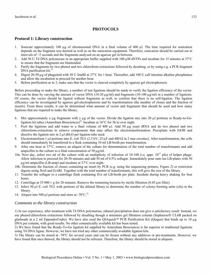
PROTOCOLS Protocol 1: Library construction 1. Sonicate approximately 100 µg of chromosomal DNA in a final volume of 400 µl. The time required for sonication
depends on the fragment size desired as well as on the sonication equipment. Therefore, sonication should be carried out in intervals of ~5 seconds and the fragments analysed on an agarose gel in between.
2. Add 50 U T4 DNA polymerase in an appropriate buffer supplied with 100 µM dNTPs and incubate for 15 minutes at 37°C to ensure that the fragments are bluntended.
3. Purify the fragments by two phenol and two chloroform-extractions followed by desalting, or by using e.g. a PCR-fragment DNA purification kit.1)
4. Digest 20-50 µg of phagemid with 50 U SnaBI at 37°C for 1 hour. Thereafter, add 100 U calf intestine alkaline phosphatase and allow the incubation to proceed for another hour.
5. Before purification as in 3, make sure that the vector is cleaved completely by agarose gel electrophoresis.
Before proceeding to make the library, a number of test ligations should be made to verify the ligation efficiency of the vector. This can be done by varying the amount of vector DNA (10-20 µg/ml) and fragments (10-100 µg/ml) in a number of ligations. Of course, the vector should be ligated without fragments as well, to confirm that there is no self-ligation. The ligation efficiency can be investigated by agarose gel-electrophoresis and by transformation (the number of clones and the fraction of inserts). From these results, it can be determined what amount of vector and fragments that should be used and how many ligations that are required to make the library.
6. Mix approximately x µg fragments with y µg of the vector. Divide the ligation mix into 20 µl portions in Ready-to-Go-
ligation kit tubes (Amersham Biosciences)2). Incubate at 16°C for 3h to over night. 7. Pool the ligations and add water to a final volume of 400 µl. Add 50 µg yeast tRNA and do two phenol and two
chloroform-extractions to remove components that may affect the electrotransformation. Precipitate with EtOH and dissolve the ligation mix in 2 µl dH2O per ligation tube used.
8. Electrotransform 1 µl portions into E. coli TG1 (2.5 kV, 25 µF and 400 Ω in 2 mm cuvettes). After transformation, the cells should immediately be transferred to a flask containing 10 ml LB-broth per transformation.
9. After one hour at 37°C, remove an aliquot of the culture for determination of the total number of transformants and add ampicillin to the culture to a final concentration of 50 µg/ml.
10a. Next day, infect two ml of the culture with an multiplicity of infection of 10-100 (i.e. appr. 1011 pfu) of helper phage. Allow infection to proceed for 20-30 minutes and add 50 ml of 0.5% softagar. Immediately pour onto ten LB-plates with 50 µg/ml ampicillin (LB-amp) and incubate at 37°C over night.
10b. Determine the fraction of clones containing an insert by PCR (e.g. using the sequencing primers, Figure 2) or restriction digests using NcoI and EcoRI. Together with the total number of transformants, this will give the size of the library.
11. Transfer the softagar to a centrifuge flask containing five ml LB-broth per plate. Incubate during heavy shaking for four hours.
12. Centrifuge at 35 000 × g for 20 minutes. Remove the remaining bacteria by sterile filtration (0.45 µm filter).
13. Infect 50 µl E. coli TG1 with portions of the diluted library to determine the number of colony-forming units (cfu) in the library.
14. Aliquot into 500 µl portions and store at -70°C.3)
Comments to the library construction 1) In our experience, after treatment with T4 DNA polymerase, ethanol precipitation does not give a satisfactory result. Instead, we use phenol/chloroform extractions followed by desalting though a miniature gel filtration column (Sepharose CL6B packed on glasbeads in a 2 ml Eppendorf-tube). We have also used the QIAquick PCR Purification Kit (Qiagen) that binds up to 10 µg DNA per column, with good results. No other commercially available kit has been tested. 2) We have found that the Ready-To-Go ligation kit supplied by Amersham Biosciences is far superior to traditional ligations using T4 DNA ligase. However, we have not tried any other commercially available ligation kits. 3) The library can be stored at -70°C for several years and can be frozen without any additives or pre-treatments. However, we have found that once thawed, the library should not be refrozen. Therefore, the library should be stored in aliquots.
Jacobsson et al.
Biological Procedures Online Vol. 5 No. 1 May 1, 2003 www.biologicalprocedures.com
134
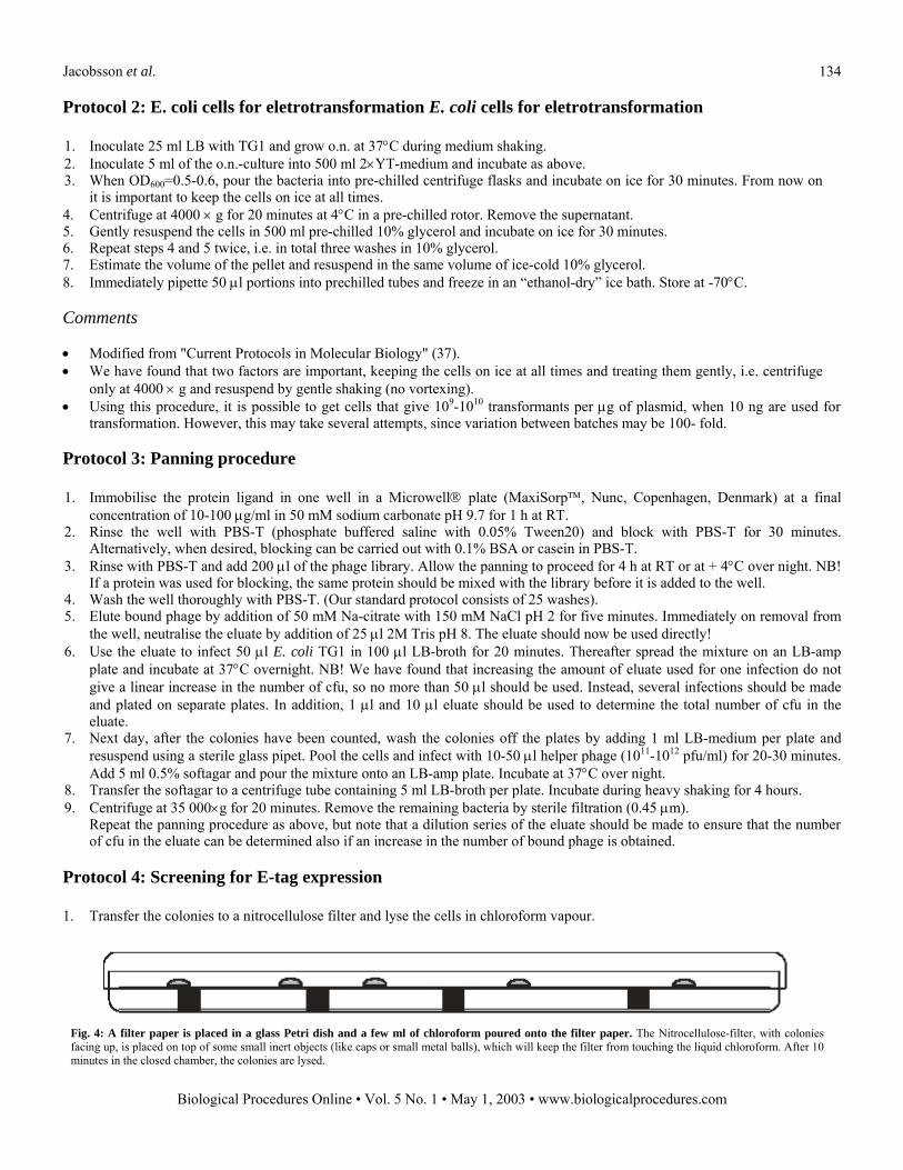
Protocol 2: E. coli cells for eletrotransformation E. coli cells for eletrotransformation 1. Inoculate 25 ml LB with TG1 and grow o.n. at 37°C during medium shaking. 2. Inoculate 5 ml of the o.n.-culture into 500 ml 2×YT-medium and incubate as above. 3. When OD600=0.5-0.6, pour the bacteria into pre-chilled centrifuge flasks and incubate on ice for 30 minutes. From now on
it is important to keep the cells on ice at all times. 4. Centrifuge at 4000 × g for 20 minutes at 4°C in a pre-chilled rotor. Remove the supernatant. 5. Gently resuspend the cells in 500 ml pre-chilled 10% glycerol and incubate on ice for 30 minutes. 6. Repeat steps 4 and 5 twice, i.e. in total three washes in 10% glycerol. 7. Estimate the volume of the pellet and resuspend in the same volume of ice-cold 10% glycerol. 8. Immediately pipette 50 µl portions into prechilled tubes and freeze in an ethanol-dry ice bath. Store at -70°C.
Comments • Modified from "Current Protocols in Molecular Biology" (37). • We have found that two factors are important, keeping the cells on ice at all times and treating them gently, i.e. centrifuge
only at 4000 × g and resuspend by gentle shaking (no vortexing). • Using this procedure, it is possible to get cells that give 109-1010 transformants per µg of plasmid, when 10 ng are used for
transformation. However, this may take several attempts, since variation between batches may be 100- fold. Protocol 3: Panning procedure 1. Immobilise the protein ligand in one well in a Microwell plate (MaxiSorp, Nunc, Copenhagen, Denmark) at a final
concentration of 10-100 µg/ml in 50 mM sodium carbonate pH 9.7 for 1 h at RT. 2. Rinse the well with PBS-T (phosphate buffered saline with 0.05% Tween20) and block with PBS-T for 30 minutes.
Alternatively, when desired, blocking can be carried out with 0.1% BSA or casein in PBS-T. 3. Rinse with PBS-T and add 200 µl of the phage library. Allow the panning to proceed for 4 h at RT or at + 4°C over night. NB!
If a protein was used for blocking, the same protein should be mixed with the library before it is added to the well. 4. Wash the well thoroughly with PBS-T. (Our standard protocol consists of 25 washes). 5. Elute bound phage by addition of 50 mM Na-citrate with 150 mM NaCl pH 2 for five minutes. Immediately on removal from
the well, neutralise the eluate by addition of 25 µl 2M Tris pH 8. The eluate should now be used directly! 6. Use the eluate to infect 50 µl E. coli TG1 in 100 µl LB-broth for 20 minutes. Thereafter spread the mixture on an LB-amp
plate and incubate at 37°C overnight. NB! We have found that increasing the amount of eluate used for one infection do not give a linear increase in the number of cfu, so no more than 50 µl should be used. Instead, several infections should be made and plated on separate plates. In addition, 1 µl and 10 µl eluate should be used to determine the total number of cfu in the eluate.
7. Next day, after the colonies have been counted, wash the colonies off the plates by adding 1 ml LB-medium per plate and resuspend using a sterile glass pipet. Pool the cells and infect with 10-50 µl helper phage (1011-1012 pfu/ml) for 20-30 minutes. Add 5 ml 0.5% softagar and pour the mixture onto an LB-amp plate. Incubate at 37°C over night.
8. Transfer the softagar to a centrifuge tube containing 5 ml LB-broth per plate. Incubate during heavy shaking for 4 hours. 9. Centrifuge at 35 000×g for 20 minutes. Remove the remaining bacteria by sterile filtration (0.45 µm).
Repeat the panning procedure as above, but note that a dilution series of the eluate should be made to ensure that the number of cfu in the eluate can be determined also if an increase in the number of bound phage is obtained.
Protocol 4: Screening for E-tag expression 1. Transfer the colonies to a nitrocellulose filter and lyse the cells in chloroform vapour.
Fig. 4: A filter paper is placed in a glass Petri dish and a few ml of chloroform poured onto the filter paper. The Nitrocellulose-filter, with colonies facing up, is placed on top of some small inert objects (like caps or small metal balls), which will keep the filter from touching the liquid chloroform. After 10 minutes in the closed chamber, the colonies are lysed.

Jacobsson et al.
Biological Procedures Online Vol. 5 No. 1 May 1, 2003 www.biologicalprocedures.com
135
2. Transfer the filter to a container with PBS-T and wash the filter for 30 minutes under shaking. 3. Block the filter with e.g. 0.1% casein or BSA in 10 ml PBS-T in a Petri-dish for 20 minutes at R.T. 4. Add 10 µl of mouse anti-E tag antibodies (Amersham Biosciences) and incubate for 2 h at R.T. 5. Rinse the filters in PBS-T a few times. Add 10 ml PBS-T with 10 µl horse-radish peroxidase labelled anti-mouse antibodies
and incubate for 1 h at R.T. 6. Wash the filter 3 × 10 minutes in PBS-T. 7. Rinse a few times in PBS to remove Tween20. 8. Develop for example using 2 ml 4-chloro-1-naphtol (3 mg/ml in methanol) and 10 µl H2O2 in 10 ml PBS.


![ABCDEFGHIJKLMNOPQRSTUVWXYZ~!@#$%^&*() +{}|:ł? …...frutiger_light_abcdefghijklmnopqrstuvwxyz`1234567890-=[]\; ,./ ABCDEFGHIJKLMNOPQRSTUVWXYZ~!@#$%^&*()_+{}|:ł? ac(C)˙˚ˇo˛(R)sY`!ıLc•ao⁄Ł](https://img.pdfslide.tips/doc/110x75/5ea7d8985175bd4afe13944d/abcdefghijklmnopqrstuvwxyz-frutigerlightabcdefghijklmnopqrstuvwxyz1234567890-.jpg)


![ç ‚ EBG -W˝‚ · 2011-06-08 · 3 ßyjgzc zc#Ö} ¯ÝgzZDäs¯yZZü äÛn¿´äÛnÛŁŠ’+flZgY#aÚŽ ŠÜ ß × ł › ł æ ł ä ô n ß × ł ´ ł o F Ö^ˆ ł ł i ö†]o](https://img.pdfslide.tips/doc/110x75/5f74168c4c5c664bb05a8064/-a-ebg-wa-2011-06-08-3-yjgzc-zc-gzzdsyzz-nnaizgya.jpg)


![łoˇF $ßçöÖ»] ØôæłƒßˆöÖ ß^ e ô Ôł −ł Û j ł›» ] ‡ôˇ ł ˚ ł †ô ... · łoˇF $ßçöÖ»] ØôæłƒßˆöÖ ß^ e ô Ôł −ł Û j ł›» ] ‡ôˇ](https://img.pdfslide.tips/doc/110x75/5fd782fff3093401417f9028/of-e-a-j-a-a.jpg)


![ß ×ł› ł æ ł ä ô n ß ×ł´ ł o F Ö^ˆł iłö†]o ß ×ł‚ ł ^mł ƒ ... · 2011-06-08 · 4 ßyjgzc zc#Ö} ¯ÝgzZDäs¯yZZü Š’+flZgY#aÚŽ ŠÜß ×ł›ł](https://img.pdfslide.tips/doc/110x75/5f74168c4c5c664bb05a8065/-a-n-o-f-iao-a-m.jpg)