Embed Size (px)
Citation preview

A calcium-dependent protease as a potentialtherapeutic target for Wolfram syndromeSimin Lua,b, Kohsuke Kanekuraa, Takashi Haraa, Jana Mahadevana, Larry D. Spearsa, Christine M. Oslowskic,Rita Martinezd, Mayu Yamazaki-Inouee, Masashi Toyodae, Amber Neilsond, Patrick Blannerd, Cris M. Browna,Clay F. Semenkovicha, Bess A. Marshallf, Tamara Hersheyg, Akihiro Umezawae, Peter A. Greerh, and Fumihiko Uranoa,i,1
aDepartment of Medicine, Division of Endocrinology, Metabolism, and Lipid Research, Washington University School of Medicine, St. Louis, MO 63110;bGraduate School of Biomedical Sciences, University of Massachusetts Medical School, Worcester, MA 01655; cDepartment of Medicine, Boston UniversitySchool of Medicine, Boston, MA 02118; dDepartment of Genetics, iPSC core facility, Washington University School of Medicine, St. Louis, MO 63110;eDepartment of Reproductive Biology, National Center for Child Health and Development, Tokyo 157-8535, Japan; fDepartment of Pediatrics, WashingtonUniversity School of Medicine, St. Louis, MO 63110; gDepartments of Psychiatry, Neurology, and Radiology, Washington University School of Medicine,St. Louis, MO 63110; hDepartment of Pathology and Molecular Medicine, Queen’s University, Division of Cancer Biology and Genetics, Queen’s CancerResearch Institute, Kingston, Ontario K7L3N6, Canada; and iDepartment of Pathology and Immunology, Washington University School of Medicine,St. Louis, MO 63110
Edited by Stephen O’Rahilly, University of Cambridge, Cambridge, United Kingdom, and approved November 7, 2014 (received for review November 4, 2014)
Wolfram syndrome is a genetic disorder characterized by diabetesand neurodegeneration and considered as an endoplasmic re-ticulum (ER) disease. Despite the underlying importance of ERdysfunction in Wolfram syndrome and the identification of twocausative genes, Wolfram syndrome 1 (WFS1) and Wolfram syn-drome 2 (WFS2), a molecular mechanism linking the ER to deathof neurons and β cells has not been elucidated. Here we implicatecalpain 2 in the mechanism of cell death in Wolfram syndrome.Calpain 2 is negatively regulated by WFS2, and elevated activationof calpain 2 byWFS2-knockdown correlates with cell death. Calpainactivation is also induced by high cytosolic calciummediated by theloss of function of WFS1. Calpain hyperactivation is observed in theWFS1 knockout mouse as well as in neural progenitor cells derivedfrom induced pluripotent stem (iPS) cells of Wolfram syndromepatients. A small-scale small-molecule screen targeting ER calciumhomeostasis reveals that dantrolene can prevent cell death in neu-ral progenitor cells derived from Wolfram syndrome iPS cells. Ourresults demonstrate that calpain and the pathway leading its acti-vation provides potential therapeutic targets for Wolfram syn-drome and other ER diseases.
Wolfram syndrome | endoplasmic reticulum | diabetes |neurodegeneration | treatment
The endoplasmic reticulum (ER) takes center stage for proteinproduction, redox regulation, calcium homeostasis, and cell
death (1, 2). It follows that genetic or acquired ER dysfunctioncan trigger a variety of common diseases, including neurodegen-erative diseases, metabolic disorders, and inflammatory boweldisease (3, 4). Breakdown in ER function is also associated withgenetic disorders such as Wolfram syndrome (5–8). It is chal-lenging to determine the exact effects of ER dysfunction on thefate of affected cells in common diseases with polygenic andmultifactorial etiologies. In contrast, we reasoned that it shouldbe possible to define the role of ER dysfunction in mechanisti-cally homogenous patient populations, especially in rare diseaseswith a monogenic basis, such as Wolfram syndrome (9).Wolfram syndrome (OMIM 222300) is a rare autosomal re-
cessive disorder characterized by juvenile-onset diabetes mellitusand bilateral optic atrophy (7). Insulin-dependent diabetes usu-ally occurs as the initial manifestation during the first decade oflife, whereas the diagnosis of Wolfram syndrome is invariablylater, with onset of symptoms in the second and ensuing decades(7, 10, 11). Two causative genes for this genetic disorder havebeen identified and named Wolfram syndrome 1 (WFS1) andWolfram syndrome 2 (WFS2) (12, 13). It has been shown thatmultiple mutations in the WFS1 gene, as well as a specific muta-tion in theWFS2 gene, lead to β cell death and neurodegenerationthrough ER and mitochondrial dysfunction (5, 6, 14–16). WFS1
gene variants are also associated with a risk of type 2 diabetes (17).Moreover, a specificWFS1 variant can cause autosomal dominantdiabetes (18), raising the possibility that this rare disorder is rel-evant to common molecular mechanisms altered in diabetes andother human chronic diseases in which ER dysfunction is involved.Despite the underlying importance of ER malfunction in
Wolfram syndrome, and the identification of WFS1 and WFS2genes, a molecular mechanism linking the ER to death of neu-rons and β cells has not been elucidated. Here we show that thecalpain protease provides a mechanistic link between the ER anddeath of neurons and β cells in Wolfram syndrome.
ResultsThe causative genes for Wolfram syndrome, WFS1 and WFS2,encode transmembrane proteins localized to the ER (5, 12, 13).Mutations in the WFS1 or WFS2 have been shown to induceneuronal and β cell death. To determine the cell death pathwaysemanating from the ER, we sought proteins associated withWolfram syndrome causative gene products. HEK293 cells weretransfected with a GST-tagged WFS2 expression plasmid. TheGST-WFS2 protein was purified along with associated proteinson a glutathione affinity resin. These proteins were separated by
Significance
Wolfram syndrome is an autosomal recessive disorder character-ized by juvenile diabetes and neurodegeneration, and is consid-ered a prototype of human endoplasmic reticulum (ER) disease.Wolfram syndrome is caused by loss of function mutations ofWolfram syndrome 1 orWolfram syndrome 2 genes, which encodetransmembrane proteins localized to the ER. Despite its rarity,Wolfram syndrome represents the best human disease modelcurrently available to identify drugs and biomarkers associatedwith ER health. Furthermore, this syndrome is ideal for studyingthe mechanisms of ER stress-mediated death of neurons and βcells. Here we report that the pathway leading to calpain activa-tion offers potential drug targets for Wolfram syndrome andsubstrates for calpainmight serve as biomarkers for this syndrome.
Author contributions: S.L., P.A.G., and F.U. designed research; S.L., K.K., T. Hara, J.M., L.D.S.,C.M.O., R.M., M.Y.-I., M.T., A.N., P.B., and C.M.B. performed research; S.L., B.A.M., T. Hershey,A.U., and F.U. contributed new reagents/analytic tools; S.L., K.K., T. Hara, J.M., L.D.S., C.M.O.,R.M., M.Y.-I., M.T., A.N., P.B., C.M.B., C.F.S., P.A.G., and F.U. analyzed data; and S.L., C.F.S.,P.A.G., and F.U. wrote the paper.
The authors declare no conflict of interest.
This article is a PNAS Direct Submission.
Freely available online through the PNAS open access option.1To whom correspondence should be addressed. Email: [email protected].
This article contains supporting information online at www.pnas.org/lookup/suppl/doi:10.1073/pnas.1421055111/-/DCSupplemental.
E5292–E5301 | PNAS | Published online November 24, 2014 www.pnas.org/cgi/doi/10.1073/pnas.1421055111

SDS/PAGE and visualized by Coomassie staining. Matrix-assistedlaser desorption/ionization-time of flight (MALDI-TOF) massspectroscopic analysis revealed 13 interacting proteins (Table S1),and one of the WFS2-associated polypeptides was CAPN2, thecatalytic subunit of calpain 2, a member of the calcium dependentcysteine proteases family whose members mediate diverse biologicalfunctions including cell death (19–21) (Fig. 1A). Previous studieshave shown that calpain 2 activation is regulated on the ERmembrane and it plays a role in ER stress-induced apoptosis and βcell death (20, 22–24), which prompted us to study the role of WFS2in calpain 2 activation.
Calpain 2 is a heterodimer consisting the CAPN2 catalytic sub-unit and the CAPNS1 (previously known as CAPN4) regulatorysubunit. We first verified that WFS2 interacts with calpain 2 byshowing that endogenous calpain 2 subunits CAPN2 (Fig. 1B) andCAPNS1 (Fig. 1C) each associated with GST-tagged WFS2 ex-pressed in HEK293 cells. Endogenous CAPN2 was also foundto be coimmunoprecipated with N- or C-terminal FLAG-taggedWFS2 expressed in HEK293 cells (Fig. S1 A and B, respectively).To further confirm these findings, we performed a coimmuno-precipitation experiment in Neuro2a cells (a mouse neuroblas-toma cell line) and INS-1 832/13 cells (a rat pancreatic β cell line)
A
D
E
C Input IP CAPN2___________ ____________IgG IgGCAPN2
CAPN2
IB: WFS2
IB: CAPN2
IB: Actin
B
Calpain 2 ER Merge
F
IB: WFS2
IB: CAPN2
IB: GAPDH
Input IP CAPN2_____________ _____________
IgG CAPN2
IgG CAPN2
Gp=0.0055
Fig. 1. WFS2 interacts with CAPN2. (A) Affinity purification ofWFS2-associated proteins from HEK293 cells transfected with GST or GST-WFS2 expression plasmid.Proteins were separated by SDS/PAGE and visualized by Coomassie blue staining. CAPN2 was identified by MALDI-TOF analysis and denoted by an arrow. (B) GST-tagged WFS2 was pulled down on a glutathione affinity resin from lysates of HEK293 cells transfected with a GST-WFS2 expression plasmid, and the pulled-downproducts were analyzed for CAPN2 by immunoblotting with anti-CAPN2 antibody. (C) GST-tagged WFS2 was pulled down on a glutathione affinity resin fromlysates of HEK293 cells transfected with GST-WFS2 expression plasmid and the pulled-down products were analyzed for CAPNS1 by immunoblotting with anti-CAPNS1 antibody. (D) Lysates of Neuro2a cells were immunoprecipitated with IgG or anti-calpain 2 antibodies. Lysates of IgG and anti-calpain 2 immunopre-cipitates were analyzed for WFS2, CAPN2 or actin by immunoblotting. (E) Lysates of INS-1 832/13 cells were immunoprecipitated with IgG or anti-calpain 2antibody. Lysates of IgG and anti-calpain 2 immunoprecipitates were analyzed for WFS2, CAPN2 or actin by immunoblotting. (F) COS7 cells were transfected withpDsRed2-ER vector (Center) and stained with anti-calpain 2 antibody (Left). (Right) A merged image is shown. (G) HEK293 cells were transfected with emptyexpression plasmid or a CAPN2 expression plasmid. Apoptosis was monitored by immunoblotting analysis of caspase 3 cleavage. (Left) Expression levels of CAPN2and actin were measured by immunoblotting. (Right) Quantification of immunoblot is shown (n = 3, *P < 0.05).
Lu et al. PNAS | Published online November 24, 2014 | E5293
MED
ICALSC
IENCE
SPN
ASPL
US

and found that endogenous WFS2 interacted with endogenousCAPN2 (Fig. 1 D and E). WFS2 is known to be a transmembraneprotein localized to the ER. We therefore explored the possibilitythat calpain 2 might also localize to the ER. We transfected COS7cells with pDsRed2-ER vector to visualize ER. Immunofluores-cence staining of COS7 cells showed that endogenous calpain 2was mainly localized to the cytosol, but also showed that a smallportion colocalized with DsRed2-ER protein at the ER (Fig. 1F).Cell fractionation followed by immunoblot further confirmed thisobservation (Fig. S1C). Collectively, these results suggest thatcalpain 2 interacts with WFS2 at the cytosolic face of the ER.Calpain hyperactivation has been shown to contribute to cell loss
in various diseases (19), raising the possibility that calpain 2 mightbe involved in the regulation of cell death. To verify this issue, weoverexpressed CAPN2, the catalytic subunit of calpain 2, and ob-served increase of cleaved caspase-3 in HEK293 cells indicating thathyperactivation of calpain 2 induces cell death (Fig. 1G).To determine whether WFS2 plays a role in cell survival, we
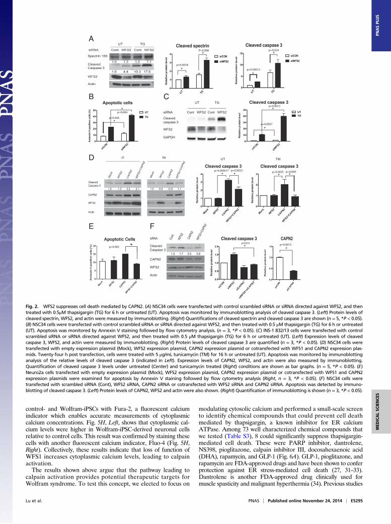
suppressed WFS2 expression in mouse neuronal NSC34 cells usingsiRNA and measured cell death under normal and ER stressconditions. WFS2 knockdown was associated with increasedcleavage of caspase-3 in normal or ER stressed conditions (Fig. 2 Aand B). We subsequently evaluated calpain 2 activation by mea-suring the cleavage of alpha II spectrin, a substrate for calpain 2.RNAi-mediated knockdown of WFS2 induced calpain activation,especially under ER stress conditions (Fig. 2A).In patients with Wolfram syndrome, destruction of β cells
leads to juvenile-onset diabetes (25). This finding prompted us toexamine whether WFS2 was also involved in pancreatic β celldeath. As was seen in neuronal cells, knockdown of WFS2 inrodent β cell lines INS1 832/13 (Fig. 2C) and MIN6 (Fig. S2) wasalso associated with increased caspase-3 cleavage under bothnormal and ER stress conditions. The association of WFS2 withcalpain 2 and their involvement in cell viability suggested thatcalpain 2 activation might be the cause of cell death in WFS2-deficient cells. To further explore the relationship between WFS2and calpain 2, we expressed WFS2 together with the calpain 2catalytic subunit CAPN2 and measured apoptosis. Ectopic ex-pression of WFS2 significantly suppressed calpain 2-associatedapoptosis under normal and ER stress conditions (Fig. 2D, lane 4and lane 8, and Fig. 2E). Next, we tested whether CAPN2mediates cell death induced by WFS2 deficiency. When CAPN2was silenced in WFS2-deficient cells, apoptosis was partially sup-pressed compared with untreated WFS2-deficient cells (Fig. 2F).Taken together, these results suggest that WFS2 is a negativeregulator of calpain 2 proapoptotic functions.To further confirm that loss of function of WFS2 leads to cell
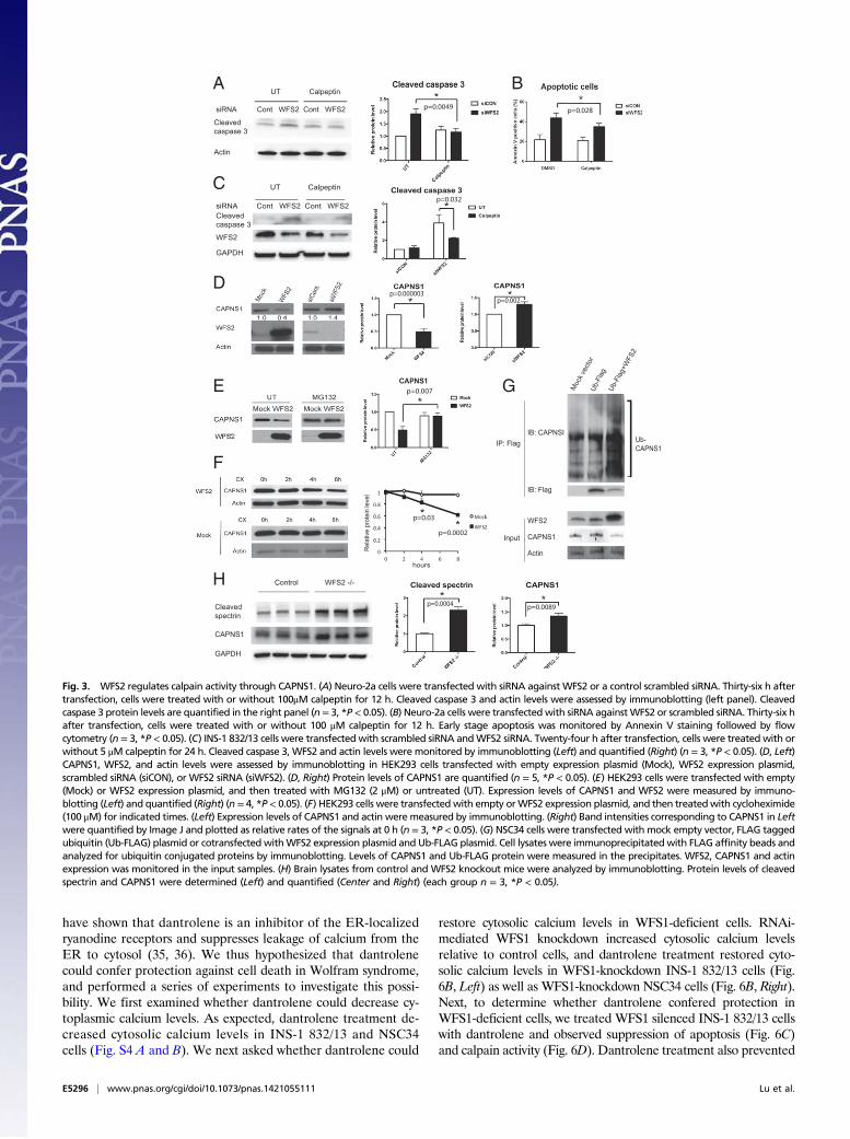
death mediated by calpain 2, we tested if calpeptin, a calpain in-hibitor, could prevent cell death in WFS2-deficient cells. In agree-ment with previous observations, calpeptin treatment preventedWFS2-knockdown-mediated cell death in neuronal (Fig. 3 A and B)and β cell lines (Fig. 3C and Fig. S3A). Collectively, these resultsindicate that WFS2 is a suppressor of calpain 2-mediated cell death.CAPN2 is the catalytic subunit of calpain 2. CAPN2 forms
a heterodimer with the regulatory subunit, CAPNS1, which isrequired for protease activity and stability. We next explored therole of WFS2 in CAPN2 and CAPNS1 protein stability. Ectopicexpression or RNAi-mediated knockdown of WFS2 did notcorrelate with changes in the steady-state expression of CAPN2(Fig. S3B). By contrast, overexpression of WFS2 significantlyreduced CAPNS1 protein expression (Fig. 3D) and transientsuppression of WFS2 slightly increased CAPNS1 protein ex-pression (Fig. 3D). These data suggest that WFS2 might be in-volved in CAPNS1 protein turnover, which is supported by thedata showing that GST-tagged WFS2 expressed in HEK293 cellsassociated with endogenous CAPNS1 (Fig. 1C). To investigatewhether WFS2 regulates CAPNS1 stability through the ubiq-uitin-proteasome pathway, we treated HEK293 cells ectopically
expressing WFS2 with a proteasome inhibitor, MG132, and thenmeasured CAPNS1 protein level. MG132 treatment stabilizedCAPNS1 protein in cells ectopically expressing WFS2 (Fig. 3E).Furthermore, we performed cycloheximide chase experimentsusing HEK293 cells ectopically expressing WFS2 and quantifiedCAPNS1 protein levels at different time points. Ectopic expressionof WFS2 was associated with significantly accelerated CAPNS1protein loss, indicating that WFS2 contributes to posttranslationalregulation of CAPNS1 (Fig. 3F). To further assess whether WFS2is involved in the ubiquitination of CAPNS1, we measured thelevels of CAPNS1 ubiquitination in cells ectopically expressingWFS2 and observed that CAPNS1 ubiquitination was increased byectopic expression of WFS2 (Fig. 3G).To further investigate the role of WFS2 in calpain 2 regulation,
we collected brain lysates from WFS2 knockout mice. Measuredlevels of cleaved spectrin, a well characterized substrate for cal-pain (26). Notably, protein expression levels of cleaved spectrin,as well as CAPNS1, were significantly increased in WFS2knockout mice compared with control mice (Fig. 3H). Collec-tively, these results indicate that WFS2 inhibits calpain 2 activationby regulating CAPNS1 degradation mediated by the ubiquitin-proteasome system.Calpain 2 is a calcium-dependent protease. WFS1, the other
causative gene for Wolfram syndrome, has been shown to beinvolved in calcium homeostasis (27, 28), suggesting that the lossof function of WFS1 may also cause calpain activation. Toevaluate this possibility, we measured calpain activation levels inbrain tissues from WFS1 brain-specific knockout and controlmice. We observed a significant increase in a calpain-specificspectrin cleavage product, reflecting higher calpain activationlevels in WFS1 knockout mice compared with control mice (Fig.4A). The suppression levels of WFS1 in different parts of thebrain were shown in Fig. 4B. To further confirm that calpain isactivated by the loss of WFS1, we looked for other calpainsubstrates in brain tissues from WFS1 knockout mice usinga proteomics approach. Two-dimensional fluorescence gel elec-trophoresis identified 12 proteins differentially expressed be-tween cerebellums of WFS1 knockout mice and those of controlmice (Fig. 4 C and D). Among these, myelin basic protein (MBP)is a known substrate for calpain in the brain (29). We measuredmyelin basic protein levels in brain lysates from WFS1 knockoutand control mice. Indeed, the cleavage and degradation of my-elin basic protein was increased in WFS1 knockout mice relativeto control mice (Fig. 4E).Next, we looked for evidence of increased calpain activity in
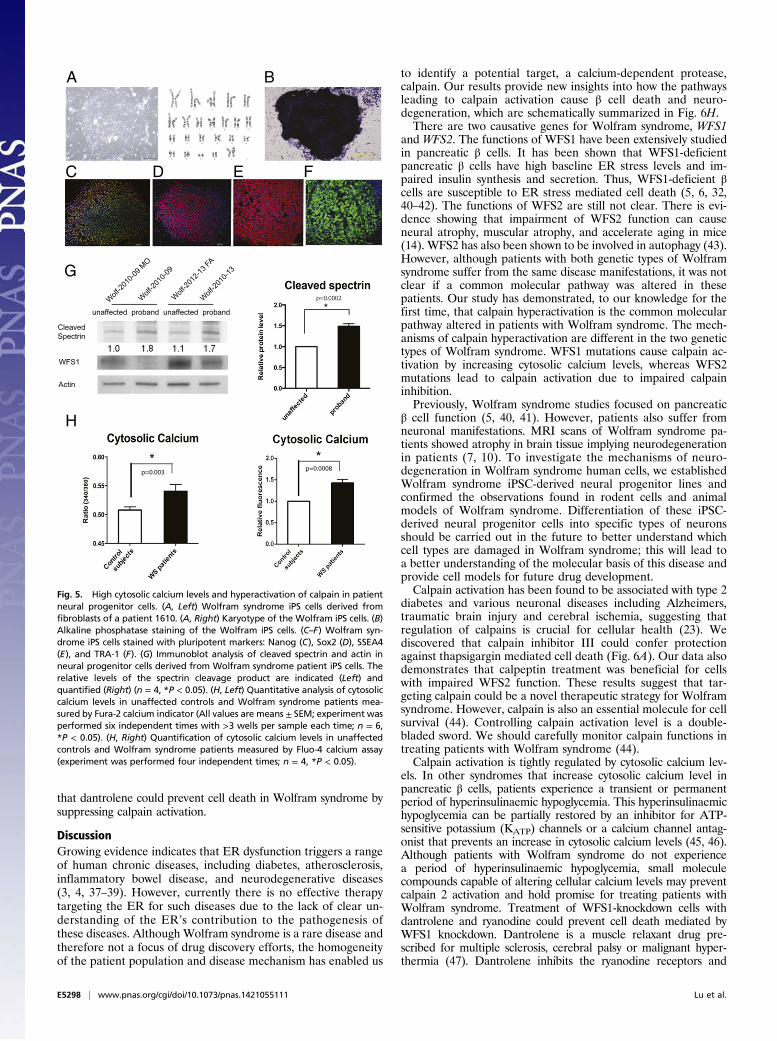
Wolfram syndrome patient cells. We created neural progenitorcells derived from induced pluripotent stem cells (iPSCs) ofWolfram syndrome patients with mutations in WFS1. Fibroblastsfrom four unaffected controls and five patients with Wolframsyndrome were transduced with four reprogramming genes(Sox2, Oct4, c-Myc, and Klf4) (30) (Table S2). We produced atleast 10 iPSC clones from each control and Wolfram patient. Allcontrol- and Wolfram-iPSCs, exhibited characteristic humanembryonic stem cell morphology, expressed pluripotency mark-ers including ALP, NANOG, SOX2, SSEA4, TRA-1–81, andhad a normal karyotype (Fig. 5 A–F). To create neural pro-genitor cells, we first formed neural aggregates from iPSCs.Neural aggregates were harvested at day 5, replated onto newplates to give rise to colonies containing neural rosette struc-tures. At day 12, neural rosette clusters were collected, replated,and used as neural progenitor cells. Consistent with the datafrom WFS1 and WFS2 knockout mice, we observed that spectrincleavage was increased in neural progenitor cells derived fromWolfram-iPSCs relative to control iPSCs, which indicates in-creased calpain activity (Fig. 5G).Because calpain is known to be activated by high calcium, we
explored the possibility that cytoplasmic calcium may be increasedin patient cells by staining neural progenitor cells derived from
E5294 | www.pnas.org/cgi/doi/10.1073/pnas.1421055111 Lu et al.
control- and Wolfram-iPSCs with Fura-2, a fluorescent calciumindicator which enables accurate measurements of cytoplasmiccalcium concentrations. Fig. 5H, Left, shows that cytoplasmic cal-cium levels were higher in Wolfram-iPSC-derived neuronal cellsrelative to control cells. This result was confirmed by staining thesecells with another fluorescent calcium indicator, Fluo-4 (Fig. 5H,Right). Collectively, these results indicate that loss of function ofWFS1 increases cytoplasmic calcium levels, leading to calpainactivation.The results shown above argue that the pathway leading to
calpain activation provides potential therapeutic targets forWolfram syndrome. To test this concept, we elected to focus on
modulating cytosolic calcium and performed a small-scale screento identify chemical compounds that could prevent cell deathmediated by thapsigargin, a known inhibitor for ER calciumATPase. Among 73 well characterized chemical compounds thatwe tested (Table S3), 8 could significantly suppress thapsigargin-mediated cell death. These were PARP inhibitor, dantrolene,NS398, pioglitazone, calpain inhibitor III, docosahexaenoic acid(DHA), rapamycin, and GLP-1 (Fig. 6A). GLP-1, pioglitazone, andrapamycin are FDA-approved drugs and have been shown to conferprotection against ER stress-mediated cell death (27, 31–33).Dantrolene is another FDA-approved drug clinically used formuscle spasticity and malignant hyperthermia (34). Previous studies
* CleavedCaspase 3
CAPN2
WFS2
Actin
1.0 1.7 0.5 0.8
siRNA Cont
WFS2
CAPN
2WFS
2+CA
PN2
Cleavedcaspase 3
WFS2
GAPDH
siRNA Cont WFS2 Cont WFS2
UT TG __________ _________
p=0.0007
p=0.0018
p=0.00012
p=0.0055p=0.000017
p=0.0015p=0.014
A
CB
D
FE
*
*
P=0.008 *
*
p=0.024
*
*
*
*
p=0.0015
**p=0.0023
**p=0.004
* *
p=0.036
p=0.004
p=0.002
MTTU
Fig. 2. WFS2 suppresses cell death mediated by CAPN2. (A) NSC34 cells were transfected with control scrambled siRNA or siRNA directed against WFS2, and thentreated with 0.5μM thapsigargin (TG) for 6 h or untreated (UT). Apoptosis was monitored by immunoblotting analysis of cleaved caspase 3. (Left) Protein levels ofcleaved spectrin, WFS2, and actin were measured by immunoblotting. (Right) Quantifications of cleaved spectrin and cleaved caspase 3 are shown (n = 5, *P < 0.05).(B) NSC34 cells were transfected with control scrambled siRNA or siRNA directed against WFS2, and then treated with 0.5 μM thapsigargin (TG) for 6 h or untreated(UT). Apoptosis was monitored by Annexin V staining followed by flow cytometry analysis. (n = 3, *P < 0.05). (C) INS-1 832/13 cells were transfected with controlscrambled siRNA or siRNA directed against WFS2, and then treated with 0.5 μM thapsigargin (TG) for 6 h or untreated (UT). (Left) Expression levels of cleavedcaspase 3, WFS2, and actin were measured by immunoblotting. (Right) Protein levels of cleaved caspase 3 are quantified (n = 3, *P < 0.05). (D) NSC34 cells weretransfected with empty expression plasmid (Mock), WFS2 expression plasmid, CAPN2 expression plasmid or cotransfected with WFS1 and CAPN2 expression plas-mids. Twenty-four h post transfection, cells were treated with 5 μg/mL tunicamycin (TM) for 16 h or untreated (UT). Apoptosis was monitored by immunoblottinganalysis of the relative levels of cleaved caspase 3 (indicated in Left). Expression levels of CAPN2, WFS2, and actin were also measured by immunoblotting.Quantification of cleaved caspase 3 levels under untreated (Center) and tunicamycin treated (Right) conditions are shown as bar graphs. (n = 5, *P < 0.05). (E)Neuro2a cells transfected with empty expression plasmid (Mock), WFS2 expression plasmid, CAPN2 expression plasmid or cotransfected with WFS1 and CAPN2expression plasmids were examined for apoptosis by Annexin V staining followed by flow cytometry analysis (Right, n = 3, *P < 0.05). (F) NSC34 cells weretransfected with scrambled siRNA (Cont), WFS2 siRNA, CAPN2 siRNA or cotransfected with WFS2 siRNA and CAPN2 siRNA. Apoptosis was detected by immuno-blotting of cleaved caspase 3. (Left) Protein levels of CAPN2, WFS2 and actin were also shown. (Right) Quantification of immunoblotting is shown (n = 3, *P < 0.05).
Lu et al. PNAS | Published online November 24, 2014 | E5295
MED
ICALSC
IENCE
SPN
ASPL
US
have shown that dantrolene is an inhibitor of the ER-localizedryanodine receptors and suppresses leakage of calcium from theER to cytosol (35, 36). We thus hypothesized that dantrolenecould confer protection against cell death in Wolfram syndrome,and performed a series of experiments to investigate this possi-bility. We first examined whether dantrolene could decrease cy-toplasmic calcium levels. As expected, dantrolene treatment de-creased cytosolic calcium levels in INS-1 832/13 and NSC34cells (Fig. S4 A and B). We next asked whether dantrolene could
restore cytosolic calcium levels in WFS1-deficient cells. RNAi-mediated WFS1 knockdown increased cytosolic calcium levelsrelative to control cells, and dantrolene treatment restored cyto-solic calcium levels in WFS1-knockdown INS-1 832/13 cells (Fig.6B, Left) as well as WFS1-knockdown NSC34 cells (Fig. 6B, Right).Next, to determine whether dantrolene confered protection inWFS1-deficient cells, we treated WFS1 silenced INS-1 832/13 cellswith dantrolene and observed suppression of apoptosis (Fig. 6C)and calpain activity (Fig. 6D). Dantrolene treatment also prevented
3
WFS2
CAPNS1
Actin
IB: CAPNSI
IB: Flag
_________________ _____ __________ ____ _ _____
Moc
k vec
tor
Ub-F
lag
Ub-F
lag+
WFS
2
p=0.0049
Cleavedcaspase 3
WFS2
GAPDH
siRNA Cont WFS2 Cont WFS2
UT Calpeptin __________ _________ p=0.032
p=0.002p=0.000003
p=0.007
hours
levelnietorp
evitaleR
BA
Control WFS2 -/-__________________ __________________
Cleavedspectrin
CAPNS1
GAPDH
p=0.0004 p=0.0089
C
D
E
F
G
H
IP: Flag
Input
p=0.028
UT Calpeptin
siRNA Cont WFS2 Cont WFS2
Cleavedcaspase 3
Actin
Ub-CAPNS1
p=0.03
p=0.0002
Fig. 3. WFS2 regulates calpain activity through CAPNS1. (A) Neuro-2a cells were transfected with siRNA against WFS2 or a control scrambled siRNA. Thirty-six h aftertransfection, cells were treated with or without 100μM calpeptin for 12 h. Cleaved caspase 3 and actin levels were assessed by immunoblotting (left panel). Cleavedcaspase 3 protein levels are quantified in the right panel (n = 3, *P < 0.05). (B) Neuro-2a cells were transfected with siRNA againstWFS2 or scrambled siRNA. Thirty-six hafter transfection, cells were treated with or without 100 μM calpeptin for 12 h. Early stage apoptosis was monitored by Annexin V staining followed by flowcytometry (n = 3, *P < 0.05). (C) INS-1 832/13 cells were transfected with scrambled siRNA andWFS2 siRNA. Twenty-four h after transfection, cells were treated with orwithout 5 μM calpeptin for 24 h. Cleaved caspase 3, WFS2 and actin levels were monitored by immunoblotting (Left) and quantified (Right) (n = 3, *P < 0.05). (D, Left)CAPNS1, WFS2, and actin levels were assessed by immunoblotting in HEK293 cells transfected with empty expression plasmid (Mock), WFS2 expression plasmid,scrambled siRNA (siCON), or WFS2 siRNA (siWFS2). (D, Right) Protein levels of CAPNS1 are quantified (n = 5, *P < 0.05). (E) HEK293 cells were transfected with empty(Mock) or WFS2 expression plasmid, and then treated with MG132 (2 μM) or untreated (UT). Expression levels of CAPNS1 and WFS2 were measured by immuno-blotting (Left) and quantified (Right) (n = 4, *P < 0.05). (F) HEK293 cells were transfected with empty orWFS2 expression plasmid, and then treatedwith cycloheximide(100 μM) for indicated times. (Left) Expression levels of CAPNS1 and actin were measured by immunoblotting. (Right) Band intensities corresponding to CAPNS1 in Leftwere quantified by Image J and plotted as relative rates of the signals at 0 h (n = 3, *P < 0.05). (G) NSC34 cells were transfected with mock empty vector, FLAG taggedubiquitin (Ub-FLAG) plasmid or cotransfected withWFS2 expression plasmid and Ub-FLAG plasmid. Cell lysates were immunoprecipitated with FLAG affinity beads andanalyzed for ubiquitin conjugated proteins by immunoblotting. Levels of CAPNS1 and Ub-FLAG protein were measured in the precipitates. WFS2, CAPNS1 and actinexpression was monitored in the input samples. (H) Brain lysates from control and WFS2 knockout mice were analyzed by immunoblotting. Protein levels of cleavedspectrin and CAPNS1 were determined (Left) and quantified (Center and Right) (each group n = 3, *P < 0.05).
E5296 | www.pnas.org/cgi/doi/10.1073/pnas.1421055111 Lu et al.

calpain activation and cell death in WFS1-knockdown NSC34 cells(Fig. 6E). To verify these observations in patient cells, we pre-treated neural progenitor cells derived from iPSCs of a Wolframsyndrome patient and an unaffected parent with dantrolene, andthen challenged these cells with thapisgargin. Thapsigargin-inducedcell death was increased in neural progenitor cells derived from the
Wolfram syndrome patient relative to those derived from the un-affected parent, and dantrolene could prevent cell death in thepatient iPSC-derived neural progenitor cells (Fig. 6F). In addition,we treated brain-specific WFS1 knockout mice with dantroleneand observed evidence of suppressed calpain activation in brainlysates from these mice (Fig. 6G). Collectively, these results argue
Merge
1
4
3
2
Myelin basic protein (-2.49)
Myelin basic protein (-2.51)
Calbindin (-1.31) SNAP-25
110968475
50
40
30
20
14
10
pI
(kDa)
3 4 5 6 7 8 9 10
1
2
3
45
6 7
8
9
1112 11
10987
65 GFAP (1.22)
Cytochrome c oxidase subunit6B1 (>1.5)
FKBP4 (1.32)
Syntaxin-binding protein 1 (1.38)
Cytochrome C1 (-1.48)Phosphatidylethanolamine-binding protein 1 (1.7)
Stathmin (-2.51)
Purkinje cell protein 4 (1.33)12 Hemoglobin subunit aplha (-1.81)
10
or increased/decreased
Cleaved spectrin
WFS1
WFS1+/+ WFS1-/-
WFS1+/+ WFS1-/-
BA
DC
WFS1 +/+ -/-
Cleaved MBP
E
GAPDH
p=0.039
p=0.012FL-MBPFL-MBP
WFS1
Fig. 4. Evidence of Calpain 2 activation in a mouse model of Wolfram syndrome. (A) Protein was extracted from brain tissues of WFS1 brain-specific knockout(−/−) and control (+/−) mice. (Left) Cleaved alpha II spectrin and actin levels were determined by immunoblot analysis. (Right) Quantification of cleavedspectrin is shown (each group n = 10, *P < 0.05). (B) WFS1 mRNA levels in different parts of brain in WFS1−/− and WFS1+/− mice were measured by qRT-PCR. (C)Two-dimensional fluorescence difference gel electrophoresis of cerebellum proteins from WFS1 knockout (WFS1−/−, labeled in red) and control (WFS1+/+,labeled in green) mice showing common (Merge, labeled in yellow) and unique proteins (circled). (D) The protein expression ratios between WFS1 knockoutand control mice were generated, and differentially expressed spots were analyzed by MALDI-TOF mass spectrometry. Quantitative diagrams of spots #2and #3, identified by mass spectrometry as myelin basic protein, showing lower levels of expression in WFS1 knockout mice compared with control mice. (E)Protein was extracted from cerebellums of WFS1 brain-specific knockout (−/−) and control (+/+) mice. Cleaved myelin basic protein (black arrow), cleavedspectrin, WFS1 and GAPDH levels were determined by immunoblot analysis (left panel) and quantified in the right panel (each group n = 3, *P < 0.05).
Lu et al. PNAS | Published online November 24, 2014 | E5297
MED
ICALSC
IENCE
SPN
ASPL
US
that dantrolene could prevent cell death in Wolfram syndrome bysuppressing calpain activation.
DiscussionGrowing evidence indicates that ER dysfunction triggers a rangeof human chronic diseases, including diabetes, atherosclerosis,inflammatory bowel disease, and neurodegenerative diseases(3, 4, 37–39). However, currently there is no effective therapytargeting the ER for such diseases due to the lack of clear un-derstanding of the ER’s contribution to the pathogenesis ofthese diseases. Although Wolfram syndrome is a rare disease andtherefore not a focus of drug discovery efforts, the homogeneityof the patient population and disease mechanism has enabled us
to identify a potential target, a calcium-dependent protease,calpain. Our results provide new insights into how the pathwaysleading to calpain activation cause β cell death and neuro-degeneration, which are schematically summarized in Fig. 6H.There are two causative genes for Wolfram syndrome, WFS1
and WFS2. The functions of WFS1 have been extensively studiedin pancreatic β cells. It has been shown that WFS1-deficientpancreatic β cells have high baseline ER stress levels and im-paired insulin synthesis and secretion. Thus, WFS1-deficient βcells are susceptible to ER stress mediated cell death (5, 6, 32,40–42). The functions of WFS2 are still not clear. There is evi-dence showing that impairment of WFS2 function can causeneural atrophy, muscular atrophy, and accelerate aging in mice(14). WFS2 has also been shown to be involved in autophagy (43).However, although patients with both genetic types of Wolframsyndrome suffer from the same disease manifestations, it was notclear if a common molecular pathway was altered in thesepatients. Our study has demonstrated, to our knowledge for thefirst time, that calpain hyperactivation is the common molecularpathway altered in patients with Wolfram syndrome. The mech-anisms of calpain hyperactivation are different in the two genetictypes of Wolfram syndrome. WFS1 mutations cause calpain ac-tivation by increasing cytosolic calcium levels, whereas WFS2mutations lead to calpain activation due to impaired calpaininhibition.Previously, Wolfram syndrome studies focused on pancreatic
β cell function (5, 40, 41). However, patients also suffer fromneuronal manifestations. MRI scans of Wolfram syndrome pa-tients showed atrophy in brain tissue implying neurodegenerationin patients (7, 10). To investigate the mechanisms of neuro-degeneration in Wolfram syndrome human cells, we establishedWolfram syndrome iPSC-derived neural progenitor lines andconfirmed the observations found in rodent cells and animalmodels of Wolfram syndrome. Differentiation of these iPSC-derived neural progenitor cells into specific types of neuronsshould be carried out in the future to better understand whichcell types are damaged in Wolfram syndrome; this will lead toa better understanding of the molecular basis of this disease andprovide cell models for future drug development.Calpain activation has been found to be associated with type 2
diabetes and various neuronal diseases including Alzheimers,traumatic brain injury and cerebral ischemia, suggesting thatregulation of calpains is crucial for cellular health (23). Wediscovered that calpain inhibitor III could confer protectionagainst thapsigargin mediated cell death (Fig. 6A). Our data alsodemonstrates that calpeptin treatment was beneficial for cellswith impaired WFS2 function. These results suggest that tar-geting calpain could be a novel therapeutic strategy for Wolframsyndrome. However, calpain is also an essential molecule for cellsurvival (44). Controlling calpain activation level is a double-bladed sword. We should carefully monitor calpain functions intreating patients with Wolfram syndrome (44).Calpain activation is tightly regulated by cytosolic calcium lev-
els. In other syndromes that increase cytosolic calcium level inpancreatic β cells, patients experience a transient or permanentperiod of hyperinsulinaemic hypoglycemia. This hyperinsulinaemichypoglycemia can be partially restored by an inhibitor for ATP-sensitive potassium (KATP) channels or a calcium channel antag-onist that prevents an increase in cytosolic calcium levels (45, 46).Although patients with Wolfram syndrome do not experiencea period of hyperinsulinaemic hypoglycemia, small moleculecompounds capable of altering cellular calcium levels may preventcalpain 2 activation and hold promise for treating patients withWolfram syndrome. Treatment of WFS1-knockdown cells withdantrolene and ryanodine could prevent cell death mediated byWFS1 knockdown. Dantrolene is a muscle relaxant drug pre-scribed for multiple sclerosis, cerebral palsy or malignant hyper-thermia (47). Dantrolene inhibits the ryanodine receptors and
H
G
* *
WFS1
Wolf-20
10-09
MO
Wolf-20
10-09
Wolf-20
12-13
FA
Wolf-20
10-13
unaffected proband unaffected proband
CleavedSpectrin
Actin
*
p=0.003p=0.0008
A B
C D E F
Fig. 5. High cytosolic calcium levels and hyperactivation of calpain in patientneural progenitor cells. (A, Left) Wolfram syndrome iPS cells derived fromfibroblasts of a patient 1610. (A, Right) Karyotype of the Wolfram iPS cells. (B)Alkaline phosphatase staining of the Wolfram iPS cells. (C–F) Wolfram syn-drome iPS cells stained with pluripotent markers: Nanog (C), Sox2 (D), SSEA4(E), and TRA-1 (F). (G) Immunoblot analysis of cleaved spectrin and actin inneural progenitor cells derived from Wolfram syndrome patient iPS cells. Therelative levels of the spectrin cleavage product are indicated (Left) andquantified (Right) (n = 4, *P < 0.05). (H, Left) Quantitative analysis of cytosoliccalcium levels in unaffected controls and Wolfram syndrome patients mea-sured by Fura-2 calcium indicator (All values are means ± SEM; experiment wasperformed six independent times with >3 wells per sample each time; n = 6,*P < 0.05). (H, Right) Quantification of cytosolic calcium levels in unaffectedcontrols and Wolfram syndrome patients measured by Fluo-4 calcium assay(experiment was performed four independent times; n = 4, *P < 0.05).
E5298 | www.pnas.org/cgi/doi/10.1073/pnas.1421055111 Lu et al.

CleavedCaspase 3
WFS1
GAPDH
siRNA Cont. Cont. WFS1 WFS1 Cont. Cont. WFS1 WFS1
_______________________ ____________________ UT TG
Dantrolene - + - + - + - +
CleavedSpectrinWFS1
GAPDH
siRNA Cont. Cont. WFS1 WFS1Dantrolene - + - +
CleavedCaspase3
GAPDH
unaffected proband unaffected proband
UT Dantrolene________________ _______________
_ _______________ ___________
)UFL(
7/ 3esapsaC
I UT I TG I
Dantrolene
A B
ED
F
H
WFS2 deficiencyWFS1 deficiency
CAPNS1/CAPN2 Ca2+
Apoptosis
ER
Ca2+
Increased cytosolic
CAPNS1
Cleavedcaspase 3
WFS1
GAPDH
Cleavedspectrin
siRNA Cont WFS1 Cont WFS1
UT Dantrolene ________ _______
p=0.001
p=0.018
Cleavedspectrin
GAPDH
Control WFS1 -/-___________________________ __________________________
- - - + + + + - - - + + + +Dantrolene p=0.02
p=0.00006p=0.038
p=0.0044p=0.036
p=0.018
p=0.017
G
*
*
*
*
***p=0.0006
p=0.0002
p=0.04
*
*
*
*
*
eavedspase 3
FS1
APDH
RNA ntrolene
aC
WFS
____
+
II_
3
Cont. Cont. WFS1 WFS1 Cont. Cont. WFS1 W
_______________________ _________________ UT TG
e - + - + - + -
I UT I TGI UT I TG_ _______________ ____________ _______________GTTUC
Fig. 6. Dantrolene prevents cell death in iPS cell-derived neural progenitor cells of Wolfram syndorme by inhibiting the ER calcium leakage to the cytosol. (A)INS-1 832/13 cells were pretreated with DMSO or drugs for 24 h then incubated in media containing 20 nM of thapsigargin (TG) overnight. Apoptosis wasdetected by caspase 3/7-Glo luminescence. (B) Cytosolic calcium levels were determined by Fura-2 in control and WFS1-deficient INS-1 832/13 (Left) and NSC34(Right) cells treated or untreated with 10 μM dantrolene for 24 h (All values are means ± SEM; experiment was performed 6 independent times with >3 wellsper sample each time n = 6, *P < 0.05). (C) INS-1 832/13 cells were transfected with scrambled siRNA or siRNA against WFS1. Cells were pretreated with orwithout 10 μM dantrolene for 48 h, then incubated in media with or without 0.5 μM TG for 6 h. Expression levels of cleaved caspase-3, WFS1, GAPDH weremeasured by immunoblotting (Left). Protein levels of caspase3 under untreated (Center) and TG treated (Right) conditions are quantified and shown as bargraphs (n = 3, *P < 0.05). (D) INS-1 832/13 cells were transfected with scrambled siRNA or siRNA against WFS1, pretreated with or without 10 μM dantrolenefor 48 h, then incubated in media containing 0.5 μM TG for 6 h. Protein levels of cleaved spectrin, WFS1, GAPDH were analyzed by immunoblotting (Left) andquantified (Right) (n = 3, *P < 0.05). (E) NSC34 cells were transfected with scrambled siRNA or siRNA against WFS1. Then treated with or without10 μMdantrolene for 24 h. Protein levels of cleaved spectrin, cleaved caspase 3, WFS2 and GAPDH were determined by immunoblotting (Left) and quantified (Right)(n = 3, *P < 0.05). (F) Wolfram patient neural progenitor cells were pretreated with or without 10 μM dantrolene for 48 h. Then, cells were treated with 0.125μM TG for 20 h. (Left) Apoptosis was monitored by immunoblotting. (Right) Quantification of cleaved caspase 3 protein levels are indicated (n = 3, *P < 0.05).(G) Control and WFS1 brain-specific knockout mice were treated with water or dantrolene for 4 wk at 20 mg/kg. Brain lysates of these mice were examined byimmunoblotting. Protein levels of cleaved spectrin and GAPDH were monitored (Left) and quantified (Right) (All values are means ± SEM; each group n > 3,*P < 0.05). (H) Scheme of the pathogenesis of Wolfram syndrome.
Lu et al. PNAS | Published online November 24, 2014 | E5299
MED
ICALSC
IENCE
SPN
ASPL
US

reduces calcium leakage from the ER to cytosol, lowering cytosoliccalcium level. The protective effect of dantrolene treatment onWFS1-deficient cells suggests that dysregulated cellular calciumhomeostasis plays a role in the disease progression of Wolframsyndrome. In addition, it has been shown that stabilizing ER cal-cium channel function could prevent the progression of neuro-degeneration in a mouse model of Alzheimer’s disease (48).Therefore, modulating calcium levels may be an effective way totreat Wolfram syndrome or other ER diseases.Dantrolene treatment did not block cell death mediated by
WFS2 knockdown, suggesting that WFS2 does not directly affectthe ER calcium homeostasis (Fig. S4 D and E). RNAi-mediatedWFS1 knockdown in HEK293 cells significantly reduced theactivation levels of sarco/endoplasmic reticulum calcium trans-port ATPase (SERCA), indicating that WFS1 may play a role inthe modulation of SERCA activation and ER calcium levels (Fig.S5). It has been shown that WFS1 interacts with the Na+/K+
ATPase β1 subunit and the expression of WFS1 parallels that ofNa+/K+ ATPase β1 subunit in a variety of settings, suggesting thatWFS1 may function as an ion channel or regulator of existingchannels (42). Further studies on this topic would be necessary tocompletely understand the etiology of Wolfram syndrome.Our study reveals that dantrolene can prevent ER stress-
mediated cell death in human and rodent cell models as well asmouse models of Wolfram syndrome. Thus, dantrolene andother drugs that regulate ER calcium homeostasis could be usedto delay the progression of Wolfram syndrome and other dis-eases associated with ER dysfunction, including type 1 and type2 diabetes.
Materials and MethodsHuman Subjects. Wolfram syndrome patients were recruited through theWashington University Wolfram Syndrome International Registry website(wolframsyndrome.dom.wustl.edu). The clinic protocol was approved by theWashington University Human Research Protection Office and all subjectsprovided informed consent if adults and assent with consent by parents ifminor children (IRB ID 201107067 and 201104010).
Animal Experiments. WFS1 brain-specific knockout mice were generated bybreeding the Nestin-Cre transgenic mice (Jackson Laboratory) with WFS1floxed mice (40). WFS2 whole body knockout mice are purchased from MRCHarwell. All animal experiments were performed according to proceduresapproved by the Institutional Animal Care and Use Committee at theWashington University School of Medicine (A-3381-01).
Calcium Levels. Calcium levels in cells were measured by Fura-2 AM dye andFluo-4 AM dye (Life Technology) Inifinite M1000 (Tecan). Cells were plated in96-well plates at 25,000 cells per well and stained with 4 μg/mL Fura-2 dyealong with 2.5 mM probenecid for 30 min, then the cells were washed withPBS and kept in the dark for another 30 min to allow cleavage of AM ester.Fluorescence was measured at excitation wavelength 510 nm and emissionwavelengths 340 nm and 380 nm. Then background subtractions were per-formed with both emission wavelengths. The subtraction result was used tocalculate 340/380 ratios.
For Fluo-4 AM staining, neural progenitor cells were plated in 24-wellplates at 200,000 cells per well. After staining with Fluo-4 AM dye for 30minalong with 2.5 mM probenecid, cells were washed and resuspended inPBS. Incubation for a further 30 min was performed to allow completedeesterification of intracellular AM esters. Then, samples were measuredby flow cytometry at the FACS core facility of Washington University Schoolof Medicine using a LSRII instrument (BD). The results were analyzed byFlowJo ver.7.6.3.
Statistical Analysis. Two-tailed t tests were used to compare the two treat-ments. P values below 0.05 were considered significant. All values are shownas means ± SD if not stated. Please see SI Materials and Methods forcomplete details.
ACKNOWLEDGMENTS. We thank all of the participants in the WashingtonUniversity Wolfram Registry and Clinic and their families for their time andeffort (wolframsyndrome.dom.wustl.edu/). We also thank Mai Kanekura,Mariko Hara, and Karen Sargent for technical support and the WashingtonUniversity Wolfram Study Group Members and the study staff for advice andsupport in the greater research program. This work was supported by NIHGrants DK067493, P60 DK020579, and UL1 TR000448; Juvenile Diabetes Re-search Foundation Grants 47-2012-760 and 17-2013-512; American DiabetesAssociation Grant 1-12-CT-61, the Team Alejandro, The Team Ian, the EllieWhite Foundation for Rare Genetic Disorders, and the Jack and J. T. SnowScientific Research Foundation (F.U.).
1. Ron D, Walter P (2007) Signal integration in the endoplasmic reticulum unfoldedprotein response. Nat Rev Mol Cell Biol 8(7):519–529.
2. Tabas I, Ron D (2011) Integrating the mechanisms of apoptosis induced by endo-plasmic reticulum stress. Nat Cell Biol 13(3):184–190.
3. Hetz C, Chevet E, Harding HP (2013) Targeting the unfolded protein response indisease. Nat Rev Drug Discov 12(9):703–719.
4. Wang S, Kaufman RJ (2012) The impact of the unfolded protein response on humandisease. J Cell Biol 197(7):857–867.
5. Fonseca SG, et al. (2005) WFS1 is a novel component of the unfolded protein responseand maintains homeostasis of the endoplasmic reticulum in pancreatic beta-cells.J Biol Chem 280(47):39609–39615.
6. Fonseca SG, et al. (2010) Wolfram syndrome 1 gene negatively regulates ER stresssignaling in rodent and human cells. J Clin Invest 120(3):744–755.
7. Barrett TG, Bundey SE, Macleod AF (1995) Neurodegeneration and diabetes: UK na-tionwide study of Wolfram (DIDMOAD) syndrome. Lancet 346(8988):1458–1463.
8. Wolfram DJ, Wagener HP (1938) Diabetes mellitus and simple optic atrophy amongsiblings: Report of four cases. Mayo Clin Proc 1:715–718.
9. Urano F (2014) Diabetes: Targeting endoplasmic reticulum to combat juvenile di-abetes. Nat Rev Endocrinol 10(3):129–130.
10. Hershey T, et al.; Washington University Wolfram Study Group (2012) Early brainvulnerability in Wolfram syndrome. PLoS ONE 7(7):e40604.
11. Marshall BA, et al.; Washington University Wolfram Study Group (2013) Phenotypiccharacteristics of early Wolfram syndrome. Orphanet J Rare Dis 8(1):64.
12. Inoue H, et al. (1998) A gene encoding a transmembrane protein is mutated in pa-tients with diabetes mellitus and optic atrophy (Wolfram syndrome). Nat Genet 20(2):143–148.
13. Amr S, et al. (2007) A homozygous mutation in a novel zinc-finger protein, ERIS, isresponsible for Wolfram syndrome 2. Am J Hum Genet 81(4):673–683.
14. Chen YF, et al. (2009) Cisd2 deficiency drives premature aging and causes mito-chondria-mediated defects in mice. Genes Dev 23(10):1183–1194.
15. Wiley SE, et al. (2013) Wolfram Syndrome protein, Miner1, regulates sulphydryl redoxstatus, the unfolded protein response, and Ca2+ homeostasis. EMBO Mol Med 5(6):904–918.
16. Shang L, et al. (2014) β-cell dysfunction due to increased ER stress in a stem cell modelof Wolfram syndrome. Diabetes 63(3):923–933.
17. Sandhu MS, et al. (2007) Common variants in WFS1 confer risk of type 2 diabetes. NatGenet 39(8):951–953.
18. Bonnycastle LL, et al. (2013) Autosomal dominant diabetes arising from a Wolframsyndrome 1 mutation. Diabetes 62(11):3943–3950.
19. Goll DE, Thompson VF, Li H, Wei W, Cong J (2003) The calpain system. Physiol Rev83(3):731–801.
20. Tan Y, et al. (2006) Ubiquitous calpains promote caspase-12 and JNK activation duringendoplasmic reticulum stress-induced apoptosis. J Biol Chem 281(23):16016–16024.
21. Tan Y, Wu C, De Veyra T, Greer PA (2006) Ubiquitous calpains promote both apoptosisand survival signals in response to different cell death stimuli. J Biol Chem 281(26):17689–17698.
22. Nakagawa T, Yuan J (2000) Cross-talk between two cysteine protease families. Acti-vation of caspase-12 by calpain in apoptosis. J Cell Biol 150(4):887–894.
23. Cui W, et al. (2013) Free fatty acid induces endoplasmic reticulum stress and apoptosisof β-cells by Ca2+/calpain-2 pathways. PLoS ONE 8(3):e59921.
24. Huang CJ, et al. (2010) Calcium-activated calpain-2 is a mediator of beta cell dys-function and apoptosis in type 2 diabetes. J Biol Chem 285(1):339–348.
25. Barrett TG, Bundey SE (1997) Wolfram (DIDMOAD) syndrome. J Med Genet 34(10):838–841.
26. Liu MC, et al. (2006) Comparing calpain- and caspase-3-mediated degradation pat-terns in traumatic brain injury by differential proteome analysis. Biochem J 394(Pt 3):715–725.
27. Hara T, et al. (2014) Calcium efflux from the endoplasmic reticulum leads to β-celldeath. Endocrinology 155(3):758–768.
28. Takei D, et al. (2006) WFS1 protein modulates the free Ca(2+) concentration in theendoplasmic reticulum. FEBS Lett 580(24):5635–5640.
29. Liu MC, et al. (2006) Extensive degradation of myelin basic protein isoforms by calpainfollowing traumatic brain injury. J Neurochem 98(3):700–712.
30. Takahashi K, Yamanaka S (2006) Induction of pluripotent stem cells from mouseembryonic and adult fibroblast cultures by defined factors. Cell 126(4):663–676.
31. Yusta B, et al. (2006) GLP-1 receptor activation improves beta cell function and sur-vival following induction of endoplasmic reticulum stress. Cell Metab 4(5):391–406.
32. Akiyama M, et al. (2009) Increased insulin demand promotes while pioglitazoneprevents pancreatic beta cell apoptosis in Wfs1 knockout mice. Diabetologia 52(4):653–663.
33. Bachar-Wikstrom E, et al. (2013) Stimulation of autophagy improves endoplasmicreticulum stress-induced diabetes. Diabetes 62(4):1227–1237.
34. Dykes MH (1975) Evaluation of a muscle relaxant: Dantrolene sodium (Dantrium).JAMA 231(8):862–864.
E5300 | www.pnas.org/cgi/doi/10.1073/pnas.1421055111 Lu et al.

35. Wei H, Perry DC (1996) Dantrolene is cytoprotective in two models of neuronal cell
death. J Neurochem 67(6):2390–2398.36. Luciani DS, et al. (2009) Roles of IP3R and RyR Ca2+ channels in endoplasmic reticulum
stress and beta-cell death. Diabetes 58(2):422–432.37. Hotamisligil GS (2010) Endoplasmic reticulum stress and atherosclerosis. Nat Med
16(4):396–399.38. Hotamisligil GS (2010) Endoplasmic reticulum stress and the inflammatory basis of
metabolic disease. Cell 140(6):900–917.39. Ozcan L, Tabas I (2012) Role of endoplasmic reticulum stress in metabolic disease and
other disorders. Annu Rev Med 63:317–328.40. Riggs AC, et al. (2005) Mice conditionally lacking the Wolfram gene in pancreatic islet
beta cells exhibit diabetes as a result of enhanced endoplasmic reticulum stress and
apoptosis. Diabetologia 48(11):2313–2321.41. Ishihara H, et al. (2004) Disruption of the WFS1 gene in mice causes progressive beta-
cell loss and impaired stimulus-secretion coupling in insulin secretion. HumMol Genet
13(11):1159–1170.
42. Zatyka M, et al. (2008) Sodium-potassium ATPase 1 subunit is a molecular partner ofWolframin, an endoplasmic reticulum protein involved in ER stress. Hum Mol Genet17(2):190–200.
43. Chang NC, Nguyen M, Germain M, Shore GC (2010) Antagonism of Beclin 1-dependentautophagy by BCL-2 at the endoplasmic reticulum requires NAF-1. EMBO J 29(3):606–618.
44. Dutt P, et al. (2006) m-Calpain is required for preimplantation embryonic develop-ment in mice. BMC Dev Biol 6:3.
45. Arya VB, Mohammed Z, Blankenstein O, De Lonlay P, Hussain K (2014) Hyper-insulinaemic hypoglycaemia. Hormone Metabolic Res 46(3):157–170.
46. Shah P, Demirbilek H, Hussain K (2014) Persistent hyperinsulinaemic hypoglycaemia ininfancy. Semin Pediatr Surg 23(2):76–82.
47. Krause T, Gerbershagen MU, Fiege M, Weisshorn R, Wappler F (2004) Dantrolene—a review of its pharmacology, therapeutic use and new developments. Anaesthesia59(4):364–373.
48. Chakroborty S, et al. (2012) Stabilizing ER Ca2+ channel function as an early pre-ventative strategy for Alzheimer’s disease. PLoS ONE 7(12):e52056.
Lu et al. PNAS | Published online November 24, 2014 | E5301
MED
ICALSC
IENCE
SPN
ASPL
US