Embed Size (px)
Citation preview
CASE REPORT Open Access
A case of a horseshoe appendixKazuya Takabatake* , Jun Ikeda, Hirotaka Furuke, Chikage Kato, Takuya Kishimoto, Tatsuya Kumano,Kenichiro Imura, Katsumi Shimomura, Takeshi Kubota, Fumihiro Taniguchi and Yasuhiro Shioaki
Abstract
Anomalies of the appendix are extremely rare, and a horseshoe appendix is even rarer. A literature searchhas revealed only five reported cases. In this report, we present a case of a horseshoe appendix.A 78-year-old man was referred for further examination following a positive fecal occult blood test. A massin his ascending colon was detected on colonoscopy, while computed tomography showed that it wasconnected to the appendix. Tumor invasion derived from the ascending colon or appendix was suspected.We diagnosed ascending colon cancer prior to laparoscopic ileocecal resection. Macroscopic findingsshowed that the appendix connected to the back side of the mass, while microscopic findings showed thatthe mucosa and submucosa were continuous from the appendiceal orifice in the cecum to the other orifice inthe ascending colon, where a type 1 tumor was observed on the orifice. We eventually diagnosed the patientwith tubulovillous adenoma and a horseshoe appendix.A horseshoe appendix communicates with the colon at both ends and is supplied by a single fan-shapedmesentery. Cases are classified by the disposal of the mesentery and the location of the orifice. Anatomicalanomalies should be considered despite the rarity of horseshoe appendices.
Keywords: Anomalies of the appendix, Horseshoe appendix
BackgroundAnomalies of the appendix are extremely rare. Therehave been several reports on the absence or duplica-tion of the appendix. However, a literature search re-vealed only five reported cases of a horseshoe-shapedappendix [1–5]. In this report, we present a case of ahorseshoe appendix that was incidentally found dur-ing resection of an adenoma in the ascending colon.
Case presentationA 78-year-old man was referred to us for further exam-ination following a positive fecal occult blood test re-sult. A mass that was possibly malignant was detectedby colonoscopy in the ascending colon. There were noparticular findings from physical examinations orhematological examinations, including the followingtumor markers: cancer embryonic antigen and cancerantigen 19–9. Colonoscopy showed a type 1 mass in
the ascending colon (Fig. 1) with submucosal inva-sion suspected from poor mobility. Computed tom-ography showed a 30-mm-wide mass in theascending colon (Fig. 2) that was connected to theappendix. Tumor invasion derived from the ascend-ing colon or appendix was suspected (Fig. 3). Wepreoperatively diagnosed ascending colon cancer, asfollows: cT1, cN0, cM0, cStage1 (UICC/AJCC 7th).A standard laparoscopic ileocecal resection was thenperformed. Intraoperative findings showed that theappendix was connected to the ascending colon. Itwas suspected to be a tumor invasion and was there-fore mobilized and resected carefully. Macroscopicfindings showed the appendix connected to the backside of the mass, inserting along the appendiceal ori-fice and reaching the adenoma of the ascending colon(Figs. 4 and 5). Microscopic findings revealed that themucosa and submucosa were continuous from theappendiceal orifice in the cecum to the other orificein the ascending colon with a seamless muscular layer(Fig. 6). There was no evidence of inflammation ormalignancy, and pathologically, the appendix was nor-mal. There was a type 1 tumor on the orifice in the
* Correspondence: [email protected] case report was presented at the poster presentation of The 77rdAnnual Congress of Japan Surgical Association. Kazuya Takabatake is the firstauthor.Department of Surgery, Japanese Red Cross Kyoto Daiichi Hospital, 15-749Honmachi, Higashiyama, Kyoto, Kyoto, Japan
© The Author(s). 2016 Open Access This article is distributed under the terms of the Creative Commons Attribution 4.0International License (http://creativecommons.org/licenses/by/4.0/), which permits unrestricted use, distribution, andreproduction in any medium, provided you give appropriate credit to the original author(s) and the source, provide a link tothe Creative Commons license, and indicate if changes were made.
Takabatake et al. Surgical Case Reports (2016) 2:140 DOI 10.1186/s40792-016-0261-3
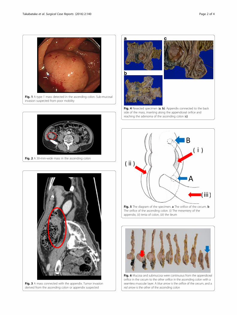
Fig. 3 A mass connected with the appendix. Tumor invasionderived from the ascending colon or appendix suspected
Fig. 2 A 30-mm-wide mass in the ascending colon
Fig. 4 Resected specimen (a, b). Appendix connected to the backside of the mass, inserting along the appendiceal orifice andreaching the adenoma of the ascending colon (c)
Fig. 5 The diagram of the specimen. a The orifice of the cecum. bThe orifice of the ascending colon. (i) The mesentery of theappendix, (ii) tenia of colon, (iii) the ileum
Fig. 1 A type 1 mass detected in the ascending colon. Sub-mucosalinvasion suspected from poor mobility
Fig. 6 Mucosa and submucosa were continuous from the appendicealorifice in the cecum to the other orifice in the ascending colon with aseamless muscular layer. A blue arrow is the orifice of the cecum, and ared arrow is the other of the ascending colon
Takabatake et al. Surgical Case Reports (2016) 2:140 Page 2 of 4
ascending colon, which was pathologically diagnosedas a tubulovillous adenoma with moderate atypia,along with an appendiceal extension. There was noevidence of lymph node metastasis. We finally diag-nosed the patient with a tubulovillous adenoma anda horseshoe appendix. After undergoing the previ-ously described surgery, the patient experienced aparalytic ileus and required fasting. He was dis-charged home on the 15th day after surgery.
DiscussionAnomalies of the appendix are extremely rare. In a studyby Collins, from among 50,000 appendix specimens,
there were four cases of agenesis and two of duplication[6]. Duplications of the appendix were classified by Cavein 1936 [7] and modified by Wallbridge in 1963 [8]and Biermann in 1993 [9]. However, there weresome cases that could not be classified using thisclassification (e.g., triplets of the appendix, horseshoeappendix).Based on our review of the literature, our patient is the
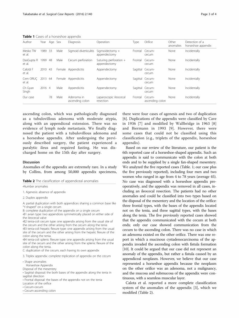
6th reported case of a horseshoe-shaped appendix. Such anappendix is said to communicate with the colon at bothends and to be supplied by a single fan-shaped mesentery.We analyzed the five reported cases (Table. 1; our case plusthe five previously reported), including four men and twowomen who ranged in age from 4 to 78 years (average 45).No case was diagnosed with a horseshoe appendix pre-operatively, and the appendix was removed in all cases, in-cluding an ileocecal resection. The patients had no otheranomalies and could be classified into two types based onthe disposal of the mesentery and the location of the orifice:three frontal types, with the bases of the appendix locatednot on the tenia, and three sagittal types, with the basesalong the tenia. The five previously reported cases showedthat the appendix communicated with the cecum at bothends; only our case showed communication from thececum to the ascending colon. There was no case in whichan adenoma existed on the other orifice. There was one re-port in which a mucinous cystadenocarcinoma of the ap-pendix invaded the ascending colon with fistula formation[10]. It could be argued that our case did not represent ananomaly of the appendix, but rather a fistula caused by anappendiceal neoplasm. However, we believe that our caserepresented a horseshoe appendix because the neoplasmon the other orifice was an adenoma, not a malignancy,and the mucosa and submucosa of the appendix were con-tinuous, with a seamless muscular layer.Calota et al. reported a more complete classification
system of the anomalies of the appendix [3], which wemodified (Table 2).
Table 1 Cases of a horseshoe appendix
Author Year Age Sex Diagnosis Operation Type Orifice Otheranomalies
Detection of ahorseshoe appendix
Mesko TWet al.
1989 33 Male Sigmoid diverticulitis Sigmoidectomy +appendectomy
Frontal Cecum-cecum
None Incidentally
DasGupta Ret al.
1999 48 Male Cecum perforation Suturing perforation +appendectomy
Frontal Cecum-cecum
None Incidentally
Calotă Fet al.
2010 43 Female Appendicitis Appendectomy Sagittal Cecum-cecum
None Incidentally
Cem ORUÇet al.
2013 64 Female Appendicitis Appendectomy Sagittal Cecum-cecum
None Incidentally
Ch GyanSingh
2016 4 Male Appendicitis Appendectomy Sagittal Cecum-cecum
None Incidentally
Our case 78 Male Adenoma inascending colon
Laparoscopic ileocecalresection
Frontal Cecum-ascending colon
None Incidentally
Table 2 The classification of appendiceal anomalies
•Number anomalies
1. Agenesis: absence of appendix
2. Duplex appendix
A: partial duplication with both appendices sharing a common base like“Y-shaped” on a single cecumB: complete duplication of the appendix on a single cecum•B1 avian type: two appendices symmetrically placed on either side ofthe ileocecal valve•B2 tenia-coli cecum type: one appendix arising from the usual site ofthe cecum and the other arising from the cecum along the tenia•B3 tenia-coli hepatic flexure type: one appendix arising from the usualsite of the cecum and the other arising from the hepatic flexure of thecolon along the tenia.•B4 tenia-coli splenic flexure type: one appendix arising from the usualsite of the cecum and the other arising from the splenic flexure of thecolon along the tenia.C: duplication of the cecum, each having its own appendix
3. Triplex appendix: complete triplication of appendix on the cecum
• Shape anomaliesHorseshoe Appendix
Disposal of the mesentery• Sagittal disposal: the both bases of the appendix along the tenia insagittal direction• Frontal disposal: the bases of the appendix not on the teniaLocation of the orifice• Cecum-cecum• Cecum-ascending colon
Takabatake et al. Surgical Case Reports (2016) 2:140 Page 3 of 4
In this classification, anomalies of the appendix areclassified by number (e.g., agenesis, duplication, andtriplet) and shape (e.g., horseshoe), while anomalies ofthe horseshoe appendix are further classified by the dis-posal of the mesentery and the location of the orifice.
ConclusionsAlthough most surgeons will not experience anomaliesof the appendix, including the horseshoe appendix, ana-tomical anomalies of appendix should nevertheless beconsidered, despite their rarity.
Authors’ contributionsAll authors participated in the management of the patient in this casereport. KT performed literature review and drafted the manuscript. JIsupervised the case and also supervised the writing of the manuscript.YS is a chairperson of our department and supervised the entire process.All authors read and approved the final manuscript.
Competing interestsThe authors declare that they have no competing interests.
Consent for publicationWritten informed consent was obtained from the patient for the publicationof this case report and any accompanying images.
Received: 13 July 2016 Accepted: 9 November 2016
References1. Mesko TW, Lugo R, Breitholtz T. Horseshoe anomaly of the appendix: a
previously undescribed entity. Surgery. 1989;106(3):563–6.2. DasGupta R, Reber PU, Patel AG. Horseshoe appendicitis. Eur J Surg.
1999;165(11):1095–6.3. Calotă F, Vasile I, Mogoantă S, Zavoi R, Paşalega M, Moraru E, Stoicea C.
Horseshoe appendix: an extremely rare anomaly. Chirurgia (Bucur).2010;105:271–4.
4. Cem ORUÇ, Özgen IŞIK, Orhan ÜREYEN, Oytun Saffet KAHYAOĞLU, AyhanKÖSEOĞLU. An extremely rare appendidfceal anomaly: horseshoeappendicitis. Ulus Travma Acil Cerrahi Derg. 2013;19(4):385–6.
5. Singh CG, Nyuwi KT, Rangaswamy R, Ezung YS, Singh HM. Horseshoeappendix: an extremely rare appendiceal anomaly. J Clin Diagn Res.2016;10(3):25–6.
6. Collins DC. A study of 50,000 specimens of the human vermiform appendix.Surg Gynecol Obstet. 1955;101(4):437–45.
7. Cave AJE. Appendix vermiformis duplex. J Anat. 1936;70:283–92.8. Wallbridge PH. Double appendix. Br J Surg. 1963;50:346–7.9. Biermann R, Borsky D, Gogora M. Die appendicitis duplex-eine pathologische
raritat. Chirurg. 1993;64:1059–61.10. Miyakura Y, Iwai H, Togashi K, Horie H, Nagai H, Kishaba Y, Sato K, Azuma H.
Mucinous cystadenocarcinoma of the appendix invading the ascendingcolon with fistula formation: report of a case. Surg Today. 2007;37(9):806–10.
Submit your manuscript to a journal and benefi t from:
7 Convenient online submission
7 Rigorous peer review
7 Immediate publication on acceptance
7 Open access: articles freely available online
7 High visibility within the fi eld
7 Retaining the copyright to your article
Submit your next manuscript at 7 springeropen.com
Takabatake et al. Surgical Case Reports (2016) 2:140 Page 4 of 4