Embed Size (px)
Citation preview
Kobe University Repository : Kernel
タイトルTit le
A novel t (8;18)(q13;q21) in acute monocyt ic leukemia evolving fromconst itut ional t risomy 8 mosaicism
著者Author(s)
Yamamoto, Katsuya / Okamura, Atsuo / Kawano, Hiroki / Katayama,Yoshio / Shimoyama, Manabu / Matsui, Toshimitsu
掲載誌・巻号・ページCitat ion Cancer Genet ics and Cytogenet ics,176(2):144-149
刊行日Issue date 2007-07
資源タイプResource Type Journal Art icle / 学術雑誌論文
版区分Resource Version author
権利Rights
DOI 10.1016/j.cancergencyto.2007.04.008
JaLCDOI
URL http://www.lib.kobe-u.ac.jp/handle_kernel/90000863
PDF issue: 2019-04-05
- 1 -
A novel translocation t(8;18)(q13;q21) in acute mo-
nocytic leukemia evolving from constitutional tri-
somy 8 mosaicism
Katsuya Yamamoto, Atsuo Okamura, Hiroki Kawano, Yoshio Katayama, Manabu Shimoya-
ma, Toshimitsu Matsui*
Hematology/Oncology, Department of Medicine, Kobe University Graduate School of Med-
icine, 7-5-1 Kusunoki-cho, Chuo-ku, Kobe 650-0017, Japan
*Corresponding author: Toshimitsu Matsui
Address: Hematology/Oncology, Department of Medicine, Kobe University Graduate School
of Medicine, 7-5-1 Kusunoki-cho, Chuo-ku, Kobe 650-0017, Japan
TEL: +81-78-382-5885
FAX: +81-78-382-5899
E-mail: [email protected]
- 2 -
Abstract
Constitutional trisomy 8 mosaicism (CT8M) has been considered as the first mutation in
multistep carcinogenesis. We describe here a 38-year-old woman with a normal phenotype,
who developed to acute monocytic leukemia with a novel translocation t(8;18)(q13;q21).
Chromosome analysis and spectral karyotyping showed 47,XX,+8,t(8;18)(q13;q21)[20].
Fluorescence in situ hybridization (FISH) demonstrated that the breakpoint at 18q21 was
centromeric to the MALT1 and BCL2 genes. FISH also revealed that trisomy 8 was detected
in buccal mucosa cells, indicating that trisomy 8 was a constitutional abnormality. These re-
sults suggest that t(8;18)(q13;q21) had a crucial role in the development of leukemia as the
second mutation following CT8M.
Keywords:
constitutional trisomy 8 mosaicism (CT8M); acute monocytic leukemia; t(8;18)(q13;q21);
fluorescence in situ hybridization; buccal mucosa
- 3 -
1. Introduction
Constitutional trisomy 8 mosaicism (CT8M) occurs in approximately one in 35000 new-
born children, and more than 80 certain cases of CT8M have been reported in the literature
[1, 2]. Affected individuals often have mild to severe mental retardation and multiple pheno-
typic anomalies including facial dysmorphism, deep palmar and plantar creases, clinodactyly
and scoliosis, although some patients with CT8M have normal intelligence and phenotypes
[2, 3]. The mosaicism is usually investigated in phytohemagglutinin (PHA)-stimulated peri-
pheral blood lymphocytes and cultured skin fibroblasts, but the distribution of the mosaicism
is different in each tissue [2]. On the other hand, acquired trisomy 8 is commonly detected in
the bone marrow cells of myeloid malignancies including myelodysplastic syndrome (MDS)
and acute myeloid leukemia (AML). Trisomy 8 as a sole abnormality is observed in 11% and
6% of cytogenetically abnormal MDS and AML cases, respectively, and is considered to
confer an intermediate prognosis [4].
Recently, it has been reported that CT8M could have a causative role in the development
of hematological malignancies and embryonal childhood tumors, especially of myeloid ma-
lignancies [5-13]. Seghezzi et al. [11] summarized 11 cases of CT8M who developed to ma-
lignancies and defined CT8M as a genetically important first mutation in multistep carcino-
genesis. However, subsequent mutations leading to myeloid malignancies with CT8M re-
main to be completely elucidated. We describe here an additional case of CT8M, who de-
veloped to AML with a novel translocation involving chromosome 8, t(8;18)(q13;q21).
- 4 -
2. Materials and methods
2.1. Case History
A 38-year-old woman was admitted to our hospital because of severe pneumonia and
thrombocytosis in January 2006. She had no mental retardation and no physical anomaly.
Peripheral blood showed hemoglobin 92 g/L, platelets 433 x 109/L and white blood cells 8.5
x 109/L with 1% myelocytes, 2% metamyelocytes, 9% band forms, 81% segmented neutro-
phils, 5% eosinophils and 2% lymphocytes. Bone marrow was hypercellular with 34.2%
erythroblasts, 52% myeloid cells with normal differentiation, 8.0% eosinophils and 2.0%
lymphocytes. Considering cytogenetic findings described in Results, we diagnosed the dis-
ease as reactive thrombocytosis due to pneumonia in a patient with CT8M. She was moved
to the nearest hospital to her home for further treatment of pneumonia.
Six months later, she was readmitted because of rapid increase of white blood cells with
monoblasts. Peripheral blood showed hemoglobin 122 g/L, platelets 136 x 109/L and white
blood cells 93.5 x 10⁹/L with 37% monoblasts, 33% promonocytes, 1% promyelocytes, 9%
myelocytes, 5% metamyelocytes, 7% band forms, 6% segmented neutrophils, 1% basophils
and 1% lymphocytes. Bone marrow was markedly hypercellular with 64.6% monoblasts and
20.8% promonocytes. These large-sized monocytic cells had subtly convoluted nuclei with
prominent nucleoli and basophilic cytoplasm with vacuoles and fine azurophilic granules
(Fig. 1A). They were positive for myeloperoxidase and α-naphthyl butyrate (ANB) esterase
but negative for chloroacetate esterase stainings (Fig. 1B, 1C). The ANB positivity was to-
tally inhibited by NaF (Fig. 1D). Therefore, we made the diagnosis as AML M5b in the
French-American-British classification or acute monocytic leukemia in the World Health
Organization classification. Unfortunately, surface marker analysis by flow cytometry could
not be performed because of the admission on holiday. An induction therapy with daunoru-
bicin and cytosine arabinoside was immediately started on admission, but she could not
- 5 -
achieve a complete remission because of the resistance to chemotherapy. She died of disease
progression in August 2006. Autopsy showed massive infiltration of leukemic cells into skin,
liver, spleen, lung and heart as well as bone marrow.
2.2. Chromosome analyses and spectral karyotyping
Chromosome analyses were performed by the G-banding technique on short-term culture
of the cells obtained from bone marrow. Karyotypes were described according to the Inter-
national System for Human Cytogenetic Nomenclature [14]. Spectral karyotyping (SKY)
was carried out with SkyPaint kit (Applied Spectral Imaging, Migdal Ha’Emek, Israel).
2.3. Fluorescence in situ hybridization (FISH) analysis
We used LSI MALT1 Dual Color, Break Apart Rearrangement Probe and LSI IGH/BCL2
Dual Color, Dual Fusion Translocation Probe (Vysis, Downers Grove, IL, USA) to charac-
terize the 18q21 breakpoint of t(8;18)(q13;q21). The LSI MALT1 Probe consists of the 5’
side 460 kb probe labeled with SpectrumOrange and the 3’ side 660 kb probe labeled with
SpectrumGreen. The expected pattern in a normal nucleus is two fusion (orange/green) sig-
nals. The LSI IGH/BCL2 Probe is a mixture of the IGH probe labeled with SpectrumGreen
spanning 1.5 Mb containing the entire IgH locus at 14q32 and the BCL2 probe labeled with
SpectrumOrange covering a region of 750 kb including the entire BCL2 gene at 18q21. The
expected pattern in a normal nucleus is two orange and two green signals. FISH analyses
were performed on metaphase spreads of the bone marrow cells at the diagnosis of AML.
We also used CEP8 SpectrumOrange probe (Vysis), specific for the α-satellite region
8p11.1-q11.1, to examine the distribution of trisomy 8. FISH analyses with CEP8 probe were
performed on 100 interphase nuclei of the buccal mucosa and bone marrow cells. Buccal
mucosa cells were obtained by scratching the inner surface of the cheek. The cut-off values

- 6 -
for false positive of one signal and three signals were set at 7.0% and 2.0%, respectively.
- 7 -
3. Results
Chromosome analysis at the initial diagnosis of reactive thrombocytosis showed
47,XX,+8[19]/46,XX[1], whereas the karyotype at the diagnosis of AML evolved to
47,XX,+8,t(8;18)(q13;q21)[20] (Fig. 2A). Spectral karyotyping confirmed t(8;18)(q13;q21)
with +8, and there was no additional cryptic abnormality (Fig. 2B). For further characteriza-
tion of t(8;18)(q13;q21), we performed FISH analyses with the MALT1 and BCL2 probes on
metaphase spreads. The MALT1 gene is shown to be located about 5 Mb centromeric of the
BCL2 gene in chromosome 18q21 [15, 16]. Both the MALT1 and BCL2 signals were trans-
located to the der(8)t(8;18)(q13;q21) (Fig. 3A and 3B), indicating that the breakpoint at
18q21 in t(8;18)(q13;q21) was centromeric to the MALT1 and BCL2 genes.
At the initial diagnosis, trisomy 8 as a sole aberration was detected in almost all bone
marrow cells, although there was no apparent hematological abnormality for possible diag-
nosis of myeloid malignancies. Then, to examine whether the patient had CT8M in spite of a
normal phenotype, we also performed FISH with CEP8 probe on interphase nuclei of
non-hematopoietic cells. As shown in Fig. 4 and Table 1, trisomy 8 was detected in buccal
mucosa as well as bone marrow cells. These results indicated that trisomy 8 was not acquired
but a constitutional abnormality found in hematopoietic and non-hematopoietic cells as mo-
saicism.
- 8 -
4. Discussion
We have identified a novel translocation t(8;18)(q13;q21) in a patient with AML evolving
from CT8M. This translocation has never been described in the literature to date [17]. The
result indicates that CT8M was the first mutation in the multistep leukemogenesis and that
t(8;18)(q13;q21) had a crucial role in the development of AML as a subsequent mutation.
This case further emphasizes the association between CT8M and myeloid malignancies. In
addition, FISH on interphase nuclei from buccal mucosa cells obtained by non-invasive pro-
cedure appears to be more practical for detection of CT8M than cell culture technique of
skin fibroblasts or peripheral blood lymphocytes [18]. Even if patients of myeloid malignan-
cies with trisomy 8 show no congenital anomalies, exclusion of CT8M by this method may
be important to avoid erroneous use of trisomy 8 as a tumor marker.
To our knowledge, a total of 13 cases of hematological malignancies with CT8M, in-
cluding seven cases of MDS, two cases of AML, two cases of atypical chronic myelogenous
leukemia (CML), one case of idiopathic myelofibrosis and one case of acute lymphoblastic
leukemia, have been reported; four of them had normal phenotypes [5-13]. Among these
cases, only two had additional chromosome abnormalities besides trisomy 8, that is,
47,XY,del(5)(q?),+8 in juvenile CML and 46,XY,+8,der(17;18)(p10;p10),-18 in MDS [7, 9],
whereas other 11 cases had trisomy 8 as a sole abnormality. It is likely that del(5)(q?) and
der(17;18)(p10;p10) were the second mutations leading to myeloid malignancies with CT8M.
In both cases, these two additional aberrations were detected in cells with trisomy 8. In the
present case, t(8;18)(q13;q21) also appeared only in cells with trisomy 8 at the diagnosis of
AML. These results indicate that leukemic cells could develop exclusively from the popula-
tion of trisomic cells but not normal diploid cells, and confirm cytogenetically that patients
with CT8M have an increased risk of myeloid malignancies.
Possible mechanisms of trisomy 8 contributing to myeloid malignancies include gene
- 9 -
dosage effects and duplication of mutated genes on chromosome 8. However, no evidence
for cryptic abnormalities has been obtained in patients with constitutional as well as acquired
trisomy 8 [4]. Therefore, it is unknown whether the translocation involving 8q13 was genet-
ically associated with trisomy 8 in the present case. Rearrangements involving 8q13 are rela-
tively rare cytogenetic aberrations in hematological malignancies. Besides inv(8)(p11q13) in
AML and t(7;8)(q22;q13) in CML as a recurrent aberration, several sporadic translocations,
such as t(8;12)(q13;p13) and t(8;20)(q13;q13), have been reported in AML [17, 19-21].
Among these abnormalities, the TIF2 gene, encoding a nuclear receptor coactivator, was
identified at 8q13 and shown to form a fusion with the MOZ gene at 8p11 in AML M4/5 with
inv(8)(p11q13) [19]. This gene might be involved in t(8;18)(q13;q21), but unfortunately we
could not examine the 8q13 breakpoint.
On the other hand, translocations involving 18q21 are frequently observed in lymphoid
malignancies, especially t(14;18)(q32;q21) with IgH/BCL2 in follicular lymphoma and
t(11;18)(q21;q21) with API2/MALT1 in MALT lymphoma [15]. We showed that the break-
point at 18q21 in our case was centromeric to the MALT1 and BCL2 genes. In addition, sev-
eral chromosome aberrations involving 18q21, such as t(18;21)(q21;q22), del(18)(q21) and
t(5;18)(q33-35;q21), have been detected in myeloid malignancies [22-25]. The possible in-
volvement of the BCL2 gene was examined and discussed in cases with del(18)(q21) and
t(5;18), respectively [23, 24]. However, the breakpoints at 18q21 in these cases have never
been identified. The genes, located at 18q21 and proximal to MALT1, include DCC and
DPC4 tumor suppressor genes [16]. It is possible that these genes at 18q21 might be asso-
ciated with t(8;18)(q13;q21), although genomic alterations of these genes have been rarely
detected in myeloid malignancies [26, 27]. Characterization of t(8;18)(q13;q21) more mi-
nutely would contribute to the clarification of the mechanisms in multistep leukemogenesis
from CT8M.
- 10 -
References
[1] Neilsen J, Wohlert M. Chromosome abnormalities found among 34910 newborn child-
ren: results from a 13-year incidence study in Årthus, Denmark. Hum Genet
1991;87:81-83.
[2] Secker-Walker LM, Fitchett M. Commentary. Constitutional and acquired trisomy 8.
Leuk Res 1995;19:737-740.
[3] Habecker-Green J, Naeem R, Goh W, Pflueger S, Murray M, Cohn G. Reproduction in a
patient with trisomy 8 mosaicism: case report and literature review. Am J Med Genet
1998;75:382-385.
[4] Paulsson K, Johansson B. Trisomy 8 as the sole chromosomal aberration in acute myelo-
id leukemia and myelodysplastic syndromes. Pathol Biol (Paris) 2007;55:37-48.
[5] Gafter U, Shabtai F, Kahn Y, Halbrecht I, Djaldetti M. Aplastic anemia followed by leu-
kemia in congenital trisomy 8 mosaicism. Clin Genet 1976;9:134-142.
[6] Riccardi YM, Humbert JR, Peakman D. Acute leukemia associated with trisomy 8 mo-
saicism and a familial translocation 46,XY,t(7;20)(p13;p12). Am J Med Genet
1978;2:15-21.
[7] Palmer CG, Provisor AJ, Weaver DD, Hodes ME, Heerema N. Juvenile chronic granulo-
cytic leukemia in a patient with trisomy 8, neurofibromatosis, and prolonged Eps-
tein-Barre virus infection. J Pediatr 1983;102:888-892.
[8] Kapaun P, Kabisch H, Held KR, Walter TA, Hegewisch S, Zander AR. Atypical chronic
myelogenous leukemia in a patient with trisomy 8 mosaicism syndrome. Ann Hematol
1993;66:57-58.
[9] Hasle H, Clausen N, Pedersen B, Bendix-Hansen K. Myelodysplastic syndrome in a
child with constitutional trisomy 8 mosaicism and normal phenotype. Cancer Genet Cy-
togenet 1995;79:79-81.
- 11 -
[10] Zollino M, Genuardi M, Bajer J, Tornesello A, Mastrangelo S, Zampino G, Mastrangelo
R, Neri G. Constitutional trisomy 8 and myelodysplasia: report of a case and review of
the literature. Leuk Res 1995;19:733-736.
[11] Seghezzi L, Maserati E, Minelli A, Dellavecchia C, Addis P, Locatelli F, Angioni A,
Balloni P, Miano C, Cavalli P, Danesino C, Pasquali F. Constitutional trisomy 8 as first
mutation in multistep carcinogenesis: clinical, cytogenetic, and molecular data on three
cases. Genes Chromosomes Cancer 1996;17:94-101.
[12] Maserati E, Aprili F, Vinante F, Locatelli F, Amendola G, Zatterale A, Milone G, Minelli
A, Bernardi F, Lo Curto F, Pasquali F. Trisomy 8 in myelodysplasia and acute leukemia is
constitutional in 15-20% of cases. Genes Chromosomes Cancer 2002;33:93-97.
[13] Ando S, Maemori M, Sakai H, Ando S, Shiraishi H, Sakai K, Ruhnke GW. Constitu-
tional trisomy 8 mosaicism with myelodysplastic syndrome complicated by intestinal
Behcet disease and antithrombin III deficiency. Cancer Genet Cytogenet
2005;162:172-175.
[14] ISCN. In: Mitelman F, editor. An international system for human cytogenetic nomen-
clature. Basel: S. Karger; 1995.
[15] Sanchez-Izquierdo D, Buchonnet G, Siebert R, Gascoyne RD, Climent J, Karran L, Ma-
rin M, Blesa D, Horsman D, Rosenwald A, Staudt LM, Albertson DG, Du MQ, Ye H,
Marynen P, Garcia-Conde J, Pinkel D, Dyer MJS, Martinez-Climent JA. MALT1 is dere-
gulated by both chromosomal translocation and amplification in B-cell non-Hodgkin
lymphoma. Blood 2003;101:4539-4546.
[16] Stoffel A, Rao PH, Louie DC, Krauter K, Liebowitz DN, Koeppen H, Le Beau MM,
Chaganti RSK. Chromosome 18 breakpoint in t(11;18)(q21;q21) translocation associated
with MALT lymphoma is proximal to BCL2 and distal to DCC. Genes Chromosomes
Cancer 1999;24:156-159.
- 12 -
[17] Mitelman F, Johansson B, Mertens F. Mitelman Database of Chromosome Aberrations
in Cancer, available at: http://cgap.nci.nih.gov/Chromosomes/Mitelman.; 2007 [accessed
February 20, 2007].
[18] Shouman N, Pabst B, Arslan-Kirchner M, Eckardt A, Schönweiler R, Ptok M, Mehraein
Y, Schmidtke J, Miller K. Search for deletion 22q11.2 in interphase nuclei of buccal mu-
cosa of patients ascertained by isolated cleft palate: a new diagnostic approach. Int J Oral
Maxillofac Surg 2003;32:198-200.
[19] Carapeti M, Aguiar RCT, Goldman JM, Cross NCP. A novel fusion between MOZ and
the nuclear receptor coactivator TIF2 in acute myeloid leukemia. Blood
1998;91:3127-3133.
[20] Yamamoto K, Nagata K, Tsurukubo Y, Inagaki K, Ono R, Taki T, Hayashi Y, Hamagu-
chi H. Translocation (8;12)(q13;p13) during disease progression in acute myelomonocyt-
ic leukemia with t(11;19)(q23;p13.1). Cancer Genet Cytogenet 2002;137:64-67.
[21] Jorgenson KF, Antoun GR, Childs CC, Felix EA, Cork A, Yee G, Trujillo JM, Pinkel DP,
Zipf TF. 8;20 chromosomal translocation in a case of acute leukemia. Cytogenetic, im-
munophenotypic, ultrastructural, and molecular characteristics. Cancer Genet Cytogenet
1991;52:1-9.
[22] Hromas R, Shopnick R, Jumean HG, Bowers C, Varella-Garcia M, Richkind K. A novel
syndrome of radiation-associated acute myeloid leukemia involving AML1 gene translo-
cations. Blood 2000;95:4011-4013.
[23] Berger R, Le Coniat M, Derré J, Flexor MA, Hillion J. Abnormalities of chromosome
18 in myelodysplastic syndromes and secondary leukemia. Cancer Genet Cytogenet
1992;63:97-99.
[24] Sashida G, Tauchi T, Ando K, Kimura Y, Kodama A, Fukutake K, Ohyashiki K. Trans-
location (5;18) in a patient with myelodysplastic syndrome: refractory anemia with
- 13 -
excess of blasts in transformation. Cancer Genet Cytogenet 2000;121:230-231.
[25] Wang ES, Maslak P, Cathcart K, Jurcic JG. Acute myeloid leukemia with
t(5;18)(q35;q21). Cancer Genet Cytogenet 2001;127:71-73.
[26] Miyake K, Inokuchi K, Dan K, Nomura T. Alterations in the deleted in colorectal car-
cinoma gene in human primary leukemia. Blood 1993;82:927-930.
[27] Kaneko H, Horiike S, Sasai Y, Iwai T, Nakao M, Yokota S, Taniwaki M, Kashima K,
Misawa S. Rare alteration of genomic structure or expression of the DPC4 gene in mye-
logenous leukemias. Acta Haematol 1998;99:187-190.
- 14 -
Figure legends
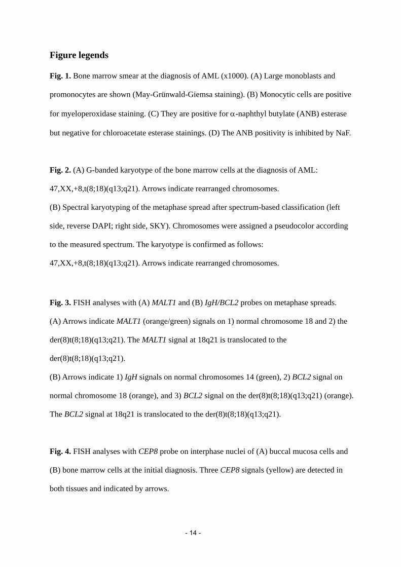
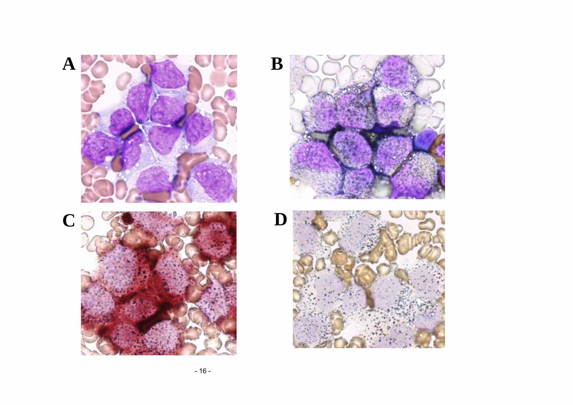
Fig. 1. Bone marrow smear at the diagnosis of AML (x1000). (A) Large monoblasts and
promonocytes are shown (May-Grünwald-Giemsa staining). (B) Monocytic cells are positive
for myeloperoxidase staining. (C) They are positive for α-naphthyl butylate (ANB) esterase
but negative for chloroacetate esterase stainings. (D) The ANB positivity is inhibited by NaF.
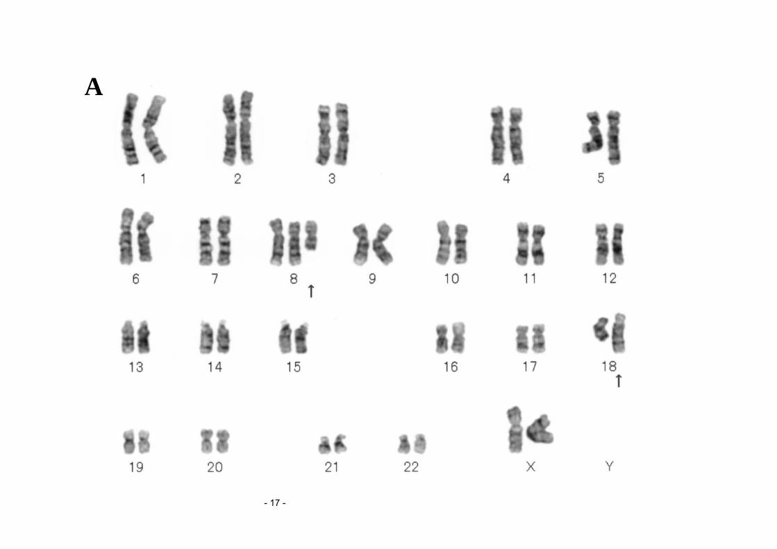
Fig. 2. (A) G-banded karyotype of the bone marrow cells at the diagnosis of AML:
47,XX,+8,t(8;18)(q13;q21). Arrows indicate rearranged chromosomes.
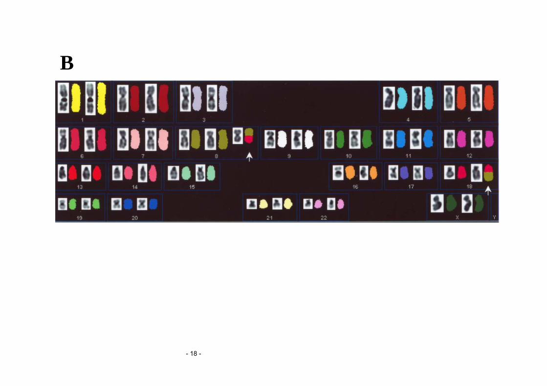
(B) Spectral karyotyping of the metaphase spread after spectrum-based classification (left
side, reverse DAPI; right side, SKY). Chromosomes were assigned a pseudocolor according
to the measured spectrum. The karyotype is confirmed as follows:
47,XX,+8,t(8;18)(q13;q21). Arrows indicate rearranged chromosomes.
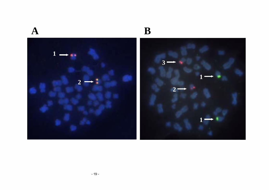
Fig. 3. FISH analyses with (A) MALT1 and (B) IgH/BCL2 probes on metaphase spreads.
(A) Arrows indicate MALT1 (orange/green) signals on 1) normal chromosome 18 and 2) the
der(8)t(8;18)(q13;q21). The MALT1 signal at 18q21 is translocated to the
der(8)t(8;18)(q13;q21).
(B) Arrows indicate 1) IgH signals on normal chromosomes 14 (green), 2) BCL2 signal on
normal chromosome 18 (orange), and 3) BCL2 signal on the der(8)t(8;18)(q13;q21) (orange).
The BCL2 signal at 18q21 is translocated to the der(8)t(8;18)(q13;q21).
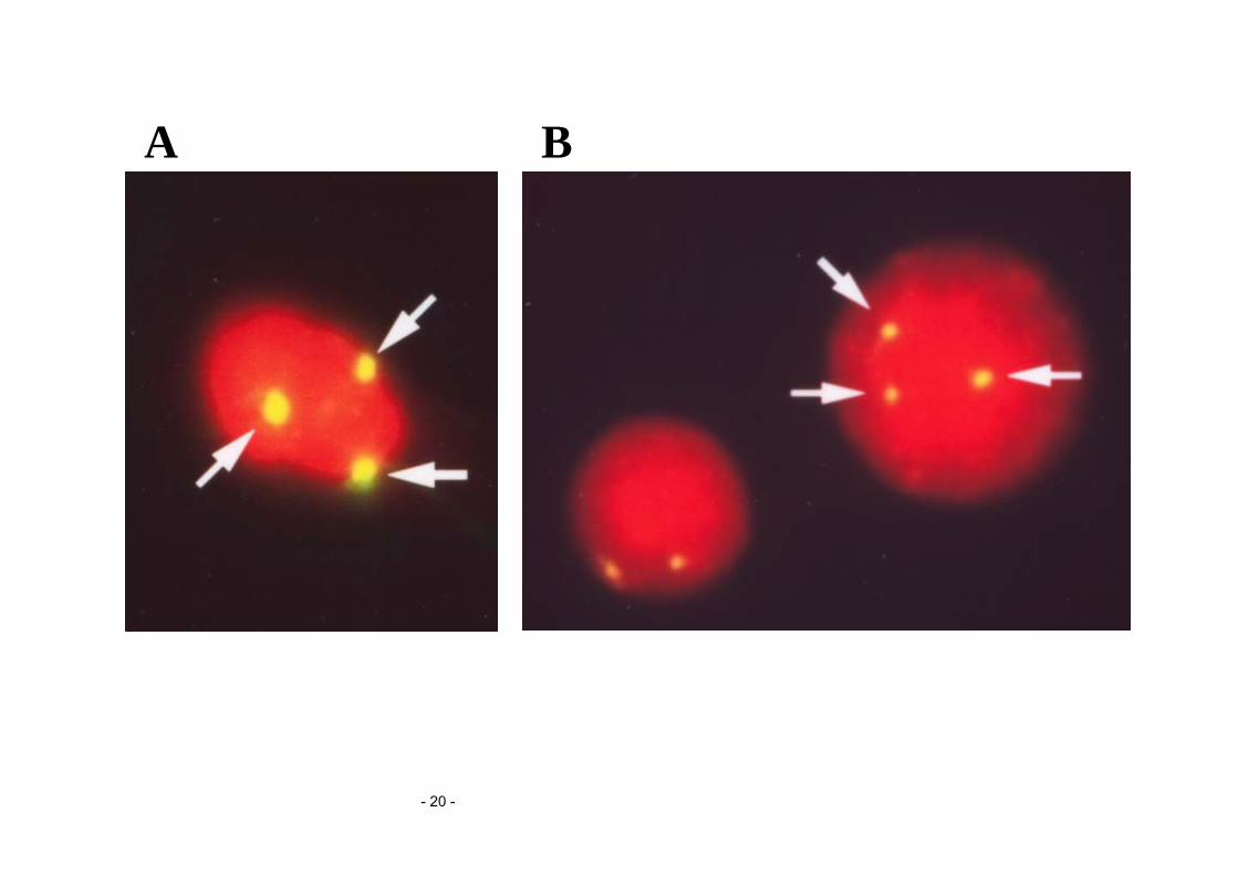
Fig. 4. FISH analyses with CEP8 probe on interphase nuclei of (A) buccal mucosa cells and
(B) bone marrow cells at the initial diagnosis. Three CEP8 signals (yellow) are detected in
both tissues and indicated by arrows.

- 15 -
- 16 -
A
C
B
D
- 17 -
A
- 18 -
B
- 19 -
A B
1
21
1
2
3
- 20 -
A B