Embed Size (px)
Citation preview

A
fImb(iiitHaJe©
K
1
tch(Tagtn
0d
Molecular Immunology 44 (2007) 3355–3363
A novel type-1 cytokine receptor from fish involved in the Januskinase/Signal transducers and activators of transcription
(Jak/STAT) signal pathway
Mudjekeewis D. Santos, Motoshige Yasuike, Hidehiro Kondo, Ikuo Hirono, Takashi Aoki ∗Laboratory of Genome Science, Tokyo University of Marine Science and Technology, Konan 4-5-7 Minato-ku, Tokyo 108-8477, Japan
Received 10 January 2007; accepted 18 February 2007Available online 26 March 2007
bstract
Type I cytokine receptors mediate the action of the members of the long chain cytokines canonically involved in numerous physiologicalunction. Here we report a novel cytokine receptor termed Japanese flounder glycoprotein 130 homologue or JfGPH, exhibiting the unique typecytokine receptor motifs i.e. having a cytokine binding domain (CBD) containing two pairs of conserved cysteine (C) residues, a WSXWSotif, three fibronectin domains all in the extracellular region. It is also composed of the Jak binding domains Box 1 and Box 2, and a STAT 3
inding motif (Box 3) in the cytoplasmic region suggesting its mediatory role for Janus kinase/Signal transducers and activators of transcriptionJak/STAT) signal pathway. The JfGPH cDNA is about 3 kb encoding 801 amino acid residues with a predicted molecular weight of 90 kDa andts gene has an 11-exon/10-intron architecture. While JfGPH shows significant homology with the members of type-1 cytokine receptor familyncluding IL6ST (or gp130), IL31� (or GLMR), CSF3R (or GCSFR), LIFR, OSMR, IL12R�1 and LEPR, structural and phylogenetic analysis ofts protein revealed that it is a novel and an ancestral cytokine receptor found in teleost. JfGPH gene is ubiquitously expressed in Japanese flounderissues and in a natural embryo (HINAE) cell line showing its critical role in teleost physiological functions similar to gp130 in higher vertebrates.igh expression of JfGPH transcripts in immune-related tissues and, in ovary and embryo-derived cell line suggest its role in immune responses,
nd reproduction/development, respectively. In vitro stimulation of spleen, kidney, peripheral blood leukocytes (PBLs) and HINAE revealed thatfGPH is down-regulated by polyinosinic:polycytidylic acid (poly I:C), an interferon (IFN) inducer, suggesting an apparent control of the JfGPH’sxpression during IFN-induced Jak/STAT signaling.
2007 Elsevier Ltd. All rights reserved.
r; No
oliita
vt
eywords: Japanese flounder (Paralichthys olivaceus); Type-1 cytokine recepto
. Introduction
Type-1 cytokine receptors are a group of related moleculeshat mediate the signaling action of class-1 helical cytokines,lassified as such based on a shared modular architecture i.e.aving a cytokine-binding domain (CBD), fibronectin type-IIIFn3) domains, and a signature WSXWS motif (Bazan, 1990;aga and Kishimoto, 1997). The cytokines these receptors medi-te, which include interleukin 6 signal transducer (IL6ST) or
lycoprotein 130 (gp130), granulocyte colony-stimulating fac-or (CSF3), interleukin 6 (IL-6), interelukin 11 (IL-11), ciliaryeurotrophic factor (CNTF), leukemia inhibiting factor (LIF),∗ Corresponding author. Tel.: +81 3 5463 0556; fax: +81 3 5463 0690.E-mail address: [email protected] (T. Aoki).
Arfdotp
161-5890/$ – see front matter © 2007 Elsevier Ltd. All rights reserved.oi:10.1016/j.molimm.2007.02.018
vel; Poly I:C; Jak/STAT signal pathway; Immunity; Development
ncostatin M (OSM), cardiotrophin-1 (CT-1) and cardiotrophin-ike cytokine (CLC) likewise share a common tertiary structure.e. composed of four bundles of �-helices and are involvedn numerous physiological processes including immune regula-ion, host defense, reproduction, development, blood formationnd energy metabolism (for review see Huising et al., 2006).
Type-1 cytokines and their cognate receptors form complexesia three interaction epitopes; a site I, located at the distal por-ion of the D helix (CBD), a site II, composed of residues in the
and C helices that allows for heterodimerization with othereceptors and, a site III, a feature only shown in gp130 receptoramily located at the N-terminal tip of the D helix- and Ig-like
omain (as reviewed by Bravo and Heath, 2000). Upon bindingf the cytokine ligand to its receptor, the Janus kinase/Signalransducers and activators of transcription (Jak/STAT) signalathway is activated. Janus kinases or Jaks are brought to the
3 Immu
Btbtvr(Stba
i(hcfofitdwi(tr2ti((tra(h(rep
sccstir
2
2
cia
KtB(G(GN
2
S(maAwJgp
2
mTddpIinpu(w(oiwm(Pd
2
caG5
356 M.D. Santos et al. / Molecular
ox 1 or Jak binding site, a proline rich motif, and in some recep-ors, to Box 2, a cluster of hydrophobic amino acids followedy positively charged amino acids. Jaks are then subsequentlyrans-phosphorylated and activated following binding. The acti-ated Jaks then phosphorylate the receptor chain and STATs areecruited through the interaction of the Src homology 2 domainSH2) with sites of receptor tyrosine phosphorylation such asTAT3 binding motif (Box 3). STATs then form dimers through
he intermolecular association of the SH2 domains with the car-oxyl sites of tyrosine phosphorylation (Ihle, 1996; Heinrich etl., 1998).
Fish class-1 helical cytokine orthologues have been reportedncluding IL6, IL11, carp cytokine-like (M17), interleukin 12IL12), leptin, erythropoietin (EPO), prolactin (PRL) and growthormone (GH) (Huising et al., 2006), and recently, the granulo-yte colony-stimulating factors (CSF3s) from Japanese flounder,ugu and pufferfish (Santos et al., 2006). Some cognate receptorsf these group of signal molecules have likewise been reported insh particularly from Tetraodon nigroviridis genome although
heir orthology is not yet clear since assignment of names wasone only up to the in silico-prediction level and the genesere compared only to human (Jaillon et al., 2004). These
nclude growth hormone receptor (GHR), prolactin receptorPRLR), erythropoietin receptor (EPOR), interleukin 12 recep-or � (IL2R�), interleukin 7 receptor A (IL7R�), interleukin 12eceptor �/interleukin 4 receptor A (IL2R�/IL4RA), interleukin1 receptor (IL21R), interleukin 12 p40 (IL12p40), ciliary neu-rophic factor (CNTFR), interleukin 11 receptor A (IL11R�),nterleukin 13 receptor A (IL13R�), interleukin 6 receptor AIL6R�), thrombopoietin receptor, interleukin 12 receptor �2IL12R�2), glycoprotein 130 (gp130), leukemia inhibiting fac-or receptor (LIFR) and obese protein receptor (OBR) or leptineceptor (LEPR). Fish cytokine receptors that have been clonednd characterized so far include LIFR and PRLR of gold fishHanington and Belosevic, 2005; Tse et al., 2000) and the growthormones of fugu, zebrafish, Southern catfish and Nile tilapiaJiao et al., 2006). Information about the kinds of type-1 cytokineeceptor molecules in fish and their function in cell signalling,.g. Jak/STAT pathway, which is important in understanding fishhysiological processes such as immunity, is very much lacking.
Here, we report a novel cytokine receptor gene that istructurally and phylogenetically related to the class-1 helicalytokine receptors. Because of its close similarity with gp130ompared with other cytokine receptors, we termed it as JfGPH,hort for Japanese flounder gp130 homologue. JfGPH appearso be a critical fish receptor because it is ubiquitously expressedn tissues and in an embryo-derived cell line, and is apparentlyegulated during IFN-induced Jak/STAT signalling.
. Materials and methods
.1. Cell culture
Primary cultures of Japanese flounder peripheral blood leuko-ytes were prepared using Percoll gradient isolation and placedn Medium B containing RPMI, 5% fetal bovine serum (FBS),nd 100 IU ml−1 penicillin G and 100 �g ml−1 streptomycin.
bcta
nology 44 (2007) 3355–3363
idney and spleen cells were prepared by slowly mash-filteringissues using a sterile mesh net and then placed in Medium. Japanese flounder-derived cell line Hirame Natural Embryo
HINAE) were grown in Leibovitz’s L-15 medium (Gibco-BRL,rand Island, NY) supplemented with 10% fetal bovine serum
FBS) (JRH Bioscience, Lenexa, KS) and 100 IU ml−1 penicillinand 100 �g ml−1 streptomycin (Gibco-BRL, Grand Island,
Y).
.2. Molecular cloning
The full-length JfGPH cDNA was determined followingantos et al. (2006). An expressed sequence tag (EST) cloneAccession no: AU050570) showing putative homology toouse CSF3 receptor (CSF3R) was used as a probe to screenJapanese flounder kidney cDNA library. 5′ SMART Randommplification of cDNA Ends (RACE) PCR and PCR cloningas used to complete the cDNA fragment generated from the
apanese flounder cDNA library. On the other hand, the JfGPHene was completed by “primer walking” using overlappingrimers designed from the JfGPH cDNA (Table S1).
.3. In silico analysis
The nucleotide sequence, translated amino acids, and averageolecular weight were analyzed and determined using GENE-YX 7.0.3 (GENETYX Corporation). SignalP (http://www.cbs.tu.dk/services/SignalP/) and NetNGlyc 1.0 (http://www.cbs.tu.dk/services/NetNGlyc/) servers were used to predict signaleptide cleavage and N-glycosylation sites, respectively.dentities were calculated using BLASTp (BLOSUM 62)mplemented in BLAST 2 SEQUENCES (http://www.ncbi.lm.nih.gov/blast/bl2seq/wblast2.cgi) and the complete multi-le amino acid alignments were carried out in CLUSTAL X 1.81sing default parameters. The ProDom Server, release 2005.1http://protein.toulouse.inra.fr/prodom/current/html/home.php/)as used to predict protein domains while Phobius
http://phobius.cgb.ki.se/) and TMpred (http://www.ch.embnet.rg/software/TMPRED form.html) servers were used todentify the transmembrane region. For phylogenetic analysis,e used the unweighted pair group method with arithmeticean (UPGMA) algorithm implemented in the MEGA3
http://www.megasoftware.net/index.html/) employing theoisson method with 1000 bootstrap tests and with completeeletion of gap sites. The bootstrap consensus tree was shown.
.4. Constitutive expression in tissues and cell lines
Regular RT- and semi-quantitative RT-PCR analysis wasarried out following Santos et al. (2006). Primers used formplification—JfGPH: Forward 5′-GTGCCACTGTGAGCT-GATCA-3′ ı̌and Reverse 3′-CTCTGACTCCGATAGGGGCT-′. For constitutive expression, total RNA was extracted from
rain, eyes, gills, kidney, heart, intestine, peripheral blood leuko-ytes (PBLs), liver, muscle, ovary, skin, spleen, stomach fromhree (3) apparently healthy Japanese flounder. �-actin was useds a positive control. PCR conditions were: initial denaturation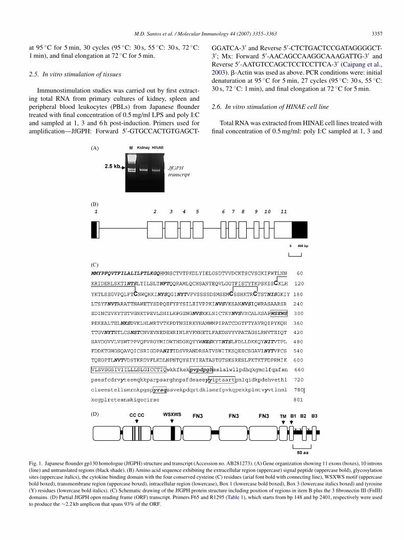
Immu
a1
2
iptaa
G3R2d3
F(sb(dt
M.D. Santos et al. / Molecular
t 95 ◦C for 5 min, 30 cycles (95 ◦C: 30 s, 55 ◦C: 30 s, 72 ◦C:min), and final elongation at 72 ◦C for 5 min.
.5. In vitro stimulation of tissues
Immunostimulation studies was carried out by first extract-ng total RNA from primary cultures of kidney, spleen and
eripheral blood leukocytes (PBLs) from Japanese flounderreated with final concentration of 0.5 mg/ml LPS and poly I:Cnd sampled at 1, 3 and 6 h post-induction. Primers used formplification—JfGPH: Forward 5′-GTGCCACTGTGAGCT-2
fi
ig. 1. Japanese flounder gp130 homologue (JfGPH) structure and transcript (Accessiline) and untranslated regions (black shade). (B) Amino acid sequence exhibiting theites (uppercase italics), the cytokine binding domain with the four conserved cysteineold boxed), transmembrane region (uppercase boxed), intracellular region (lowercasY) residues (lowercase bold italics). (C) Schematic drawing of the JfGPH protein stromains. (D) Partial JfGPH open reading frame (ORF) transcript. Primers F65 and Ro produce the ∼2.2 kb amplicon that spans 93% of the ORF.
nology 44 (2007) 3355–3363 3357
GATCA-3′ and Reverse 5′-CTCTGACTCCGATAGGGGCT-′; Mx: Forward 5′-AACAGCCAAGGCAAAGATTG-3′ andeverse 5′-AATGTCCAGCTCCTCCTTCA-3′ (Caipang et al.,003). �-Actin was used as above. PCR conditions were: initialenaturation at 95 ◦C for 5 min, 27 cycles (95 ◦C: 30 s, 55 ◦C:0 s, 72 ◦C: 1 min), and final elongation at 72 ◦C for 5 min.
.6. In vitro stimulation of HINAE cell line
Total RNA was extracted from HINAE cell lines treated withnal concentration of 0.5 mg/ml: poly I:C sampled at 1, 3 and
on no. AB281273). (A) Gene organization showing 11 exons (boxes), 10 intronsextracellular region (uppercase) signal peptide (uppercase bold), glycosylation(C) residues (arial font bold with connecting line), WSXWS motif (uppercase
e), Box 1 (lowercase bold boxed), Box 3 (lowercase italics boxed) and tyrosineucture including position of regions in item B plus the 3 fibronectin III (FnIII)1295 (Table 1), which starts from bp 148 and bp 2401, respectively were used
3 Immu
6ta1
3
3
fcsuP(go
ftwtacc
TCo
G
I
I
C
D
L
O
I
I
L
–
W(gatdBS6bDba
oebt
tft
358 M.D. Santos et al. / Molecular
h post-induction. Primers for JfGPH, Mx and �-actin werehe same as above. PCR conditions were: initial denaturationt 95 ◦C for 5 min, 27 cycles (95 ◦C: 30 s, 55 ◦C: 30 s, 72 ◦C:min), and final elongation at 72 ◦C for 5 min.
. Results
.1. Japanese flounder gp130 homologue (JfGPH)
Screening of a Japanese flounder cDNA library using an ESTragment corresponding to a CSF3R as a probe yielded a partialDNA fragment of about 2 kb that includes the polyA tail. Sub-equent RACE PCR of the 5′ region (confirmed with RT-PCRsing specific primers) completed the 3 kb cDNA fragment. RT-CR likewise verified the existence of the full JfGPH transcriptFig. 1A) BLAST analysis showed that it was most similar top130, thus it was named as Japanese flounder gp130 homologuer JfGPH (Table 1).
JfGPH cDNA is about 3 kb long and has an open readingrame (ORF) of 2406 bp (Fig. 1B, Fig. S1). It encodes for a pro-ein with 801 amino acid residues having a predicted moleculareight of 90 kDa. In silico analysis revealed that the JfGPH pro-
ein has an extracellular region of 600 amino acids that includespotential signal peptide of 21 amino acids, followed by a
ytokine binding domain (CBD) consisting of two (2) pairs ofonserved cysteine residues (aa 117 and 134; 163 and 170), a
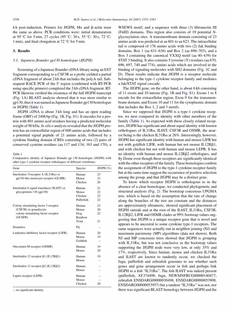
able 1omparative identity of Japanese flounder gp 130 homologue (JfGPH) withther type-1 cytokine receptor orthologues of different vertebrates
ene Species JfGPH (%)
nterleukin 31receptor A (IL31R�) orgp130-like monocyte receptor (GLMR)
Human 23Mouse 23Chicken 26
nterleukin 6 signal transducer (IL6ST) orglycoprotein 130 (gp130)
Human 21Mouse 22Chicken 22Pufferfish 22
olony stimulating-factor 3 receptor(CSF3R) or granulocytecolony-stimulating factor receptor(GCSFR)
Human 23Mouse 20Frog 22RainbowTrout 21
omeless Fly –
eukemia inhibitory factor receptor (LIFR) Human 20Mouse 19Goldfish –
ncostatin M receptor (OSMR) Human 19Mouse 20
nterleukin 12 receptor �1 (IL12R�1) Human 19Mouse –
nterleukin 12 receptor �2 (IL12R�2) Human –Mouse –
eptin receptor (LEPR) Human –Mouse –Chicken 20
: no significant identity.
oeJnanflwtba
astaaJIgasmNws1afgJ(zEt
nology 44 (2007) 3355–3363
SEWS motif, and a sequence with three (3) fibronectin IIIFnIII) domains. This region also consists of 19 potential N-lycosylation sites. A transmembrane domain consisting of 23mino acids was predicted at aa 601 to aa 623. The intracellularail is composed of 178 amino acids with two (2) Jak bindingomains, Box 1 (aa 631–636) and Box 2 (aa 696–703), and aox 3 containing the canonical YXXQ motif (aa 40–439) forTAT 3 binding. It also contains 5 tyrosine (Y) residues (aa 670,96, 697, 740 and 774), amino acids which are involved in theinding of signaling molecules with SH2 domains (Fig. 1C and). These results indicate that JfGPH is a receptor moleculeelonging to the type-1 cytokine receptor family and mediatesJak/STAT signal cascade.
The JfGPH gene, on the other hand, is about 6 kb consistingf 11 exons and 10 introns (Fig. 1B and Fig. S1). Exons 1 to 8ncode for the extracellular region, Exon 9 for the transmem-rane domain, and Exons 10 and 11 for the cytoplasmic domainhat includes the Box 1, 2 and 3 motifs.
Since we supposed that JfGPH is a type-1 cytokine recep-or, we next compared its identity with other members of theamily (Table 1). As expected with these closely related recep-ors, JfGPH has significant and about equal identity with knownrthologues of IL31R�, IL6ST, CSF3R and OSMR, the near-st being is the chicken IL31R� at 26%. Interestingly, however,fGPH has significant identity with human and mouse LIFR butot with goldfish LIFR, with human but not mouse IL12R�1,nd with chicken but not with human and mouse LEPR. It haso identity with human and mouse IL12R�2 orthologues, andy Dome even though these receptors are significantly identicalith the other receptors of the family. These homologies confirm
he assignment of JfGPH to the type-1 cytokine receptor familyut at the same time suggest the occurrence of positive selectionmong the group, and that JfGPH may be a distinct gene.
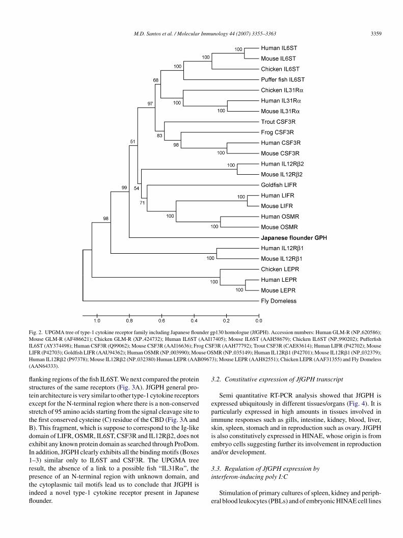
To know which receptor JfGPH is orthologous to in thebsence of a clear homologue, we conducted phylogenetic andtructural analysis (Fig. 2). The bootstrap concensus UPGMAree, which is based on the assumption that the rate of changelong the branches of the tree are constant and the distancesre approximately ultrameric, showed significant placement offGPH outside and at the root of the IL6ST, IL31R�, CSF3R,L12R�2, LIFR and OSMR clades at 99% bootsrap values sug-esting that JfGPH is a unique receptor gene that is novel andppears to be ancestral to some cytokine type-1 receptors. Theame sequences were actually run in neighbor-joining (NJ) andaximum parsimony (MP) algorithms (data not shown). BothJ and MP concensus trees showed that JfGPH is groupingith IL31R�, but was not conclusive as the bootstrap values
upporting the JfGPH node were very low, at only 35% and7%, respectively. Since human, mouse and chicken IL31R�nd IL6ST are known to tandemly occur, we checked theugu, pufferfish and zebrafish genomes to see whether suchenes and gene arrangement occur in fish and perhaps linkfGPH to a fish “IL31R�”. The fish IL6ST was indeed present
pufferfish, AY374498; fugu, NEWSINFRUG00000140477;ebrafish ENSDARG00000030498, ENSDARG00000053908,NSDARG00000053957) but a tandem “IL31R�” was not, norhere was significant BLAST homology between JfGPH and the
M.D. Santos et al. / Molecular Immunology 44 (2007) 3355–3363 3359
Fig. 2. UPGMA tree of type-1 cytokine receptor family including Japanese flounder gp130 homologue (JfGPH). Accession numbers: Human GLM-R (NP 620586);Mouse GLM-R (AF486621); Chicken GLM-R (XP 424732); Human IL6ST (AAI17405); Mouse IL6ST (AAH58679); Chicken IL6ST (NP 990202); PufferfishI g CSL se OSH 0967(
flstestBdeI1rptifl
3
episiea
3
L6ST (AY374498); Human CSF3R (Q99062); Mouse CSF3R (AAI16636); FroIFR (P42703); Goldfish LIFR (AAU94362); Human OSMR (NP 003990); Mouuman IL12R�2 (P97378); Mouse IL12R�2 (NP 032380) Human LEPR (AAB
AAN64333).
anking regions of the fish IL6ST. We next compared the proteintructures of the same receptors (Fig. 3A). JfGPH general pro-ein architecture is very similar to other type-1 cytokine receptorsxcept for the N-terminal region where there is a non-conservedtretch of 95 amino acids starting from the signal cleavage site tohe first conserved cysteine (C) residue of the CBD (Fig. 3A and). This fragment, which is suppose to correspond to the Ig-likeomain of LIFR, OSMR, IL6ST, CSF3R and IL12R�2, does notxhibit any known protein domain as searched through ProDom.n addition, JfGPH clearly exhibits all the binding motifs (Boxes–3) similar only to IL6ST and CSF3R. The UPGMA treeesult, the absence of a link to a possible fish “IL31R�”, the
resence of an N-terminal region with unknown domain, andhe cytoplasmic tail motifs lead us to conclude that JfGPH isndeed a novel type-1 cytokine receptor present in Japaneseounder.i
e
F3R (AAH77792); Trout CSF3R (CAE83614); Human LIFR (P42702); MouseMR (NP 035149); Human IL12R�1 (P42701); Mouse IL12R�1 (NP 032379);
3); Mouse LEPR (AAH82551); Chicken LEPR (AAF31355) and Fly Domeless
.2. Constitutive expression of JfGPH transcript
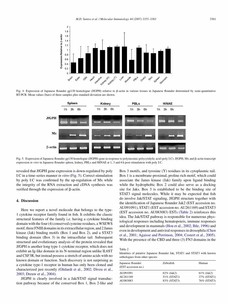
Semi quantitative RT-PCR analysis showed that JfGPH isxpressed ubiquitously in different tissues/organs (Fig. 4). It isarticularly expressed in high amounts in tissues involved inmmune responses such as gills, intestine, kidney, blood, liver,kin, spleen, stomach and in reproduction such as ovary. JfGPHs also constitutively expressed in HINAE, whose origin is frommbryo cells suggesting further its involvement in reproductionnd/or development.
.3. Regulation of JfGPH expression by
nterferon-inducing poly I:CStimulation of primary cultures of spleen, kidney and periph-ral blood leukocytes (PBLs) and of embryonic HINAE cell lines

3360 M.D. Santos et al. / Molecular Immunology 44 (2007) 3355–3363
Fig. 3. Comparison of class 1 helical cytokine receptors with significant identity with Japanese flounder gp130 homologue (JfGPH). (A) Schematic drawing ofprotein motifs and domains (similar to Fig. 2C) are shown. Figure with question mark corresponds to the 93 amino acids in the N-terminal region with no knowndomain. (B) Multiple sequence alignment of the Ig-like domain of IL6ST and CSF3R, and the sequences upstream of the first cysteine residue of IL31R� and JfGPHminus the leader peptide. Conserved amino acids (dots), introduced gaps (dash) and the first conserved cysteine residue of the cytokine binding domain (boxed) areshown.
M.D. Santos et al. / Molecular Immunology 44 (2007) 3355–3363 3361
Fig. 4. Expression of Japanese flounder gp130 homologue (JfGPH) relative to �-actin in various tissues in Japanese flounder determined by semi-quantitativeRT-PCR. Mean values (bars) of three samples plus standard deviation are shown.
F onsee 3 and
rIbtv
4
1sdmkbsJeakac2
t
BBawsSdtA(iiand development in mammals (Hou et al., 2002; Ihle, 1996) andeven in development and antiviral responses in drosophila (Chenet al., 2001; Agaisse and Perrimon, 2004; Costert et al., 2005).With the presence of the CBD and three (3) FN3 domains in the
Table 2Identities of putative Japanese flounder Jak, STAT1 and STAT3 with knownorthologues from other species
Japanese flounder Zebrafish Human
ig. 5. Expression of Japanese flounder gp130 homologue (JfGPH) gene in respxpression in vitro in Japanese flounder spleen, kidney, PBLs and HINAE at 1,
evealed that JfGPH gene expression is down-regulated by poly:C in a time-series manner in vitro (Fig. 5). Correct stimulationy poly I:C was confirmed by the up-regulation of Mx whilehe integrity of the RNA extraction and cDNA synthesis waserified through the expression of �-actin.
. Discussion
Here we report a novel molecule that belongs to the type-cytokine receptor family found in fish. It exhibits the classic
tructural features of the family i.e. having a cytokine bindingomain with the four (4) conserved cysteine residues, a WSEWSotif, three FNIII domains in its extracellular region, and 2 Janus
inase (Jak) binding motifs (Box 1 and Box 2), and a STAT3inding domain (Box 3) in the intracellular tail. Subsequenttructural and evolutionary analysis of the protein revealed thatfGPH is another long type-1 cytokine receptor, which does notxhibit an Ig-like domain in its N-terminal region unlike IL6STnd CSF3R, but instead possess a stretch of amino acids with nonown domain or function. Such discovery is not surprising ascytokine type-1 receptor in human has only been cloned and
haracterized just recently (Ghilardi et al., 2002; Diveu et al.,003; Dreuw et al., 2004).
JfGPH is clearly involved in a Jak/STAT signal transduc-ion pathway because of the conserved Box 1, Box 2-like and
(
AAA
to polyinosinic:polycytidylic acid (poly I:C). JfGPH, Mx and �-actin transcript6 h post-stimulation with poly I:C.
ox 3 motifs, and tyrosine (Y) residues in its cytoplasmic tail.ox 1 is a membrane-proximal, proline rich motif, which couldssociate the Janus kinase (Jak) family upon ligand bindinghile the hydrophobic Box 2 could also serve as a docking
ite for Jaks. Box 3 is established to be the binding site ofTAT3 signal molecules. While it may be expected that fisho involve Jak/STAT signaling, JfGPH structure together withhe identification of Japanese flounder Jak2 (EST accession no.U091091), STAT1 (EST accession no. AU261169) and STAT3
EST accession no. AU083083) ESTs (Table 2) reinforces thisdea. The Jak/STAT pathway is responsible for numerous phys-ological responses including hematopoieis, immune responses
EST accession no.)
U091091 62% (Jak2) 61% (Jak2)U261169 51% (STAT1) 37% (STAT1)U083083 83% (STAT3) 76% (STAT3)
3362 M.D. Santos et al. / Molecular Immunology 44 (2007) 3355–3363
F f tranfl apanel func
eIcHttnptstatIhs
ahoJvwetc
srto2tJscToi
ewaitbtte
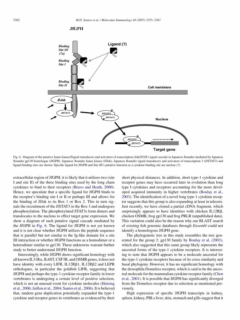
ig. 6. Diagram of the putative Janus kinase/Signal transducers and activators oounder gp130 homologue (JfGPH). Japanese flounder Janus kinase (JfJak), J
igand binding sites are shown. Specific ligand for JfGPH and Site III’s putative
xtracellular region of JfGPH, it is likely that it utilizes two (siteand site II) of the three binding sites used by the long chainytokines to bind to their receptors (Bravo and Heath, 2000).ence, we speculate that a specific ligand for JfGPH binds to
he receptor’s binding site I or II or perhaps III and allows forhe binding of JfJak to its Box 1 or Box 2. This in turn sig-als the recruitment of the JfSTAT3 in the Box 3 and undergoeshosphorylation. The phosphorylated STAT3s form dimers andranslocates to the nucleus to effect target gene expression. Wehow a diagram of such putative signal cascade mediated byhe JfGPH in Fig. 6. The ligand for JfGPH is not yet knownnd it is not clear whether JfGPH utilizes the peptide sequencehat is parallel but not similar to the Ig-like domain for a siteII interaction or whether JfGPH functions as a homodimer or aeterodimer similar to gp130. These unknowns warrant furthertudy to better understand JfGPH function.
Interestingly, while JfGPH shows significant homology withll known IL31R�, IL6ST, CSF3R, and OSMR genes, it does notave identity with every LIFR, IL12R�1, IL12R�2 and LEPRrthologues, in particular the goldfish LIFR, suggesting thatfGPH and perhaps the type-1 cytokine receptor family in lowerertebrates is undergoing a certain level of positive selection,
hich is not an unusual event for cytokine molecules (Huisingt al., 2006; Jaillon et al., 2004; Santos et al., 2006). It is believedhat, tandem gene duplication potentially expanded the type-1ytokine and receptor genes in vertebrates as evidenced by their
fv
s
scription (Jak/STAT) signal cascade in Japanese flounder mediated by Japanesese flounder signal transducers and activators of transcription 3 (JfSTAT3) andtion as a cytokine binding site are unclear (?).
hort physical distances. In addition, short type-1 cytokine andeceptor genes may have occurred later in evolution than longype I cytokines and receptors accounting for the more devel-ped acquired immunity in higher vertebrates (Boulay et al.,003). The identification of a novel long type-1 cytokine recep-or suggests that this group is also expanding at least in teleosts.ust recently, we have cloned a partial cDNA fragment, whichurprisingly appears to have identities with chicken IL12R�,hicken OSMR, frog gp130 and frog PRLR (unpublished data).his variation could also be the reason why our BLAST searchf existing fish genomic databases through Ensembl could notdentify a homologous JfGPH gene.
The phylogenetic tree in this study resembles the tree gen-rated for the group 2: gp130 family by Boulay et al. (2003),hich also suggested that this same group likely represents the
ncestral forms of the type-1 cytokine receptors. It is interest-ng to note that JfGPH appears to be a molecule ancestral forhe type-1 cytokine receptors because of its cross similarity andasal phylogeny. However, it has no significant homology withhe drosophila Domeless receptor, which is said to be the ances-ral molecule for the mammalian cytokine receptor family (Chent al., 2001). It is possible that JfGPH has significantly diverged
rom the Domeless receptor due to selection as mentioned pre-iously.High expression of specific JfGPH transcripts in kidney,pleen, kidney, PBLs liver, skin, stomach and gills suggest that it

Immu
iecTpwbo(
trtkitstoII2aa
ciJ(haur
A
SSta
A
i
R
A
B
B
B
C
C
C
D
D
G
H
H
H
H
I
J
J
K
N
P
S
M.D. Santos et al. / Molecular
s involved in immune responses. Moreover, JfGPH’s significantxpression in ovary and in a cell line whose origin is from embryoells shows that it has a role in reproduction and development.his highlights the important role of JfGPH in the physiologicalrocesses in fish similar to gp130, a type-1 cytokine receptorell studied in higher vertebrate. Gp130 has been established toe a critical receptor molecule to an organism such that mutationr knockdown of the said gene has been fatal to the organismKishimoto et al., 1995).
The involvement of JfGPH in immunity and particularly inhe Jak/STAT pathway is further confirmed through the down-egulation of its expression following poly I:C stimulation inissues and in cell lines. poly I:C is a double stranded RNAnown to induce IFN-�/� production. We speculate that the IFN-nduced Jak/STAT pathway somehow inhibits the expression andhus the function of JfGPH-dependent Jak/STAT signaling pre-umably to regulate and balance the system. Actual mechanismhough should be confirmed and fully explored. Down regulationf JfGPH by poly I:C is in contrast to the up-regulation of GPL,L12R�2 and IL23R �1, receptors homologous to JfGPH, byFN-� treatment in monocytes and dendritic cells (Diveu et al.,003; Parham et al., 2002). The difference in JfGPH expressions compared with other receptors further reflects its uniquenesst the transcriptional level.
It is important to identify the ligand(s) that could specifi-ally bind to JfGPH and confirm the Jak/STAT signal cascadet would induce. Four (4) cytokines of the IL-6 cytokines fromapanese flounder namely poCSF3 (Santos et al., 2006), IL6Nam et al., 2007), IL11 type b (unpublished data) and an M17omologue (submitted) have been cloned and are potential lig-nds. Identification of the JfGPH ligand could greatly help innderstanding further the biological function of JfGPH and otherelated receptors.
cknowledgements
This study was supported in part by the Grants-in-Aid forcientific Research (S) from the Ministry of Education, Culture,ports, Science and Technology of Japan. We would also like to
hank Fernand Fagutao for his assistance in doing the statisticalnalysis.
ppendix A. Supplementary data
Supplementary data associated with this article can be found,n the online version, at doi:10.1016/j.molimm.2007.02.018.
eferences
gaisse, H., Perrimon, N., 2004. The roles of Jak/STAT signaling in Drosophilaimmune responses. Immunol. Rev. 198, 72–82.
azan, J.F., 1990. Structural design and molecular evolution of a cytokine recep-tor superfamily. Proc. Natl. Acad. Sci. 87, 6934–6938.
T
T
nology 44 (2007) 3355–3363 3363
oulay, J.L., O’Shea, J.J., Paul, W.E., 2003. Molecular phylogeny within type Icytokines and their cognate receptors. Immunity 19, 159–163.
ravo, J., Heath, J.K., 2000. Receptor recognition by gp130 cytokines. EMBOJ. 19, 2399–2411.
aipang, C.M.A., Hirono, I., Aoki, T., 2003. In vitro inhibition of fish rhab-doviruses by Japanese flounder, Paralichthys olivaceus Mx. Virology 317,373–382.
hen, H.W., Chen, X., Oh, S.W., Marinissen, M.J., Gutkind, J.S., Hou, S.X.,2001. mom identifies a receptor for the Drosophila JAK/STAT signal trans-duction pathway and encodes a protein distantly related to the mammaliancytokine receptor family. Genes Dev. 16, 388–398.
ostert, C., Jouanguy, E., Irving, P., Troxler, L., Galiana-Arnoux, D., Hetru, C.,Hoffman, J.A., Imler, J.L., 2005. The Jak-STAT pathway is required but notsufficient for the antiviral response of drosophila. Nat. Immunol. 6, 946–953.
iveu, C., Lelievre, E., Perret, D., Lak-Hal, A.L., Froger, J., Guillet, C., Cheva-lier, S., Rousseau, F., Wesa, A., Preisser, L., Chabbert, M., Gauchat, J.,Galy, A., Gascan, H., Morel, A., 2003. GPL, a novel cytokine receptorrelated to gp130 amd leukemia inhibiting factor receptor. J. Biol. Chem.278, 49850–49859.
reuw, A., Radtke, S., Pflanz, S., Lippok, B.E., Heinrich, P.C., Hermanns, H.M.,2004. Characterization of the signaling capacities of the novel gp130-likecytokine receptor. J. Biol. Chem. 279, 36112–36120.
hilardi, N., Li, J., Hongo, J.A., Yi, S., Gurney, A., de Sauvage, F.J., 2002. Anovel type 1 cytokine receptor is expressed on monocytes, signals prolifer-ation, and activates STAT-3 and STAT-5. J. Biol. Chem. 277, 16831–16836.
anington, P.C., Belosevic, M., 2005. Characterization of the leukemiainhibitory factor receptor in the goldfish (Carassius auratus). Fish ShellfishImmunol. 18, 359–369.
einrich, P.C., Behrmann, I., Muller-Newen, G., Schaper, F., Graeve, L., 1998.Interleukin-6-type cytokine signaling through the gp130/Jak/STAT pathway.Biochem. J. 334, 297–314.
ou, S.X., Zheng, Z., Chen, X., Perrimon, N., 2002. The Jak/STAT Pathway inmodel organisms: emerging roles in cell movement. Dev. Cell 3, 765–778.
uising, M.O., Kruiswijk, C.P., Flik, G., 2006. Phylogeny and evolution ofclass-I helical cytokines. J. Endocrinol. 189, 1–25.
hle, J.N., 1996. STATs: signal transducers and activators of transcription. Cell84, 331–334.
aillon, O., Aury, J.M., Brunet, F., Petit, J.L., Stange-Thomann, N., Mauceli,E., Bouneau, L., Fischer, C., Ozouf-Costaz, C., Bernot, A., et al., 2004.Genome duplication in the teleost fish Tetraodon nigroviridis reveals theearly vertebrate proto-karyotype. Nature 431, 946–957.
iao, B., Huang, X., Chan, C.B., Zhang, L., Wang, D., Cheng, C.H.K., 2006.The co-existence of two growth hormone receptors in teleost fish and theirdifferential signal transduction, tissue distribution and hormonal regulationof expression in seabream. J. Mol. Endocrinol. 36, 23–40.
ishimoto, T., Akira, S., Narazaki, M., Taga, T., 1995. Interleukin-6 family ofcytokines and gp130. Blood 86, 1243–1254.
am, B.H., Byon, J.Y., Kim, Y.O., Park, E.M., Cho, Y.C., Cheong, J.C., 2007.Molecular cloning and characterization of the flounder (Paralichthys oli-vaceus) interleukin-6 gene. Fish Shellfish Immunol. 23, 231–236.
arham, C., Chirica, M., Timans, J., Vaisberg, E., Travis, M., Cheung, J., Pflanz,S., Zhang, R., Singh, K.P., Vega, F., et al., 2002. A receptor for the het-erodimeric cytokine IL-23 is composed of IL-12R�1 and a novel cytokinereceptor subunit, IL-23R. J. Immunol. 168, 5699–5708.
antos, M.D., Yasuike, M., Hirono, I., Aoki, T., 2006. The granulocyte colonystimulating factors (CSF3s) of fish and chicken. Immunogenetics 56,422–432.
aga, T., Kishimoto, T., 1997. Gp130 and the interleukin-6 family of cytokines.Ann. Rev. Immunol. 15, 797–819.
se, D.L.Y., Chow, B.K.C., Chan, C.B., Lee, L.Y.O., Cheng, C.H.K., 2000.Molecular cloning and expression studies of a prolactin receptor in goldfish(Carassius auratus). Life Sci. 66, 593–605.