Embed Size (px)
Citation preview

Acute Kidney Injury in the ICU
Sept 8, 2016
Ed Gould, MD Assistant Professor of Medicine
Vanderbilt Division of Nephrology


“Oh the places we’ll go…”
• Definition
• Epidemiology
• Risk Factors, Etiology, and Pathophysiology
• Conservative Management
• Contrast Nephropathy
• Renal Replacement Therapy

Definitions
(Cruz, Ricci et al. 2009)

“A Rose By Any Other Name…”
64% 16%
14%
6% RIFLE
None
Risk/ 1
63% 18%
10%
9%
AKIN
Large New Zealand Cohort. Similar Incidence of AKI between categories.
Perhaps more importantly, the Mortality Odds Ratio between the categories was very similar
(Bagshaw, George et al. 2008)

Take Home Point #1
Can stratify AKI according to degree of injury; which system you use probably doesn’t matter.

Breadth of the Problem
AKI Among All Hospitalized Patients:: 5 - 7.5%
AKI Among ICU Patients:: ?
USRDS database

Breadth of the Problem
(Hoste, Clermont et al. 2006)
33%
12% 27%
28%
Degree of Acute Kidney Injury in ICU
No Renal Failure
Risk
Injury
Failure
UPMC had 7 ICUs • Collected data from 5383
patients • Excluded those who went
on to need HD
• No AKI: 33% • Risk: 12% • Injury: 27% • Failure: 28%

Depth of the Problem AKI also statistically changes the prognosis of patients in the ICU…
The risk of death for all ICU patients: ~8-19%
Risk of death for ICU patients with AKI: 25-40%
Risk of developing HD requirement: 5-6%
Risk of death for patients requiring HD: 50-80%

Depth of the Problem
USRDS database
43%
And if they survive the hospital?
This is based on Medicare data, so the included patients had AKI coded at the time of discharge.

Take Home Point #2
AKI is common, and may significantly impact mortality both in and after the hospitalization
Alternatively, it may be a marker for mortality “susceptibility”

So what causes it and how?
0
20
40
60
80
Prerenal Intrarenal Obstruct Idiopath
Outpatient
Inpatient
(Nash, Hafeez et al. 2002)

Etiology - Risk Factors
• Old Age (> 75 yrs)
• Chronic kidney disease (eGFR < 60 mls/min/1.73m2)
– The worse your function is at baseline, the greater the risk
• Cardiovascular Disease
– CHF
– PVD
• Liver Disease
• Diabetes Mellitus
Note that by the time you all meet these folks,
these risks are non-modifiable

Etiology – Making Urine
• 25% of all Cardiac Output (CO) goes straight to the kidney.
• Blood moves through the arteriolar system into the glomeruli.
• The glomerular filtration barrier allows for production of urinary filtrate.
• The tubules modify that filtrate by reabsorbing things of value and secreting things too large to be filtered
• Urine moves into the renal pelvis, through the ureters and into the bladder.

Etiology - Broad Strokes
1 2
3
1. Prerenal (Hemodynamic Causes): Anything that disturbs blood flood to the glomeruli.
2. Intrarenal: Anything that disturbs the glomerular or tubular architecture.
3. Postrenal: Acute obstruction with increased pressure referred back to Bowmans Space.
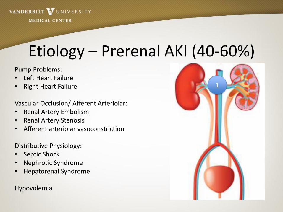
Etiology – Prerenal AKI (40-60%)
1
Pump Problems: • Left Heart Failure • Right Heart Failure Vascular Occlusion/ Afferent Arteriolar: • Renal Artery Embolism • Renal Artery Stenosis • Afferent arteriolar vasoconstriction Distributive Physiology: • Septic Shock • Nephrotic Syndrome • Hepatorenal Syndrome Hypovolemia

Etiology – Intrarenal AKI (20-40%)
2
Glomerulonephritides • Too many to list Vascular Diseases • Thrombotic Microangiopathy • HUS • HTN, DMII • Calcineurin Inhibitor Toxicity Tubular Diseases • Acute Tubular Necrosis • Allergic Interstitial Nephritis • Direct Tubular Toxins • Tumor Lysis Syndrome

Etiology – Obstructive (<10%)
3
High Obstruction (Renal Pelvis) • Papillary Necrosis • Struvite Stones Bilateral Ureteral Obstruction • Nephrolithiasis • Extrinsic Compression Bladder Outlet Obstruction • Prostatic Disease • Bladder CA • Ureteral diseases

Great… I Have Another Stupid List
The question immediately becomes, how do you parse through it? How do you meaningfully narrow it into something intervenable?
1. History and physical examination
2. Identify your toolset and apply deliberately…

Evaluation of Kidney Function…
Clearance:
1. Serum Cr (Cystatin C)
2. 24 hour Cr Clearance
Perfusion:
1. Urine Output
Tubular Function:
1. Urinalysis
2. FENa
3. Urine Osmolarity
Structural Injury:
1. Renal Ultrasound
2. Urinalysis
3. Proteinuria quantification

Serum Creatinine
• MDRD is meaningless in AKI
• A small change in Creatinine is VERY meaningful
(HP Lefebvre 2016 International Renal Interest Society )

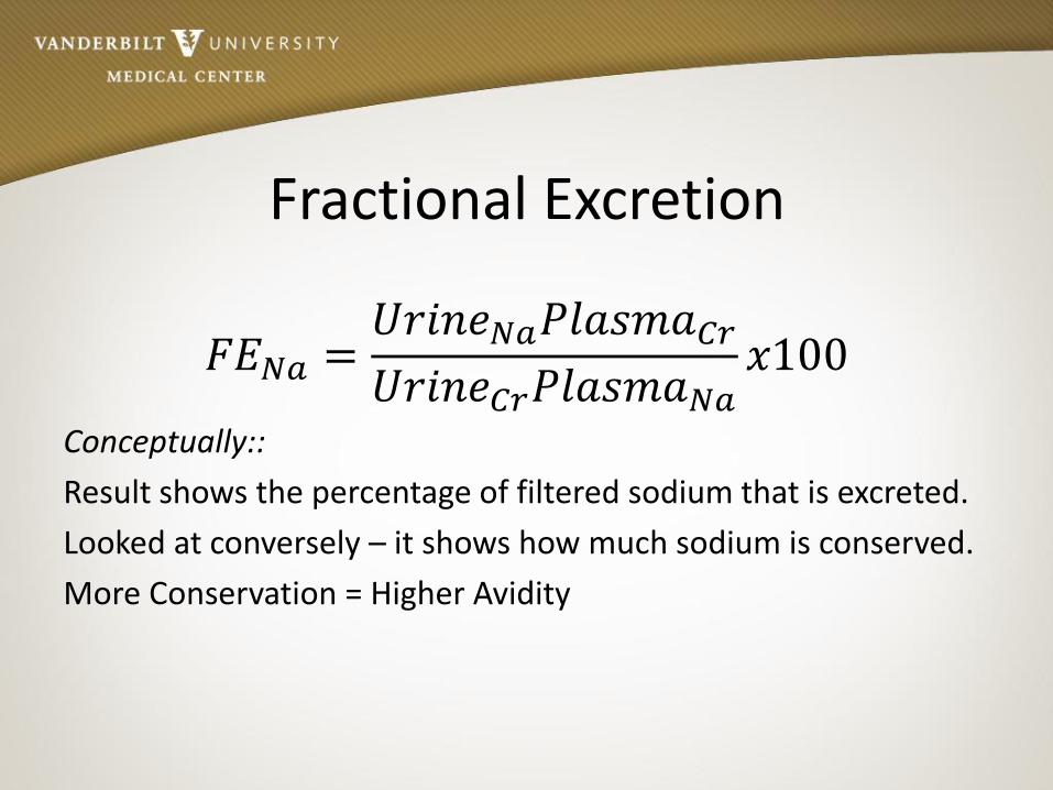
Fractional Excretion
Fraction Excretion of Sodium (or Urea) gives you insight into the avidity of the kidney for solute.
A highly avid kidney is trying to reclaim solute (and therefore water) maximally…
The kidney is “thirsty”…

Fractional Excretion
Interpretation, simplified:
Less than 1% sodium excreted (or 35% Urea) means the kidney is working as hard as it can to conserve solute and water.
Prerenal Etiology
Above 1% = Something else
Fractional Excretion
𝐹𝐸𝑁𝑎 =𝑈𝑟𝑖𝑛𝑒𝑁𝑎𝑃𝑙𝑎𝑠𝑚𝑎𝐶𝑟𝑈𝑟𝑖𝑛𝑒𝐶𝑟𝑃𝑙𝑎𝑠𝑚𝑎𝑁𝑎
𝑥100
Conceptually::
Result shows the percentage of filtered sodium that is excreted.
Looked at conversely – it shows how much sodium is conserved.
More Conservation = Higher Avidity

Fractional Excretion - Pitfalls
FENa is most useful early in ICU stay: 1. AKI with oliguria
2. Minimal background CKD
3. Absence of Metabolic Alkalosis
4. Absence of nonreabsorbable ions
5. No diuretics
FEUrea has similar restrictions, except, diuretics less of an impact.
(Nguyen, Maynard et al. 2009)

Proteinuria
Proteinuria is a marker of glomerular breakdown.
In AKI, some small amount of proteinuria is “acceptable”
If large protein losses, this suggests glomerular disease…

Prerenal AKI
ATN AIN Postrenal AKI
Etiology Shock, hypovolemia
Ischemia Anaphylaxis, Allergic Drug Rx
Obstruction
Urine Na < 20 > 20 Variable Variable
FeNa < 1% > 1% Variable Variable
Urine osms Concentrated Isoosmolar Isoosmolar to dilute
Iso to dilute
Urinary sediment
Hyaline casts Muddy Brown Casts
White Cells, White Cell Casts, maybe eos
Variable
Renal U/S Nl kidneys Nl or echogenic
Echogenic Dilated urinary space

Take Home Point #3
Develop a differential based on the available history, then refine with deliberate testing…

Conservative Management Prerenal AKI Step 1: Identify Underlying Cause and Treat
Step 2: Attempt to perfuse glomeruli and tubules to prevent development of ATN.
a. Fluid challenge - Crystalloid: 15-30mL/kg x1, repeat based on exam and monitoring
- Colloid: No proven benefit (though…)
b. If clinically volume overloaded - Consider Lasix challenge: one dose of 1mg/kg
c. Uncertainty? - Evaluate with CVP, echo for IVC fullness
- Empirical trial of a or b

Conservative Management Intrarenal AKI ATN:
Challenging. No “Magic Bullet” Treat the cause! Support: - Optimize hemodyn. - Ensure volume replete - Follow electrolytes - Treat aberrancies as
they arise - When the time comes,
initiate hemodialysis
Glomerular Diseases: Challenging. Varies according the specific cause. Thankfully rare in the ICU. Consult Nephrology for help.
AIN: Challenging. Steroids might benefit. Support: - Optimize
hemodynamics - Remove the offending
agent. - Support with
Hemodialysis if needed

Conservative Management
Obstructive Nephropathy Relieve the obstruction.

Take Home Point #4
Narrow the differential, treat what you can, support through what you can’t.

A word about Contrast Nephropathy
Common in certain ICU settings.
Classically:
2% of the general population develop AKI, up to 50% of people with multiple comorbidities.
Risk Factors Exposure Recovery AKI
No change

Contrast Nephropathy
Focus on prevention:
Essentially data has shown that only intravascular volume seems to matter.
No benefit (or harm) for NAC, nor alkalinization (add’l slides)

Take Home Point #5
The only proven intervention for Contrast Nephropathy is pre-procedural euvolemia.

When to dialyze?
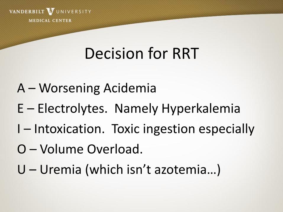
Decision for RRT
A – Worsening Acidemia
E – Electrolytes. Namely Hyperkalemia
I – Intoxication. Toxic ingestion especially
O – Volume Overload.
U – Uremia (which isn’t azotemia…)

Decision for RRT
Decision to start hemodialysis is highly dependent on the details of the case.
(Gaudry, Hajage et al. 2016)
One of Many…

Decision for RRT – personal word
Dialysis is a significant threshold.
Given that it generally signifies a worsening underlying process, it provides an opportunity to revisit goals of care.

Take Home Point #6
No data on when to start HD in a patient in ICU with AKI. Seek guidance of Nephrologist, but
leverage your opinion.

“Oh the places we’ve been…”
• Definition
• Epidemiology
• Risk Factors, Etiology, and Pathophysiology
• Conservative Management
• Contrast Nephropathy
• Renal Replacement Therapy

Questions?
Ed Gould, MD Assistant Professor of Medicine
Vanderbilt Division of Nephrology

References
1. Bagshaw SM, George C, Bellomo R, Committe ADM. A comparison of the RIFLE and AKIN criteria for acute kidney injury in critically ill patients. Nephrology, dialysis, transplantation : official publication of the European Dialysis and Transplant Association - European Renal Association 2008;23:1569-74.
2. Case J, Khan S, Khalid R, Khan A. Epidemiology of acute kidney injury in the intensive care unit. Critical care research and practice 2013;2013:479730.
3. Cruz DN, Ricci Z, Ronco C. Clinical review: RIFLE and AKIN--time for reappraisal. Critical care 2009;13:211.
4. Gaudry S, Hajage D, Schortgen F, et al. Initiation Strategies for Renal-Replacement Therapy in the Intensive Care Unit. The New England journal of medicine 2016;375:122-33.
5. Goldberg R, Dennen P. Long-term outcomes of acute kidney injury. Advances in chronic kidney disease 2008;15:297-307.
6. Hoste EA, Clermont G, Kersten A, et al. RIFLE criteria for acute kidney injury are associated with hospital mortality in critically ill patients: a cohort analysis. Critical care 2006;10:R73.
7. International Renal Interest Society Dog Creatinine. 2016 International Renal Interest Society (Accessed August 19, 2016,
8. Nash K, Hafeez A, Hou S. Hospital-acquired renal insufficiency. American journal of kidney diseases : the official journal of the National Kidney Foundation 2002;39:930-6.
9. Nguyen MT, Maynard SE, Kimmel PL. Misapplications of commonly used kidney equations: renal physiology in practice. Clinical journal of the American Society of Nephrology : CJASN 2009;4:528-34.
10. Rennie TJ, Patton A, Dreischulte T, Bell S. Incidence and Outcomes of Acute Kidney Injury Requiring Renal Replacement Therapy: A Retrospective Cohort Study. Nephron 2016.