Embed Size (px)
Citation preview
BioMed CentralRespiratory Research
ss
Open AcceResearchAcute lung inflammation and ventilator-induced lung injury caused by ATP via the P2Y receptors: an experimental studyHiroki Matsuyama1, Fumimasa Amaya1, Soshi Hashimoto1, Hiroshi Ueno1, Satoru Beppu1, Mitsuhiko Mizuta1, Nobuaki Shime1, Akitoshi Ishizaka2 and Satoru Hashimoto*1Address: 1Department of Anesthesiology and Intensive Care, Kyoto Prefectural University of Medicine, Kyoto, Japan and 2Pulmonary Division, Department of Medicine, Keio University School of Medicine, Tokyo, Japan
Email: Hiroki Matsuyama - [email protected]; Fumimasa Amaya - [email protected]; Soshi Hashimoto - [email protected]; Hiroshi Ueno - [email protected]; Satoru Beppu - [email protected]; Mitsuhiko Mizuta - [email protected]; Nobuaki Shime - [email protected]; Akitoshi Ishizaka - [email protected]; Satoru Hashimoto* - [email protected]
* Corresponding author
AbstractBackground: Extracellular adenosine 5'-triphosphate (ATP) is an endogenous signaling moleculeinvolved in multiple biological phenomena, including inflammation. The effects of extracellular ATPin the lung have not been fully clarified. This study examined 1) the biological roles of extracellularATP in the pathogenesis of lung inflammation and 2) the possibility of involvement of extracellularATP in mechanical ventilation-induced lung injury.
Methods: The effects of intratracheal ATP on lung permeability, edema or lung inflammation wereassessed by measurements of the lung wet-to-dry weight ratio and lung permeability index,immunohistochemistry and expression of key cytokines by real-time polymerase chain reaction.The ATP concentration in broncho-alveolar lavage (BAL) fluid from mice mechanically ventilatedwas measured by luciferin-luciferase assay. The suppressive effects of a P2 receptor antagonist onventilator-induced lung inflammation were also examined.
Results: ATP induced inflammatory reactions in the lung mainly via the ATP-P2Y receptor system.These reactions were alleviated by the co-administration of a specific P2 receptor antagonist.Mechanical ventilation with a large tidal volume caused lung inflammation and increased the ATPconcentration in BAL fluid. P2 receptor antagonism partially mitigated the inflammatory effects oflarge tidal volume ventilation.
Conclusion: Our observations suggest that the ATP-P2Y receptor system is partially involved inthe pathogenesis of ventilator-induced lung injury.
BackgroundAcute lung injury and acute respiratory distress syndromeare major causes of acute respiratory failure, and are char-acterized by pulmonary edema, neutrophil infiltration
with hemorrhage and increased production of inflamma-tory mediators [1]. Although mechanical ventilation isindispensable for the survival of critically ill patients pre-senting with acute lung injury (ALI)/acute respiratory dis-
Published: 13 December 2008
Respiratory Research 2008, 9:79 doi:10.1186/1465-9921-9-79
Received: 15 August 2008Accepted: 13 December 2008
This article is available from: http://respiratory-research.com/content/9/1/79
© 2008 Matsuyama et al; licensee BioMed Central Ltd. This is an Open Access article distributed under the terms of the Creative Commons Attribution License (http://creativecommons.org/licenses/by/2.0), which permits unrestricted use, distribution, and reproduction in any medium, provided the original work is properly cited.
Page 1 of 13(page number not for citation purposes)
Respiratory Research 2008, 9:79 http://respiratory-research.com/content/9/1/79
tress syndrome (ARDS) [2], clinical trials have shown thatimproperly delivered mechanical ventilation may worsenor cause lung injury [3]. Lungs exposed to ineffective ven-tilator settings often develop diffuse alveolar injury [4],pulmonary edema [5] and activation of inflammatorycells [6]. The development of ventilator-induced lunginjury (VILI) has been closely related to an increased pro-duction of pro-inflammatory cytokines [7], and to theleakage of inflammatory mediators into the systemic cir-culation [8]. Ventilation with a small tidal volume lowersthe pulmonary and systemic concentrations of inflamma-tory mediators [9], and has beneficial effects in patientswith ALI/ARDS [10], as well as in patients without lungdisease undergoing mechanical ventilation [11].
Adenosine 5'-triphosphate (ATP), a nucleotide normallypresent in the cytoplasm, plays a prominent role in energymetabolism. Besides its intracellular role, extracellularATP is involved in the regulation of several biologicalprocesses such as nociception [12], renal cell growth [13],and bone remodeling [14] via P2 purinergic receptors inthe cell surface. Purinergic receptors are present in thelung [15], and the alveolar epithelial cells release ATP inresponse to various stimuli [16]. Bronchial hyper-respon-siveness in asthmatic patients is triggered by intrinsic ATP,suggesting an important role played by ATP in the inflam-mation of the airways [17]. The purinergic system partici-pates in the mechano-sensory functions of the urinarysystem [18,19] and of the pain- and stretch-sensing neu-rons [20]. Since mechanical stress causes the release ofATP by the lung epithelial cells [21], and since ATP stim-ulates the release of inflammatory cytokines by culturedmacrophages, dendritic cells, or both [22-26], the purin-ergic system may be involved in the development ofinflammatory reactions from mechanical stress in thelung.
To define the role played by extracellular ATP in thepathogenesis of lung inflammation due to mechanicalventilation, we 1) examined the effects of ATP exoge-nously instilled in the airways, 2) measured the concen-trations of extracellular ATP in broncho-alveolar lavage(BAL) fluid after mechanical ventilation, 3) determinedwhether a purinergic receptor antagonist can alleviate thelung injury caused by mechanical ventilation, and 4) doc-umented the expression of the P2Y2 and P2Y4 ATP recep-tors in lung tissue. Some of the results of these studieshave been previously reported in the form of an abstract[27].
MethodsBiochemicalsAdenosine 5'-triphosphate (ATP), selective P2Xs, P2Y2and P2Y4 antagonist pyridoxal-5'-phosphate-6-azophe-nyl-2', 4 '-disulfonic acid (PPADS), selective P2Y agonist
uridine 5'-triphosphate (UTP) and selective P2X agonistα,β-methylene ATP (α,β-MeATP) were obtained fromSigma-Aldrich (St. Louis, MO).
AnimalsAll experimental procedures and protocols were approvedby the Animal Care Committee of the Kyoto PrefecturalUniversity of Medicine. The experiments included 308male, specific, pathogen-free, 6- to 8-week-old Institute ofCancer Research mice (Japan S.L.C. Co. LTD., Shizuoka,Japan).
ATP instillationUnder general anesthesia with inhaled sevoflurane, themice were intubated with a 24 gauge, modified animalgavage needle (Popper & Sons, Inc., New Hyde Park, NY).First we performed a 6–48-h time course study and a 100–200-mM dose-response study to determine the properresponse time and amount of ATP instillation. In somemice, 50 μl of 100 mM ATP was instilled into the left mainbronchus via the needle. Other mice received a) a mixtureof 100 mM ATP and 50 mM PPADS, b) 200 mM UTP, orc) 200 mM α,β-MeATP. Control mice received the sameamount of saline. Mice recovered from the anesthesiawithin 1 min, were returned to their cages, and were pro-vided with unrestricted food and water. They wereallowed to survive for 60 min or 24 h, then sacrificed withdeep sevoflurane anesthesia for further experiments.
Wet-to-dry lung weight ratioThe lung wet-to-dry (W/D) weight ratio was used as anindex of lung water accumulation after the instillation ofATP. To measure the total amount of lung water, the ani-mals were dissected under deep sevoflurane anesthesia,and the lung weight was measured immediately after itsexcision (wet weight). The lung tissue was then dried in anoven at 60°C for 5 days and re-weighed as dry weight. TheW/D weight ratio was calculated by dividing the wet bythe dry weight as described previously [28].
Permeability indexThe permeability index, an index of alveolar epithelialand endothelial permeability [29], was calculated byinjecting 100 μl containing 25 μg of human serum albu-min intravenously, via a tail vein, 23 h after the instilla-tion of ATP. The mice were anesthetized with sevoflurane1 h after the injection, blood was sampled from the infe-rior vena cava, and BAL was twice performed with 0.5 mlof normal saline. To avoid the contamination of bloodinto BAL fluid, the catheter was inserted into the tracheaand BAL was performed through the catheter. The totalrecovery volume of lavage fluid was regularly in the rangefrom 0.8 to 0.9 ml in each mouse. The whole blood andBAL fluid were centrifuged at 1,000 g for 10 min at 4°C,to obtain plasma and cell-free BAL fluid. The plasma sam-
Page 2 of 13(page number not for citation purposes)
Respiratory Research 2008, 9:79 http://respiratory-research.com/content/9/1/79
ples and the cell-free BAL fluid supernatant were kept at -80°C until further analysis. The concentration of humanalbumin in each solution was determined by enzyme-linked immunosorbent assay, using a human serum albu-min kit (Cygnus Technologies, Southport, NC). The per-meability index was calculated as the human albuminconcentration in BAL fluid/plasma ratio × 1,000.
Histological examinationsThe mice were sacrificed 24 h after the instillation of ATP,and the left lung was excised, fixed with 4% paraformal-dehyde for 6 h, embedded in paraffin, and sectioned in 4μm thick slices, which were stained with hematoxylin andeosin. Immunohistochemical staining was also carriedout to detect the distribution of P2Y2 and P2Y4 receptorsin the lung of untreated mice. The lung sections weredeparaffinized in toluene and hydrated by passagethrough decreasing concentrations of ethanol solutions.The antigen was activated by autoclave at 121°C for 15min, immersed in 10 mM sodium citrate buffer followedby a 20-min cool-down, and incubated with rabbit anti-P2Y2 antibodies (1:300, AlphaGenix, Sioux Falls, SD) orrabbit anti-P2Y4 receptor antibodies (1:100, Biomol Inter-national, L.P., Plymouth Meeting, PA) at 4°C for 3 days.Staining was performed using the biotin-streptavidintechnique and developed with diaminobenzidine. Coun-terstaining was performed with methyl green.
BAL fluid analysesThe mice were sacrificed 24 h after the instillation of ATP,and the left lung was twice lavaged with 0.5 ml of saline.In all of the mice, the recovery volume was >0.8 ml. Aftercentrifugation of the BAL fluid at 400 g for 10 min at 4°C,the cell pellets were resuspended in 1 ml of saline. Thetotal number of cells in BAL fluid was counted with ahemocytometer. Cytospins were prepared from resus-pended BAL fliud cells, using a Shandon Cytospin® 3Cytocentrifuge (Shandon, Astmoore, UK). Cell differen-tials were counted on the slides stained with Diff-Quik(Sysmex, Kobe, Japan).
Expression of cytokine mRNAQuantitative real-time reverse transcription (RT) polymer-ase chain reaction (PCR) was performed to measure therelative levels of expression of lung inflammatorycytokine gene. Total RNA was extracted from the left lunghomogenates, using the TRIzol® reagent (Invitrogen,Carlsbad, CA) according to the manufacturer's recom-mendations. The RNA concentration was measured byspectrophotometry. First-strand cDNA was synthesizedfrom total RNA using a SuperScript Platinum® Two-Step qRT PCR reaction Kit (Invitrogen, Carlsbad, CA) asinstructed by the manufacturer. PCR primers for targetgene were purchased from Takara Bio Inc. (Otsu, Shiga,Japan). Relative mRNA levels were measured with a
SYBER green detection system on an ABI 7300 Real-TimePCR system (Applied Biosystems, Foster City, CA). Allsamples were measured in triplicate. We measured theexpression levels of macrophage inflammatory protein-2(MIP-2), tumor necrosis factor-α (TNF-α), interleukin-6(IL-6) and IL-1β. The relative amount of expression ofeach gene was calculated as a ratio compared with the ref-erence gene, glyceraldehyde-3-phosphate dehydrogenase(GAPDH).
Mechanical ventilationThe mice were anesthetized with inhaled sevoflurane andintraperitoneal injection of pentobarbital (Abbot Labora-tories, North Chicago, IL), 50 mg/kg. A vertical midlinecervical incision was used for cannulation of the tracheawith a blunt 18-gauge endotracheal tube. Immediatelyafter the cannulation, the mice were connected to a model683 mechanical ventilator (Harvard Apparatus, SouthNatick, MA) for the delivery of lung injurious ventilationwith a 40-ml/kg tidal volume, or to a HSE-Harvard Mini-Vent (Hugo Sachs Elektronik-Harvard Apparatus GmbH,March-Hugstetten, Germany) for room air ventilationwith an 8-ml/kg tidal volume, for 60 min. Positive end-expiratory pressure was set at 0 cmH2O for large, and 3 cmH2O for small tidal volumes ventilation. We chose a 40-mg/kg tidal volume for injurious ventilation, since ourpreliminary study showed no significant change of lungW/D weight ratio by the ventilation with 10–20 ml/kgtidal volume (data not shown). The control group under-went tracheotomy only. In some mice, both lungs wereexcised for measurement of the W/D weight ratio andanalysis of cytokine mRNA expression. Others were proc-essed to measure the alveolar ATP concentration in BALfluid. Some mice received 60 μl of either sterile saline or50 mM PPADS into the lung, 60 min before the onset ofmechanical ventilation.
ATP assay in BAL fluidFollowing mechanical ventilation, 1 ml of sterile salinewas slowly instilled from the endotracheal tube, and BALfluid was collected, centrifuged at 800 g for 10 min at 4°Cto prevent cytolysis, and the supernatant was used for theATP assay. ATP in BAL fluid was measured by a luciferin-luciferase assay (Toyo Ink Co., Tokyo, Japan). The relativelight intensity was recorded in a Lumat LB9507 luminom-eter (Berthold Technologies GmbH & Co. KG, Wildbad,Germany).
Statistical analysesAll data are presented as means ± SEM. Between-groupscomparisons were made by one-way analysis of variancewith the parametric Student-Newman-Keuls multiplecomparison post-test or the non-parametric Kruskal-Wal-lis test with Dunn's multiple comparison post-test. Instat3 software (GraphPad Software Inc. San Diego, CA) was
Page 3 of 13(page number not for citation purposes)
Respiratory Research 2008, 9:79 http://respiratory-research.com/content/9/1/79
used for all analyses. p values < 0.05 were considered sta-tistically significant.
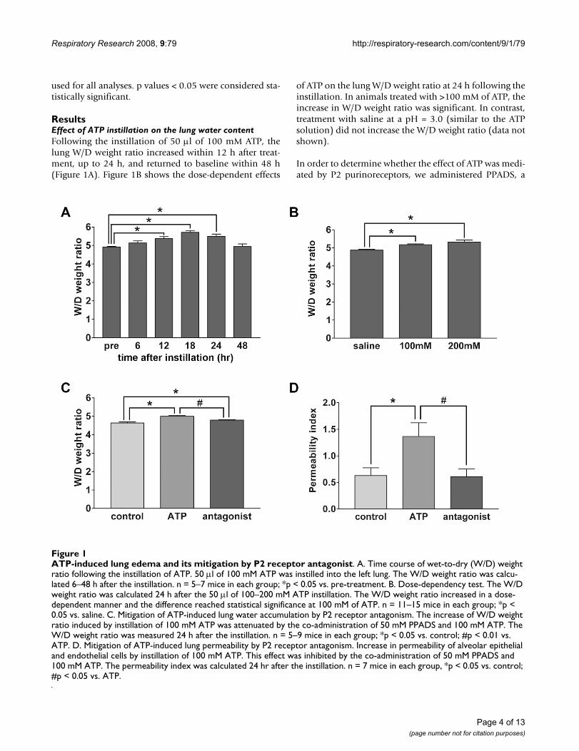
ResultsEffect of ATP instillation on the lung water contentFollowing the instillation of 50 μl of 100 mM ATP, thelung W/D weight ratio increased within 12 h after treat-ment, up to 24 h, and returned to baseline within 48 h(Figure 1A). Figure 1B shows the dose-dependent effects
of ATP on the lung W/D weight ratio at 24 h following theinstillation. In animals treated with >100 mM of ATP, theincrease in W/D weight ratio was significant. In contrast,treatment with saline at a pH = 3.0 (similar to the ATPsolution) did not increase the W/D weight ratio (data notshown).
In order to determine whether the effect of ATP was medi-ated by P2 purinoreceptors, we administered PPADS, a
ATP-induced lung edema and its mitigation by P2 receptor antagonistFigure 1ATP-induced lung edema and its mitigation by P2 receptor antagonist. A. Time course of wet-to-dry (W/D) weight ratio following the instillation of ATP. 50 μl of 100 mM ATP was instilled into the left lung. The W/D weight ratio was calcu-lated 6–48 h after the instillation. n = 5–7 mice in each group; *p < 0.05 vs. pre-treatment. B. Dose-dependency test. The W/D weight ratio was calculated 24 h after the 50 μl of 100–200 mM ATP instillation. The W/D weight ratio increased in a dose-dependent manner and the difference reached statistical significance at 100 mM of ATP. n = 11–15 mice in each group; *p < 0.05 vs. saline. C. Mitigation of ATP-induced lung water accumulation by P2 receptor antagonism. The increase of W/D weight ratio induced by instillation of 100 mM ATP was attenuated by the co-administration of 50 mM PPADS and 100 mM ATP. The W/D weight ratio was measured 24 h after the instillation. n = 5–9 mice in each group; *p < 0.05 vs. control; #p < 0.01 vs. ATP. D. Mitigation of ATP-induced lung permeability by P2 receptor antagonism. Increase in permeability of alveolar epithelial and endothelial cells by instillation of 100 mM ATP. This effect was inhibited by the co-administration of 50 mM PPADS and 100 mM ATP. The permeability index was calculated 24 hr after the instillation. n = 7 mice in each group, *p < 0.05 vs. control; #p < 0.05 vs. ATP.
Page 4 of 13(page number not for citation purposes)
Respiratory Research 2008, 9:79 http://respiratory-research.com/content/9/1/79
specific antagonist against the P2X and P2Y receptors [15],along with ATP. The simultaneous administration ofPPADS, 50 mM, and ATP attenuated the increase in W/Dweight ratio induced by ATP (Figure 1C).
The instillation, 24 h before the assay, of ATP, 100 mM,caused a significant increase in the albumin permeabilityindex, a measure of the permeability of alveolar epithelialand endothelial cells (Figure 1D). The concomitantadministration of PPADS and ATP inhibited the effects ofATP.
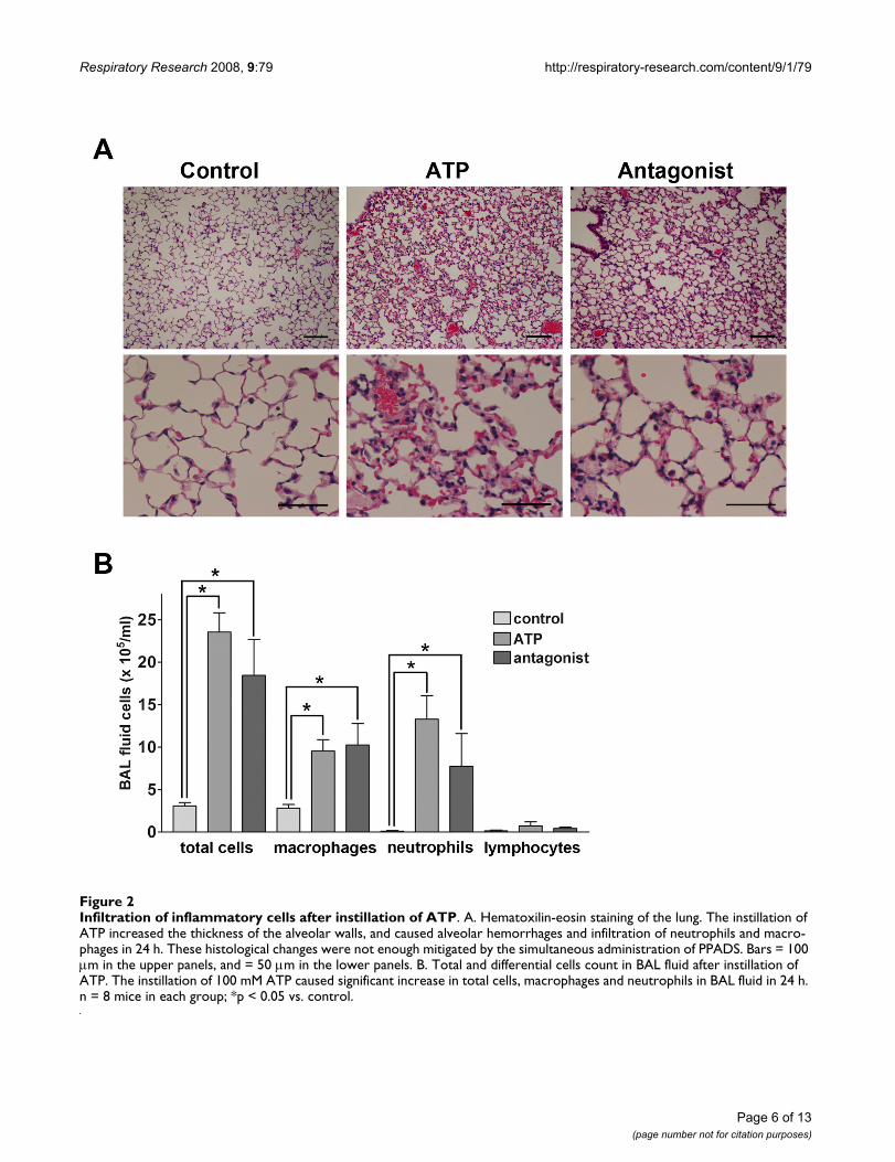
ATP-induced inflammatory response in the lungOn histological examination, 24 h after the instillation ofATP, 100 mM, the thickness of the alveolar walls, theamount of alveolar hemorrhage and the numbers of neu-trophils and macrophages infiltrating the lung wereincreased (Figure 2A). In contrast, the instillation of salinedid not induce this inflammatory response in the controlgroup. The histological derangement of alveolar architec-ture was partially mitigated by the simultaneous adminis-tration of PPADS and ATP. Therefore, the total number ofcells in the BAL fluid was significantly increased in themice instilled with 100 mM ATP 24 h before the assay.The differential cell counts showed that there wereincreased numbers of macrophages and neutrophils inATP-treated mice (Figure 2B). The co-administration ofPPADS limited the neutrophil infiltration, though thedecrease was not statistically significant.
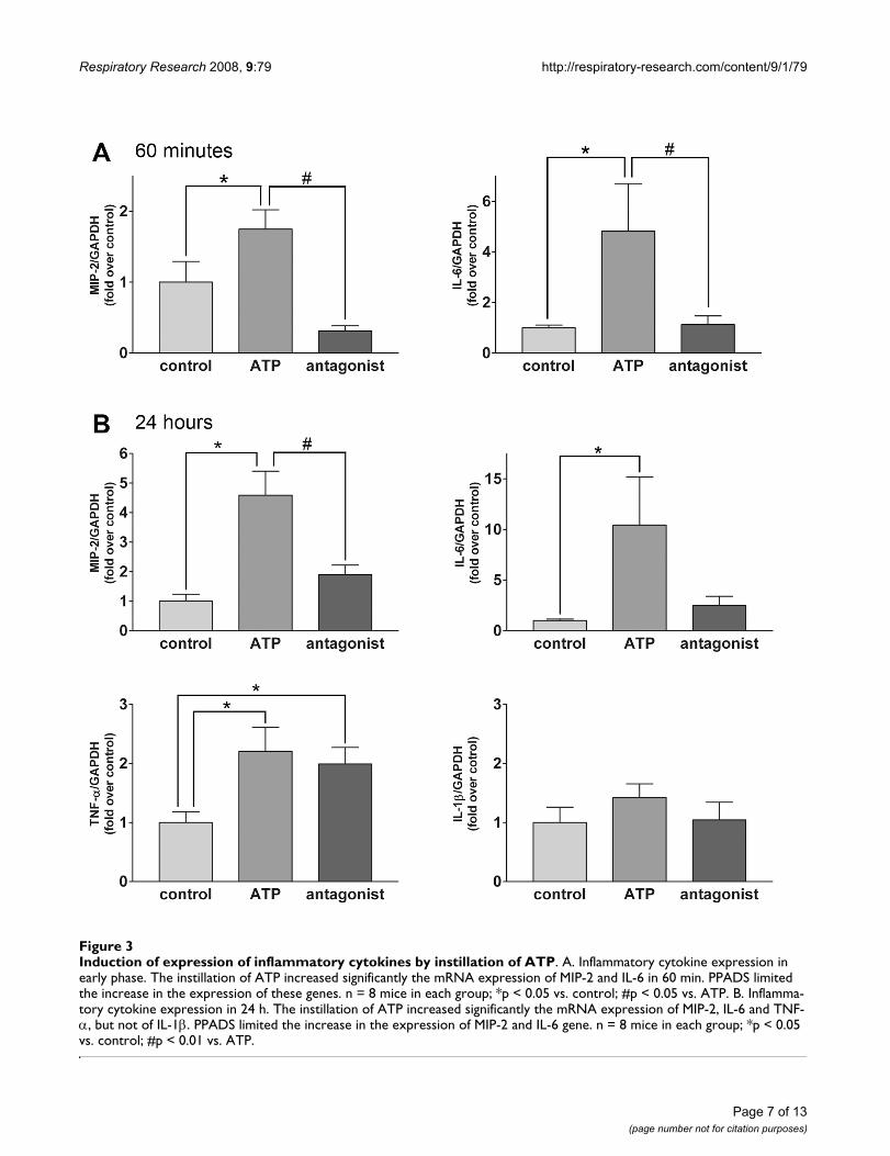
Real-time PCR revealed that the induction of multipleinflammatory cytokines and chemokines is involved inthe pulmonary inflammatory reaction induced by ATP.Exogenous ATP increase significantly the expression ofMIP-2 and IL-6 mRNA within 60 min after its intratra-cheal instillation, and the simultaneous administration ofPPADS and ATP inhibited the expression of these genes(Figure 3A). There was no significant change in the expres-sion of TNF-α mRNA (data not shown). The instillation ofATP caused a significant increase in the expression of MIP-2, IL-6 and TNF-α mRNA within 24 h. The simultaneousadministration of PPADS and ATP inhibited the inductionof MIP-2 and expression of the IL-6 gene, however notthat of TNF-α. The expression of IL-1β mRNA in the lungtissue, by contrast, was not modified by treatment withATP (Figure 3B).
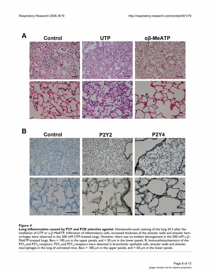
Mediation of the effect of ATP on lung inflammation by the P2Y receptorTo identify the signal transduction of ATP-induced lunginflammation, we examined the effects caused by theinstillation of UTP, an ATP analog selective for P2Y recep-tors, and α,β-MeATP, selective for P2X receptors, on thelung status. The instillation of UTP, 200 mM, caused asimilar infiltration of inflammatory cells as that caused by
ATP (Figure 4A). On the other hand, the instillation ofα,β-MeATP caused no significant histological change inthe lung. The instillation of UTP also induced a significantincrease in W/D weight ratio (control; 4.781 ± 0.050,UTP; 5.065 ± 0.069, p = 0.0063) and permeability index(control; 0.4600 ± 0.0915, UTP; 0.8018 ± 0.2003, p =0.0285).
Immunohistochemistry identified P2Y2 and P2Y4 receptorexpressions in bronchiolar epithelial cells, alveolar wallsand alveolar macrophages in the lung of untreated mice(Figure 4B).
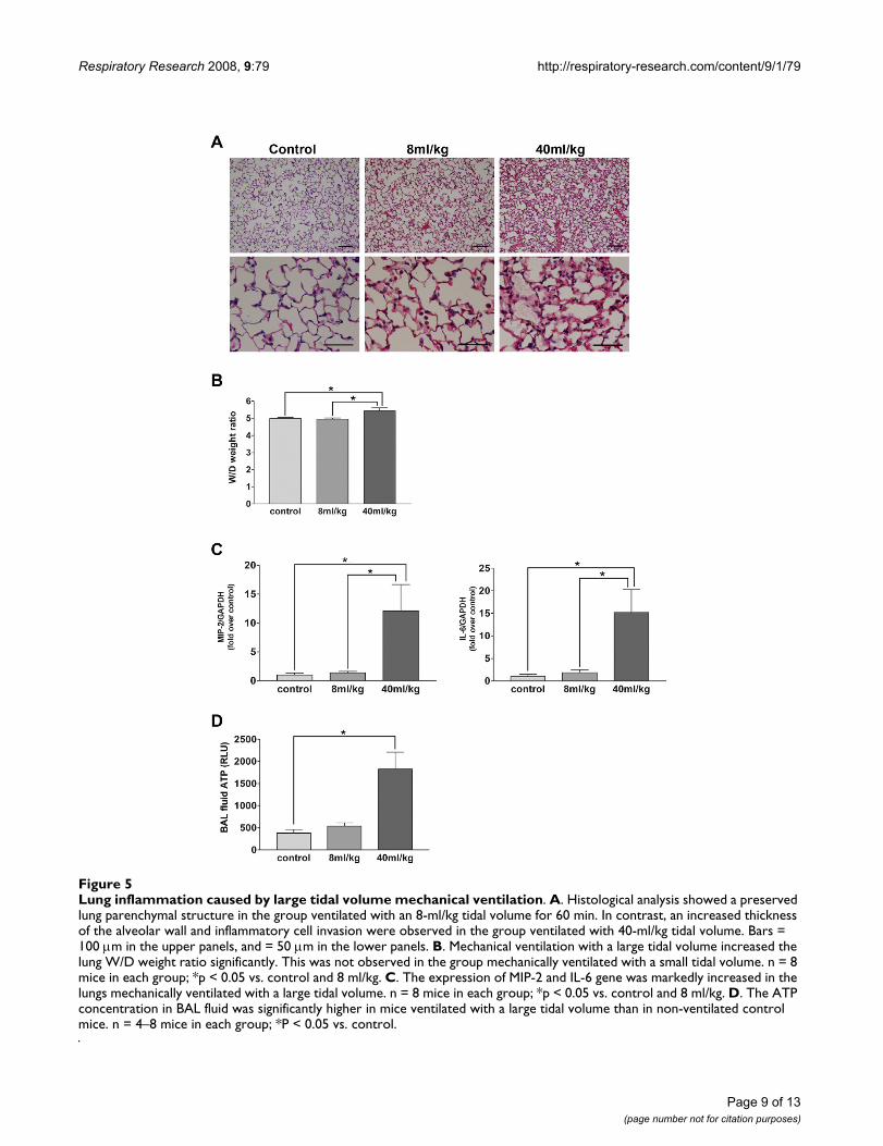
ATP secretion induced by large volume ventilationOn histological examination, the thickness of the alveolarwall was increased and an invasion by inflammatory cellswas observed in the lung of the group ventilated with alarge tidal volume (Figure 5A). The lung W/D weight ratioincreased significantly following 60 min of mechanicalventilation with the 40-ml/kg tidal volume, while noapparent change was observed in the group ventilatedwith the 8-ml/kg tidal volume (Figure 5B). The cytokinemRNA assay revealed a significant increase in the expres-sion of MIP-2 and IL-6 mRNA that was limited to thelungs of mice ventilated with large tidal volumes (Figure5C). These observations confirmed that mechanical venti-lation with a large tidal volume constituted a suitablemodel of lung injury.
Compared to the spontaneously breathing control mice,the ATP concentration in BAL fluid, measured photomet-rically, was significantly increased in the animals exposedto the large tidal volume, but not those ventilated with asmall tidal volume (Figure 5D).
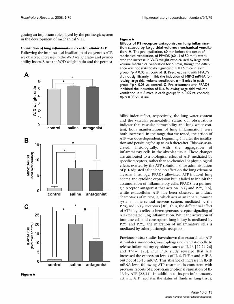
Mitigation of the ventilation-induced pulmonary inflammatory response by PPADSTo confirm the involvement of the purinergic system inVILI, we instilled 60 μl of 50 mM PPADS or saline beforethe onset of mechanical ventilation. This administrationof PPADS caused a significant blockade of the expressionof IL-6 in the lung (Figure 6C), though did not limit theincrease in the W/D weight ratio (Figure 6A), expressionof MIP-2 (Figure 6B) and permeability index (data notshown).
DiscussionATP is believed to act in the intercellular signal transduc-tion as a "purinergic system" in multiple organs, and isinvolved as an energy source in cellular metabolism. Inthe present study, exogenously applied ATP caused aninflammatory reaction by activating the P2Y purinergicreceptors. The extracellular concentrations of ATP in thealveolar space increased as a result of the injury inflictedby mechanical ventilation with a large tidal volume, sug-
Page 5 of 13(page number not for citation purposes)
Respiratory Research 2008, 9:79 http://respiratory-research.com/content/9/1/79
Page 6 of 13(page number not for citation purposes)
Infiltration of inflammatory cells after instillation of ATPFigure 2Infiltration of inflammatory cells after instillation of ATP. A. Hematoxilin-eosin staining of the lung. The instillation of ATP increased the thickness of the alveolar walls, and caused alveolar hemorrhages and infiltration of neutrophils and macro-phages in 24 h. These histological changes were not enough mitigated by the simultaneous administration of PPADS. Bars = 100 μm in the upper panels, and = 50 μm in the lower panels. B. Total and differential cells count in BAL fluid after instillation of ATP. The instillation of 100 mM ATP caused significant increase in total cells, macrophages and neutrophils in BAL fluid in 24 h. n = 8 mice in each group; *p < 0.05 vs. control.
Respiratory Research 2008, 9:79 http://respiratory-research.com/content/9/1/79
Page 7 of 13(page number not for citation purposes)
Induction of expression of inflammatory cytokines by instillation of ATPFigure 3Induction of expression of inflammatory cytokines by instillation of ATP. A. Inflammatory cytokine expression in early phase. The instillation of ATP increased significantly the mRNA expression of MIP-2 and IL-6 in 60 min. PPADS limited the increase in the expression of these genes. n = 8 mice in each group; *p < 0.05 vs. control; #p < 0.05 vs. ATP. B. Inflamma-tory cytokine expression in 24 h. The instillation of ATP increased significantly the mRNA expression of MIP-2, IL-6 and TNF-α, but not of IL-1β. PPADS limited the increase in the expression of MIP-2 and IL-6 gene. n = 8 mice in each group; *p < 0.05 vs. control; #p < 0.01 vs. ATP.
Respiratory Research 2008, 9:79 http://respiratory-research.com/content/9/1/79
Page 8 of 13(page number not for citation purposes)
Lung inflammation caused by P2Y and P2X selective agonistFigure 4Lung inflammation caused by P2Y and P2X selective agonist. Hematoxilin-eosin staining of the lung 24 h after the instillation of UTP or α,β-MeATP. Infiltration of inflammatory cells, increased thickness of the alveolar walls and alveolar hem-orrhages were observed in the 200 mM UTP-treated lungs. However, there was no evident derangement in the 200 mM α,β-MeATP-treated lungs. Bars = 100 μm in the upper panels, and = 50 μm in the lower panels. B. Immunohistochemistry of the P2Y2 and P2Y4 receptors. P2Y2 and P2Y4 receptors were detected in bronchiolar epithelial cells, alveolar walls and alveolar macrophages in the lung of untreated mice. Bars = 100 μm in the upper panels, and = 50 μm in the lower panels.
Respiratory Research 2008, 9:79 http://respiratory-research.com/content/9/1/79
Page 9 of 13(page number not for citation purposes)
Lung inflammation caused by large tidal volume mechanical ventilationFigure 5Lung inflammation caused by large tidal volume mechanical ventilation. A. Histological analysis showed a preserved lung parenchymal structure in the group ventilated with an 8-ml/kg tidal volume for 60 min. In contrast, an increased thickness of the alveolar wall and inflammatory cell invasion were observed in the group ventilated with 40-ml/kg tidal volume. Bars = 100 μm in the upper panels, and = 50 μm in the lower panels. B. Mechanical ventilation with a large tidal volume increased the lung W/D weight ratio significantly. This was not observed in the group mechanically ventilated with a small tidal volume. n = 8 mice in each group; *p < 0.05 vs. control and 8 ml/kg. C. The expression of MIP-2 and IL-6 gene was markedly increased in the lungs mechanically ventilated with a large tidal volume. n = 8 mice in each group; *p < 0.05 vs. control and 8 ml/kg. D. The ATP concentration in BAL fluid was significantly higher in mice ventilated with a large tidal volume than in non-ventilated control mice. n = 4–8 mice in each group; *P < 0.05 vs. control.
Respiratory Research 2008, 9:79 http://respiratory-research.com/content/9/1/79
gesting an important role played by the purinergic systemin the development of mechanical VILI.
Facilitation of lung inflammation by extracellular ATPFollowing the intratracheal instillation of exogenous ATP,we observed increases in the W/D weight ratio and perme-ability index. Since the W/D weight ratio and the permea-
bility index reflect, respectively, the lung water contentand the vascular permeability status, our observationsindicate that vascular permeability and lung water con-tent, both manifestations of lung inflammation, wereboth increased. In the range that we tested, the action ofATP was dose-dependent, beginning 6 h after the instilla-tion and persisting for up to 24 h thereafter. This was asso-ciated, histologically, with the aggregation ofinflammatory cells in the alveolar tissue. These changesare attributed to a biological effect of ATP mediated byspecific receptors, rather than to chemical or physiologicaleffects exerted by the ATP solution, since administrationof pH-adjusted saline had no effect on the lung edema oralveolar histology. PPADS alleviated ATP-induced lungedema and cytokine expression but it failed to inhibit theaccumulation of inflammatory cells. PPADS is a puriner-gic receptor antagonist that acts on P2Y2 and P2Y4 [15],while extracellular ATP has been observed to inducechemotaxis of microglia, which acts as an innate immunesystem in the central nervous system, mediated by theP2X4 and P2Y12 receptors [30]. Thus, the differential effectof ATP might reflect a heterogeneous receptor signaling ofATP-mediated lung inflammation. While the activation ofimmune cell and consequent lung injury is mediated byP2Y2 and P2Y4, the migration of inflammatory cells ismediated by other purinergic receptors.
Previous in vitro studies have shown that extracellular ATPstimulates monocytes/macrophages or dendritic cells torelease inflammatory cytokines, such as IL-1β [22,24-26]and TNF-α [23]. Our PCR study revealed that ATPincreased the expression levels of IL-6, TNF-α and MIP-2,but not of IL-1β mRNA. This absence of increase in IL-1βmRNA level following ATP treatment is consistent withprevious reports of a post-transcriptional regulation of IL-1β by ATP [22,31]. In addition to its pro-inflammatoryactivity, ATP regulates the status of fluids in lung tissue,
Figure 6
Effects of P2 receptor antagonist on lung inflammation caused by large tidal volume mechanical ventilationFigure 6Effects of P2 receptor antagonist on lung inflamma-tion caused by large tidal volume mechanical ventila-tion. A. The pre-instillation, 60 min before the onset of mechanical ventilation, of PPADS (60 μl of 50 mM) attenu-ated the increase in W/D weight ratio caused by large tidal volume mechanical ventilation for 60 min, though the differ-ence was not statistically significant. n = 16 mice in each group; *p < 0.05 vs. control. B. Pre-treatment with PPADS did not significantly inhibit the induction of MIP-2 mRNA fol-lowing large tidal volume ventilation. n = 8 mice in each group; *p < 0.05 vs. control. C. Pre-treatment with PPADS inhibited the induction of IL-6 following large tidal volume ventilation. n = 8 mice in each group; *p < 0.05 vs. control; #p < 0.05 vs. saline.
Page 10 of 13(page number not for citation purposes)
Respiratory Research 2008, 9:79 http://respiratory-research.com/content/9/1/79
stimulating the release of mucin and surfactant frombronchial epithelial and type II alveolar epithelial cells[32,33]. Therefore, the increase in lung edema that fol-lowed the instillation of ATP was the mixed consequenceof an inflammatory reaction and a derangement of fluidexchange, both of which are due to the direct action ofextracellular ATP.
Involvement of the P2Y receptor in ATP-induced lung inflammationThe two subtypes of the purinergic receptor family areP2X, which is coupled with the ion channel, and P2Y,which activates the intracellular G-protein. To identify thereceptor primarily involved in ATP-induced lung injury,we used the ATP analogue, α,β-MeATP, which acts selec-tively against the P2X receptor, and UTP, which acts selec-tively against the P2Y receptor [34]. While UTP inducedan inflammatory response similar to ATP, α,β-MeATP hadno apparent effect on the lung, suggesting that the activa-tion of the P2Y receptor system was sufficient to promotelung injury. Among several subtypes of P2Y receptors,P2Y2 and P2Y4 are the most abundant in lung tissueextracts [15] and are expressed on alveolar macrophagesin BAL fluid [35]. Our immunohistochemical analysisidentified the expression of P2Y2 and P2Y4 in bronchiolarand alveolar epithelial cells and alveolar macrophages,which are both believed to be sources of inflammatorycytokines during acute lung injury [36,37]. Consistentwith these observations, PPADS, a selective purinergicantagonist against P2X, P2Y2 and P2Y4, mitigated theinflammatory effects of the instillation of ATP. Therefore,ATP might activate epithelial cells, macrophages or bothvia P2Y2 and P2Y4 receptors to promote the production ofinflammatory cytokines associated with lung injury.
Involvement of ATP in mechanical lung injuryWorsening or induction of acute lung injury by mechani-cal ventilation is known as "VILI, and also as "ventilator-associated" lung injury. VILI is characterized by anincreased alveolar permeability, pulmonary edema, infil-tration of neutrophils, and the release of inflammatorymediators [38,39]. An increased cytokine expressionaccompanied by migration of inflammatory cells wasobserved in lungs ventilated with large tidal volumes. Theconcentration of ATP in BAL fluid was markedly increasedunder these circumstances, consistent with the release ofATP in response to alveolar epithelial cell stretch in vitro[40], or the increase in ATP or purine concentrations inBAL fluid after mechanical ventilation in vivo [41-43]. Theconcentration of ATP did not increase after lung protectiveventilation, suggesting an essential role of ATP in mediat-ing VILI. Alveolar epithelial cells or macrophages can pro-duce pro-inflammatory cytokines such as IL-6, IL-8 andTNF-α when stretched in vitro [44-47] and promote VILI[48,49]. Since the instillation of ATP induced proinflam-
matory cytokines, ATP-P2Y signaling might act as a bio-logical sensor that translates mechanical stimuli intoproduction of cytokines. Yoshikawa et al. have shown thatlung edema induces VILI independently [50]. Since exog-enous ATP directly controls the fluid status in the lung, thelung edema caused by ATP might be another mechanismof VILI.
The antagonism of ATP-P2Y signaling by PPADS blockedthe production of IL-6 induced by mechanical ventilation,illustrating the, at least partial, involvement of ATP signal-ing in the mechano-transduction and pathophysiology ofVILI. PPADS prevented neither the production of MIP-2,nor the changes in W/D weight ratio and permeabilityindex following ventilation. This might reflect the com-plexity of pathogenesis of VILI, even in ATP signaling.
In contrast to our observations, a recent study found thatintravenous ATP enhanced endothelial integrity and alle-viated LPS-induced lung injury in mice [51]. In vitro stud-ies have shown that the biological effects of ATP aremultiple, including monocyte chemotaxis [52] andenhanced endothelial integrity [53]. Discrepanciesbetween our observations and those made by Kolosova etal. are probably attributable to a multimodal effect ofATP. We injected ATP intratracheally, which might havehad a direct effect on the alveolar tissue. The limited effi-cacy of PPADS in the prevention of mechanical lunginjury is, therefore, likely to reflect multiple and site-spe-cific biological effects of ATP.
We found, in this study, that considerable amounts of ATPare released into the alveolar space following injuriousventilation, which are sufficient to promote an alveolarinflammatory reaction. The efficacy of ATP antagonism inthe treatment of VILI should be tested in a clinically-ori-ented animal model.
ConclusionIn the present study, we found that extracellular ATP pro-motes lung inflammation in mice in vivo, and that theATP-P2Y receptor system is involved in the pathogenesisof VILI. The blockade of ATP signaling might, therefore, bea promising treatment of VILI.
Competing interestsThe authors declare that they have no competing interests.
Authors' contributionsHM performed the experimental studies and drafted themanuscript. FA designed and planned the experiments.SoH, HU, SB and MM assisted with several phases of thestudy. NS and AI participated in the design of the study.FA and SaH designed the experimental set up, supervisedthe experimental work, participated in the manuscript
Page 11 of 13(page number not for citation purposes)
Respiratory Research 2008, 9:79 http://respiratory-research.com/content/9/1/79
preparation and contributed important intellectual con-tent. SaH coordinated the research group. All authors haveread and approved the final manuscript.
AcknowledgementsThe authors thank Dr Junji Magae for contributing insightful advice.
References1. Matthay MA, Zimmerman GA: Acute lung injury and the acute
respiratory distress syndrome: four decades of inquiry intopathogenesis and rational management. Am J Respir Cell MolBiol 2005, 33(4):319-327.
2. Wheeler AP, Bernard GR: Acute lung injury and the acute res-piratory distress syndrome: a clinical review. Lancet 2007,369(9572):1553-1564.
3. Fernandez-Perez ER, Keegan MT, Brown DR, Hubmayr RD, Gajic O:Intraoperative tidal volume as a risk factor for respiratoryfailure after pneumonectomy. Anesthesiology 2006,105(1):14-18.
4. Dreyfuss D, Soler P, Basset G, Saumon G: High inflation pressurepulmonary edema. Respective effects of high airway pres-sure, high tidal volume, and positive end-expiratory pres-sure. Am Rev Respir Dis 1988, 137(5):1159-1164.
5. Webb HH, Tierney DF: Experimental pulmonary edema due tointermittent positive pressure ventilation with high inflationpressures. Protection by positive end-expiratory pressure.Am Rev Respir Dis 1974, 110(5):556-565.
6. Sugiura M, McCulloch PR, Wren S, Dawson RH, Froese AB: Ventila-tor pattern influences neutrophil influx and activation inatelectasis-prone rabbit lung. J Appl Physiol 1994,77(3):1355-1365.
7. Tremblay L, Valenza F, Ribeiro SP, Li J, Slutsky AS: Injurious venti-latory strategies increase cytokines and c-fos m-RNAexpression in an isolated rat lung model. J Clin Invest 1997,99(5):944-952.
8. Haitsma JJ, Uhlig S, Goggel R, Verbrugge SJ, Lachmann U, LachmannB: Ventilator-induced lung injury leads to loss of alveolar andsystemic compartmentalization of tumor necrosis factor-alpha. Intensive Care Med 2000, 26(10):1515-1522.
9. Ranieri VM, Suter PM, Tortorella C, De Tullio R, Dayer JM, BrienzaA, Bruno F, Slutsky AS: Effect of mechanical ventilation oninflammatory mediators in patients with acute respiratorydistress syndrome: a randomized controlled trial. Jama 1999,282(1):54-61.
10. Ventilation with lower tidal volumes as compared with tra-ditional tidal volumes for acute lung injury and the acute res-piratory distress syndrome. The Acute Respiratory DistressSyndrome Network. N Engl J Med 2000, 342(18):1301-1308.
11. Schultz MJ, Haitsma JJ, Slutsky AS, Gajic O: What tidal volumesshould be used in patients without acute lung injury? Anesthe-siology 2007, 106(6):1226-1231.
12. Burnstock G, Wood JN: Purinergic receptors: their role in noci-ception and primary afferent neurotransmission. Curr OpinNeurobiol 1996, 6(4):526-532.
13. Ishikawa S, Higashiyama M, Kusaka I, Saito T, Nagasaka S, Fukuda S,Saito T: Extracellular ATP promotes cellular growth of renalinner medullary collecting duct cells mediated via P2ureceptors. Nephron 1997, 76(2):208-214.
14. Bowler WB, Buckley KA, Gartland A, Hipskind RA, Bilbe G, GallagherJA: Extracellular nucleotide signaling: a mechanism for inte-grating local and systemic responses in the activation ofbone remodeling. Bone 2001, 28(5):507-512.
15. Burnstock G, Knight GE: Cellular distribution and functions ofP2 receptor subtypes in different systems. Int Rev Cytol 2004,240:31-304.
16. Guyot A, Hanrahan JW: ATP release from human airway epi-thelial cells studied using a capillary cell culture system. JPhysiol 2002, 545(Pt 1):199-206.
17. Idzko M, Hammad H, van Nimwegen M, Kool M, Willart MA, MuskensF, Hoogsteden HC, Luttmann W, Ferrari D, Di Virgilio F, et al.: Extra-cellular ATP triggers and maintains asthmatic airway inflam-mation by activating dendritic cells. Nat Med 2007,13(8):913-919.
18. Ferguson DR, Kennedy I, Burton TJ: ATP is released from rabbiturinary bladder epithelial cells by hydrostatic pressurechanges – a possible sensory mechanism? J Physiol 1997, 505(Pt2):503-511.
19. Knight GE, Bodin P, De Groat WC, Burnstock G: ATP is releasedfrom guinea pig ureter epithelium on distension. Am J PhysiolRenal Physiol 2002, 282(2):F281-288.
20. Cook SP, Vulchanova L, Hargreaves KM, Elde R, McCleskey EW: Dis-tinct ATP receptors on pain-sensing and stretch-sensingneurons. Nature 1997, 387(6632):505-508.
21. Grygorczyk R, Hanrahan JW: CFTR-independent ATP releasefrom epithelial cells triggered by mechanical stimuli. Am JPhysiol 1997, 272(3 Pt 1):C1058-1066.
22. Ferrari D, Chiozzi P, Falzoni S, Dal Susino M, Melchiorri L, BaricordiOR, Di Virgilio F: Extracellular ATP triggers IL-1 beta releaseby activating the purinergic P2Z receptor of human macro-phages. J Immunol 1997, 159(3):1451-1458.
23. Ferrari D, La Sala A, Chiozzi P, Morelli A, Falzoni S, Girolomoni G,Idzko M, Dichmann S, Norgauer J, Di Virgilio F: The P2 purinergicreceptors of human dendritic cells: identification and cou-pling to cytokine release. Faseb J 2000, 14(15):2466-2476.
24. Grahames CB, Michel AD, Chessell IP, Humphrey PP: Pharmaco-logical characterization of ATP- and LPS-induced IL-1betarelease in human monocytes. Br J Pharmacol 1999,127(8):1915-1921.
25. Perregaux D, Gabel CA: Interleukin-1 beta maturation andrelease in response to ATP and nigericin. Evidence thatpotassium depletion mediated by these agents is a necessaryand common feature of their activity. J Biol Chem 1994,269(21):15195-15203.
26. Pizzirani C, Ferrari D, Chiozzi P, Adinolfi E, Sandona D, Savaglio E, DiVirgilio F: Stimulation of P2 receptors causes release of IL-1beta-loaded microvesicles from human dendritic cells.Blood 2007, 109(9):3856-3864.
27. Matsuyama H, Hashimoto S, Amaya F, Mizuta M, Ueno H, HashimotoS: Adenosine triphosphate instillation induces lung inflam-mation in mouse. Am J Resp Crit Care Med 2007, 173:A839.
28. Kitamura Y, Hashimoto S, Mizuta N, Kobayashi A, Kooguchi K, Fuji-wara I, Nakajima H: Fas/FasL-dependent apoptosis of alveolarcells after lipopolysaccharide-induced lung injury in mice. AmJ Respir Crit Care Med 2001, 163(3 Pt 1):762-769.
29. Ueno H, Matsuda T, Hashimoto S, Amaya F, Kitamura Y, Tanaka M,Kobayashi A, Maruyama I, Yamada S, Hasegawa N, et al.: Contribu-tions of high mobility group box protein in experimental andclinical acute lung injury. Am J Respir Crit Care Med 2004,170(12):1310-1316.
30. Ohsawa K, Irino Y, Nakamura Y, Akazawa C, Inoue K, Kohsaka S:Involvement of P2X4 and P2Y12 receptors in ATP-inducedmicroglial chemotaxis. Glia 2007, 55(6):604-616.
31. Bianco F, Pravettoni E, Colombo A, Schenk U, Moller T, Matteoli M,Verderio C: Astrocyte-derived ATP induces vesicle sheddingand IL-1 beta release from microglia. J Immunol 2005,174(11):7268-7277.
32. Gilfillan AM, Rooney SA: Functional evidence for involvement ofP2 purinoceptors in the ATP stimulation of phosphatidyl-choline secretion in type II alveolar epithelial cells. BiochimBiophys Acta 1988, 959(1):31-37.
33. Kemp PA, Sugar RA, Jackson AD: Nucleotide-mediated mucinsecretion from differentiated human bronchial epithelialcells. Am J Respir Cell Mol Biol 2004, 31(4):446-455.
34. Brunschweiger A, Muller CE: P2 receptors activated by uracilnucleotides – an update. Curr Med Chem 2006, 13(3):289-312.
35. Bowler JW, Bailey RJ, North RA, Surprenant A: P2X4, P2Y1 andP2Y2 receptors on rat alveolar macrophages. Br J Pharmacol2003, 140(3):567-575.
36. Lomas-Neira J, Chung CS, Perl M, Gregory S, Biffl W, Ayala A: Roleof alveolar macrophage and migrating neutrophils in hemor-rhage-induced priming for ALI subsequent to septic chal-lenge. Am J Physiol Lung Cell Mol Physiol 2006, 290(1):L51-58.
37. Sharma AK, Fernandez LG, Awad AS, Kron IL, Laubach VE: Proin-flammatory response of alveolar epithelial cells is enhancedby alveolar macrophage-produced TNF-alpha during pulmo-nary ischemia-reperfusion injury. Am J Physiol Lung Cell Mol Phys-iol 2007, 293(1):L105-113.
Page 12 of 13(page number not for citation purposes)
Respiratory Research 2008, 9:79 http://respiratory-research.com/content/9/1/79
Publish with BioMed Central and every scientist can read your work free of charge
"BioMed Central will be the most significant development for disseminating the results of biomedical research in our lifetime."
Sir Paul Nurse, Cancer Research UK
Your research papers will be:
available free of charge to the entire biomedical community
peer reviewed and published immediately upon acceptance
cited in PubMed and archived on PubMed Central
yours — you keep the copyright
Submit your manuscript here:http://www.biomedcentral.com/info/publishing_adv.asp
BioMedcentral
38. Dreyfuss D, Saumon G: Ventilator-induced lung injury: lessonsfrom experimental studies. Am J Respir Crit Care Med 1998,157(1):294-323.
39. Belperio JA, Keane MP, Lynch JP 3rd, Strieter RM: The role ofcytokines during the pathogenesis of ventilator-associatedand ventilator-induced lung injury. Semin Respir Crit Care Med2006, 27(4):350-364.
40. Patel AS, Reigada D, Mitchell CH, Bates SR, Margulies SS, Koval M:Paracrine stimulation of surfactant secretion by extracellu-lar ATP in response to mechanical deformation. Am J PhysiolLung Cell Mol Physiol 2005, 289(3):L489-496.
41. Douillet CD, Robinson WP 3rd, Milano PM, Boucher RC, Rich PB:Nucleotides induce IL-6 release from human airway epithe-lia via P2Y2 and p38 MAPK-dependent pathways. Am J PhysiolLung Cell Mol Physiol 2006, 291(4):L734-746.
42. Rich PB, Douillet CD, Mahler SA, Husain SA, Boucher RC: Adenos-ine triphosphate is released during injurious mechanical ven-tilation and contributes to lung edema. J Trauma 2003,55(2):290-297.
43. Verbrugge SJ, de Jong JW, Keijzer E, Vazquez de Anda G, LachmannB: Purine in bronchoalveolar lavage fluid as a marker of ven-tilation-induced lung injury. Crit Care Med 1999, 27(4):779-783.
44. Pugin J, Dunn I, Jolliet P, Tassaux D, Magnenat JL, Nicod LP, ChevroletJC: Activation of human macrophages by mechanical ventila-tion in vitro. Am J Physiol 1998, 275(6 Pt 1):L1040-1050.
45. Vlahakis NE, Schroeder MA, Limper AH, Hubmayr RD: Stretchinduces cytokine release by alveolar epithelial cells in vitro.Am J Physiol 1999, 277(1 Pt 1):L167-173.
46. Mourgeon E, Isowa N, Keshavjee S, Zhang X, Slutsky AS, Liu M:Mechanical stretch stimulates macrophage inflammatoryprotein-2 secretion from fetal rat lung cells. Am J Physiol LungCell Mol Physiol 2000, 279(4):L699-706.
47. Pugin J: Molecular mechanisms of lung cell activation inducedby cyclic stretch. Crit Care Med 2003, 31(4 Suppl):S200-206.
48. Goodman RB, Pugin J, Lee JS, Matthay MA: Cytokine-mediatedinflammation in acute lung injury. Cytokine Growth Factor Rev2003, 14(6):523-535.
49. Frank JA, Parsons PE, Matthay MA: Pathogenetic significance ofbiological markers of ventilator-associated lung injury inexperimental and clinical studies. Chest 2006,130(6):1906-1914.
50. Yoshikawa S, King JA, Lausch RN, Penton AM, Eyal FG, Parker JC:Acute ventilator-induced vascular permeability and cytokineresponses in isolated and in situ mouse lungs. J Appl Physiol2004, 97(6):2190-2199.
51. Kolosova IA, Mirzapoiazova T, Moreno-Vinasco L, Sammani S, GarciaJG, Verin AD: Protective effect of purinergic agonist ATPgam-maS against acute lung injury. Am J Physiol Lung Cell Mol Physiol2008, 294(2):L319-324.
52. Wu LJ, Vadakkan KI, Zhuo M: ATP-induced chemotaxis ofmicroglial processes requires P2Y receptor-activated initia-tion of outward potassium currents. Glia 2007, 55(8):810-821.
53. Kolosova IA, Mirzapoiazova T, Adyshev D, Usatyuk P, Romer LH,Jacobson JR, Natarajan V, Pearse DB, Garcia JG, Verin AD: Signalingpathways involved in adenosine triphosphate-inducedendothelial cell barrier enhancement. Circ Res 2005,97(2):115-124.
Page 13 of 13(page number not for citation purposes)


![ARDS & SEVERE HYPOXEMIAevaggelismos-hosp.gr/files/epistimoniki_enosi/02... · absence of Pneumothorax or ↑ Vt? ... [=Vt/Crs] LUNG: What do we need to avoid? • Hypoxemia • Ventilator-associated](https://img.pdfslide.tips/doc/110x75/5e9ac115fd0edd1d2c61726a/ards-severe-hypoxemiaevaggelismos-hospgrfilesepistimonikienosi02.jpg)