Embed Size (px)
Citation preview

Mechanism of Electrical De�brillation: Current Status and Future Perspective
Nitaro SHIBATA,*, # Shin INADA,** Kazuo NAKAZAWA,** Naoki TOMII,*** Masatoshi YAMAZAKI,*** Hiroshi SENO,*** Haruo HONJO,† Ichiro SAKUMA***
Abstract Ventricular �brillation (VF) is currently a major cause of sudden cardiac death (SCD). To cure VF, electrical de�brillation is the only therapy. However, strong energy is required. Thus, to reduce the energy or develop a new method is desired. The mechanism of how the electric shock sweeps VF is still controversial. In this article, we summarize evidence and remaining problems of this topic. There are three issues in time se-quence of VF: how to initiate, how to continue, and how to terminate. Many investigations to achieve VF-free heart have been reported, but there are currently no de�nite methods to prevent VF. Thus, to terminate VF is one of the big challenges to prevent SCD. There are two strategies to improve electrical de�brillation: elucidate the substantial mechanism and reduce the energy. (1) Substantial mechanism proposed: In a failed de�brillation ep-isode, at the energy level of the near de�brillation threshold, the initial activation site is related to the repolar-ization phase of the location. However, it is still not clear whether it is part of the continuous VF activity or ini-tiation of re-VF. It is well known that strong �eld electric shock (including cathodal and anodal stimuli) has many effects on the cardiac tissue, such as electroporation, virtual electrode effects, and electrophysiological responses, which are in�uenced by tissue geometry (including �ber orientation and bifurcation of tissues). These phenomena should modify the de�brillation effect. Finally, the characteristics of dynamic spiral wave (SW; the sources of continuity of re-entries) in�uence the continuity of VF. (2) Efforts to reduce the de�brilla-tion energy: To reduce the de�brillation energy, biphasic pulse, regional cooling, modi�ed stimuli programs, and automated local stimuli to SW are proposed. The superiority of biphasic pulse to monophasic pulse was es-tablished in the late 20th century; however, the mechanism is still not well understood. Cooling of some region of the heart ventricles widens the route of SW trajectory and terminates SW. Programming high frequency stim-ulus or double stimuli according to computer simulation of the heart model could reduce the de�brillation threshold. Automated local stimulus to the site between the tail of SW activation and the next activation front could terminate the SW.
Keywords: ventricular de�brillation, ventricular �brillation, spiral wave, sudden cardiac death, heart excitation simulation model, virtual electrode effect.
Adv Biomed Eng. 9: pp. 125–137, 2020.
1. Introduction
Sudden cardiac death (SCD) is one of the important is-sues to be solved in order to preserve life expectancy. The major cause of SCD is ventricular �brillation (VF), which indicates a completely disorganized contraction phase of the heart muscle. In the healthy state, the heart ventricles contract simultaneously within the range of <100 ms that causes the pump function of the heart, by which blood perfuses all over the body and supply oxy-gen and nutrition to individual organs.
When VF occurs, electric de�brillation is the only effective therapy to restore the normal heartbeat. Chemi-cal de�brillation using potassium chloride or other drugs has been reported [1]. However, this procedure is practi-cally not useful. Because strong energy of approximately
Received on April 15, 2020; revised on May 22, 2020; accepted on June 2, 2020.
* Department of Cardiology, Shinjuku Mitsui Building Clinic, Tokyo, Japan.
** Faculty of Health Sciences, Morinomiya University of Medical Sciences, Osaka, Japan.
*** Department of Bioengineering, The University of Tokyo, Tokyo, Japan.
† Research Institute of Environmental Medicine, Nagoya Universi-ty, Aichi, Japan.
# Shinjuku Mitsui Building Clinic, 2–1–1 Nishi-Shinjuku, Shin-juku-ku, Tokyo 163–0404, Japan. E-mail: [email protected]
Copyright: ©2020 The Author(s). This is an open access article distributed under the terms of the
Creative Commons BY 4.0 International (Attribution) License (https://creativecommons.org/licenses/by/4.0/legalcode), which permits the unrestricted distribution, reproduction and use of the article provided the original source and authors are credited.
Review PaperAdvanced Biomedical Engineering9: 125–137, 2020.
DOI:10.14326/abe.9.125
20 J is required for de�brillation, the size of an implant-able automatic de�brillator is large, the time to re-im-plantation of the device is short, and candidate of the device implantation may have fear because of a strong impulse to the body near the shock electrodes as well as to the heart.
In the late 20th century, many research efforts have been made to elucidate the mechanism of �brillation, and with the advancement of electromechanical systems, the size of implantable de�brillator showed signi�cant reduction [2, 3]. However, the basic electrophysiologic mechanisms of electrical de�brillation remain incom-pletely understood. In this paper, we summarize the up-date and future of investigations of the de�brillation mechanisms.
2. Mechanism of VF
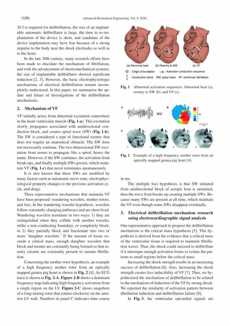
VF initially arises from abnormal excitation somewhere in the heart ventricular muscle (Fig. 1-a). This excitation slowly propagates associated with unidirectional con-duction block, and creates spiral wave (SW) (Fig. 1-b). The SW is considered a type of functional reentry that does not require an anatomical obstacle. The SW does not necessarily continue. The two-dimensional SW exci-tation front seems to propagate like a spiral, hence the name. However, if the SW continues, the activation front break-ups, and �nally multiple SWs persist, which main-tain VF (Fig. 1-c) that never terminates spontaneously.
It is also known that these SWs are modi�ed by many factors such as autonomic nerve tone, electrophys-iological property changes to the previous activation cy-cle, and drugs.
Three representative mechanisms that maintain VF have been proposed: wandering wavelets, mother rotors, and foci. In the wandering wavelet hypothesis, wavelets follow constantly changing pathways and are short lived. Wandering wavelets terminate in two ways: 1) they are extinguished when they collide with another wavelet, strike a non-conducting boundary, or completely block; or 2) they partially block and fractionate into two or more “daughter wavelets.” If the amount of tissue ex-ceeds a critical mass, enough daughter wavelets that block and reenter are constantly being formed so that re-entry circuits are constantly present to sustain �brilla-tion.
Concerning the mother rotor hypothesis, an example of a high frequency mother rotor from an optically mapped guinea pig heart is shown in Fig. 2 [4]. An ECG trace is shown in Fig. 2-A. Figure 2-B shows a dominant frequency map indicating high frequency activation from a single region on the LV. Figure 2-C shows snapshots of a long-lasting rotor that rotates clockwise on the ante-rior LV wall. Numbers in panel C indicates time course
in ms.The multiple foci hypothesis is that SW initiated
from unidirectional block of ectopic beat is sustained, then the wave front breaks up creating multiple SWs. Be-cause many SWs are present at all time, which maintain the VF even though some SWs disappear eventually.
3. Electrical de�brillation mechanism research using electrocardiographic signal analysis
One representative approach to propose the de�brillation mechanism is the critical mass hypothesis [5]. This hy-pothesis is derived from the evidence that a critical mass of the ventricular tissue is required to maintain �brilla-tion waves. Thus, the shock could succeed to de�brillate if it interrupts enough activation fronts to isolate the pat-terns to small regions below the critical mass.
Increasing the shock strength results in an increasing success of de�brillation [6]. Also, increasing the shock strength creates less inducibility of VF [7]. Thus, we hy-pothesized the mechanism of de�brillation to be related to the mechanism of induction of the VF by strong shock. We reported the similarity of activation pattern between �brillation induction and de�brillation failure [8].
In Fig. 3, the ventricular epicardial signals are
Fig. 1 Abnormal activation sequences. Abnormal beat (a), reentry to SW (b), and VF (c).
Fig. 2 Example of a high frequency mother rotor from an optically mapped guinea pig heart [4].
Advanced Biomedical Engineering. Vol. 9, 2020.(126)

shown. The left side shows induction of ventricular �brillation episode, and the right side shows failure of de�brillation in the same experiment using the same shock energy. Upper �gures show representative epicar-dial signals from 8 epicardial sites, and bottom �gures, activation mappings. Earliest activation sites, activation timing, and propagation of the activations were similar between these two episodes. We also found a relation-ship between the energy required for de�brillation (DFT) and the upper limit of the energy inducing �brillation (ULV).
Therefore, we proposed the hypothesis that sub-threshold de�brillation shock swipes out all SWs, and then induces new activation that re-induces VF, i.e., re-initiation hypothesis. In these studies, we used the epicardial activation mapping system; thus, signals ~25 milliseconds immediately after the shock could not be recorded, and the repolarization phase could not be mea-sured.
4. Current representative hypotheses of electri-cal de�brillation
With the advancement of research tools of cardiac activa-tions, such as optical measurement of transmembrane action potentials, phase analysis of SW, and computer
simulation of 3-D heart activation mapping, many de-�brillation hypotheses have been proposed.
Kwaku and Dillon [9] suggested that de�brillation success is related to the ability of the shock to increase the refractoriness at the border of shock-depolarized ar-eas so as to prevent wave front propagation from these areas after the shock.
Many de�brillation mechanisms have been proposed since 1990. The main concerns are the mechanism of ter-mination of SW, and the mechanism of failure of VF ter-mination regardless of the continuity of one of the SW sources or re-initiation of SW. Representative concepts are shown below.
4.1 Focal activation arises at near DFT energyChattipakorn et al. [10] analyzed optical mapping sig-nals from all epicardial surfaces of heart ventricles for near-de�brillation energy level. Figure 4 shows maps of failure of de�brillation episode (FDF) on the left side, and successful de�brillation (SDF) on the right side, us-ing the same energy in the same experiment. The top row shows the coupling interval (CI) preceding the shock, the middle row, the half repolarization time (RT50), and the bottom row, 75% repolarization time (RT75).
CI for all SDF was 59 ± 7 ms, and was signi�cantly longer than that of FDF, 52 ± 6 ms. RT50 and RT75 were
Fig. 3 Similarities of activation sequence between induc-tion of VF and failure of de�brillation in a dog heart [7]. The activation map shows similar activa-tion waveforms (upper panels) and activation map-ping (lower panels) between the induction of VF (left side) and failure of de�brillation (right side) in the same experiment at the same delivered energy. See text for details.
Fig. 4 Mechanism of ventricular de�brillation for near-de-�brillation threshold shocks [12]. Figures show the optical mapping �ndings recorded from both sides of the ventricles. Left side indicates failure of de-�brillation episode (FDF), and the right side, suc-cessful de�brillation episode (SDF). The upper, middle, and lower panels show coupling interval (CI) from the previous activity, 50% repolarization time (RT50), and 75% repolarization time (RT75), respectively. See text for details.
Nitaro SHIBATA, et al: De�brillation Mechanism (127)

signi�cantly longer for SDF. Thus, the de�brillation out-come correlated with the electrical state of the heart at the time of the shock and with RT. In conclusion, the re-polarization pattern of the �rst post-shock cycle may in-�uence the shock outcome.
There are controversial reports that the activation(s) after failed de�brillation with subthreshold shock is the continuous activity of part of the VF waveform. For ex-ample, using a computer simulation heart activation model, Ashihara et al. [11] presented the mechanism of re-�brillation to be continuous electrical activity.
4.2 Virtual electrode polarizationOne of the functions of strong electrical stimulus is the electroporation of the cell membrane. Using the comput-er simulation 2-D cardiac tissue bi-domain model, Ashi-hara et al. [12] showed that with electroporation, even weak energy (16 V/cm shock) can halt SW (far right col-umn in Fig. 5), but energy of 24 V/cm cannot halt SW if the electroporation effect is not applied (far left column of Fig. 5) (j). Electroporation-mediated anodal break ex-citation may be important to terminate SW.
4.3 Virtual electrode phenomenon (VEP) as the mechanism of de�brillation
During unipolar cathodal stimulation, cardiac tissue is depolarized in a dog-bone-shaped region (the virtual
cathode) that lies underneath and adjacent to the cathod-al electrode, and a pair of hyperpolarized regions (virtual anodes) exist adjacent to the cathode along the directions parallel to the myocardial �bers (Fig. 6). These respons-es are called the virtual electrode effect [13].
E�mov et al. [15] showed the effect of monophasic and biphasic shocks on sustained ventricular arrhythmias in a rabbit heart model. Monophasic shocks extinguished arrhythmic pattern via a virtual electrode polarization ef-fect. However, the virtual electrode polarization effect could produce phase singularities, leading to arrhythmia which resulted in de�brillation failure. In contrast, opti-mal biphasic shocks were successful because the �rst phase erased the arrhythmia via the virtual electrode ef-fect, whereas the second phase canceled the virtual elec-trodes, eliminating the substrates for phase singularities (Fig. 7).
Fig. 6 Schematic representation of the theoretical predic-tion of action potential propagation for point stimu-lation of cardiac tissue [13]. A: Cathodal stimulation in an equal anisotropy model. An elliptical region of the tissue would be directly depolarized by a strong point stimulus. B to E: Cathodal/anodal make/break stimulation in a model with different anisotropic conductivities for the intracellular and extracellular spaces.
Fig. 5 Snapshots of transmembrane potential maps (red: 0 mV, blue: −100 mV) in a 2-D heart ventricular tis-sue bidomain model [12]. In each column, 24, 20 or 16 V/cm monophasic shocks were applied. EP(+) indicates application of electroporation (EP) in sim-ulation model. Time course from the shock timing is shown from top (0 ms) to bottom (100 ms) [12]. With EP(-) (A), reentry does not stop at 100 ms after application of 24 V/cm. However, with EP(+) (D), reentry terminates at 50 ms after applying 16 V/cm.
Advanced Biomedical Engineering. Vol. 9, 2020.(128)

4.4 Graded response of the action potentialGraded response or progressive cellular depolarization is a property of cardiac cells, which is manifested when a strong electrical stimulus is applied during the relative refractory period. Figure 8 shows how an S2 initiates ac-tivation distal to the stimulus site with unidirectional block developing at the S2 site and reentry initiated [16]. Because this response is not an all or none phenomenon, unidirectional block with re-entry can be easily created. An example of S2-induced re-entry is shown in the re-port [16].
4.5 The effect of tissue geometry in the changes of action potential by �eld stimulation shows the secondary source
It is well known that reverse polarity of electrical stimu-lation does not simply reverse the electrophysiological responses to the action potential. Gilles et al. [17] ob-served the in�uence of tissue geometry on the transmem-brane action potential of cultured 2-D myocardial tis-sues. They found that electrical �eld stimulation (EFS) produced changes in transmembrane action potential at the resistive boundaries in bifurcations of the tissue strand. Extracellular �eld stimulation (EFS) produced 2 delta Vm maxima (secondary source) at the shoulder of each limb that was separated by a decrease of either hy-perpolarization or depolarization at the intersection of
the stem strand (Fig. 9).
4.6 Restitution hypothesisTerminating VF by pharmacological method is one of the dreams of de�brillation strategies. VF is created and sustained by the property of restitution of the cardiac ac-tion potential duration (APD). The restitution hypothesis states that steeply sloped restitution curves create unsta-ble wave propagation that results in wave brake leading to VF. Changing the electrophysiological characteristics by modifying cardiac restitution property using bretyli-um, Gar�nkel et al. [18] showed the recovery of VF to monomorphic ventricular tachycardia (Fig. 10). Effec-tive anti-arrhythmic drugs might be developed using res-titution curve modi�cation.
4.7 Mechanism of vortex termination in the cardiac muscle
It was found that a single vortex in a heart can be termi-nated with an electric pulse within a certain time interval, called the critical window, or vulnerable window (VW) [19]. Hornung et al. [20] investigated termination of multiple vortices in the heart. They scanned the phases of all pinned vortices in parallel with electric �eld pulses. In order to control vortices, electric �eld pulses were giv-en to stimulate the VWs of all pinned vortices to over-come the geometric positions of vortex cores. Figure 11 shows termination of pinned vortices by electric �eld pulses in two different periods. This can be achieved ge-nerically for any parameter of the vortices, without
Fig. 7 Snapshots of anterior epicardial transmembrane voltage during the 2nd phase of biphasic shock [14, 15]. The second phase sweeps the repolarized area, which prevents the initiation of SW. See the refer-ence for details. LV: left ventricle, RV: right ventri-cle, LA: left atrium, RA: right atrium.
Fig. 8 Cellular graded responses and ventricular vulnera-bility to reentry by a premature stimulus in isolated canine ventricle [16]. A: Effects of increasing cur-rent strength from 40 mA (top) to 80 mA (bottom). B: Effects of increasing S-2 coupling interval from 140 ms (top) to 154 ms (bottom). Note the increase of action potential duration when S2 stimulus strength or coupling interval increase. C: Relation-ship between graded response-induced additional prolongation of total action potential duration (APD). D: Relationship between take-off potential and graded response amplitude is shown in .
Nitaro SHIBATA, et al: De�brillation Mechanism (129)

knowing the geometric location and time position of the VWs. Moreover, they also suggested that a similar mech-anism terminates an unpinned vortex from a series of ap-proximately 500 experiments with termination of ven-tricular �brillation by electric �eld pulses in isolated pig hearts.
4.8 Uni�ed hypothesisApart from the above 7 stories, many other �ndings have been discovered. However, the main mechanism of elec-trical de�brillation has not been elucidated. Dillon and Kwaku [21] introduced the uni�ed hypothesis, which is the progressive depolarization theory. After introducing upper limit of the vulnerability hypothesis, the progres-sive depolarization hypothesis using optical mapping technique uni�es de�brillation and shock-induced �bril-lation.
5. Reducing the de�brillation energy
Besides studies to clarify the de�brillation mechanism,
many efforts have been made to reduce the de�brillation energy, and evidence to reduce the energy required for de�brillation should be related to the mechanism of de-�brillation. In the practical manner, efforts to improve the de�brillation process is crucial even the background
Fig. 9 Effect of extracellular �eld stimulation (EFS) on bi-furcation strands (100 μm width) of cultured myo-cardial cells [15]. A: Phase contrast image of bifur-cation. B: Shaded background illustrates bifurcation, and superimposed grid denotes position of photodi-ode array. Area of each square is 60 × 60 μm2. Iso-chronal map (isochrone interval, 500 μs) shows ac-tion potential propagation from main stem into 2 bifurcating branches. C: Isopotential map of ΔVm/APA (in %) induced by EFS with �eld strength of 7 V/cm applied parallel to the strand (arrow). Back-ground shading outlines bifurcation. Each color rep-resents an isopotential step of 10%. + indicates an-ode; - indicates cathode. EFS was applied for 20 ms after the pacing stimulus. D: Isopotential map in-duced by EFS of opposite polarity. Format corre-sponds to that shown in C. Fig. 10 Computer simulation of the effects of APD restitu-
tion (APDR) on scroll wave stability [18]. Aa: APD restitution curves (APD vs. DI; diastolic in-terval) of the ventricular action potential model for two different values of Gsi (maximal value of the slow inward Ca2+ current). Prolonging APD by in-creasing Gsi increased the slope of the APDR curve. b and c: Snapshots of voltage (coded from red, denoting depolarized tissue) to blue for resting tissue. Note that scroll waves break up in A (left side), but do not break up in B (right side). d: Transmembrane action potential from one cell in simulation Ab. Ba: Flattened APDR curves after modi�cation of the Luo-Rudy action potential model for two different mean value of APD. Bb-d: snapshots of voltage and transmembrane potential from simulation Ba, corresponding to A. See Text for details.
Advanced Biomedical Engineering. Vol. 9, 2020.(130)

mechanism is unknown. So far, the most effective meth-od established is the use of biphasic waveform. Other representative examples to lower the energy are elec-trode numbers, local cooling, and electrical stimulation to the SW.
5.1 Biphasic waveformThe de�brillation threshold using biphasic waveform is known to be signi�cantly lower than that of monophasic shock. Many reports concerning the ef�cacy, mecha-nism, and clinical value of biphasic waveform have been published [15, 22]. However, it is still not clear which is the most important part of the function of biphasic wave-form. One of the representative data concerning the ef-fect of biphasic waveform is shown in Fig. 7 [15]. Opti-cal mapping at the epicardial surface during biphasic shock is shown. The second phase of the biphasic wave-form prevents re-creation of new activation front. Details were shown at Section 4.4.
5.2 Regional coolingLocal cooling of the heart may be one option to termi-nate the SW. In Fig. 12, low intensity DC shock with lo-cal cooling succeeded cardioversion in the presence of regional cooling, as shown by the dotted circle in the center of optical mapping area [23]. Top panel shows the action potential trace. After 25 V DC, marked as “b,” ac-tion potentials were stabilized compared to before the shock, and terminated at nearly 4 seconds later. The mapping pattern showed that after the shock, unpinning of rotors were observed, which terminated the SW.
5.3 High-frequency and low voltage de�brillationHigh frequency alternating current (HFAC) electric �eld can reversibly block the activation front propagation. Weinberg et al. [24] applied HFAC �eld to an experi-mental study using optical mapping, and, also performed simulation studies with an anatomically accurate ventric-
ular model (Fig. 13). HFAC �elds increase a �eld-depen-dent state of conduction block, and de�brillation success is related to the degree and location of the conduction block. In Fig. 13-A (15 V/cm HFCA �eld), successful de�brillation was observed, in which loss of conduction power (LCP, %) (left and middle) is greater than that of Fig. 12-B (10 V/cm HFCA).
5.4 Terminating VF by two stage low voltage stimuliRantner et al. [25] developed a detailed MRI-based com-putational model of the rabbit right ventricular wall and applied multiple low-voltage far-�eld stimuli to VF (Fig. 14). A novel two-stage de�brillation protocol was proposed. The �rst stage converted VF into VT by apply-ing low-voltage stimuli at times of maximal excitable gap, capturing large tissue volume, and synchronizing depolarization; the second stage terminated VT. The en-ergy required for de�brillation using this method is less than 60% of the de�brillation threshold using standard stimulation.
5.5 Interaction of SW by local stimulation(1) Effect of local electrical stimulus to modify the
SW con�guration.Recently, Yatabe et al. [26] reported that three conditions are required to induce the SW shift. These factors are (a) stimulus location to be close to SW center, (b) stimulus location to be along the �ber orientation of SW center, and (c) electric stimulation should be applied in the repo-larization phase of action potential.
(2) Interaction of PS on the SW tailThe interaction between the location of the SW tail and electrical stimulation may contribute to the SW shift, which causes annihilation of the SW. Tomii et al. [27] published a paper concerning effective stimulation to ter-minate SW. They hypothesized that the action mecha-nism of capturing excitable area of SW by stimulation can also be explained as the collision of the original phase singularity (PS) with PSs induced on the wave tail of SW by stimulation. Low energy electrical stimulation at the point near the SW center during the relative refrac-tory period annihilates the reentry focus. Electrical stim-ulation applied during the relative refractory period of the SW induced the SW shift. This method may contrib-ute to the reduction of energy required to halt ventricular �brillation. The experimental results con�rmed this hy-pothesis (Fig. 15).
6. Discussion
6.1 Function of high intensity electrical stimulationHigh intensity electrical shock is commonly used for atrial and ventricular de�brillation (epicardial, implant-
Fig. 11 Parallel termination of two pinned vortices, both with unknown geometrical location and time posi-tion of the critical (vulnerable) window (VW) [18]. Color code: red is a wave, green is the wavefront. Time is measured from the start of pacing. At 440 ms, E-pulses terminate two vortices [20].
Nitaro SHIBATA, et al: De�brillation Mechanism (131)

able, and external). The effects of this shock are anodal/cathodal stimulus, virtual electrode effect, and �eld stim-ulation. Also, this shock is commonly used for induction of SW reentry. The mechanism of SW induction was the-oretically predicted by Winfree [28] and clearly con-�rmed by our group [7]. In addition, this shock is used to predict the de�brillation threshold, because the upper limit of the shock inducing VF is related to DFT [29]. In other words, high intensity electrical shock has many clinical applications. However, these functions do not di-rectly relate to the de�brillation mechanism.
6.2 Limitation of de�brillation researchTo �nd out the mechanism of successful de�brillation is dif�cult, because no electrical signals are shown after the electrical shock.
The other problem is the limitation of each evalua-tion tool. Epicardial mapping system cannot measure the refractory phase or obtain the signal tens of milliseconds after a strong electric shock. It is impossible to measure the real value of transmembrane potential level by the optical mapping system. Also, motion uncoupler is re-quired to minimize the motion artifact in optical signals. The results of realistic computer simulation studies re-quire con�rmation by experimental studies.
Fig. 12 Rotor modi�cation by DC shock applied to sustained VT in the absence and presence of regional cooling (RC). A: Failure of cardioversion by shock without RC. Top, action potential trace; middle, phase maps before and after shock application; lower, phase singularity (PS) trajectories. B: Success of cardioversion by the same shock strength in the presence of RC. Shock generates new PSs, �nally colliding with the atrioventricular groove [23].
Advanced Biomedical Engineering. Vol. 9, 2020.(132)

6.3 Key mechanism to succeed de�brillation effec-tively
In order to understand the dynamics of �brillation, derive the phenomenon from the viewpoint of imaging analysis may be a useful strategy. For example, visualization of electrocardiographic information such as the rotor during unstable SW rotation in persistent atrial �brillation is useful in order to interrupt SWs by catheter ablation ther-apy [30], and analyzing the characteristics of intracardi-ac ECG signals during ventricular tachyarrhythmias in ICD patients [31] is useful in order to interrupt SWs by ablation or DC shock.
Many hypotheses from many researchers have been proposed, as shown above. However, we still do not know whether any of them is the major mechanism of electrical de�brillation. It is possible that a combination of many theories is needed to suf�ciently cover the elec-trical de�brillation mechanism. All the proposed de-�brillation mechanisms may be partially correct, but not the true mechanism.
As many established researchers mentioned [32, 33], we may need to �nd different strategies to elucidate the true mechanism of �brillation and de�brillation, such as by increasing the conduction velocity of excitation,
preventing break-up of activation front, and prolonging the action potential duration. In addition, we may need other intervention methods such as autonomic nerve
Fig. 13 Conduction block on the surface and subsurface of myocardium during high frequency alternating current (HFAC) [24]. (A) Successful and (B) failed termination of VF, following 15-V/cm and 10-V/cm HFAC �elds, respectively. Spatial maps of loss of conduction power (LCP) on the epicardial sur-face (left) and in a short-axis slice (middle) are shown. (Right) Transmembrane potential (Vm) traces at sites a-e, shown in the spatial maps in im-age A, with the corresponding LCP values. Gray bar indicates time of the HFAC �eld. The time win-dows used for LCP analysis before and during the HFAC �eld are shown in red and blue, respectively.
Fig. 14 A: Comparison of mean number of stimuli and en-ergy required for successful de�brillation follow-ing 250 mV/cm stimuli at 88% VF cycle length and using the two-stage de�brillation protocol (means taken over electrode setups). B: Vm maps of successful de�brillation with the two-stage de-�brillation protocol compared to control Vm maps. With this two-stage de�brillation protocol, VF was converted into VT, which was then successfully terminated. Stimuli applied at 16 or 88% of BT cy-cle length to the simulation model were most effec-tive in cardioverting VT [25].
Nitaro SHIBATA, et al: De�brillation Mechanism (133)

modulation, arti�cial intelligence, and/or dynamics modulation.
6.4 How to elucidate true mechanism of electrical de�brillation
With recent advance in immunology, several kinds of cancers can be cured using speci�c immune check-point modi�cation agents which have no serious side effects to the patients, because immunologists found the key to control the reproduction of cancer cells. We do not need to use strong energy if we �nd the key mechanism by which VF is sustained. Fibrillation requires the sequence of abnormal heart conduction such as unidirectional block, creation of SW, break-up of the wave front lead-ing to multiple SWs, and perpetual asynchrony of the contraction phase. We hope to explore the comprehen-sive mechanisms that sustain VF and that allows termi-nation of VF in the near future.
7. Conclusion
Many works have been published about the mechanism of electrical de�brillation. However, there are no reports that clarify all the de�brillation phenomena using a sim-ple explanation. The mechanism of failure of electrical de�brillation, whether re-initiation or part continuation of �brillation activity, remains unclear.
Given the limitation of the above background, we attempted to reduce the energy required for de�brillation in many ways. Many factors are considered to terminate �brillation, such as virtual electrode effect, number of electrodes, de�brillation waveform, and antiarrhythmic drugs. However, we have not yet found a signi�cant method to remove VF easily.
Still we need to continue the investigation of the mechanism of de�brillation to overcome VF.
Acknowledgement
This study was supported by JSPS KAKENHI Grant Number JP18H04161.
Ethical approval
No ethical approval was required for this study.
References 1. Wann S-R, Weil MH, Sun S, Tang W, Pellis T: Pharmacological
de�brillation. Crit Care Med. 30, S154–156, 2002.
2. Chen PS, Swerdlow CD, Hwang C, Karagueuzian H: Current concepts of ventricular de�brillation. J Cardiovasc Electrophysi-ol. 9, 553–562, 1998.
3. Kavanagh KM, Tang ASL, Rollins Dl, et al. Comparison of the internal de�brillation thresholds for monophasic and double and single capacitor biphasic waveform. J Am Coll Cardiol. 14, 1343–1349, 1989.
4. Samie FH, Berenfeld O, Mironov SF, Udassi S, Beaumont J, Taf-fet S, Pertsov AM, Jalife J. Recti�cation of the background potas-sium current: a determinant of rotor dynamics in ventricular �brillation. Circ Res. 89, 1216–1223, 2001.
5. Zipes DP, Fischer J, King RM, Nicoll A deB, Jolly WW: Termi-nation of ventricular �brillation in dogs by depolarizing a critical amount of myocardium. Am J Cardiol. 36, 37–44, 1975
6. Schuder JC, Rahmoeller GA, Stoeckle H: Transthoracic ventric-ular de�brillation with triangular and trapezoidal waveforms. Circ Res. 19, 689–694, 1966.
7. Shibata N, Chen P-S, Dixon EG, Wolf PD, Danieley ND, Smith WM, Ideker RE: In�uence of epicardial shock strength and tim-ing on the induction of ventricular arrhythmias in dogs. Am J Physiol. 255, H891–901, 1988.
8. Shibata N, Chen PS, Dixon EG, Wolf PD, Danieley ND, Smith WM, Ideker RE: Epicardial activation following unsuccessful de�brillation shocks in dogs. Am J Physiol. 255, H902–909, 1988.
9. Kwaku KF, Dillon SM: Shock-induced depolarization of refrac-tory myocardium prevents wave-front propagation in de�brilla-tion. Circ Res. 79, 957–973, 1996.
10. Chattipakorn N, Banville I, Gray RA, Ideker RE: Mechanism of ventricular de�brillation for near-de�brillation threshold shocks. A whole-heart optical mapping study in swine. Circulation. 104, 1313–1319, 2001.
11. Ashihara T, Constantino J, Trayanova N: Tunnel propagation of postshock activities as a hypothesis for �brillation induction and isoelectric window. Circ Res, 102, 737–745, 2008
12. Ashihara T, Yao T, Namba T, Ito M, Ikeda T, Kawase A, Toda S, Suzuki T, Inagaki M, Sugimachi M, Kinoshita M, Nakazawa K:
Fig. 15 An example of stimulation where shift of SW was observed in the epicardial surface of the heart ven-tricle (extended view). Integrated map of the de-rived action potential (color), wave front (WF)/wave tail (WT), and PS (white points with white identi�cation numbers) in an isolated rabbit heart. The star indicates the position and timing of S3. Numbers shown in left upper corner indicate the time from the stimulus during continuous SW in the isolated rabbit heart ventricle. Multiple PSs were induced (2 and 3), and one of them (2) collid-ed with the original PS and both disappeared. As a result, PS of the SW shifted right and upward (3).
Advanced Biomedical Engineering. Vol. 9, 2020.(134)
Electroporation in a model of cardiac de�brillation. J Cardiovasc Electrophysiol. 12, 1393–1403, 2001.
13. Wikswo JP Jr., Lin S-F, Abbas RA: Virtual electrodes in cardiac tissue. A common mechanism for anodal and cathodal stimula-tion. Biophys J. 69, 2195–2210, 1995.
14. Kroll MW, E�mov IR, Tchou PJ: Present understanding of shock polarity for internal de�brillation: The obvious and non-obvious clinical implications. PACE. 29, 885–891, 2006.
15. E�mov IR, Cheng Y, Yamanouchi Y, Tchou PJ: Direct evidence of the role of virtual electrode induced phase singularity in suc-cess and failure of de�brillation. J Cardiovasc Electrophysiol. 11, 861–868, 2000.
16. Gotoh M, Uchida T, Mandel WJ, Fishbein MC, Chen PS, Kara-gueuzian HS: Cellular graded responses and ventricular vulnera-bility to reentry by a premature stimulus in isolated canine ventri-cle. Circulation. 95, 2141–2154, 1997.
17. Gillis AM, Fast VG, Rohr S, Kleber AG: Mechanism of ventricu-lar de�brillation: Tissue Geometry in the changes in transmem-brane potential in patterned myocyte cultures. Circulation. 101, 2438–2445, 2000.
18. Gar�nkel A, Kim YH, Voroshilovsky O, Qu Z, Kil JR, Lee MH, Karagueuzian HS, Weiss JN, Chen PS: Preventing ventricular �brillation by �attening cardiac restitution. PNAS. 97, 6061–6066, 2000.
19. Mines GR: On circulating excitations in heart muscles and their possible relation to tachycardia and �brillation. Trans R Soc Can. 8, 43–52, 1914.
20. Hornung D, Biktashev VN, Otani NF, Shajahan TK, Baig T, Berg S, Han S, Krinsky VI, Luther S: Mechanisms of vortices termina-tion in the cardiac muscle. R Soc Open Sci. 4, p. 170024, 2017.
21. Dillon SM, Kwaku KF: Progressive depolarization: A uni�ed hy-pothesis for de�brillation and �brillation induction by shocks. J Cardiovasc Electrophysiol. 9, 529–552, 1998.
22. Hwang G-S, Tang L, Joung B, Morita N, Hayashi H, Karagueuz-ian HS, Weiss JN, Lin SF, Chen PS: Superiority of biphasic over monophasic de�brillation shocks is attributable to less intracellu-lar calcium transient heterogeneity. J Am Coll Caridiol. 52, 828–835, 2008.
23. Yamazaki M, Honjo H, Ashihara T, Harada M, Sakuma l, Naka-zawa K, Trayanova N, Horie M, Kalifa J, Jalife J, Kamiya K, Kodama I: Regional cooling facilitates termination of spiral-wave reentry thorough unpinning of rotors in rabbit hearts. Heart Rhythm. 9, 107–114, 2012.
24. Weinberg SH, Chang KC, Ahu R, Tandri H, Berger RD, Trayano-va NA: De�brillation success with high frequency electric �eld is related to degree and location of conduction block. Heart Rhythm. 10, 740–748, 2013.
25. Rantner LJ, Tice BM, Trayanova NA: Terminating ventricular tachyarrhythmias using far-�eld low-voltage stimuli: Mecha-nisms and delivery protocols. Heart Rhythm. 10, 1209–1217, 2013.
26. Yatabe J, Yamazaki M, Shibata N, Tomii N, Sakuma I, Honjo H, Honma A, Arafune T: Spiral wave reentry control by electrical stimulus-induced virtual electrode polarization. Jap Soc Med Biol Eng. 57, 49–57, 2019 (Jap with English abst).
27. Tomii N, Yamazaki M, Arafune T, Kamiya K, Nakazawa K, Honjo H, Shibata N, Sakuma I: Interaction of phase singularities on spiral wave tail: Reconsideration of capturing the excitable gap. Am J Physiol Heart Circ Physiol. 315, H318–H326, 2018.
28. Winfree AT. When time breaks down: The three dimensional dy-namics of electrochemical waves and cardiac arrhythmia. Prince-ton University Press, Princeton, NJ, U.S.A 1987.
29. Chen PS, Shibata N, Dixon EG, Martin RO, Ideker RE: Compar-ison of the de�brillation threshold and the upper limit of ventric-ular vulnerability. Circulation. 73, 1022–1028, 1986.
30. Sakata K, Okuyama Y, Ozawa T, Haraguchi R, Nakazawa K, Tsuchiya T, Horie M, Ashihara T: Not all rotors, effective abla-tion targets for nonparoxysmal atrial �brillation, are included in areas suggested by conventional indirect indicators of atrial �brillation drivers: ExTRa Mapping project. J Arrhythm. 34, 176–184, 2018.
31. Abe M, Yoshizawa M, Sugita N, Homma N, Shimizu K, Goto M, Inagaki M, Sugimachi M, Sunagawa K. Veri�cation of a method of detecting life-threatening arrhythmias from human data for use in implantable cardioverter-de�brillator. Adv Biomed Eng. 3, 59–64, 2014.
32. Ideker RE, Chattipakorn N, Gray RA: De�brillation mecha-nisms: the parable of the blind men and elephant. J Cardiovasc Electrophysiol. 11, 1008–1013, 2000.
33. Trayanova NA: Are we closer to understand de�brillation in the whole heart? J Cardiovasc Electrophysiol. 13, 1128–1130, 2002.
Nitaro SHIBATA
He received the M.D. degree from Toho University
in 1975 and the Ph.D. degree from Tokyo Women’s
Medical College in 1981, He was a Cardiology
Fellow of the Heart Institute of Japan, Tokyo
Women’s Medical College between 1979 and
1995. He worked as a clinical cardiologist at Saka-
kibara Heart institute between 1980 and 1982, Cardiovascular Institute
of Sendai between 1986 and 1988, Saiseikai Kurihashi Hospital be-
tween 1989 and 1995. He was a Research Associate at Basic Arrhyth-
mia Laboratory, Duke University from 1983 to 1985. He was the Direc-
tor of Cardiology, Tokyo Metropolitan Ohkubo Hospital from 1995 to
2007. He has been a clinical cardiologist at the Department of Cardiol-
ogy, Shinjuku Mitsui Building clinic since 2007 to present. He was
certi�ed as a teacher for medical internship and internal medicine, He
is a member of Japanese Circulation Society, Japanese Society of Inter-
nal Medicine, Japanese Heart Rhythm Society, Japanese Society for
Medical Engineering, and Japanese Medical Association. His research
interests include electrocardiography, clinical pharmacology, simula-
tion of cardiac electrophysiology, and phase analysis of cardiac phe-
nomena.
Nitaro SHIBATA, et al: De�brillation Mechanism (135)

Hiroshi SENO
He received the B.S, M.S. in Engineering from The
University of Tokyo in 2017, and 2019. He is cur-
rently a Ph.D candidate at the University of Tokyo,
in the Graduate School of Engineering. His re-
search interests include arrhythmia simulation,
cardiac ablation, neural network.
Haruo HONJO
He graduated from the School of Medicine, Na-
goya University in 1983 and received the Ph.D.
degree from Nagoya University in 1989. He was
an Assistant Professor from 1993 to 1997 and an
Associate Professor from 1998 to 2019 in the Re-
search Institute of Environmental Medicine, Na-
goya University. He is currently an occupational health physician in
Toyota Auto Body Co., Ltd. His research interests include cardiac elec-
trophysiology and arrhythmia. He is a board member of the Japanese
Heart Rhythm Society and a member of the Japanese Circulation Soci-
ety.
Masatoshi YAMAZAKI
He completed his M.D. at the Kawasaki Medical
School, Japan in 1997. After clinical training in
cardiology/electrophysiology, he graduated from
Nagoya University Graduate School of Medicine
and received the Ph.D. degree in 2007. Then, he
moved to the United States to work as a postdoc-
toral fellow the SUNY Upstate Medical University in Syracuse, NY,
and University of Michigan in Ann Arbor, MI. He was an assistant
professor in Research Institute of Environmental Medicine, Nagoya
University from 2013–2017. From 2017, he was a visiting assistant
professor at Brown University and Rhode Island Hospital in Provi-
dence, RI. He is currently an associate professor of the Medical Device
Development and Regulation Research Center, the University of To-
kyo, Japan. He focuses on the understanding of the mechanisms of
life-threatening cardiac arrhythmias, from the cell to bedside. He is a
board-certi�ed member of Japanese Society of Internal Medicine, Jap-
anese Circulation Society and a councilor of Japanese Heart Rhythm
Society. He is also a member of Japanese Society for Medical and Bio-
logical Engineering and Heart Rhythm Society, USA.
Kazuo NAKAZAWA
He received the B.S., M.S. and Ph.D. degrees in
Engineering from Osaka University in 1980, 1987
and 2000, respectively. He is currently a Professor
in Morinomiya University of Medical Sciences.
His research interests include biomedical comput-
er simulation, cardiac arrhythmias, and medical
informatics. He is a Board Member of Japanese Society for Medical
and Biological Engineering. He is also members of the Japanese Circu-
lation Society and Japanese Heart rhythm Society.
Naoki TOMII
He received B.S., M.S., in Precision Engineering
and Ph.D. in Bioengineering from The University
of Tokyo, in 2008, 2010, and 2017 respectively. He
has been an Assistant Professor since 2017 in Cen-
ter for Disease Biology and Integrative Medicine
in Faculty of Medicine, The University of Tokyo.
His research interests include biomedical signal processing, pattern
recognition, and cardiac arrhythmias. He is a Member of Japanese So-
ciety for Medical and Biological Engineering (JSMBE), and Japanese
Heart Rhythm Society (JHRS).
Shin INADA
Shin INADA graduated from School of Engineering,
Tokyo Denki University in 1997. He received the
Ph.D. degree from Tokyo Denki University in
2003. He was a research associate at University of
Leeds from 2004 to 2005, University of Manches-
ter from 2005 to 2010, National Cerebral and Car-
diovascular Center Research Institute from 2010 to 2016. He was an
associate professor in Faculty of Health Care Sciences, Himeji Dokkyo
University from 2016 to 2018. He is currently a professor in Faculty of
Health Sciences, Morinomiya University of Medical Sciences. His re-
search interests include biomedical engineering, cardiac electrophysi-
ology, and computational science. He is a member of the Japanese So-
ciety for Medical and Biological Engineering, the Japanese Heart
Rhythm Society and the Japanese Circulation Society.
Advanced Biomedical Engineering. Vol. 9, 2020.(136)

Ichiro SAKUMA
He received B.S., M.S., Ph.D. in Precision Engi-
neering from The University of Tokyo, in 1982,
1984, and 1989 respectively. He was a Research
Associate from 1985 to 1987 in Department Preci-
sion Engineering in Department of Precision Engi-
neering, The University of Tokyo. He was a Re-
search Associate from 1997–1991, a Lecturer from 1991 to 1992, and
an Associate Professor from 1992 to 1998 in School of Science and
Engineering, Tokyo Denki University. He was an Associate Professor
from 1998 to 1999 in School of Engineering, The University of Tokyo.
He was an Associate Professor from 1999 to 2001, and Professor from
2001 to 2006 in Graduate School of Frontier Sciences, in the same
University respectively. He has been a Professor in School of Engineer-
ing in the same university since 2006. He was the Vice Dean of School
of Engineering from 2014 to 2017. He has been the director of Re-
search Institute for Biomedical Science and Engineering in the same
university. His research interests include biomedical instrumentation,
cardiac arrhythmias, computer-assisted intervention, and surgical ro-
botics. He is a Board Member of Japanese Society for Medical and
Biological Engineering (JSMBE), and President of JSMBE from 2015
to 2016. He is an editorial board member of IEEE Transactions on Bio-
medical Engineering. He is a fellow of International Academy of Med-
ical and Biological Engineering (IAMBE). He is a Board member of
Japan Society of Computer Aided Surgery, International Society for
Computer Aided Surgery, Japanese Heart Rhythm Society. He is also
members of Japan Society for Precision Engineering, Japan Society of
Mechanical Engineers, Robotic Society of Japan, The Japanese Circu-
lation Society, Heart rhythm Society.
Nitaro SHIBATA, et al: De�brillation Mechanism (137)