Embed Size (px)
Citation preview
ADVERTIMENT. Lʼaccés als continguts dʼaquesta tesi queda condicionat a lʼacceptació de les condicions dʼúsestablertes per la següent llicència Creative Commons: http://cat.creativecommons.org/?page_id=184
ADVERTENCIA. El acceso a los contenidos de esta tesis queda condicionado a la aceptación de las condiciones de usoestablecidas por la siguiente licencia Creative Commons: http://es.creativecommons.org/blog/licencias/
WARNING. The access to the contents of this doctoral thesis it is limited to the acceptance of the use conditions setby the following Creative Commons license: https://creativecommons.org/licenses/?lang=en
Enzyme-Benzalkonium chloride combined
strategies to remove Listeria monocytogenes
mixed-species biofilms
PEDRO RODRÍGUEZ LÓPEZ
PhD Thesis

Enzyme-Benzalkonium chloride combined strategies to
remove Listeria monocytogenes mixed-species biofilms
by
PEDRO RODRÍGUEZ LÓPEZ
A dissertation submitted in the Department of Genetics and Microbiology
of the Autonomous University of Barcelona in partial fulfilment
of the requirements for the degree of Doctor of Philosophy in Microbiology
Pedro Rodríguez López Dr. Marta López Cabo Dr. Jordi Mas Gordi
(IIM-CSIC) (DGM-UAB)
Bellaterra (Cerdanyola del Vallès), 2017

If I set out to prove something I am no real scientist
– I have to learn to follow where the facts lead me –
I have to learn to whip my prejudices…
Lazzaro Spallanzani (Scandiano 1729 – Pavia 1799)
Biologist and Catholic Priest
A scientist in his laboratory is not a mere technician:
he is also a child confronting natural phenomena
that impress him as though they were fairy tales.
Maria Sklodowska (Warsaw 1867 – Sallanches 1934)
Physicist and Chemist

VII
Table of contents
List of abbreviations ...................................................................................... 1
Summary ....................................................................................................... 5
Resumen ........................................................................................................ 9
1. Introduction and objectives ...................................................................... 13
Listeria monocytogenes: from environment to human disease ........................................ 15
Historical facts, taxonomy and general characteristics .................................................. 15
Ecological aspects of L. monocytogenes ......................................................................... 17
L. monocytogenes as a foodborne pathogen ................................................................... 18
Biofilm formation in Listeria monocytogenes .................................................................. 20
Bacterial biofilms: sessile but not stuck communities ................................................... 20
Steps in biofilm formation ............................................................................................... 21
Step I: Attachment ........................................................................................................ 21
Step II: Maturation and growth ...................................................................................24
Step III: Dispersion and detachment ........................................................................... 25
L. monocytogenes mixed-species biofilms ...................................................................... 25
Methods for biofilm quantification and structural studies ............................................ 27
Listeria monocytogenes in the food industry .................................................................... 28
Incidence of L. monocytogenes. A major concern in food processing plants ............... 28
Strategies to control L. monocytogenes ..........................................................................29
Hazard analysis and critical control points (HACCP) .................................................29
Non-chemical agents for L. monocytogenes control ..................................................29
Chemically-based agents for L. monocytogenes control ............................................ 32
L. monocytogenes resistance to disinfection .............................................................. 35
Effects of antimicrobial sublethal exposure ................................................................36
Motivation and general objectives....................................................................................... 37
VIII
2. Listeria monocytogenes-carrying consortia present in the food industry .. 39
Introduction ......................................................................................................................... 41
Methods ................................................................................................................................ 41
Sample collection ............................................................................................................. 41
Isolation of Listeria monocytogenes and accompanying microbiota ............................ 42
Bacterial identification ..................................................................................................... 44
PFGE subtyping................................................................................................................ 45
Listeria monocytogenes serotyping ................................................................................ 45
RAPD-PCR for accompanying microbiota ...................................................................... 45
Setup of biofilm formation............................................................................................... 46
Assays to evaluate Listeria monocytogenes mixed-species association in biofilms ..... 47
Fluorescence microscopy assays and image analysis ..................................................... 47
Results .................................................................................................................................. 48
Detection, isolation and identification of Listeria monocytogenes in fish, meat and
dairy industry surfaces ..................................................................................................... 48
Subtyping of isolated Listeria monocytogenes ............................................................... 48
PFGE and serotyping results ....................................................................................... 48
Fingerprint analysis...................................................................................................... 50
Characterisation of biofilms formed by Listeria monocytogenes isolates by
BIOFILMDIVER............................................................................................................... 50
Isolation, characterisation and RAPD-PCR subtyping of accompanying microbiota
present in Listeria monocytogenes-positive samples .................................................... 55
Association capacity assays of dual-species biofilms onto stainless steel surfaces ....... 56
Discussion ............................................................................................................................ 58
3. Enzymes-Benzalkonium chloride combined treatments against L.
monocytogenes-carrying early-stage biofilms ............................................... 63
Introduction ......................................................................................................................... 65
Methods ................................................................................................................................ 65
Bacterial strains ................................................................................................................ 65
Introduction and objectives
IX
Construction of fluorescent-tagged stains ...................................................................... 66
Modification of L. monocytogenes ............................................................................. 66
Modification of E. coli .................................................................................................. 67
Biofilms setup .................................................................................................................. 68
Biofilm formation kinetics .............................................................................................. 68
Determination of the number of adhered viable cultivable cells (AVC).................... 68
Epifluorescence microscopy visualisation .................................................................. 69
Effect of enzymatic solutions on dual-species biofilms ................................................. 69
Effect of benzalkonium chloride combined with either PRN or DNaseI on L.
monocytogenes-E. coli biofilms ......................................................................................70
Determination of BAC effect: Calculation of lethal dose 90 (LD90) ...........................70
Influence of L. monocytogenes accompanying species in the resistance to DNaseI-BAC
treatments ......................................................................................................................... 71
Statistical analysis ............................................................................................................ 72
Results .................................................................................................................................. 72
L. monocytogenes-E. coli biofilm formation kinetics on AISI 316 stainless steel ......... 72
Effects of pronase, cellulase and DNaseI on the elimination of mixed biofilms formed
by L. monocytogenes-E. coli ............................................................................................ 73
Combined effects of BAC and PRN or DNaseI solutions for 48 h L. monocytogenes-E.
coli biofilm elimination .................................................................................................... 76
Role of the accompanying species (E. coli, P. fluorescens) in the adhesion and
resistance of L. monocytogenes to DNaseI and DNaseI-BAC treatments in dual-species
biofilms ............................................................................................................................. 78
Discussion............................................................................................................................ 82
4. Pronase-Benzalkonium chloride combined treatments against L.
monocytogenes-carrying late-stage biofilms ................................................ 87
Introduction ........................................................................................................................ 89
Methods ............................................................................................................................... 89
Bacterial strains ............................................................................................................... 89
X
Setup of dual-species biofilms ......................................................................................... 89
Plate count assays ............................................................................................................90
Fluorescence microscopy and image analysis assays ..................................................... 91
Repeatability and reproducibility assays ........................................................................ 91
Dual-species biofilm formation kinetics ......................................................................... 92
Effects of sequential pronase-benzalkonium chloride treatments on 168 h A1-A14
biofilms. ............................................................................................................................ 92
Preparation of the solutions ......................................................................................... 92
Experimental design..................................................................................................... 92
Statistical analysis ............................................................................................................ 93
Results .................................................................................................................................. 94
Repeatability and reproducibility of OA and agar plate count assays in quantification
of L. monocytogenes mixed-species biofilm formation ................................................. 94
Combination of OA and plate count assays for determination of L. monocytogenes
mixed-species biofilm formation dynamics .................................................................... 96
Effectiveness of PRN-BAC sequential treatments on the removal of 168 h L.
monocytogenes A1-E. coli A14 biofilms grown on SS .................................................... 99
Occupied area ............................................................................................................... 99
Adhered viable cultivable cells (AVC) ........................................................................ 100
Released viable cells (RVC) ........................................................................................ 100
Discussion .......................................................................................................................... 103
Appendix ............................................................................................................................ 106
5. Tolerance development to PRN-BAC combined treatments in L.
monocytogenes dual-species biofilms ......................................................... 109
Introduction ........................................................................................................................ 111
Methods ............................................................................................................................... 111
Bacterial strains ............................................................................................................... 111
Set-up of dual-species biofilms ....................................................................................... 112
Adhered viable cultivable cells (AVC) quantification .................................................... 112
Introduction and objectives
XI
Microscopy assays .......................................................................................................... 113
Enzymes, BAC and neutralising solutions preparation ................................................ 113
Experimental design ...................................................................................................... 114
Phase 1: sublethal expositions. ................................................................................... 114
Phase 2: Quantification of tolerance development (TD) in L. monocytogenes-E. coli
biofilms to PRN-BAC combined treatments ..............................................................115
Statistical analysis .......................................................................................................... 116
Results ................................................................................................................................ 116
Approach 1: short term exposure ................................................................................... 116
Approach 2: short term exposure with medium renewal ............................................. 119
Approach 3: long term exposition without medium renewal ....................................... 120
Discussion........................................................................................................................... 121
General discussion ...................................................................................... 125
Conclusions ................................................................................................ 133
Reference list .............................................................................................. 137
List of original publications ........................................................................ 173
Acknowledgements ..................................................................................... 177


List of abbreviations

Abbreviations
3
Amp Ampicillin
AVC Adhered viable cultivable cells
BAC Benzalkonium chloride
BHI Brain-heart infusion
BPW Buffered peptone water
CA Covered area
CEL Cellulase
CFU Colony forming unit
Cm Chloramphenicol
CV Coefficient of variation
DI Discrimination index
DNA Deoxyribonucleic acid
eDNA Extracellular deoxyribonucleic acid
EFSA European food safety authority
EO Essential oil
EPS Exopolymeric substances
EW Electrolysed water
FISH Fluorescence in situ hybridisation
GAPs Good agricultural practices
GHPs Good hygienic practices
GLPs Good laboratory practices
GMPs Good manufacturing practices
HACCP Hazard analysis and critical control points
LAB Lactic acid bacteria
LD90 Lethal dose 90
MBEC Minimum biofilm eradication concentration
MDD Maximum diffusion distance
mTSB Modified trypticase soy broth
OA Occupied area
PBS Phosphate buffer saline
PCR Polymerase chain reaction
PFGE Pulsed-field gel electrophoresis
PFU Plaque forming unit
PNA Peptide nucleic acid
POA Percentage of occupied area
PRN Pronase
Abbreviations
4
QAC Quaternary ammonium compound
QS Quorum sensing
RAPD Random amplified polymorphic DNA
RTE Ready-to-eat
RVC Released viable culturable cells
Sm Streptomycin
SS Stainless steel
TD Tolerance development
TSA Trypticase soy agar
TSB Trypticase soy broth
VBNC Viable but not cultivable cells

Summary

Summary
7
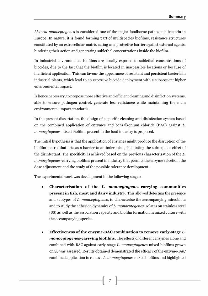
Listeria monocytogenes is considered one of the major foodborne pathogenic bacteria in
Europe. In nature, it is found forming part of multispecies biofilms, resistance structures
constituted by an extracellular matrix acting as a protective barrier against external agents,
hindering their action and generating sublethal concentrations inside the biofilm.
In industrial environments, biofilms are usually exposed to sublethal concentrations of
biocides, due to the fact that the biofilm is located in inaccessible locations or because of
inefficient application. This can favour the appearance of resistant and persistent bacteria in
industrial plants, which lead to an excessive biocide deployment with a subsequent higher
environmental impact.
Is hence necessary, to propose more effective and efficient cleaning and disinfection systems,
able to ensure pathogen control, generate less resistance while maintaining the main
environmental impact standards.
In the present dissertation, the design of a specific cleaning and disinfection system based
on the combined application of enzymes and benzalkonium chloride (BAC) against L.
monocytogenes mixed biofilms present in the food industry is proposed.
The initial hypothesis is that the application of enzymes might produce the disruption of the
biofilm matrix that acts as a barrier to antimicrobials, facilitating the subsequent effect of
the disinfectant. The specificity is achieved based on the previous characterisation of the L.
monocytogenes-carrying biofilms present in industry that permits the enzyme selection, the
dose adjustment and the study of the possible tolerance development.
The experimental work was development in the following stages:
Characterisation of the L. monocytogenes-carrying communities
present in fish, meat and dairy industry. This allowed detecting the presence
and subtypes of L. monocytogenes, to characterise the accompanying microbiota
and to study the adhesion dynamics of L. monocytogenes isolates on stainless steel
(SS) as well as the association capacity and biofilm formation in mixed culture with
the accompanying species.
Effectiveness of the enzyme-BAC combination to remove early-stage L.
monocytogenes-carrying biofilms. The effects of different enzymes alone and
combined with BAC against early-stage L. monocytogenes mixed biofilms grown
on SS was assessed. Results obtained demonstrated the efficacy of the enzyme-BAC
combined application to remove L. monocytogenes mixed biofilms and highlighted
Summary
8
that this efficacy varies with the composition and age of the biofilm, pointing out
the importance of designing strain-specific cleaning and disinfection strategies.
Quantification of the effects of pronase-BAC combined application
against L. monocytogenes-E. coli late-stage dual-species biofilms. The
individual and combined effects on the occupied surface, and the number of viable
adhered and released cells after the application of pronase and BAC against late-
stage L. monocytogenes-E. coli dual-species biofilms were assessed. Results
demonstrated a synergistic effect of pronase-BAC application against L.
monocytogenes-E. coli dual-species biofilms, a higher efficacy against L.
monocytogenes, and the need to use high BAC doses to ensure the absence of
adhered and released viable cells.
Tolerance development to pronase-BAC combined treatments in L.
monocytogenes-E. coli mixed biofilms. The effects of the frequency and
duration of consecutive sublethal exposures to pronase-BAC on the development of
tolerance in L. monocytogenes-E. coli mixed biofilms was assessed. Results showed
that only when sublethal exposures are alternated with recovery periods, a
tolerance development to the application of pronase-BAC combined treatments
takes place.

Resumen

Resumen
11
Listeria monocytogenes está considerada una de las bacterias patógenas transmitidas por
alimentos de mayor relevancia en Europa. En la naturaleza, se encuentra formando
biopelículas multiespecie, estructuras de resistencia constituidas por una matriz extracelular
que actúa de barrera protectora frente a agentes externos, dificulta su acción y genera
concentraciones subletales en el interior de la biopelícula.
En el ámbito industrial, es habitual que las biopelículas estén expuestas a concentraciones
subletales de biocidas, bien debido a que estas se encuentran en ubicaciones de difícil acceso,
bien como consecuencia de una aplicación ineficiente. Ello favorece la aparición de bacterias
resistentes y persistentes en plantas industriales, lo que ha llevado a un exceso en el uso de
biocidas y al consecuente incremento del impacto ambiental.
Es necesario, pues, proponer sistemas de limpieza y desinfección más efectivos y eficientes,
que aseguren el control de patógenos, generen menor resistencia y mantengan los cánones
de impacto ambiental.
En la presente tesis se propone el diseño de un sistema de limpieza y desinfección específico
frente a biopelículas mixtas de L. monocytogenes presentes en la industria alimentaria
basado en la aplicación combinada de enzimas y cloruro de benzalconio (CB).
La hipótesis de partida se basa en que la aplicación de las enzimas podría suponer la
disrupción de la matriz de la biopelícula que actúa como barrera frente a antimicrobianos
facilitando la acción posterior del desinfectante. La especificidad se consigue a partir de la
caracterización previa de las biopelículas portadoras de L. monocytogenes presentes en la
industria, que permite la selección de las enzimas, el ajuste de las dosis y el estudio del
posible desarrollo de tolerancia.
El trabajo se desarrolló en las siguientes etapas:
Caracterización de las comunidades portadoras de L. monocytogenes
presentes en superficies de industrias pesquera, cárnica y láctica. Esto
permitió detectar la presencia y subtipos de L. monocytogenes, caracterizar la
microbiota acompañante y estudiar las dinámicas de adhesión de los aislados de L.
monocytogenes sobre acero inoxidable (AI) así como la capacidad de su asociación
y formación de biopelículas en cultivo mixto con las especies acompañantes.
Efectividad de la combinación de enzimas-CB sobre la eliminación de
biopelículas tempranas portadoras de L. monocytogenes. Se estudiaron
los efectos de diferentes enzimas solas y combinadas con CB sobre biopelículas
Resumen
12
tempranas mixtas de L. monocytogenes formadas en AI. Los resultados obtenidos
demostraron la efectividad de la aplicación combinada enzima-CB sobre la
eliminación de biopelículas mixtas portadoras de L. monocytogenes y pusieron de
manifiesto que dicha efectividad varía con la composición y edad de la biopelícula,
señalando la importancia de diseñar sistemas específicos de limpieza y
desinfección.
Cuantificación de los efectos de la aplicación combinada de pronasa-CB
sobre la eliminación de biopelículas tardías de L. monocytogenes-E.
coli. Se cuantificaron los efectos individuales y combinados de la aplicación de
pronasa y CB sobre la superficie ocupada por las biopelículas tardías mixtas y el
número de células viables adheridas y desprendidas después de la aplicación de los
tratamientos. Los resultados demostraron un efecto sinérgico de pronasa-CB sobre
la eliminación de biopelículas de L. monocytogenes-E. coli, una mayor efectividad
frente a L. monocytogenes y la necesidad de dosificar elevadas concentraciones de
BAC para asegurar la ausencia de células viables adheridas y liberadas.
Desarrollo de tolerancia a tratamientos combinados de pronasa-CB en
biopelículas mixtas de L. monocytogenes-E. coli. Se evaluó el efecto de la
frecuencia y duración de exposiciones subletales consecutivas de pronasa-CB sobre
el desarrollo de tolerancia en biopelículas mixtas de L. monocytogenes-E. coli. Los
resultados demostraron que únicamente cuando las exposiciones subletales se
acompañan de un periodo de recuperación se produce el desarrollo de tolerancia a
la aplicación de los tratamientos combinados pronasa-CB.

1
Introduction and objectives

Introduction and objectives
15
Listeria monocytogenes: from environment to human disease
Historical facts, taxonomy and general characteristics
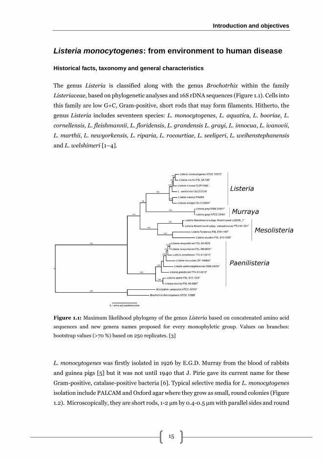
The genus Listeria is classified along with the genus Brochotrhix within the family
Listeriaceae, based on phylogenetic analyses and 16S rDNA sequences (Figure 1.1). Cells into
this family are low G+C, Gram-positive, short rods that may form filaments. Hitherto, the
genus Listeria includes seventeen species: L. monocytogenes, L. aquatica, L. booriae, L.
cornellensis, L. fleishmannii, L. floridensis, L. grandensis L. grayi, L. innocua, L. ivanovii,
L. marthii, L. newyorkensis, L. riparia, L. rocourtiae, L. seeligeri, L. weihenstephanensis
and L. welshimeri [1–4].
Figure 1.1: Maximum likelihood phylogeny of the genus Listeria based on concatenated amino acid
sequences and new genera names proposed for every monophyletic group. Values on branches:
bootstrap values (>70 %) based on 250 replicates. [3]
L. monocytogenes was firstly isolated in 1926 by E.G.D. Murray from the blood of rabbits
and guinea pigs [5] but it was not until 1940 that J. Pirie gave its current name for these
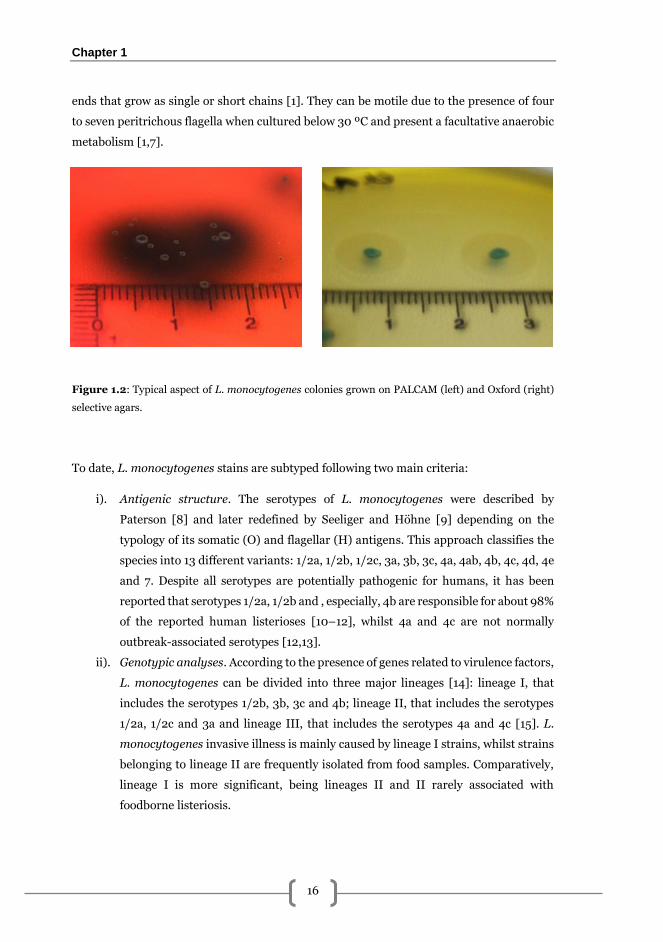
Gram-positive, catalase-positive bacteria [6]. Typical selective media for L. monocytogenes
isolation include PALCAM and Oxford agar where they grow as small, round colonies (Figure
1.2). Microscopically, they are short rods, 1-2 µm by 0.4-0.5 µm with parallel sides and round
Chapter 1
16
ends that grow as single or short chains [1]. They can be motile due to the presence of four
to seven peritrichous flagella when cultured below 30 ºC and present a facultative anaerobic
metabolism [1,7].
Figure 1.2: Typical aspect of L. monocytogenes colonies grown on PALCAM (left) and Oxford (right)
selective agars.
To date, L. monocytogenes stains are subtyped following two main criteria:
i). Antigenic structure. The serotypes of L. monocytogenes were described by
Paterson [8] and later redefined by Seeliger and Höhne [9] depending on the
typology of its somatic (O) and flagellar (H) antigens. This approach classifies the
species into 13 different variants: 1/2a, 1/2b, 1/2c, 3a, 3b, 3c, 4a, 4ab, 4b, 4c, 4d, 4e
and 7. Despite all serotypes are potentially pathogenic for humans, it has been
reported that serotypes 1/2a, 1/2b and , especially, 4b are responsible for about 98%
of the reported human listerioses [10–12], whilst 4a and 4c are not normally
outbreak-associated serotypes [12,13].
ii). Genotypic analyses. According to the presence of genes related to virulence factors,
L. monocytogenes can be divided into three major lineages [14]: lineage I, that
includes the serotypes 1/2b, 3b, 3c and 4b; lineage II, that includes the serotypes
1/2a, 1/2c and 3a and lineage III, that includes the serotypes 4a and 4c [15]. L.
monocytogenes invasive illness is mainly caused by lineage I strains, whilst strains
belonging to lineage II are frequently isolated from food samples. Comparatively,
lineage I is more significant, being lineages II and II rarely associated with
foodborne listeriosis.
Introduction and objectives
17
The ubiquity of L. monocytogenes can be attributed to the outstanding ability to cope with
different environmental conditions. In fact, this pathogen is considered one of the most
robust non-spore forming organism. It can proliferate under a broad range of temperatures,
from about 0 to 45 ºC, tolerate salt concentrations up to 12 % (w/v) and pH values from 4.3
to 9.2 [16]. This wide variety of environmental conditions under which L. monocytogenes
can grow and survive, make it a difficult pathogen to eliminate in the food industry and food-
related areas.
Ecological aspects of L. monocytogenes
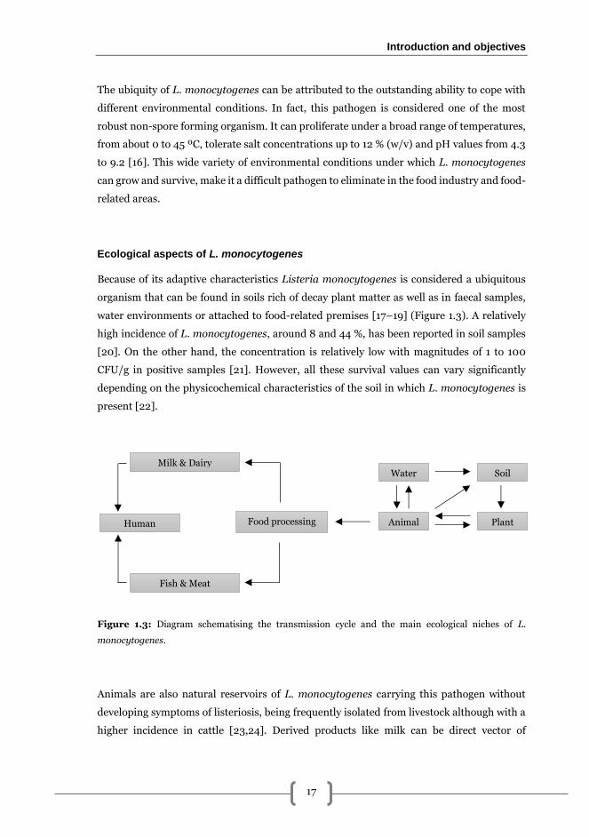
Because of its adaptive characteristics Listeria monocytogenes is considered a ubiquitous
organism that can be found in soils rich of decay plant matter as well as in faecal samples,
water environments or attached to food-related premises [17–19] (Figure 1.3). A relatively
high incidence of L. monocytogenes, around 8 and 44 %, has been reported in soil samples
[20]. On the other hand, the concentration is relatively low with magnitudes of 1 to 100
CFU/g in positive samples [21]. However, all these survival values can vary significantly
depending on the physicochemical characteristics of the soil in which L. monocytogenes is
present [22].
Figure 1.3: Diagram schematising the transmission cycle and the main ecological niches of L.
monocytogenes.
Animals are also natural reservoirs of L. monocytogenes carrying this pathogen without
developing symptoms of listeriosis, being frequently isolated from livestock although with a
higher incidence in cattle [23,24]. Derived products like milk can be direct vector of
Milk & Dairy
Fish & Meat
Human
Water
Animal Plant
Soil
Food processing
Chapter 1
18
contamination to humans, but essentially the transmission of the pathogen is due to the
routes involving food processing environments. In fact, ready-to-eat (RTE) products, soft
cheese, fish, shellfish and deli products [25,26] are some of the most common foodstuffs
through which L. monocytogenes infection takes place. Gombas et al. [27] demonstrated that
in the USA the prevalence of L. monocytogenes is generally associated with seafood salads
(4.7 %) and smoked seafood (4.3 %), whereas in the EU, non-compliance among RTE
products was significantly lower. In addition to RTE, raw products also harbour L.
monocytogenes as demonstrated in a study performed by Pagadala et al. [28] reporting a L.
monocytogenes incidence of 4.5 % in blue crab processing plants. Other authors have
reported presence of L. monocytogenes in raw meat of chicken [29,30] of pork [30,31].
Although in a lower proportion compared to other products [32], vegetables and fruits and
related processing environments are also associated with L. monocytogenes incidence
[33,34].
In food related environments, this pathogen can be a difficult pathogen to control and
become persistent [35,36] being usually associated with other microorganisms in complex
multi-species communities [37]. Remark that L. monocytogenes is among the major agents
causing death due to foodborne illnesses in the United States [38] and in Europe [39], which
justifies the importance to study and understand the different aspects regarding the life cycle
of L. monocytogenes to develop effective strategies to control this bacterium especially in
food processing facilities.
L. monocytogenes as a foodborne pathogen
Inside the genus, L. monocytogenes is the only species considered as pathogen for humans
causing mainly foodborne infections [36]. Human listeriosis typically courses as a two-phase
illness with an initial phase of mild symptoms including sub-febrile episodes that can last
from 3 to 10 days and may be concomitant with headache, ataxia, general physical discomfort
and nausea followed by a subsequent phase with severe signs of central nervous system
affection [40]. These meningeal forms usually provoke consciousness alteration, motor
disorders or even partial nervous paralysis [40].
L. monocytogenes is considered an important paradigm due to its particular replication
cycle. They are intracellular pathogens that undertake cell-to-cell spreading and therefore
they remain invisible for host defences [41]. They can proliferate within macrophages, once
the pathogen is engulfed, carrying out an early scape from the phagocytic vacuole followed
by a multiplication in the cytosol of endothelial and epithelial cells and in hepatocytes. This
Introduction and objectives
19
leads to an eventual intracytosolic mobilisation via actin filaments and a final protrusion and
invasion of the neighbouring cells where all the invasive cycle reinitiates [41].
According to the European Food Safety Authority (EFSA), L. monocytogenes appears to be
a microorganism of a great concern because even though its incidence among population is
relatively low, it is maintained throughout time with high morbidity and mortality rates
among the major risk groups: newborns, elderly people, people with weakened immune
system, and pregnant women [42,43]. In fact, the latest report of the European Food Safety
Authority shows that the incidence of confirmed European L. monocytogenes infections has
increased by 30 % regarding previous data [39]. Large food-borne listeriosis outbreaks with
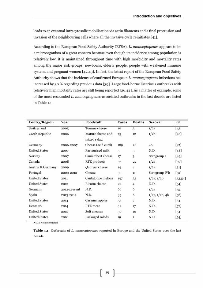
relatively high mortality rates are still being reported [36,44]. As a matter of example, some
of the most resounded L. monocytogenes-associated outbreaks in the last decade are listed
in Table 1.1.
Contry/Region Year Foodstuff Cases Deaths Serovar Ref.
Switzerland 2005 Tomme cheese 10 3 1/2a [45]
Czech Republic 2006 Mature cheese and
mixed salad
75 12 1/2b [46]
Germany 2006-2007 Cheese (acid curd) 189 26 4b [47]
United States 2007 Pasteurised milk 5 3 N.D. [48]
Norway 2007 Camembert cheese 17 3 Serogroup I [49]
Canada 2008 RTE products 57 22 1/2a [50]
Austria & Germany 2009 Quargel cheese 14 4 1/2a [51]
Portugal 2009-2012 Cheese 30 11 Serogroup IVb [52]
United States 2011 Cantaloupe melons 147 33 1/2a, 1/2b [53,54]
United States 2012 Ricotta cheese 22 4 N.D. [54]
Germany 2012-present N.D. 66 6 1/2a [55]
Spain 2013-2014 N.D. 35 6 1/2a, 1/2b, 4b [56]
United States 2014 Caramel apples 35 7 N.D. [54]
Denmark 2014 RTE meat 41 17 N.D. [57]
United States 2015 Soft cheeses 30 10 N.D. [54]
United States 2016 Packaged salads 19 1 N.D. [54]
N.D.: Not determined
Table 1.1: Outbreaks of L. monocytogenes reported in Europe and the United States over the last
decade.
Chapter 1
20
Regarding L. monocytogenes-associated foodborne epidemiology, serotype 4b appears to be
the most frequent serotype causing large outbreaks and invasive illness [58]. This serotype
has been associated with the consumption of contaminated foodstuffs such as paté, cheese
and coleslaw [59,60]. On the other hand, serotype 1/2b is the most frequent in non-invasive
listerioses and it has been isolated among outbreaks involving contaminated dairies and rice
salad [61,62].
Several reasons have been postulated to explain the apparent deficient control of this
pathogen in food industry: lack of sensitivity among methods leading to an inadequate L.
monocytogenes detection due to the existence of viable non cultivable cells [63,64],
inefficient procedures for cleaning and disinfection [65] and principally biofilm formation
by L. monocytogenes and subsequent increase of its capability to resist sanitizers [66–68].
Biofilm formation in Listeria monocytogenes
Bacterial biofilms: sessile but not stuck communities
Even though biofilms may be considered as a modern concept, the reality is that the very
first observations of these structures were carried out in 1684 by Antoni van Leeuwenhoek
in dental plaque samples. He reported those results to the Royal Society of London, referring
to his observations of the vast quantity of microorganisms present stating that: “the number
of these animicules in the scurf of a man’s teeth are so many that I believe they exceed the
number of men in a kingdom”. However, it was not until 1975 that the word “biofilm” was
not used in a scientific publication [69].
The currently accepted definition of a biofilm was coined in 2002 by Donlan and Costerton
who elegantly described them as microbially derived sessile communities characterised by
cells that are irreversively attached to a surface, an interface or to each other, are embedded
in a matrix of extracellular polymeric substances (EPS) and have an altered phenotype
regarding its growth rate and genic expression [70]. Therefore, biofilms are considered the
main structure in which bacteria can be found ubiquitously in sanitary, environmental and
industrial settings [71–74]. The capability to grown as a biofilm demonstrates somehow the
social component of bacteria even though the formation rates and the physicochemical
features of the final structure are highly variable and depend on the strain (or strains)
composing the actual biofilm and the abiotic factors involved [75,76]. Despite its sessile
nature, biofilms cannot be considered as halted structures. They are made up in a large part
of water canalicules which constitute a metabolically active and effective oxygen and
Introduction and objectives
21
nutrients distribution network [77]. The establishment of microscale chemical gradients
inside the biofilm, leads to the presence of local phenotypical and genotypical cellular
variations among the resident population [76] and, therefore, cells present are in a broad
range of physiological states [76–78].
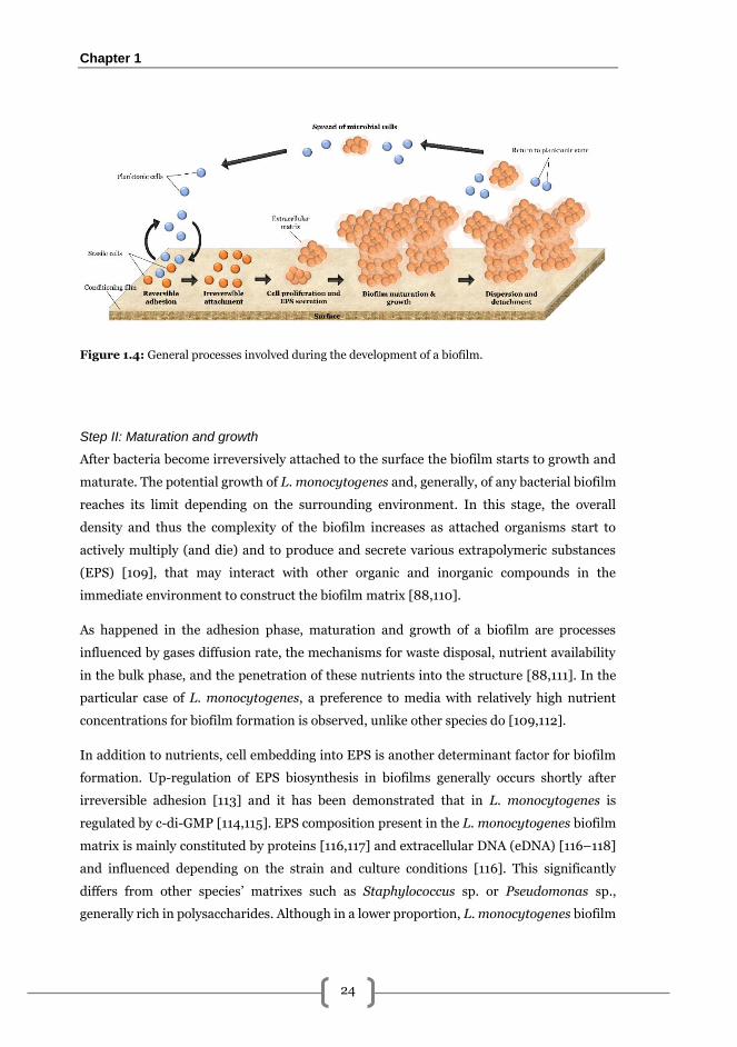
Steps in biofilm formation
The development of surface-adhered bacterial biofilms can be divided into three
fundamental steps schematised in Figure 1.4: (i) attachment; (ii) maturation and growth;
and (iii) detachment and/or dispersion [77,79]. All these phases are deeply regulated by
chemical stimuli that act as modulators modifying the communal behaviour in a
concentration-dependent manner. This mechanism of signalling and molecule recognition
known as quorum sensing (QS) still remains partially unknown to microbiology due to its
complexity [80,81]. In L. monocytogenes the main regulation pathways are dependent of the
so-called auto-inducer 2 (AI-2) [82], the agr (accessory gene regulator) system [83] and the
transcriptional regulator of stress response sigB [84]. Other factors that influence biofilm
development include medium composition and presence of antimicrobials, temperature,
bacterial concentration in the bulk phase and shear forces [70].
Step I: Attachment
Among the steps involved in the development of a biofilm, the phenomenon of initial
adhesion is the phase in which bacteria shift from a free-living (planktonic) cell to a sessile
state. This initial stage is strongly influenced by the environment and bacteria involved
undertake several physiological changes.
It is important to remark, that primary contact generally occurs between bacteria and a
conditioned surface. This conditioning is an accumulation in the solid-liquid interface of
different inorganic and organic molecules that are present in the bulk phase. This
accumulation leads to a local higher concentration of nutrients that alters the
physicochemical properties of the surface [85]. Following the formation of this conditioning
film, bacteria are deposited onto the surface either passively via Brownian motion,
sedimentation or convective transport [86] although it has been reported that active
transport via flagella and chemical sensing also plays an essential role [87].
After that, initial attachment of bacteria takes place in which van der Waals forces,
electrostatic forces and hydrophobic interactions contribute to stabilise the cell-surface
Chapter 1
22
interaction [86]. The nature of this primary adhesion is weak and cells can be effortless
removed by shear forces (e.g. rinsing). This reversible feature allows bacterial cells to move
along the surface to find an appropriate place to adhere. The duration of this initial phase
tends to be short and cells rapidly carry out transition from reversible to irreversible attached
cells in which the production of specific ligands, such as pili and fimbriae, and also secretion
of exopolymeric substances (EPS) makes bacteria to be strongly adhered to the surface and
therefore much more difficult to remove both by physical (e.g. scraping) and chemical (e.g.
cleaners) methods [88]. In L. monocytogenes, Schwab et al. [89] observed that this
phenotypical shift is produced in approximately 5 min after initial adhesion.
Besides the formation of the conditioning film, environmental conditions also modulate the
adhesion phenomena. Major factors affecting biofilm adhesion are:
i). pH. Many authors have studied the effects of the pH in culture medium on the
initial steps of L. monocytogenes biofilm formation. Nevertheless, results depict
contradictory results and are highly influenced by the rest of the conditions in each
assay and, therefore, the actual effects still remain obscure. As a matter of example
of this divergence, Herald and Zottola [90] and lately Poimenidou et al. [91],
reported that L. monocytogenes initial adhesion was hampered at acidic pH
whereas Briandet et al. [92] observed that adherences was increased at low pHs due
to a higher hydrophobicity of the cell wall in L. monocytogenes Scott A.
ii). Temperature. Even though L. monocytogenes is able to grow and adhere to food-
related surfaces in a broad range of temperatures (0 – 45 ºC), this and other
processes like flagella synthesis [93], are influenced by temperature. Briandet et al.
[92] demonstrated that L. monocytogenes Scott A adhered significantly better in
Trypticase soy-yeast extract broth (TSYE) at 37 ºC compared to lower incubation
temperatures. Despite this, subsequent studies demonstrated that this temperature
dependent favouring is produced until certain extent [94].
iii). Nutrient availability. It has been observed that the nutrients of the medium
stimulate or not the adherence of L. monocytogenes depending on the strain. Thus,
in some cases nutrient starvation promotes the initial adherence [95]. Kim and
Frank [96] reported a higher adherence in biofilms grown in chemically defined
medium compared to those grown in trypicase soy broth (TSB) while Mai and
Conner [94] observed that rich media promoted L. monocytogenes adhesion.
Glucose availability also alters adhesion in L. monocytogenes. With this regard,
Guilbaud et al. [97] used glucose supplements to enhance biofilm formation,
whereas other studies report that rich media with high glucose concentrations give
Introduction and objectives
23
rise to biofilms with fewer adhered cells and a block of the Listeria adhesion protein
(LAP) expression [98] despite a higher EPS production [99].
iv). Characteristics of the surface. Several studies have demonstrated that L.
monocytogenes is able to adhere and undertake biofilm formation on a wide range
of surfaces routinely used in food-related environments [17,73,100–103]. Among
them, stainless steel (SS) is the most common material used for food contact
purposes in the food industry because it is easy to produce, durable and
straightforwardly cleaned and disinfected [86]. However, the under scanning
electron microscopy (SEM) the surface of SS reveals cracks and crevices,
susceptible to provide bacteria a greater surface to adhere [101] and a shelter for
antimicrobials [86]. With this regard, Mosquera-Fernández et al. [101]
demonstrated that L. monocytogenes is able to adhere better to AISI 304 SS
(rough) than to polished AISI 316 SS (smooth). Due to its relevance in the food
industry, SS will be the only surface used in the present thesis to grow both mono
and multi-species biofilms.
v). Flagella and cellular motility. Contrarily to many bacterial species, flagella
synthesis in L. monocytogenes is temperature-dependent [93]. Incubation
temperatures higher than 37 ºC impede the flagellin polymerisation, and
subsequent motility failure, due to MogR repression of flagellar gene transcription.
At temperatures of 30 ºC and below, MogR is inhibited by GmaR antirepressor,
restoring flagella biosynthesis and cellular motility [104]. Studies such as those
carried out by Guerrieri et al. [105], Lemon et al. [104], Tresse et al. [106] and
Vatanyoopaisarn et al. [107], among others, demonstrated that flagella are critical
for L. monocytogenes biofilm formation during the first stages. However, in
subsequent stages of biofilm formation flagella presence seems not to have any
deleterious effects [108].
Chapter 1
24
Figure 1.4: General processes involved during the development of a biofilm.
Step II: Maturation and growth
After bacteria become irreversively attached to the surface the biofilm starts to growth and
maturate. The potential growth of L. monocytogenes and, generally, of any bacterial biofilm
reaches its limit depending on the surrounding environment. In this stage, the overall
density and thus the complexity of the biofilm increases as attached organisms start to
actively multiply (and die) and to produce and secrete various extrapolymeric substances
(EPS) [109], that may interact with other organic and inorganic compounds in the
immediate environment to construct the biofilm matrix [88,110].
As happened in the adhesion phase, maturation and growth of a biofilm are processes
influenced by gases diffusion rate, the mechanisms for waste disposal, nutrient availability
in the bulk phase, and the penetration of these nutrients into the structure [88,111]. In the
particular case of L. monocytogenes, a preference to media with relatively high nutrient
concentrations for biofilm formation is observed, unlike other species do [109,112].
In addition to nutrients, cell embedding into EPS is another determinant factor for biofilm
formation. Up-regulation of EPS biosynthesis in biofilms generally occurs shortly after
irreversible adhesion [113] and it has been demonstrated that in L. monocytogenes is
regulated by c-di-GMP [114,115]. EPS composition present in the L. monocytogenes biofilm
matrix is mainly constituted by proteins [116,117] and extracellular DNA (eDNA) [116–118]
and influenced depending on the strain and culture conditions [116]. This significantly
differs from other species’ matrixes such as Staphylococcus sp. or Pseudomonas sp.,
generally rich in polysaccharides. Although in a lower proportion, L. monocytogenes biofilm
Introduction and objectives
25
matrix contains polysaccharides such as teichoic acids equals to the ones found in the
bacterial membrane [119].
EPS secretion and, subsequently, matrix formation plays an important role in biofilms
providing protection against environmental aggressions impeding e.g. antimicrobial
molecules to reach the cells due to the reduced diffusion or by direct neutralisation of these
molecules with matrix components. In addition, the matrix also confers the biofilm a
physical stability that influences the final tridimensional conformation [110,120], favours the
genetic exchange between cells [110] and acts as a reserve of carbon, nitrogen and
phosphorus [121].
Step III: Dispersion and detachment
These two phenomena can take place separately of simultaneously in a given biofilm. The
two processes are similar, because both refer to a certain amount of cells are physically
separated from the biofilm and returned to the bulk phase, but different, since dispersion is
a process related with active genetic and metabolic processes undertaken inside the cell
whereas detachment is more of a passive phenomenon related to biofilm sloughing and
erosion produced by shear forces [122,123].
In spite of the advances in the field, dispersion phenomena in biofilms still remain as a
controversial issue. Several reasons have been attributed to this. It has been demonstrated
that can be regulated by QS [124], the production of glycolipids [125], the production of
endogenous enzymes [126] or due to nutrient depletion [109]. In these last two cases,
eventual matrix decay may take place and therefore the extrusion of parts of the biofilm
would be facilitated.
The most immediate consequence of mobilisation of parts of the biofilm in the context of the
food industry is the creation of new contamination foci [127] that could finally affect final
product safety and quality via cross contamination. Because of this, in the present PhD
thesis, the pool of live viable cells released from the biofilms after the application of cleaning
and disinfection strategies will be addressed.
L. monocytogenes mixed-species biofilms
Although considered as a relatively poor biofilm former compared to other species [128], L.
monocytogenes can easily associate with other bacterial species forming part of complex
microbial communities with both Gram-positive and Gram-negative species [37,129–132]
Chapter 1
26
and the interaction among species forming these consortia varies depending of the genera
implicated and the environmental conditions [132]. Various studies involving L.
monocytogenes multispecies biofilms have highlighted the complexity of such interactions
and the different effects that associated bacteria could have in terms of the number of
adhered cells [129,131,133] and the EPS composition of the biofilm matrix [134]. In these
polymicrobial communities, L. monocytogenes can act as primary coloniser or as later
biofilm partner establishing interactions with other microorganisms present [135], therefore
increasing the complexity of bacterial ecological niches [136].
Considering this, it seems to be clear that increasing the knowledge regarding multispecies
biofilms could provide key information to develop new cleaning and disinfection strategies
against a given target [137,138]. In the particular context of the food industry, this would
reduce the number of bacterial foci thus reducing subsequent cross-contaminations of food
products. Despite this, the number of studies regarding mixed-species biofilms in food
industry-related environments dealing with a characterisation of the whole microbiota in a
particular surface is relatively low compared to other ambits such as oral biofilms [139]. In
this line, various authors have remarked the need to characterise the bacterial interactions
among L. monocytogenes-multispecies biofilms present in real scenarios. These would
include, those influencing the biofilm formation patterns as well as other phenotypical
characteristics [140–142], especially when designing new disinfection strategies. However,
most of the studies dealing with polymicrobial biofilms, use model structures based on the
literature rather than using bacteria isolated previously from relevant environments, since
they can present unique phenotypical features [143].
Regarding interactions within L. monocytogenes mixed-species biofilms, Carpentier and
Chassaing [129] analysed 29 different L. monocytogenes dual-species biofilms and observed
how the number of adhered L. monocytogenes was increased, decreased o unaltered
depending on the accompanying bacterium. Other studies demonstrated how certain
bacteria such as Pseudomonas fluorescens clearly contributes to L. monocytogenes adhesion
in mixed biofilms [144,145]. In a posterior study, it was discussed how the effects of such
interactions affected the level of L. monocytogenes persistence in a processing plant [35].
Almeida et al. [146] characterised L. monocytogenes-Salmonella enterica-E. coli mixed
biofilms using peptide nucleic acid fluorescence in situ hybridisation (PNA-FISH) on SS
coupons describing a defined structural pattern in which S. enterica and L. monocytogenes
were in the bottom parts of the biofilm while E. coli was located on the top layer. These
results were consistent with those of Puga et al. [144] who observed the disposition in the
bottom layers of L. monocytogenes in mixed biofilms with P. fluorescens grown on glass. A
posterior work of this group, demonstrated that L. monocytogenes-P. fluorescens biofilms
Introduction and objectives
27
became more compact with the age especially in those grown at 4 ºC, despite cell viability
remained unaltered [147], showing higher resistance to chitosan [148].
The present thesis, contributes to the knowledge of the ecological aspects of L.
monocytogenes-carrying biofilms present on surfaces of the food industry characterising the
composition and distribution of L. monocytogenes polymicrobial communities in industrial
premises, gaining further insight into the accompanying species’ distribution depending on
the environmental factors [37]. In addition to these determinations, evidence on how the
accompanying species clearly affects the final morphology of the L. monocytogenes mixed
biofilm and the susceptibility to enzyme-based combined antimicrobial treatments is also
provided [145].
Methods for biofilm quantification and structural studies
The structural studies in biofilms have been highly conditioned by the technological
advances. First studies performed in the 70s and the 80s were mostly based in the
quantification of adhered cells via agar plating despite its limitations [149] and the lack of
information provided other that the number of viable-and-cultivable cells.
Light field and electron microscopy allowed the very first studies in this field. However, the
lack of resolution in the first case and the need for dehydrate the sample in the second,
limited the accuracy of such observations [150]. Therefore, biofilms were initially considered
as flat and homogeneous instead of complex and heterogeneous structures. The various
structural models currently accepted were not observed and described until the fabrication
and utilisation of higher resolution microscopy techniques. In this line, it is usual to
incorporate microscopic assays, mainly fluorescence and confocal laser scanning microscopy
(CLSM). In many studies regarding structure and spatio-temporal distribution of cells into
biofilms [101,151–153] CLSM is preferred since it permits to gather 3D data from hydrated
biofilms in vivo. However, CLSM microscopes are expensive and not available in all research
centres, which undoubtedly represents a major drawback at the time to incorporate this
technique in the habitual laboratory technique [154].
Although numerous image analysis software (COMSTAT, ImageJ, ISA, Imaris, MATLAB…),
and structural 2D and 3D parameters have been used in the literature for biofilm
quantification [101,151,152,155,156], the main issue of concern still is remains when selecting
the most appropriate parameters to give an accurate description of the structures.
Theoretically, reliable parameters should be easily related to biological processes, however,
Chapter 1
28
it is a consensus that the high complexity of biofilms makes difficult to correlate structural
changes within a specific biological process associated with a biofilm forming cycle.
Looking for solutions, some authors recommended to use image analysis only as auxiliary
information [157] whereas others came to the conclusion that an ad hoc selection of the
structural parameters for biofilm characterisation and quantification is required [153] and
the number should be as few as possible [158]. In biofilm quantification areal parameters
such as areal porosity, were described by Lewandowski et al. [159] have been considered as
good biofilm descriptors with an intuitive approach, used in many biofilm studies
[152,153,157,160]. When it comes to the study of the effects of antimicrobial substances on
biofilms Beyenal et al. [153] demonstrated how areal parameters allow to easily gather
information from microscopic data.
An analogue parameter to areal porosity, occupied area, is proposed in this PhD thesis to,
along with plate count, describe and quantify biofilm formation and antimicrobial
effectiveness of enzyme-benzalkonium chloride treatments against L. monocytogenes
mixed-species biofilms. This parameter was chosen since it was considered as the 2D
structural parameter with the most biologically meaningful, easy-to-interpret outcome.
Besides, it can be calculated with most of the commercial and home-made software.
Listeria monocytogenes in the food industry
Incidence of L. monocytogenes. A major concern in food processing plants
In Europe, it is estimated that the food industry annually invests about five trillion euros in
the implementation and application of cleaning and disinfections systems. Nonetheless,
bacterial contamination of foodstuffs is still a major problem with a remarkable increasing
incidence of L. monocytogenes over the last decade [39]. Numerous studies have
demonstrated that L. monocytogenes can be present in food processing facilities and how
some strains are able to persist in these premises for various months or even years [30,161–
165] mostly found associated with other bacteria forming mixed-species biofilms [130].
Molecular methods have been used in various surveys to point out equipment, floors and
drains as important contamination sources in food processing lines. In many cases
harbouring the same L. monocytogenes subtype in different locations therefore
demonstrating that clonal expansion due to cross-contamination occurs [37,164,166,167].
A high prevalence of L. monocytogenes has been detected in food-related facilities in North
America and Europe representing a serious concern in dairies, smoked fish houses and RTE
Introduction and objectives
29
meat processing plants [168]. In the light of these facts, various authors have suggested to
establish routing programs for of environmental sampling for L. monocytogenes detection
and control in an effort to ensure microbiological safety of the foodstuffs intended for human
consumption especially those belonging to the risk groups [162,169–171].
Strategies to control L. monocytogenes
Hazard analysis and critical control points (HACCP)
HACCP is an effective management system that ensures safety in all relevant points of the
production, storage, distribution and consumption of food products, by anticipation and
control of associated health hazards. Pre-requisite programs provide the basics in
environmental and operating conditions needed for the production of safe, wholesome
foodstuffs. The combination with Good Agricultural Practices (GAPs), Good Manufacturing
Practices (GMPs), Good Hygienic Practices (GHPs) and Good Laboratory Practices (GLPs)
is the best strategy to control potential hazards [172].
Some of these basic conditions to avoid product contamination by foodborne pathogens are
listed below [168]:
i). Good quality of the raw material.
ii). Effective training of employees in food hygiene.
iii). Appropriate design of the processing environment, including equipment, to ensure
proper cleaning and disinfection of all the in-contact surfaces.
These should be appropriately documented and regularly audited, and established and
managed separately from the HACCP plan. In L. monocytogenes, early studies reported that
L. monocytogenes is able to get access to the processing plants via operators’ shoes, clothing
and transport equipment, raw material and, probably, asymptomatic human carriers [173].
Despite great advances have been made for in HACCP, the widespread and survival capacity
of this pathogen still makes L. monocytogenes to be a difficult microorganism to control
[174].
Non-chemical agents for L. monocytogenes control
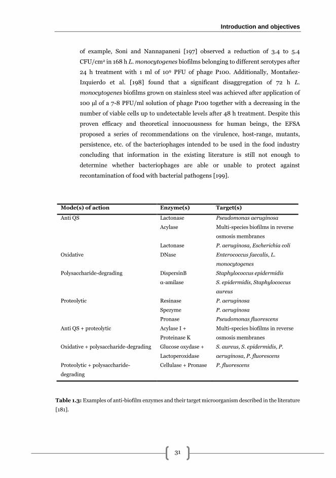
i). Enzymes. A promising strategy, especially for biofilm control, is the use of
molecules that can interfere in biofilm formation processes or even degrade specific
components of the extracellular matrix, some of them listed in Table 1.3. Following
this line of research, in the last few years the use of different enzymes has
Chapter 1
30
increasingly become a method used for biofilm control. These environmentally
friendly compounds have been shown to both prevent the initial adhesion and
remove formed structures [175–178] because of their dispersive effect on the sessile
structures acting on target molecules present in the biofilm matrix [141,179,180].
However, enzymes do not necessarily have bactericidal activity which makes them
unsuitable to be used as a strategy for disinfection [117]. To overcome this, a feasible
strategy to obtain both biofilm disinfection and removal would be the combination
of enzymes with chemical biocidals [181]. With this regard, in the present PhD
thesis the feasibility of combining enzymes with chemically-based disinfectants will
be addressed not only demonstrating that such combination is possible [145], but
also how the synergic action of these two components, effectively remove L.
monocytogenes mixed-species biofilms. ii). Essential oils (EOs). These comprise a broad family of approximately 3000
different aromatic and volatile liquid preparations extracted from plant material,
such as roots, fruits, herbs, flowers, etc. [182,183] with different antioxidant and
antimicrobial properties [184,185]. EOs cause changes in cell morphology,
physicochemical properties of membranes, as well as several intracellular
phenomena by interfering in the metabolic pathways, including cell division,
and/or altering the molecular interaction [186]. Despite the practical application of
EOs has been limited due to their alteration of organoleptic properties of foodstuffs,
poor solubility and partial volatility [184].
iii). Bacteriocins. They form part of a heterogeneous group of small, bacterially
produced, ribosomally synthesised peptides with antimicrobial properties
classified according to the post-translational modifications that they undergo once
synthesised [187]. Recent studies have shown the effectiveness of bacteriocins on
L. monocytogenes biofilms [188–190] and they have been proposed as an
environmentally friendly alternative to the currently used strategies. In addition to
bacteriocin direct use, the co-culture of L. monocytogenes with bacteriocin-
producing bacteria, have also demonstrated to be effective for biofilm control.
iv). Bacteriophages. The application of viruses infecting bacteria and, therefore,
inducing the lysis of the host is considered nowadays as a versatile biofilm control
tool, highly active and specific, without deleterious effects to mammalian cells and
relatively low cost [191–193]. In L. monocytogenes phage-therapy has been
reported to be effective both in medical and industrial environments [194]. To date,
approximately 500 Listeria phages have been identified [195] all belonging to the
Caudovirales family [196]. Among them, bacteriophage P100 is one of the best
characterised being effective against L. mononcytogenes biofilms [196]. As a matter
Introduction and objectives
31
of example, Soni and Nannapaneni [197] observed a reduction of 3.4 to 5.4
CFU/cm2 in 168 h L. monocytogenes biofilms belonging to different serotypes after
24 h treatment with 1 ml of 109 PFU of phage P100. Additionally, Montañez-
Izquierdo et al. [198] found that a significant disaggregation of 72 h L.
monocytogenes biofilms grown on stainless steel was achieved after application of
100 µl of a 7-8 PFU/ml solution of phage P100 together with a decreasing in the
number of viable cells up to undetectable levels after 48 h treatment. Despite this
proven efficacy and theoretical innocuousness for human beings, the EFSA
proposed a series of recommendations on the virulence, host-range, mutants,
persistence, etc. of the bacteriophages intended to be used in the food industry
concluding that information in the existing literature is still not enough to
determine whether bacteriophages are able or unable to protect against
recontamination of food with bacterial pathogens [199].
Mode(s) of action Enzyme(s) Target(s)
Anti QS Lactonase Pseudomonas aeruginosa
Acylase Multi-species biofilms in reverse
osmosis membranes
Lactonase P. aeruginosa, Escherichia coli
Oxidative DNase Enterococcus faecalis, L.
monocytogenes
Polysaccharide-degrading DispersinB Staphylococcus epidermidis
α-amilase S. epidermidis, Staphylococcus
aureus
Proteolytic Resinase P. aeruginosa
Spezyme P. aeruginosa
Pronase Pseudomonas fluorescens
Anti QS + proteolytic Acylase I +
Proteinase K
Multi-species biofilms in reverse
osmosis membranes
Oxidative + polysaccharide-degrading Glucose oxydase +
Lactoperoxidase
S. aureus, S. epidermidis, P.
aeruginosa, P. fluorescens
Proteolytic + polysaccharide-
degrading
Cellulase + Pronase P. fluorescens
Table 1.3: Examples of anti-biofilm enzymes and their target microorganism described in the literature
[181].
Chapter 1
32
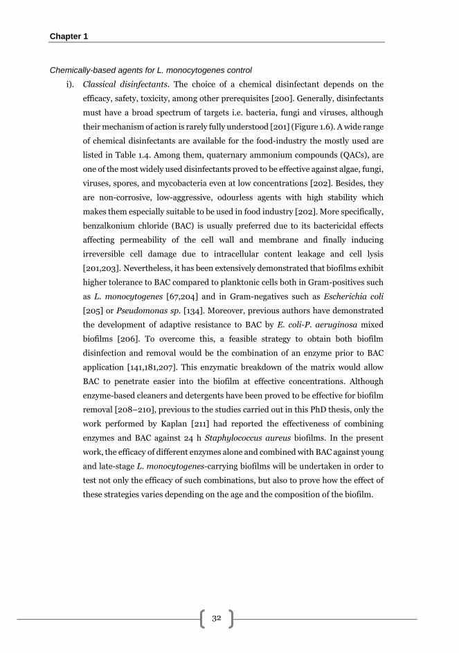
Chemically-based agents for L. monocytogenes control
i). Classical disinfectants. The choice of a chemical disinfectant depends on the
efficacy, safety, toxicity, among other prerequisites [200]. Generally, disinfectants
must have a broad spectrum of targets i.e. bacteria, fungi and viruses, although
their mechanism of action is rarely fully understood [201] (Figure 1.6). A wide range
of chemical disinfectants are available for the food-industry the mostly used are
listed in Table 1.4. Among them, quaternary ammonium compounds (QACs), are
one of the most widely used disinfectants proved to be effective against algae, fungi,
viruses, spores, and mycobacteria even at low concentrations [202]. Besides, they
are non-corrosive, low-aggressive, odourless agents with high stability which
makes them especially suitable to be used in food industry [202]. More specifically,
benzalkonium chloride (BAC) is usually preferred due to its bactericidal effects
affecting permeability of the cell wall and membrane and finally inducing
irreversible cell damage due to intracellular content leakage and cell lysis
[201,203]. Nevertheless, it has been extensively demonstrated that biofilms exhibit
higher tolerance to BAC compared to planktonic cells both in Gram-positives such
as L. monocytogenes [67,204] and in Gram-negatives such as Escherichia coli
[205] or Pseudomonas sp. [134]. Moreover, previous authors have demonstrated
the development of adaptive resistance to BAC by E. coli-P. aeruginosa mixed
biofilms [206]. To overcome this, a feasible strategy to obtain both biofilm
disinfection and removal would be the combination of an enzyme prior to BAC
application [141,181,207]. This enzymatic breakdown of the matrix would allow
BAC to penetrate easier into the biofilm at effective concentrations. Although
enzyme-based cleaners and detergents have been proved to be effective for biofilm
removal [208–210], previous to the studies carried out in this PhD thesis, only the
work performed by Kaplan [211] had reported the effectiveness of combining
enzymes and BAC against 24 h Staphylococcus aureus biofilms. In the present
work, the efficacy of different enzymes alone and combined with BAC against young
and late-stage L. monocytogenes-carrying biofilms will be undertaken in order to
test not only the efficacy of such combinations, but also to prove how the effect of
these strategies varies depending on the age and the composition of the biofilm.
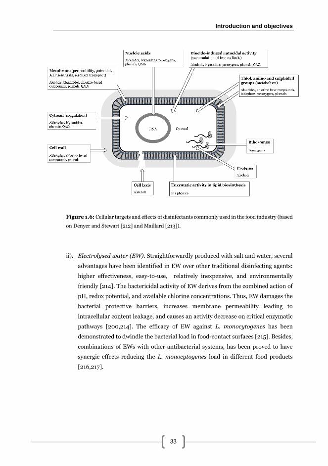
Introduction and objectives
33
Figure 1.6: Cellular targets and effects of disinfectants commonly used in the food industry (based
on Denyer and Stewart [212] and Maillard [213]).
ii). Electrolysed water (EW). Straightforwardly produced with salt and water, several
advantages have been identified in EW over other traditional disinfecting agents:
higher effectiveness, easy-to-use, relatively inexpensive, and environmentally
friendly [214]. The bactericidal activity of EW derives from the combined action of
pH, redox potential, and available chlorine concentrations. Thus, EW damages the
bacterial protective barriers, increases membrane permeability leading to
intracellular content leakage, and causes an activity decrease on critical enzymatic
pathways [200,214]. The efficacy of EW against L. monocytogenes has been
demonstrated to dwindle the bacterial load in food-contact surfaces [215]. Besides,
combinations of EWs with other antibacterial systems, has been proved to have
synergic effects reducing the L. monocytogenes load in different food products
[216,217].
Chapter 1
34
Disinfectant Pros Cons
Alcohols
(e.g. ethanol)
Cheap, fast-acting biocides of broad
microbial spectrum, non-toxic, easy-
to-use, colourless, harmless on skin,
soluble in water and volatile
Biostatics. Lack of effectiveness
against spores.
Chlorine-based
compounds
(e.g. sodium
hypochlorite)
Cheap, fast-acting oxidisers of broad
microbial spectrum. Easy-to-use and
unaffected by hard water. Effective
against planktonic cells and spores,
even at low temperatures. Non-film
forming without residues
Toxic, irritating, unstable, potentially
explosive and corrosive. Inactivated
by organic matter. pH sensitive.
Discoloration of products. Resistance
development.
Glutaraldehyde Cheap biocide of broan microbial
spectrum, non-corrosive
Biostatic. Non-biodegradable. Low
penetration in biofilms.
Idophors Sanitisers of broad activity spectrum,
non-corrosive, non-irritating and
easy-to-use. Low toxicity and stable
at a very low pH. Little affected by
organic matter
Alters flavour and odour of foodstuffs
Stain plastics and porous materials.
Highly foaming, unsuitable for
cleaning-in-place (CIP) systems.
Reduced efficacy at high pH and
temperatures >50 ºC. Expensive.
Peroxygens Strong fast acting oxidisers of broad
microbial spectrum, relatively non-
toxic and easy-to-use. Low foaming,
suitable for CIP. Effective against
bacterial biofilms and spores, even at
low temperatures. Non-corrosive to
stainless steel.
Loss of effectiveness in the presence
of organic material and some metals
contained in water. May corrode
some metals. Low efficacy against
yeasts and moulds. Relatively
expensive.
QACs (e.g. BAC) Stable, surface-active agents. Non-
toxic, non-irritating, non-corrosive,
odour and flavourless. Little affective
by organic materials. Support
microbial detachment
Limited effectiveness, which is
affected by hard water, low
temperatures and low pH.
Incompatible with most detergents.
Highly foaming. Unsuitable for CIP.
Residual antimicrobial film forming.
Resistance development. Relatively
expensive.
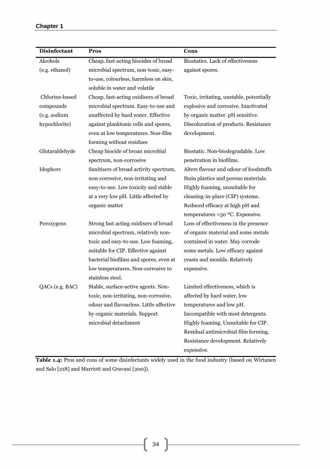
Table 1.4: Pros and cons of some disinfectants widely used in the food industry (based on Wirtanen
and Salo [218] and Marriott and Gravani [200]).
Introduction and objectives
35
L. monocytogenes resistance to disinfection
The increased resistance to disinfectants, especially to QACs, has been a topic of concern in
the context of the food industry. It has been demonstrated that resistance to BAC can be
attributed to two main factors: the expression of membrane active efflux pumps [219–222]
or the modification in the membrane fatty acid composition [223]. The effects of such
resistance have been recently investigated by Møretrø et al. [224] concluding that resistance
to BAC thanks to the presence of qacH and bcrABC genes, may contribute to an increased
growth of L. monocytogenes in food-related premises.
In biofilms, several mechanisms leading to a resistance to biocides can take place. According
to the observations of Costerton et al. [225] and Donlan and Costerton [70], some of them
may be the following:
i). Lack of penetration and further diffusion of the antimicrobial agent due to the
biofilm matrix [68,113,226]. Additionally, some authors have also pointed out that
the abiotic part of the biofilm may have a neutralising effect on many compounds
[110,120,227].
ii). Altered growth rate of cells into the biofilms [66,228].
iii). Other physiological changes due to the biofilm mode of growth supposing a
coexistence of different cell phenotypes can be present within the biofilm
[228,229].
iv). Formation of multispecies biofilms [226,230].
Regarding this last point, the association of L. monocytogenes with other microorganisms
can increase the resistance to sanitisation treatments, despite results vary depending on the
study. Van der Veen and Abee [132] observed that in L. monocytogenes-Lactobacillus
plantarum mixed biofilms in polystyrene microtiter plates, application of 100 µg/ml BAC
caused about 2.5 log CFU/well less compared with monocultures. In a similar way, Saá
Ibusquiza et al. [230] observed denser biofilm formation and a five-fold increase in the lethal
dose 90 (LD90) value to BAC of L. monocytogenes CECT 4032 in 96 h mixed biofilms with
Pseudomonas putida CECT 845 grown on SS. Contrarily, Kostaki et al. [231] did not found
any difference in the level of resistance to BAC, NaClO and peracetic acid in L.
monocytogenes-S. enterica biofilms compared with monocultures. In addition, a recent
study demonstrated that P. putida resistance to BAC is increased in co-culture with L.
monocytogenes while the resistance of the latter remains the same [134]. These
contradictory findings highlight the necessity to continue exploring the mechanisms
underneath bacterial associations in biofilms and the relationship with antimicrobial
resistance.
Chapter 1
36
Effects of antimicrobial sublethal exposure
It has been reported that continuous misuse of biocides e.g. using sublethal concentrations
unable to kill bacteria within a biofilm, can have long-term deleterious effects, contributing
to the selection of multi-drug resistant variants. Development of these resistances could be
detected as changes in the susceptibility to these biocides such as increased minimum
biofilm eradication concentration (MBEC) values, being especially relevant in the food
industry where chemical biocidals (quaternary ammonium compounds (QACs), chlorine,
etc.) are constantly deployed [113]. Besides, this incorrect use can alter the biofilm
phenotypic heterogeneity and the intra-specific relationships that take place. Therefore, the
late effects of these treatments will be the selection of the most resistant phenotypic variant
of the biofilm. Some authors have related this fact with the presence of persistent L.
monocytogenes through time, although this issue still arouses controversial discussion [35].
Introduction and objectives
37
Motivation and general objectives
Listeria monocytogenes is considered one of the main foodborne pathogens causing human
listeriosis, a rare but serious illness that, in susceptible individuals, can end into fatality.
Recent reports have highlighted an increasing incidence over the last years which turns L.
monocytogenes into a major concern in food safety.
Listeria monocytogenes, like all bacteria, lives in natural environments as bacterial
multispecies biofilms, where resident microorganisms interact with each other different
manners. Despite this, most of the studies found in the literature use collection strains, being
the number of studies regarding mixed-species biofilms in food industry-related
environments scarce. In the present PhD, the experimental design used was developed with
the aim to reproduce as much as possible the natural consortia found in real industrial
settings.
Cleaning and disinfection protocols used in food industry are non-specifically designed being
frequent that their application implies sublethal expositions to biocides, thus generating
tolerance and resistance or persistence among the pathogenic bacterial associations present.
Additionally, a high number of studies have reported cross-resistance between different
antimicrobials e.g. disinfectants and antibiotics. In this situation, the tendency is to increase
the concentration of biocides applied causing, ultimately, a major problem of environmental
pollution. Then, it is necessary to purpose specific and more efficient cleaning and
disinfection strategies that could generates lower levels of resistance and lower pollution
whereas assuring the control of the pathogenic bacteria.
Enzymes were chosen as good candidates to specifically damage and degrade the matrix
components of the L. monocytogenes mixed biofilms, in order to facilitate penetration of the
disinfectant in a synergistic manner. Additionally, if this occurs, the concentration of the
disinfectant and the subsequent generation of resistance and environmental impact could be
decreased.
In summary, the studies detailed within this PhD thesis were mainly motivated by the need
to develop specific and efficient cleaning and disinfection strategies to remove L.
monocytogenes-carrying biofilms present in food-related industrial environments. With
this global aim, the main objectives of the present thesis were:
i). Detect and characterise the composition of L. monocytogenes-carrying consortia
present in surfaces of fish and seafood, meat, and dairy industries. This
characterisation included molecular subtyping to establish ecological distributions
and further demonstration of how L. monocytogenes can associate with the

Chapter 1
38
different bacteria present to form dual-species biofilms. Besides, the dynamics of
L. monocytogenes monospecies biofilm on SS was determined using fluorescence
microscopy and image analysis. The isolates obtained were used in the subsequent
studies, and in those environments where no isolates were detected model biofilms
formed by strains also isolated from environmental samples were used.
ii). Determine the biofilm-removal effects of different enzymes aiming to degrade
specific components of the biofilm matrix in young L. monocytogenes dual-species
biofilms. The enzymes showing the highest efficacy were combined with
benzalkonium chloride to determine its biofilm removal effects in two different L.
monocytogenes dual-species biofilms also taking into consideration the amount of
viable cells released after the treatment.
iii). Ascertain the synergic effects of a pronase-benzalkonium chloride combined
treatment against late-stage L. monocytogenes-E. coli biofilms. Determination of
synergy was performed following a first order factorial design using images of
fluorescence microscopy and further determination of areal parameters as well as
the effects of viable adhered and released cells after the application of combined
treatments. In addition to this, a previous statistical study to determine the
feasibility of this method was also carried out.
iv). Determine the capacity of L. monocytogenes-E. coli biofilms to develop tolerance
after application of sublethal concentrations of pronase-benzalkonium chloride
combined treatments using different exposure approaches.

2
Listeria monocytogenes-carrying consortia
present in the food industry

Listeria monocytogenes-carrying consortia in the food industry
41
Introduction
The ability of L. monocytogenes to survive and persist for long periods in food-related
industrial environments has been extensively documented in the last decades
[30,162,164,232]. Besides, its ecological flexibility permits this pathogen to associate with
both Gram-positive and Gram-negative species [130,131] forming multispecies biofilms
[132]. Since the composition and the interaction heterogeneity in these structures
determines a series of characteristics such as the number of adhered L. monocytogenes
[129,131,133] or the capability to endure sanitation treatments [68,132,134,230,231], it
seems obvious that gaining further insight into the actual composition of multispecies
biofilms present in industrial environments, would provide valuable information for
development of novel strategies for pathogen control.
However, most of the research carried out involving L. monocytogenes-carrying biofilms,
generally uses the most representative species of the expected accompanying microbiota
instead of bacteria isolated from real food processing premises. For this reason, as a starting
point in this thesis, the main aim of this work was to detect and characterise the bacterial
communities carrying L. monocytogenes present in surfaces of fish, meat and dairy
industries. This included sampling, isolation, identification and subtyping of L.
monocytogenes isolates and related microbiota and a study of L. monocytogenes biofilm
formation dynamics on stainless steel.
Methods
Sample collection
Sampling was carried out between September 2010 and July 2011 in eight different surveys
in Northwest Spain (Galicia and Asturias) in different food-related premises obtaining total
of 270 samples from fish, meat and cheese production industries (Table 2.1). In each survey
all samples were collected the same day. A detailed list of all samples obtained for food
industry-related consortia is stated in Supplementary Table 1 (available online). Surveys in
fish industry were carried out by personnel from the Microbiology and Technology of Marine
Products personnel whereas an external company was needed to perform meat and cheese
industry samplings since they did not grant us access due to their legal and privacy policy.
Samples corresponding to 200 cm2 from every selected surface were aseptically collected by
thoroughly rubbing with a sterile sponge moistened with 10 ml of sterile LPT Neutralizing
Chapter 2
42
broth (composition per litre: 0.7 g soy lecithin, 5 g NaCl, 1 g Na2S2O3, 2.5 g NaHSO3, 1 g
HSCH2COONa, 5 g Yeast Extract, 1 g L-histidine, 5 ml Tween 80, pH 7.6 ± 0.2). Sponges
were introduced individually in auto-sealable bags, kept refrigerated at 4ºC and processed
within the 24 hours following the sample collection.
Isolation of Listeria monocytogenes and accompanying microbiota
Sponges were mixed with 50 ml of sterile buffered peptone water (BPW; Cultimed,
Barcelona, Spain) and digested with a stomacher masticator (IUL Instruments, Barcelona,
Spain) during 1 minute. An aliquot of 100 µl of the resultant suspension was directly spread
onto TSA plates (Cultimed, Barcelona, Spain) and incubated at 25 ºC for subsequent
isolation of accompanying microbiota in case of L. monocytogenes-positive sample, where
morphologically different colonies were picked and subcultured twice in TSA to obtain pure
cultures (isolates). These isolates were finally cultured in TSB (Cultimed, Barcelona, Spain)
at 25 ºC for DNA extraction.
To detect L. monocytogenes 1 ml was directly transferred to a flask containing 25 ml of sterile
Half Fraser broth (Oxoid, Hampshire, England) and incubated at 30 ºC during 24 hours. 100
µl of positive samples (changing medium from green to black) was transferred to 10 ml of
sterile Fraser broth (Oxoid, Hampshire, England) and incubated at 37 ºC for 24 hours.
Finally, 100 µl of positive samples was plated in Chromogenic (ISO) Listeria Agar (Oxoid,
Hampshire, England) and incubated for further 24 hours at 37 ºC. Presumptive L.
monocytogenes appeared blue presenting a clear halo around them. These were recovered
and subcultured twice in TSA to ensure purity of cultures and finally cultured in TSB at 37
ºC for DNA extraction.
Stock cultures of every sample were made and kept at -80 ºC in BHI (Biolife, Italy)
containing 50 % glycerol 1:1 (v/v) mixed. Work cultures were maintained at -20 ºC in TSB
(Cultimed, Barcelona, Spain) containing 50 % glycerol 1:1 (v/v) mixed.
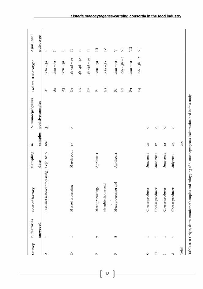
Listeria monocytogenes-carrying consortia in the food industry
43
Ap
aI,
As
cI
pu
lso
typ
e
I I I II
II
II
III
IV
V
VI
VII
VI
Ta
ble
2.1
: O
rig
in,
da
tes,
nu
mb
er o
f sa
mp
les
an
d s
ub
typ
ing
of
L.
mo
no
cyto
gen
es i
sola
tes
ob
tain
ed i
n t
his
stu
dy.
Se
ro
typ
e
1/2
a –
3a
1/2
a –
3a
1/2
a –
3a
4b
-4
d –
4e
4b
-4
d –
4e
4b
-4
d –
4e
1/2
a –
3a
1/2
a –
3a
1/2
a –
3a
½b
– 3
b –
7
1/2
a –
3a
½b
– 3
b –
7
Iso
late
ID
A1
A2
A3
D1
D2
D3
E1
E2
F1
F2
F3
F4
L.
mo
no
cy
tog
en
es
po
sit
ive
sa
mp
les
3
3
0
0
0
0
n.
sa
mp
les
106
17
24
12
12
24
27
0
Sa
mp
lin
g
da
te
Sep
t. 2
010
Ma
rch
20
01
Ap
ril
20
11
Ap
ril
20
11
Ju
ne
20
11
Ju
ne
20
11
Ju
ne
20
11
Ju
ly 2
011
So
rt
of
fac
tor
y
Fis
h a
nd
sea
foo
d p
roce
ssin
g
Mu
ssel
pro
cess
ing
Mea
t p
roce
ssin
g,
sla
ug
hte
rho
use
an
d
qu
art
erin
g
Mea
t p
roce
ssin
g a
nd
qu
art
erin
g
Ch
eese
pro
du
cer
Ch
eese
pro
du
cer
Ch
eese
pro
du
cer
Ch
eese
pro
du
cer
n.
fac
torie
s
su
rv
ey
ed
1 1 7
8
1 1 1 1
Su
rv
ey
A
D
E
F
G
H
I J
To
tal
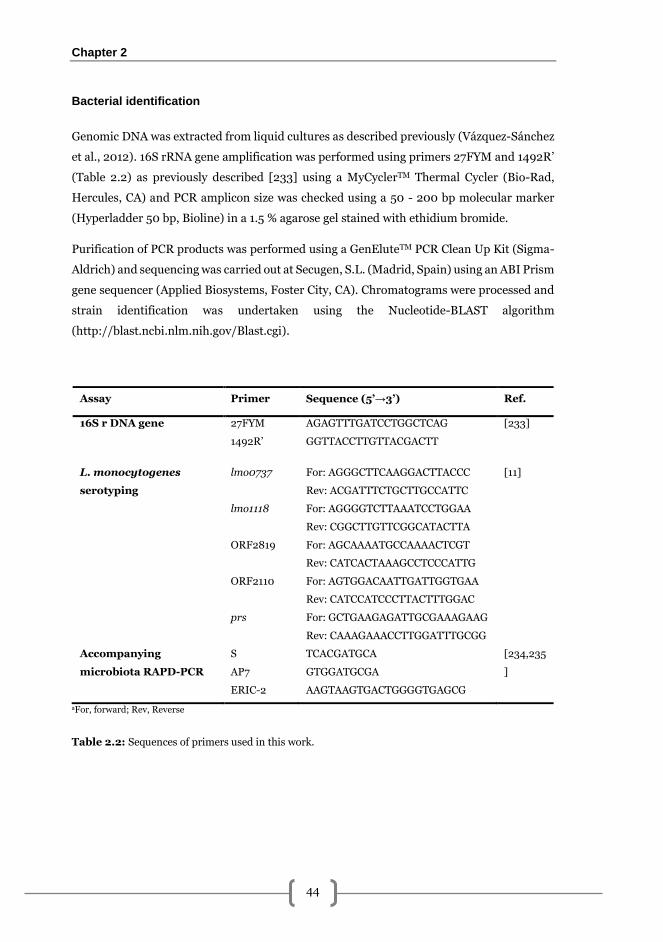
Chapter 2
44
Bacterial identification
Genomic DNA was extracted from liquid cultures as described previously (Vázquez-Sánchez
et al., 2012). 16S rRNA gene amplification was performed using primers 27FYM and 1492R’
(Table 2.2) as previously described [233] using a MyCyclerTM Thermal Cycler (Bio-Rad,
Hercules, CA) and PCR amplicon size was checked using a 50 - 200 bp molecular marker
(Hyperladder 50 bp, Bioline) in a 1.5 % agarose gel stained with ethidium bromide.
Purification of PCR products was performed using a GenEluteTM PCR Clean Up Kit (Sigma-
Aldrich) and sequencing was carried out at Secugen, S.L. (Madrid, Spain) using an ABI Prism
gene sequencer (Applied Biosystems, Foster City, CA). Chromatograms were processed and
strain identification was undertaken using the Nucleotide-BLAST algorithm
(http://blast.ncbi.nlm.nih.gov/Blast.cgi).
Assay Primer Sequence (5’→3’) Ref.
16S r DNA gene 27FYM
1492R’
AGAGTTTGATCCTGGCTCAG
GGTTACCTTGTTACGACTT
[233]
L. monocytogenes
serotyping
lmo0737
lmo1118
ORF2819
ORF2110
prs
For: AGGGCTTCAAGGACTTACCC
Rev: ACGATTTCTGCTTGCCATTC
For: AGGGGTCTTAAATCCTGGAA
Rev: CGGCTTGTTCGGCATACTTA
For: AGCAAAATGCCAAAACTCGT
Rev: CATCACTAAAGCCTCCCATTG
For: AGTGGACAATTGATTGGTGAA
Rev: CATCCATCCCTTACTTTGGAC
For: GCTGAAGAGATTGCGAAAGAAG
Rev: CAAAGAAACCTTGGATTTGCGG
[11]
Accompanying
microbiota RAPD-PCR
S
AP7
ERIC-2
TCACGATGCA
GTGGATGCGA
AAGTAAGTGACTGGGGTGAGCG
[234,235
]
aFor, forward; Rev, Reverse
Table 2.2: Sequences of primers used in this work.
Listeria monocytogenes-carrying consortia in the food industry
45
PFGE subtyping
For those confirmed L. monocytogenes samples, pulsed field gel electrophoresis (PFGE)
assays were performed in Complexo Hospitalario Universitario Xeral - Cíes (Vigo, Spain) in
a CHEF-DR®III Electrophoresis Apparatus (Bio-Rad Laboratories, Hercules, CA) using the
re-evaluation of PulseNet protocol for L. monocytogenes [236]. Agarose plugs were digested
with AscI and ApaI restriction endonucleases (NewEngland Biolabs) and Lambda Ladder
PFG Marker (NewEngland Biolabs) was used in all experiments. After electrophoresis, gels
were stained with ethidium bromide and visualised under UV light.
Similarity factors based on Dice coefficient, cluster analysis by UPGMA system and strain
dendograms (Tolerance 1%, Optimisation 0.5%) were obtained using GelComparII software
(Applied Maths NV, Belgium).
Listeria monocytogenes serotyping
So as to differentiate the major serovars (1/2a, 1/2b, 1/2c, and 4b) among the obtained L.
monocytogenes isolates, a multiplex PCR was used following a modified protocol of that
described previously [11]. Briefly, 5 μl of confirmed L. monocytogenes DNA sample was
mixed in a 50 μl PCR reaction mixture containing 0.2 mM of each dNTP (Bio-Rad, Hercules,
CA), 5 µl 10X Advanced Taq buffer without Mg2+ (5 Prime), 2 mM MgCl2, 1 µM for primers
lmo0737, ORF2819 and ORF2110, 1.5 µM for primer lmo1118 and 0.2 µM for primer prs
(Table 2.2) and 1 U Taq polymerase (5 Prime). Conditions consisted of an initial denaturing
step at 95 ºC (3 min), followed by 35 cycles of 94 ºC (1 min), 53 ºC (1:15 min) and 72 ºC (1:15
min), with a final extension of 7 min at 72 ºC. Amplicons were resolved in a 1.5 % agarose gel
stained with ethidium bromide and bands were visualised using a GelDoc 2000 Apparatus
equipped with Quantity One software (Bio-Rad, Hercules, CA) using Hyperladder 50 bp
(Bioline) as a molecular marker.
RAPD-PCR for accompanying microbiota
Sequences of oligomers used in Random Amplified Polymorphic DNA (RAPD) PCR reactions
for associated microbiota are listed in Table 2.2. Primers AP7, ERIC-2 [234] and S [235] were
used as previously described [237]. RAPD reactions were carried out using a MyCyclerTM
Thermal Cycler (Bio-Rad, Hercules, CA) in a 50 µl final volume PCR reaction mixture
containing 80 µM of each dNTP (Bio-Rad, Hercules, CA), 5 µl 10X Advanced Taq buffer (5
Chapter 2
46
Prime) (supplemented with 1 mM MgCl2 for reactions with primers AP7 and ERIC-2), 5 µM
primer (Thermo Scientific), 2.5 U Taq polymerase (5 Prime) and 200 ng of DNA sample.
Conditions for reactions containing primer S consisted of an initial denaturing step at 95 ºC
(5 min), followed by 35 cycles of 95 ºC (1 min), 37 ºC (1 min) and 72 ºC (2 min), with a final
extension of 5 min at 72 ºC. Conditions for reactions containing primers AP-7 or ERIC-2
included a denaturing step at 94 ºC (4 min), followed by 35 cycles of 94 °C (1 min), 25 °C (1
min) and 72°C (2 min), and a final extension step at 72 ºC for 7 min.
Products were resolved in 1.5 % agarose gels stained with ethidium bromide and bands were
visualised using a GelDoc 2000 Apparatus equipped with Quantity One software (Bio-Rad,
Hercules, CA).
Setup of biofilm formation
In all cases, work cultures were thawed and subcultured twice in TSB at 37 ºC for L.
monocytogenes or 25 ºC for associated microbiota prior to use.
Inocula were prepared by adjusting Abs700 to 0.1 ± 0.001 in sterile TSB using a 3000 Series
scanning spectrophotometer (Cecil Instruments, Cambridge, England), corresponding to a
concentration of 108 CFU/ml according to previous calibrations. Inocula used for dual-
species association assays were further diluted in TSB until obtaining a cellular
concentration of 104 CFU/ml and 1:1 (v/v) mixed. Controls for these assays were mono-
species cultures with the same final concentration.
Biofilms were cultured on 10 x 10 x 1 mm AISI 304 stainless steel (SS) coupons (Acerinox
S.A., Madrid, Spain). Coupons were individually cleaned with industrial soap in order to
remove any grease residue, thoroughly rinsed with tap water and finally rinsed with distilled
water. Coupons were then autoclaved at 121 ºC during 20 min, placed individually into a 24
flat-bottomed well microtiter plate and inoculated with 1 ml of each culture.
L. monocytogenes mono-species biofilms for microscopy were incubated statically at 25 ºC
whereas cultures for association assays were incubated statically during 2 hours to allow
initial adhesion and then in constant shaking at 100 rpm in saturated humidity conditions
at 25 ºC.
Listeria monocytogenes-carrying consortia in the food industry
47
Assays to evaluate Listeria monocytogenes mixed-species association in biofilms
Two coupons of each culture were harvested at 24, 48 and 72 hours for attached cell number
determination. Coupons were briefly immersed in sterile PBS to remove loosely attached
cells. Biofilms were then collected by double scrapping using BPW-moistened sterile cotton
swabs which were placed in sterile assay tubes containing 2 ml of sterile BPW and vortexed
vigorously for 1 min so as to release cells. Cells suspensions were 10-fold diluted in sterile
BPW and spread onto agar plates.
Control cultures were spread onto TSA plates (Cultimed, Barcelona, Spain). In mixed-
cultures Listeria-PALCAM agar (Liofilchem, Italy) was used to select L. monocytogenes,
Pseudomonas Agar Base with CFC Supplement (Liofilchem, Italy) for Pseudomonas sp.,
Chromogenic E. coli agar (Cultimed, Barcelona, Spain) with a 5 mg/l supplement of
Vancomycin and Cefsulodin (Sigma-Aldrich) for Escherichia coli. TSA medium was used if
no selective medium was available, in these cases number of cells of strain co-cultured with
L. monocytogenes was expressed as the number of colonies present in TSA (total cell
counting) minus the number of cells present in PALCAM cultures (L. monocytogenes cell
counting). Chromogenic and PALCAM plates were incubated at 37 ºC whereas the rest were
incubated at 25 ºC for 24-48 hours and results were expressed in log CFU/cm2.
Fluorescence microscopy assays and image analysis
In order to compare the adhesion dynamics of L. monocytogenes, six of the strains isolated
were cultured on AISI 304-type SS at 25 ºC in TSB. Two coupons were stained with
FilmTracerTM Calcein Green Biofilm Stain (Life Technologies) at 72, 96, 120, 144, 168 and
240 hours and biofilms were visualised with a Leica 4500DM epifluorescence microscope
using 10x ocular lenses and 40x objective. From each sample, images of ten randomly chosen
fields were taken using a Leica DFC365 FX camera.
Image analysis was performed using BIOFILMDIVER, a MATLAB-based code, in order to
perform dynamic analysis [101]. The structural parameters computed on binary images were
Average diffusion distance (ADD), Maximum diffusion distance (MDD) [152] and Covered
Area (CA). ADD and MDD are defined as the average and maximum euclidean distance from
the central foreground pixel of a cell-aggregate to the nearest background pixel of a given
image. Combined results of both parameters show the density of the biofilms and the mean
and maximum distances covered by cells. CA uses the ratio between the number of
foreground pixels and the total number of pixels and the actual area of each pixel to extract
the percentage of occupied area by cells.
Chapter 2
48
Results
Detection, isolation and identification of Listeria monocytogenes in fish, meat and
dairy industry surfaces
270 environmental samples were screened for the presence of L. monocytogenes and its
accompanying bacterial microbiota collected from a variety of surfaces from food industry.
Enrichment and subsequent Chromogenic (ISO) Listeria Agar cultures of the samples
allowed to primary identify putative L. monocytogenes by the presence/absence of the halo
around the colony, giving positive results in 6.30 % (n = 17) out of the 270 samples.
Presumptive L. monocytogenes colonies were further analysed so as to obtain an accurate
identification by 16S rRNA gene sequencing. Outcomes showed that among Chromogenic
(ISO) Listeria Agar-positive isolates only 12 were actually L. monocytogenes. False-positive
results were subsequently identified as L. innocua (n = 2), L. welshimeri (n = 2) and
Enterococcus faecalis (n = 1) giving an overall incidence of L. monocytogenes among the
checked surfaces of 4.44 %. Distribution of L. monocytogenes-positive samples among the
different surfaces surveyed shows a higher incidence of positive samples among those
samples coming from meat industry (8 %) when compared with those from the fish industry
(4.88 %). No L. monocytogenes-positive samples were recovered from samples harvested
among the surveyed surfaces of cheese factories.
Subtyping of isolated Listeria monocytogenes
PFGE and serotyping results
Table 2.1 summarises the results of L. monocytogenes isolates’ subtyping with regard to their
origin. Multiplex PCR serotyping demonstrated that 58.63 % (n = 7) of the isolates belonged
to serogroup 1/2a – 3a, 25 % (n = 3) to serogroup 4b – 4d – 4e and 16.67 % (n = 2) to
serogroup 1/2b – 3b – 7. Isolates from surveys A, D and E shared the same serogroup
whereas in survey F two different serogroups were obtained.
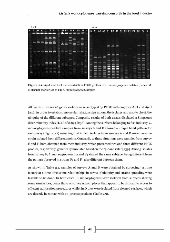
Listeria monocytogenes-carrying consortia in the food industry
49
Figure 2.1: ApaI and AscI macrorestriction PFGE profiles of L. monocytogenes isolates (Lanes: M:
Molecular marker; A1 to F4: L. monocytogenes samples)
All twelve L. monocytogenes isolates were subtyped by PFGE with enzymes AscI and ApaI
[236] in order to establish molecular relationships among the isolates and also to check the
ubiquity of the different subtypes. Composite results of both assays displayed a Simpson’s
discriminatory index (D.I.) of 0.894 [238]. Among the surfaces belonging to fish industry, L.
monocytogenes-positive samples from surveys A and D showed a unique band pattern for
each assay (Figure 2.1) revealing that in fact, isolates from surveys A and D were the same
strain isolated from different points. Contrarily to these situations were samples from survey
E and F, both obtained from meat industry, which presented two and three different PFGE
profiles, respectively, genetically unrelated based on the “3-band rule” [239]. Among isolates
from survey F, L. monocytogenes F2 and F4 shared the same subtype, being different from
the pattern observed in strains F1 and F3 also different between them.
As shown in Table 2.1, samples of surveys A and D were obtained by surveying just one
factory at a time, thus some relationships in terms of ubiquity and strains spreading were
feasible to be done. In both cases, L. monocytogenes were isolated from surfaces sharing
some similarities, being those of survey A from places that appear to be difficult to access to
efficient sanitisation procedures whilst in D they were isolated from cleaned surfaces, which
are directly in contact with on-process products (Table 2.3).
ApaAscI
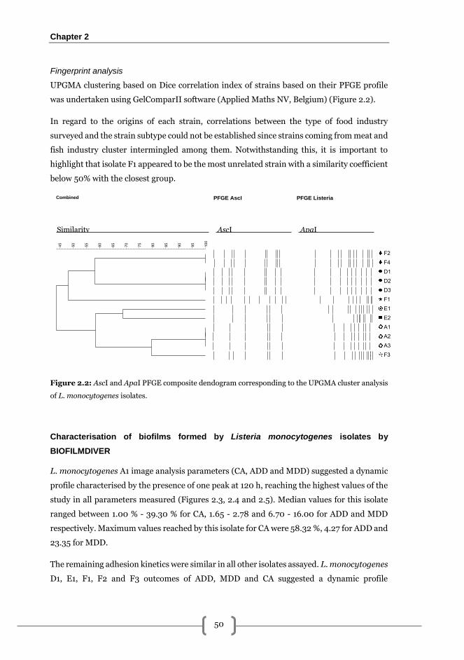
Chapter 2
50
Fingerprint analysis
UPGMA clustering based on Dice correlation index of strains based on their PFGE profile
was undertaken using GelComparII software (Applied Maths NV, Belgium) (Figure 2.2).
In regard to the origins of each strain, correlations between the type of food industry
surveyed and the strain subtype could not be established since strains coming from meat and
fish industry cluster intermingled among them. Notwithstanding this, it is important to
highlight that isolate F1 appeared to be the most unrelated strain with a similarity coefficient
below 50% with the closest group.
Figure 2.2: AscI and ApaI PFGE composite dendogram corresponding to the UPGMA cluster analysis
of L. monocytogenes isolates.
Characterisation of biofilms formed by Listeria monocytogenes isolates by
BIOFILMDIVER
L. monocytogenes A1 image analysis parameters (CA, ADD and MDD) suggested a dynamic
profile characterised by the presence of one peak at 120 h, reaching the highest values of the
study in all parameters measured (Figures 2.3, 2.4 and 2.5). Median values for this isolate
ranged between 1.00 % - 39.30 % for CA, 1.65 - 2.78 and 6.70 - 16.00 for ADD and MDD
respectively. Maximum values reached by this isolate for CA were 58.32 %, 4.27 for ADD and
23.35 for MDD.
The remaining adhesion kinetics were similar in all other isolates assayed. L. monocytogenes
D1, E1, F1, F2 and F3 outcomes of ADD, MDD and CA suggested a dynamic profile
AscI Similarity ApaI
Combined
100
9590858075706560555045
PFGE AscI PFGE Listeria
F2
F4
D1
D2
D3
F1
E1
E2
A1
A2
A3
F3
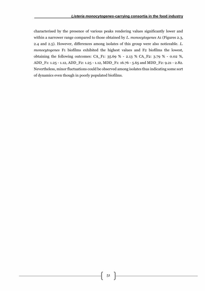
Listeria monocytogenes-carrying consortia in the food industry
51
characterised by the presence of various peaks rendering values significantly lower and
within a narrower range compared to those obtained by L. monocytogenes A1 (Figures 2.3,
2.4 and 2.5). However, differences among isolates of this group were also noticeable. L.
monocytogenes F1 biofilms exhibited the highest values and F2 biofilms the lowest,
obtaining the following outcomes: CA_F1: 35.69 % - 2.13 % CA_F2: 3.79 % - 0.02 %,
ADD_F1: 1.25 - 1.12, ADD_F2: 1.25 - 1.12, MDD_F1: 16.76 - 5.65 and MDD_F2: 9.21 - 2.82.
Nevertheless, minor fluctuations could be observed among isolates thus indicating some sort
of dynamics even though in poorly populated biofilms.
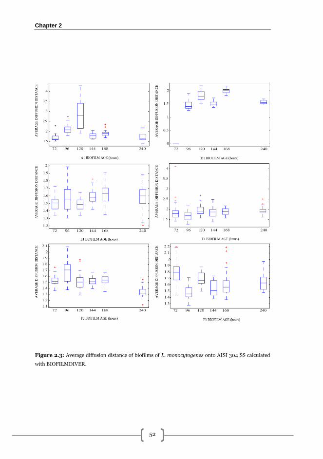
Chapter 2
52
Figure 2.3: Average diffusion distance of biofilms of L. monocytogenes onto AISI 304 SS calculated
with BIOFILMDIVER.
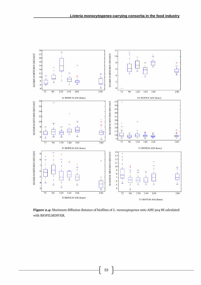
Listeria monocytogenes-carrying consortia in the food industry
53
Figure 2.4: Maximum diffusion distance of biofilms of L. monocytogenes onto AISI 304 SS calculated
with BIOFILMDIVER.
Chapter 2
54
Figure 2.5: Covered area of biofilms of L. monocytogenes onto AISI 304 SS calculated with
BIOFILMDIVER.
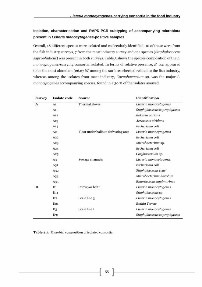
Listeria monocytogenes-carrying consortia in the food industry
55
Isolation, characterisation and RAPD-PCR subtyping of accompanying microbiota
present in Listeria monocytogenes-positive samples
Overall, 18 different species were isolated and molecularly identified, 10 of these were from
the fish industry surveys, 7 from the meat industry survey and one species (Staphylococcus
saprophyticus) was present in both surveys. Table 3 shows the species composition of the L.
monocytogenes-carrying consortia isolated. In terms of relative presence, E. coli appeared
to be the most abundant (26.27 %) among the surfaces checked related to the fish industry,
whereas among the isolates from meat industry, Carnobacterium sp. was the major L.
monocytogenes accompanying species, found in a 30 % of the isolates assayed.
Table 2.3: Microbial composition of isolated consortia.
Survey Isolate code Source Identification
A A1 Thermal gloves Listeria monocytogenes
A11 Staphylococcus saprophyticus
A12 Kokuria varians
A13 Aerococus viridans
A14 Escherichia coli
A2 Floor under halibut-defrosting area Listeria monocytogenes
A22 Escherichia coli
A23 Microbacterium sp.
A24 Escherichia coli
A25 Corybacterium sp.
A3 Sewage channels Listeria monocytogenes
A31 Escherichia coli
A32 Staphylococcus scuri
A33 Microbacterium luteolum
A35 Enterococcus aquimarinus
D D1 Conveyor belt 1 Listeria monocytogenes
D11 Staphylococcus sp.
D2 Scale line 3 Listeria monocytogenes
D21 Rothia Terrae
D3 Scale line 1 Listeria monocytogenes
D31 Staphylococcus saprophyticus
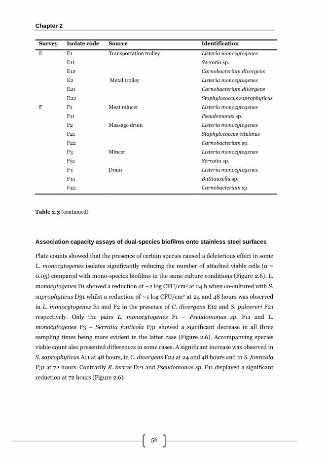
Chapter 2
56
Table 2.3 (continued)
Association capacity assays of dual-species biofilms onto stainless steel surfaces
Plate counts showed that the presence of certain species caused a deleterious effect in some
L. monocytogenes isolates significantly reducing the number of attached viable cells (α =
0.05) compared with mono-species biofilms in the same culture conditions (Figure 2.6). L.
monocytogenes D1 showed a reduction of ∼2 log CFU/cm2 at 24 h when co-cultured with S.
saprophyticus D31 whilst a reduction of ∼1 log CFU/cm2 at 24 and 48 hours was observed
in L. monocytogenes E1 and F2 in the presence of C. divergens E12 and S. pulvereri F21
respectively. Only the pairs L. monocytogenes F1 – Pseudomonas sp. F11 and L.
monocytogenes F3 – Serratia fonticola F31 showed a significant decrease in all three
sampling times being more evident in the latter case (Figure 2.6). Accompanying species
viable count also presented differences in some cases. A significant increase was observed in
S. saprophyticus A11 at 48 hours, in C. divergens F22 at 24 and 48 hours and in S. fonticola
F31 at 72 hours. Contrarily R. terrae D21 and Pseudomonas sp. F11 displayed a significant
reduction at 72 hours (Figure 2.6).
Survey Isolate code Source Identification
E E1 Transportation trolley Listeria monocytogenes
E11 Serratia sp.
E12 Carnobacterium divergens
E2 Metal trolley Listeria monocytogenes
E21 Carnobacterium divergens
E22 Staphylococcus saprophyticus
F F1 Meat mincer Listeria monocytogenes
F11 Pseudomonas sp.
F2 Massage drum Listeria monocytogenes
F21 Staphylococcus vitulinus
F22 Carnobacterium sp.
F3 Mincer Listeria monocytogenes
F31 Serratia sp.
F4 Drain Listeria monocytogenes
F41 Buttiauxella sp.
F42 Carnobacterium sp.
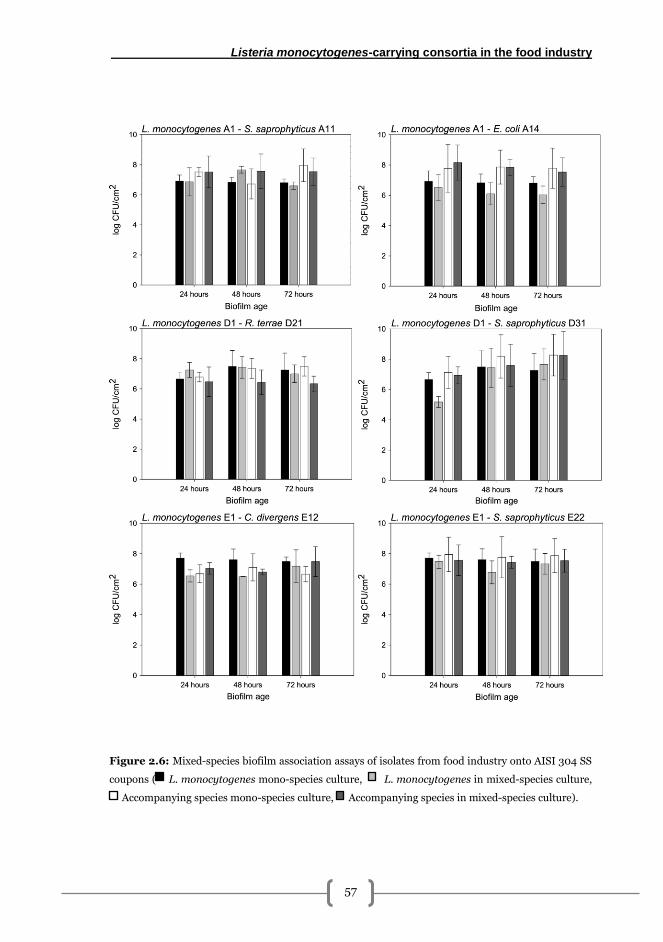
Listeria monocytogenes-carrying consortia in the food industry
57
Figure 2.6: Mixed-species biofilm association assays of isolates from food industry onto AISI 304 SS
coupons ( L. monocytogenes mono-species culture, L. monocytogenes in mixed-species culture,
Accompanying species mono-species culture, Accompanying species in mixed-species culture).
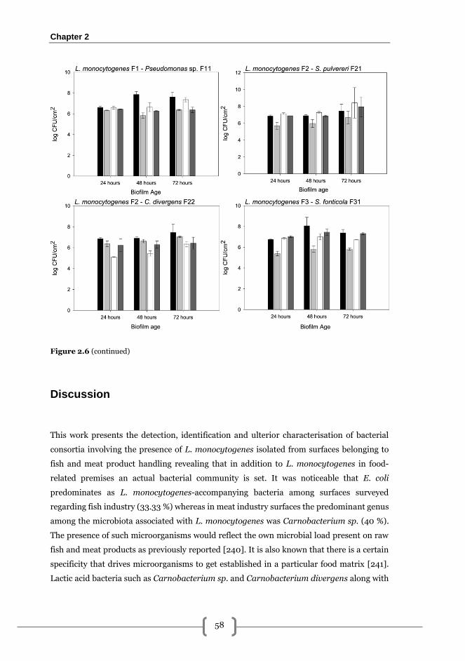
Chapter 2
58
Figure 2.6 (continued)
Discussion
This work presents the detection, identification and ulterior characterisation of bacterial
consortia involving the presence of L. monocytogenes isolated from surfaces belonging to
fish and meat product handling revealing that in addition to L. monocytogenes in food-
related premises an actual bacterial community is set. It was noticeable that E. coli
predominates as L. monocytogenes-accompanying bacteria among surfaces surveyed
regarding fish industry (33.33 %) whereas in meat industry surfaces the predominant genus
among the microbiota associated with L. monocytogenes was Carnobacterium sp. (40 %).
The presence of such microorganisms would reflect the own microbial load present on raw
fish and meat products as previously reported [240]. It is also known that there is a certain
specificity that drives microorganisms to get established in a particular food matrix [241].
Lactic acid bacteria such as Carnobacterium sp. and Carnobacterium divergens along with
Listeria monocytogenes-carrying consortia in the food industry
59
non-fermentative gram negative bacteria such as Pseudomonas sp. or Serratia sp. are
among the main microorganisms present in meat products [242]. Similarly, E. coli is known
to be associated with faecal contamination [243,244] and it is normally present in seafood
products and seafood processing industries [245].
Moreover, obtained results from this surveys have shown an overall incidence of 4.44 %, with
higher outcome of L. monocytogenes in meat industry (8 %, n = 75) compared with fish
industry (4.88 %, n = 125). Obviously, in non-standardized conditions, incidence results can
vary noticeably depending on the sampling method used [246], the number of samples
analysed, the moment when samples are collected and the size of the surface sampled, among
others. Concerning this last, previous authors have obtained values of 7.92 % when sampling
surfaces between 50 and 100 cm2 [247] and reaching values of 17 or even 26 % when 1 m2
surfaces are sampled [164]. In this article, sponge swabbing with subsequent enrichment was
chosen since is the method most commonly used among surveys to determine
presence/absence of L. monocytogenes [161,164,248–250].
In fish industry results showed that origins of L. monocytogenes isolates can be diverse
coming from surfaces typically not in contact with raw or processed product in survey A
whereas in survey D positive samples were obtained from surfaces typically in contact with
fish products. This heterogeneity in the distribution of strains could be of a multifactorial
nature and contact with manufactured product may help to maintain L. monocytogenes
prevalence along food premises [28]. Nevertheless, other authors support the hypothesis
that this distribution in a given industrial environment is independent to incoming raw
matter contact [251]. In the light of these results and due to the variation of the different
surveys, it would be recommendable to establish a common procedure of surfaces sampling
in food-related premises, so as to ensure the possibility to compare results between different
assays. In addition, identification of contamination foci for a particular microorganism
taking into account the casuistic of each processing plant may be considered to develop
preventive strategies.
When molecular confirmation of L. monocytogenes positive colonies was carried out by 16 s
rRNA gene sequencing, 29.41 % of positives were demonstrated to be false-positive and
subsequently identified as L. innocua (n = 2), L. welshimeri (n = 2) and Enterococcus
faecalis (n = 1). This lack of accuracy in regards to classical identification methods is in
accordance with results obtained by other authors [252] and highlights once again the
combination of classical and molecular methods as the optimal approach for proper bacterial
identification.
Chapter 2
60
In order to give further insight on the ecology of L. monocytogenes isolates obtained, these
were subtyped by serotyping and PFGE yielding a discrimination index (D.I.) of 0.894 which
in spite of being below the ideal D.I. value of 0.9 [238], is close enough considering the
number of the samples available thus the assay was considered as valid.
Among subtypes obtained from surveys carried out at one single fish-processing premises
(surveys A and D), a unique L. monocytogenes subtype was present at different locations
suggesting that this bacterium may be able to endure and colonise different environmental
conditions as was observed in previous studies carried out in fish industries [164]. From an
ecological perspective, the fact that L. monocytogenes isolates from surveys A and D
belonged to a single subtype and shared the same typology of surface may suggest that these
isolates have undertaken adaptation phenomena in order to be able to survive in a particular
environment [240,242,253].
Molecular serotyping demonstrated that in most cases serovars of isolates belonged to group
1/2a – 3a, followed by serogroup 4b – 4d – 4e and finally by serogroup 1/2b – 3b – 7 being
in accordance with previous published studies of surveys performed in Europe which showed
that serogroup 1/2a – 3a is the most abundant among environmental samples [254–256].
Two main structural patterns were observed from numerical characterisation of mono-
species L. monocytogenes biofilms. One was exhibited by L. monocytogenes A1. CA, ADD
and MDD profiles of this isolate showed one peak that suggest a dynamic evolution
characterised by a unique episode of attachment and detachment (Figures 2.3, 2.4 and 2.5).
The fact that ADD and MDD increase with CA, until reaching a maximum at 120 h, indicating
that A1 form biofilms with a homogeneous distribution of viable cells and high density,
according to the CA values obtained.
D1, E1, F1, F2 and F3 isolates shared a common pattern of individual cells that evolved to
cell aggregates which finally disappear. The low CA values obtained reflected the incapacity
of these isolates to form dense biofilms. However, among them F1 biofilm showed the highest
population level of this group. The generation of clusters were noted by ADD and MDD
results, however size of those clusters also varied among isolates, being bigger the clusters of
F1 regarding those exhibited by the rest. In contrast with results showed by A1, evolution
dynamics in this particular case was characterised by several episodes of attachment-
detachment. Similar results, two patterns characterised by two different dynamic profiles
and parameter values, were reported previously after studying three different L.
monocytogenes strains under same conditions [101].
Listeria monocytogenes-carrying consortia in the food industry
61
Previous studies have demonstrated that biofilm architecture influences the grade in which
diffusion takes place. Following this idea Stewart and Franklin [76] stated that physiological
heterogeneity and complex structures such as cell clusters, quantified in this study by CA,
ADD and MDD, could promote diffusional limitations to antimicrobials via establishment of
local gradients being especially remarkable in mature biofilms. In this regard, however,
Carpentier and Cerf [35] claimed that the presence of L. monocytogenes among surfaces in
food-related environments is more likely to be due to an improper design of cleaning and
disinfection routines along with erroneous manufacturing practices among plants rather
than to an enhanced biofilm forming capability. Nevertheless, recent studies suggest that
this theory could be too reductionist and the fact that L. monocytogenes can be established
in a particular industrial setting appears to be more of a multifactorial phenomenon where
genetic and physiological changes may take place [257,258]. In order to elucidate the actual
causes of presence and subsequent persistence phenomena in L. monocytogenes strains
isolated not only further sampling must be carried out at different times but also the
assessment of the composition of accompanying microbiota and its contribution to the
establishment of stable ecological niches by L. monocytogenes in industrial settings.
Interactions among bacteria forming the different consortia appear to be crucial for the
fitness of the whole structure [259]. Association capacity assays of dual-species biofilms
demonstrated how L. monocytogenes isolates were able to form biofilms along with other
microorganisms [132,134,260,261]. Outcomes obtained showed that Pseudomonas sp. F11
and Serratia fonticola F31 seemed to have a deleterious effect at all sampling times, on L.
monocytogenes present on biofilms. This is in agreement with previous results obtained by
Carpentier and Chassaing [129], who demonstrated a 3-log reduction on the number of L.
monocytogenes adhered cells in presence of Pseudomonas fluorescens. L. monocytogenes
E1 was also affected by C. divergens E12 being reduced its number of attached cells at 24 and
48 hours being in accordance with previous studies published showing that lactic acid
bacteria (LAB) and LAB-related species such as Carnobacterium sp. strains can be used as
a strategy so as to control the population of L. monocytogenes in order to avoid spoilage and
potential foodborne poisoning in meat [262] and in fish products [263]. However, no
differences in the number of attached cells of L. monocytogenes F2 co-cultured with C.
divergens F22 were observed. Since isolates E12 and F22 are the same strain according to
RAPD subtyping (see results for further detail), these results indicate that different strains
of L. monocytogenes may respond diversely to the same C. divergens strain.
Relative abundances of L. monocytogenes and its accompanying strain did not show
significant differences except when co-cultured with S. fonticola or Pseudomonas sp. being
in agreement with previous studies [226,264] showing that bacteria such as Pseudomonas
Chapter 2
62
spp. in mixed-biofilm culture appear to be dominant in dual-species biofilms with L.
monocytogenes even though the relative abundances reported by these authors were much
lower than the ones obtained in this study.
Recently, the European Food Safety Authority reported a human listeriosis incidence among
the European Member States of 0.44 cases per 100000 inhabitants which meant a 8.6 %
increase compared with previously published data [265] fact that ratifies that L.
monocytogenes control systems are, to date, insufficient. Nowadays, bacterial biofilms are
well-known to be more resistant to disinfectants and even though it is widely accepted that
bacterial species are ubiquitous in the environment, the consideration of multi-species
associations for the investigation regarding pathogen control is scarce, despite the fact that
the presence of accompanying microbiota producing significant changes in the whole
structure has been demonstrated [132,230,266,267]. This work aimed to deepen in the
knowledge on sessile bacteria present in an actual food-related industrial context so as to use
it as a starting point to perform further investigation of efficient pathogen control strategies
regarding not only the composition of the biofilms but also the choice of real targets based
on interactions among species.
Although many efforts have been put on the detection and elimination of L. monocytogenes
in industrial settings, results obtained in this work showed how this pathogen is able to grow
and survive in different food industry related surfaces. In addition to this fact, it can be
noticed that L. monocytogenes-carrying bacterial consortia follow an association pattern
from an ecological point of view depending on the industrial setting where they are present,
which represents an interesting clue when planning a cleaning and disinfection procedure.
Since L. monocytogenes has a great impact in the current society at clinical and industrial
level, screening of niches of these communities appears to be compulsory in order to identify
possible contamination foci and to design efficient, target-specific sanitisation methods to
ensure proper elimination of undesirable microbiota whereas manufactured products’
properties remain unaltered.

3
Enzymes-Benzalkonium chloride combined
treatments against L. monocytogenes-
carrying early-stage biofilms

Enzymes-BAC against early-stage biofilms
65
Introduction
Once the composition of L. monocytogenes-carrying communities was determined, the next
step in the present work was to carry out a first screening of the combined treatments and
their biofilm-removing efficacy.
For this, BAC was chosen since is one of the most used QAC in the food industry causing
membrane structure alteration upon bacteria and producing a subsequent cellular leakage
[202,203]. However, in biofilms, resistance and tolerance to this biocide has been reported
in several microorganisms such as L. monocytogenes [67,204], E. coli [205] or
Pseudomonas sp. [134].
Recently, the use of enzymes as an antibiofilm strategy has significantly increased because
their dispersal action degrading molecules present in the biofilm matrix, and proven to
impede initial adhesion and remove formed structures [175–178]. Nevertheless, enzymes do
not have bactericidal properties so they cannot be used as disinfectant compounds [117].
Consequently, in this chapter, the effects of an enzymatic treatment with different enzymes
alone and followed by benzalkonium chloride against early-stage L. monocytogenes dual-
species biofilms grown on stainless steel were assessed by means of classical agar plate
counts and epifluorescence microscopy.
Methods
Bacterial strains
Listeria monocytogenes A1 and Escherichia coli A14 were isolated from a fish processing
plant in a previous survey [37]. Pseudomonas fluorescens B52, a strong biofilm former and
associated with milk and dairy products spoilage, was kindly provided by Dr. Carmen San
José [268].
In all situations, stock cultures were kept at -80 ºC in Brain-Heart infusion broth (BHI;
Biolife, Milan, Italy) containing 50% glycerol 1:1 (v/v) mixed. Work cultures were kept at -
20 ºC in Tripticase Soy Broth (TSB; Cultimed, Barcelona, Spain) containing 50% glycerol 1:1
(v/v) mixed.
Chapter 3
66
Construction of fluorescent-tagged stains
Genetic modification for constitutive expression of a fluorescent reporter of strains L.
monocytogenes A1 and E. coli A14 was carried out in the laboratory of Prof. Colin Hill
(School of Microbiology, University College Cork (UCC), Ireland).
Modification of L. monocytogenes
L. monocytogenes was modified for Green fluorescent protein (GFP) constitutive expression.
Briefly, the fragment of pNF8 corresponding to the PdltΩgfp-mut1 [269] was amplified with
primers Pdlt For-KpnI and GFP pNF Rev-PstI (Table 3.1) containing KpnI and PstI
restriction sites, respectively, digested and cloned into pPL2 [270] previously digested with
KpnI and PstI and further treated with rAPid Alkaline Phosphatase (Roche) to avoid
religation. Ligation was performed using T4-ligase (Roche, Germany) in a PCR thermocycler
as follows: 4 ºC for 5 h, 12 h ramp increasing 1 ºC/h, 16 ºC for 2 h and back to 4 ºC giving a
plasmid of 7393 bp coded as pROLO1. The plasmid solution was dialysed in sterile deionised
water on a 0.025 µm pore nitrocellulose filter (Millipore, Germany) for 30 min and then kept
at -20 ºC until use. pROLO1 was then introduced into E. coli TOP10 cells (Thermo Fisher
Scientific, Waltham, MA) according to manufacturer’s instructions and cultured overnight
in LB (Merck, Germany) + 10 µg/ml chloramphenicol (Cm; Sigma Aldrich, Saint Louis, MO)
at 37 ºC. Plasmid extraction was then performed using a Gene JET Plasmid MiniPrep Kit
(Thermo Fisher Scientific, Waltham, MA). PdltΩgfp-mut1 integration was checked with
primers pPL2 MCS-for and pPL2 MCS-rev (Table 3.1).
Electroporation was carried out by mixing 50 µl of electrocompetent cells prepared as
previously described [271] with 2 µl of plasmid prep in 2 mm cuvettes using a BTX ECM 630
Generator (Harvard Apparatus, Holliston, MA). Conditions: field strength: 10 kV/cm; time
constant: 5 ms; voltage: 2 kV; resistance: 400 Ω; capacitance: 25 µF. Cells were then
resuspended in fresh sterile BHI + 0.5M Sucrose, incubated at 37 ºC for 1 h and then plated
on BHI + 1,5 % agar + 10 µg/ml Cm and incubated at 37 ºC for 48 h. Colonies were picked
and PCR was performed to check for plasmid integration using primers PL95 and PL102
[272] (Table 3.1). The resulting isolate was named L. monocytogenes A1-gfp.
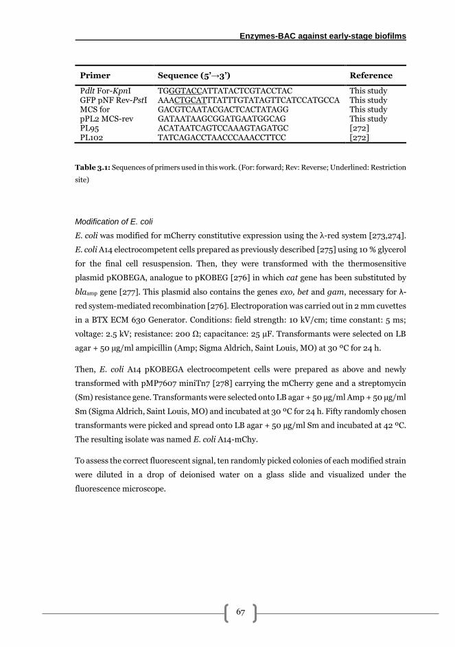
Enzymes-BAC against early-stage biofilms
67
Primer Sequence (5’→3’) Reference
Pdlt For-KpnI GFP pNF Rev-PstI
TGGGTACCATTATACTCGTACCTAC AAACTGCATTTATTTGTATAGTTCATCCATGCCA
This study This study
MCS for pPL2 MCS-rev PL95 PL102
GACGTCAATACGACTCACTATAGG GATAATAAGCGGATGAATGGCAG ACATAATCAGTCCAAAGTAGATGC TATCAGACCTAACCCAAACCTTCC
This study This study [272] [272]
Table 3.1: Sequences of primers used in this work. (For: forward; Rev: Reverse; Underlined: Restriction
site)
Modification of E. coli
E. coli was modified for mCherry constitutive expression using the λ-red system [273,274].
E. coli A14 electrocompetent cells prepared as previously described [275] using 10 % glycerol
for the final cell resuspension. Then, they were transformed with the thermosensitive
plasmid pKOBEGA, analogue to pKOBEG [276] in which cat gene has been substituted by
blaamp gene [277]. This plasmid also contains the genes exo, bet and gam, necessary for λ-
red system-mediated recombination [276]. Electroporation was carried out in 2 mm cuvettes
in a BTX ECM 630 Generator. Conditions: field strength: 10 kV/cm; time constant: 5 ms;
voltage: 2.5 kV; resistance: 200 Ω; capacitance: 25 µF. Transformants were selected on LB
agar + 50 µg/ml ampicillin (Amp; Sigma Aldrich, Saint Louis, MO) at 30 ºC for 24 h.
Then, E. coli A14 pKOBEGA electrocompetent cells were prepared as above and newly
transformed with pMP7607 miniTn7 [278] carrying the mCherry gene and a streptomycin
(Sm) resistance gene. Transformants were selected onto LB agar + 50 µg/ml Amp + 50 µg/ml
Sm (Sigma Aldrich, Saint Louis, MO) and incubated at 30 ºC for 24 h. Fifty randomly chosen
transformants were picked and spread onto LB agar + 50 µg/ml Sm and incubated at 42 ºC.
The resulting isolate was named E. coli A14-mChy.
To assess the correct fluorescent signal, ten randomly picked colonies of each modified strain
were diluted in a drop of deionised water on a glass slide and visualized under the
fluorescence microscope.
Chapter 3
68
Biofilms setup
One hundred microlitres of work cultures was grown overnight at 37 ºC in 5 ml of BHI + 10
µg/ml Cm for L. monocytogenes A1-gfp and LB + 50 µg/ml Sm for E. coli A14-mChy and
subcultured overnight so as to ensure a proper growth.
Inocula preparation was performed following a modification of a protocol previously
described [37]. Briefly, cultures were adjusted to Abs700 = 0.1 ± 0.001 in sterile phosphate
buffer saline (PBS) using a Cecil3000 scanning spectrophotometer (Cecil Instruments,
Cambridge, England), corresponding to a concentration of about 108 CFU/ml. Adjusted
cultures were further diluted in sterile mTSB (TSB supplemented with 2.5 g/l glucose
(Vorquímica, S.L., Vigo, Spain) and 0.6 % yeast extract (Cultimed, Barcelona, Spain)) to a
final concentration of about 104 CFU/ml. Then, equal volumes of these adjusted cultures
were mixed to obtain the inoculum for dual-species biofilms.
Biofilms were grown on 10 x 10 x 1 mm AISI 316 stainless steel (SS) coupons (Comevisa,
Vigo, Spain). Pre-treatment of coupons included individual washing with industrial soap
(Sutter Wash, Sutter Ibérica, S.A., Madrid), rinsing with tap water, a final rinse with
deionised water and autoclaved at 121 ºC for 20 min. Coupons were then placed individually
into a 24 flat-bottomed well plate and each well was inoculated with 1 ml of the
corresponding culture. Plates were incubated in a humidified atmosphere at 25 ºC statically
for 2 h so as to allow initial adhesion, and then in constant shaking at 100 rpm.
Biofilm formation kinetics
Samples (SS coupons) were collected at 24, 36, 48, 72 and 100 h and briefly immersed in
sterile PBS in order to remove loosely attached cells before any analysis was performed.
Determination of the number of adhered viable cultivable cells (AVC)
Three different coupons were scraped using two cotton swabs pre-moistened with buffered
peptone water (BPW; Cultimed, Barcelona, Spain). The swabs were then placed in 2 ml of
BPW vigorously vortexed for 1 min to resuspend cells. The cell suspensions were then serially
diluted in BPW and spread in duplicates onto agar plates. Listeria-PALCAM (Liofilchem,
Italy) was used to select L. monocytogenes and HiCromeTM Coliform agar (Sigma Aldrich,
Saint Louis, MO) with a supplement of 5 µg/ml of Vancomycin and Cefsulodine (Sigma
Aldrich, Saint Louis, MO) for E. coli selection. Plates were incubated at 37 ºC for 24-48 h
and results were expressed as the mean in log CFU/cm2 of samples. The accepted limit of
Enzymes-BAC against early-stage biofilms
69
detection for this and all assays involving viable cell counts was at least 25 CFU in the plate
of the lowest dilution corresponding to a total of 1.70 log CFU/cm2 [149].
Epifluorescence microscopy visualisation
At each sampling time, three coupons were air dried avoiding as much as possible direct light
exposure. Samples were then visualised under a Leica DM6000 epifluorescence microscope
using a 40x objective and 10x ocular lenses. Microscope was equipped with filter cubes L5
(Excitation 480/40) for A1-gfp and TX2 (Excitation 560/40) for A14-mChy. Images were
taken using a Leica DFC365 FX controlled with Metamorph MMAF software (Molecular
Devices, Sunnyvale, CA camera from 10 representative fields).
Effect of enzymatic solutions on dual-species biofilms
Enzyme solutions were prepared at concentrations 200, 400, 700 and 1000 µg/ml. Pronase
(PRN, from Streptomyces griseus, Roche) was dissolved in 0.1 M Tris-HCl (Sigma Aldrich)
buffer at pH = 7.5 ± 0.2. Cellulase (CEL, from Aspergillus niger, Sigma Aldrich) was
dissolved in 100 mM citrate (Sigma Aldrich) buffer at pH = 6.0 ± 0.1. Finally, DNaseI (from
bovine pancreas, Sigma Aldrich) was dissolved in 10 mM Tris-HCl (pH = 7.5 ± 0.2) buffer
also containing 2.5 mM MgCl2 and 0.1 mM CaCl2. After preparation, all solutions were filter
sterilised through a 0.2 µm syringe filter (Sartorius) and kept at -20 ºC until use.
The biofilm removal action of each enzymatic solution was evaluated against 24 h biofilms.
Three coupons were washed as before and then placed in a clean well. One millilitre of each
enzyme solution was added and allowed to act for 30 min at 37 ºC for PRN and 32 ºC for
CEL and DNaseI. Negative controls were run in parallel by adding the corresponding buffer
solution without enzyme. Solutions were then gently removed by pipetting and SS coupons
were subsequently washed with 1 ml of sterile PBS in order to remove residual enzyme.
Determination of remaining adhered cells and visualisation of coupons was performed as
described above. Results were expressed as the reduction in log CFU/cm2, calculated as the
mean of each replica difference in log CFU/cm2 before enzymatic and after enzymatic
treatment. After this, the two most effective enzymes were used in the following experiments.
Chapter 3
70
Effect of benzalkonium chloride combined with either PRN or DNaseI on L.
monocytogenes-E. coli biofilms
Benzalkonium chloride solutions (BAC; Guinama, Alboraya, Spain) were prepared in sterile
deionised water at concentrations 25, 50, 100, 250 and 500 µg/ml. Each solution was applied
after 30 min treatment with 400 µg/ml of either PRN or DNaseI solution against 48 h L.
monocytogenes A1-gfp-E. coli A14-mChy biofilms.
Fourteen different coupons washed with sterile PBS for loosely attached cells removal were
used for each enzyme series: two for the negative controls (no treatment), two for enzymes
treatment without BAC (only enzyme and deionised water were applied), and two for each
BAC concentration after enzymatic treatment performed as described above. In this latter
case, 1.5 ml of each BAC solution was added to each coupon for a 10 min contact time at room
temperature. For negative controls, buffer without enzymes and deionised water were
sequentially used. Coupons were then transferred to a new well and immersed for 30 s in 1
ml of a neutralising solution (composition per litre: 10 ml of a 34 g/l KH2PO4 buffer (pH =
7.2); soybean lecithin: 3 g; Tween 80: 30 ml; Na2S2O3: 5 g; L- histidine: 1 g) at room
temperature followed by a final 10 s wash by immersion with sterile PBS to remove any
neutraliser residues.
Following its application, neutralising solution was serially diluted in BPW and spread in
duplicate onto appropriate agar media to determine the number of released viable cells
(RVC) after treatments. Outcomes were expressed as mean of log CFU/ml. Microscopic
visualisation and determination of the remaining attached cells were performed as described
above. In the latter case, results were expressed as percentage of biofilm removal with respect
to the log CFU/cm2 obtained in control samples.
Determination of BAC effect: Calculation of lethal dose 90 (LD90)
LD90, defined as the dose of an antimicrobial required to achieve a 90 % kill of the initial
bacterial population, was used as a parameter to determine the effect of BAC on dual-species
biofilms. To assess this, a modified logistic model proposed by Cabo et al. [279] was used.
Logistic equations are widely recognised as suitable for describing dose-response kinetics
[280,281]. Firstly, outcomes were obtained by fitting of the experimental data obtained in
plate count assays, expressed in percentage of biofilm removal according to following
equation (1) using the least-squares method (quasi-Newton) of the SOLVER tool of Microsoft
Excel 2016:
Enzymes-BAC against early-stage biofilms
71
BR = K (1
1+0.11er(LD90−D) − 1
1+0.11erLD90) (1)
where BR = biofilm removal expressed in percentage; LD90 = dose of BAC that removes 90%
of the initial adhered population; D = dose of BAC used; K = maximum percentage of biofilm
removal (asymptote); and r = specific inhibition coefficient (dimensions: inverse of the
dose).
Since the equation [1] modifies the resulting Dose/Response parameters by subtracting the
intercept of the original logistic equation, results were further adjusted to obtain the new K
value (K’):
BRmax = K′ = limD→∞
BR (2)
Then, the real LD90 (RD90) was determined according to a modification of an equation
described previously [282]:
RD90 = 1
rln (9 + erD) (3)
Influence of L. monocytogenes accompanying species in the resistance to DNaseI-
BAC treatments
Two different 48 h dual-species biofilms were used: L. monocytogenes A1-E. coli A14 and L.
monocytogenes A1-P. fluorescens B52 to evaluate sequential DNaseI-BAC treatments.
400 µg/ml DNaseI + 100 µg/ml BAC treatments and plate count analysis for attached and
released cells determination were performed as described above. For P. fluorescens selection
Pseudomonas Agar Base (PAB; Liofilchem, Italy) supplemented with CFC supplement
(Liofilchem, Italy) was used and incubated at 30 ºC for 48 h.
For microscopic visualisation, samples were stained using LIVE/DEAD Bacterial viability kit
(Life Technologies) to distinguish total cells with undamaged membranes (green
fluorescence) and damaged cells (red fluorescence). Staining solution was prepared by
mixing 0.25 µl of Propidium iodide and 0.75 µl of Syto9 in 1 ml of filter sterilized deionised
water. Fifty microlitres of this solution was then poured onto each coupon and allowed to
dwell for 15 min in the dark. Coupons were then washed three times in 1 ml of sterile milliQ
water, air dried and visualised under the epifluorescence microscope to obtain images of
representative fields.
Chapter 3
72
Statistical analysis
Experimental results were analysed for statistical significance using IBM SPSS Statistics 23.
An independent-samples two-tailed Student’s t test was performed to assess differences
between species in the biofilm formation kinetics and the effects of BAC in RVC after PRN
and DNaseI treatments. Differences among the effects of the different enzymatic treatments
and treatments’ effects in different dual-species biofilms were determined using a one-way
ANOVA with a post hoc Bonferroni test. In all cases, significance was expressed at the 95 %
confidence level (α = 0.05) or greater.
In RD90 determination, correlation coefficient (r2) was calculated to quantify the discrepancy
between the observed experimental values and those expected according with the model.
Results
L. monocytogenes-E. coli biofilm formation kinetics on AISI 316 stainless steel
Dual-species biofilm formation dynamics are depicted in Figure 3.1. Plate count assays
showed a significantly higher number of AVC in E. coli with respect to that obtained for L.
monocytogenes at 24 and 100 hours of growth yielding differences of 3.11 and 2.63 log
CFU/cm2 respectively. No significance was observed among the values of the rest sampling
times.
Microscopic images displayed in Figure 3.2 showed a uniform distribution of E. coli and L.
monocytogenes over the coupon. In spite of this uniform distribution, at 24 h E. coli
presented about 3 log higher AVC counts compared to L. monocytogenes (Figure 3.1). A
tendency for aggregation was observed at 24 and 36 h yielding a final composite structure
with both species intermingled therein (Figure 3.2). From this point onward, the amount of
cells increased and the biofilm developed a cloud-shape structure which was maintained in
the last three sampling times (Figure 3.2).
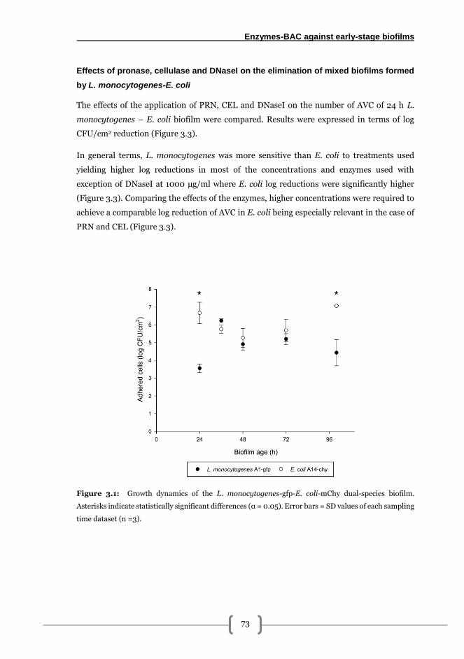
Enzymes-BAC against early-stage biofilms
73
Effects of pronase, cellulase and DNaseI on the elimination of mixed biofilms formed
by L. monocytogenes-E. coli
The effects of the application of PRN, CEL and DNaseI on the number of AVC of 24 h L.
monocytogenes – E. coli biofilm were compared. Results were expressed in terms of log
CFU/cm2 reduction (Figure 3.3).
In general terms, L. monocytogenes was more sensitive than E. coli to treatments used
yielding higher log reductions in most of the concentrations and enzymes used with
exception of DNaseI at 1000 µg/ml where E. coli log reductions were significantly higher
(Figure 3.3). Comparing the effects of the enzymes, higher concentrations were required to
achieve a comparable log reduction of AVC in E. coli being especially relevant in the case of
PRN and CEL (Figure 3.3).
Figure 3.1: Growth dynamics of the L. monocytogenes-gfp-E. coli-mChy dual-species biofilm.
Asterisks indicate statistically significant differences (α = 0.05). Error bars = SD values of each sampling
time dataset (n =3).
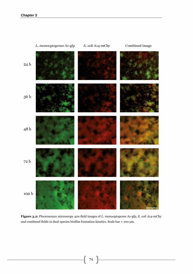
Chapter 3
74
Figure 3.2: Fluorescence microscopy 40x-field images of L. monocytogenes A1-gfp, E. coli A14-mChy
and combined fields in dual-species biofilm formation kinetics. Scale bar = 100 µm.
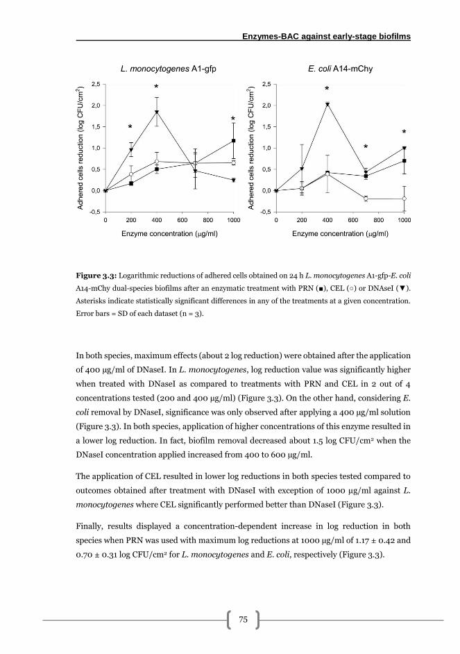
Enzymes-BAC against early-stage biofilms
75
Figure 3.3: Logarithmic reductions of adhered cells obtained on 24 h L. monocytogenes A1-gfp-E. coli
A14-mChy dual-species biofilms after an enzymatic treatment with PRN (■), CEL (○) or DNAseI (▼).
Asterisks indicate statistically significant differences in any of the treatments at a given concentration.
Error bars = SD of each dataset (n = 3).
In both species, maximum effects (about 2 log reduction) were obtained after the application
of 400 µg/ml of DNaseI. In L. monocytogenes, log reduction value was significantly higher
when treated with DNaseI as compared to treatments with PRN and CEL in 2 out of 4
concentrations tested (200 and 400 µg/ml) (Figure 3.3). On the other hand, considering E.
coli removal by DNaseI, significance was only observed after applying a 400 µg/ml solution
(Figure 3.3). In both species, application of higher concentrations of this enzyme resulted in
a lower log reduction. In fact, biofilm removal decreased about 1.5 log CFU/cm2 when the
DNaseI concentration applied increased from 400 to 600 µg/ml.
The application of CEL resulted in lower log reductions in both species tested compared to
outcomes obtained after treatment with DNaseI with exception of 1000 µg/ml against L.
monocytogenes where CEL significantly performed better than DNaseI (Figure 3.3).
Finally, results displayed a concentration-dependent increase in log reduction in both
species when PRN was used with maximum log reductions at 1000 µg/ml of 1.17 ± 0.42 and
0.70 ± 0.31 log CFU/cm2 for L. monocytogenes and E. coli, respectively (Figure 3.3).
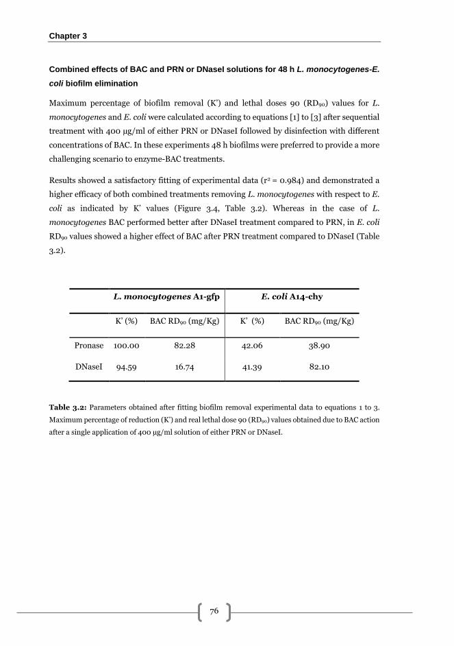
Chapter 3
76
Combined effects of BAC and PRN or DNaseI solutions for 48 h L. monocytogenes-E.
coli biofilm elimination
Maximum percentage of biofilm removal (K’) and lethal doses 90 (RD90) values for L.
monocytogenes and E. coli were calculated according to equations [1] to [3] after sequential
treatment with 400 µg/ml of either PRN or DNaseI followed by disinfection with different
concentrations of BAC. In these experiments 48 h biofilms were preferred to provide a more
challenging scenario to enzyme-BAC treatments.
Results showed a satisfactory fitting of experimental data (r2 = 0.984) and demonstrated a
higher efficacy of both combined treatments removing L. monocytogenes with respect to E.
coli as indicated by K’ values (Figure 3.4, Table 3.2). Whereas in the case of L.
monocytogenes BAC performed better after DNaseI treatment compared to PRN, in E. coli
RD90 values showed a higher effect of BAC after PRN treatment compared to DNaseI (Table
3.2).
L. monocytogenes A1-gfp E. coli A14-chy
K’ (%) BAC RD90 (mg/Kg) K’ (%) BAC RD90 (mg/Kg)
Pronase 100.00 82.28 42.06 38.90
DNaseI 94.59 16.74 41.39 82.10
Table 3.2: Parameters obtained after fitting biofilm removal experimental data to equations 1 to 3.
Maximum percentage of reduction (K’) and real lethal dose 90 (RD90) values obtained due to BAC action
after a single application of 400 µg/ml solution of either PRN or DNaseI.
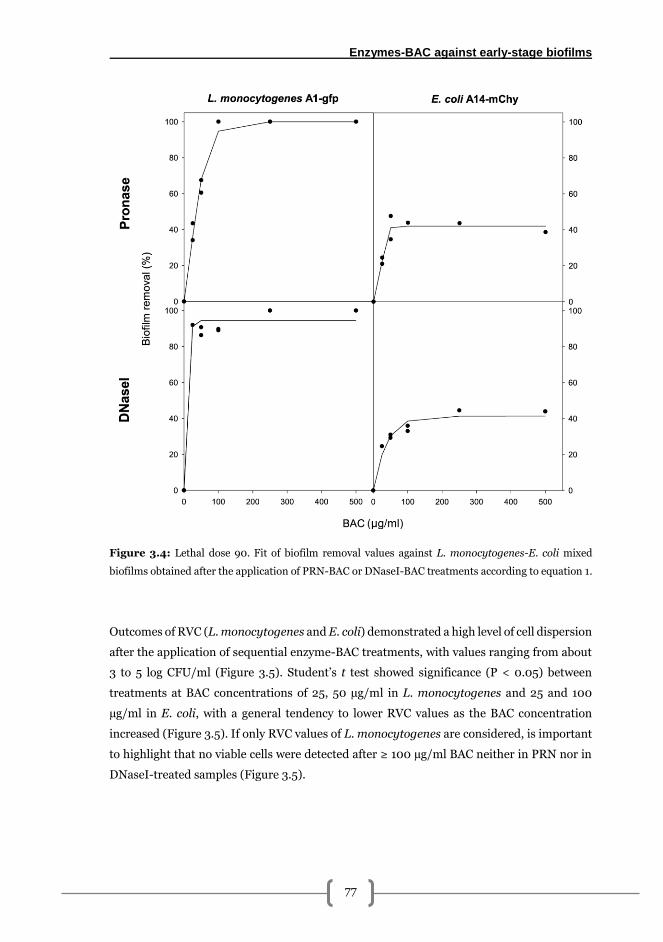
Enzymes-BAC against early-stage biofilms
77
Figure 3.4: Lethal dose 90. Fit of biofilm removal values against L. monocytogenes-E. coli mixed
biofilms obtained after the application of PRN-BAC or DNaseI-BAC treatments according to equation 1.
Outcomes of RVC (L. monocytogenes and E. coli) demonstrated a high level of cell dispersion
after the application of sequential enzyme-BAC treatments, with values ranging from about
3 to 5 log CFU/ml (Figure 3.5). Student’s t test showed significance (P < 0.05) between
treatments at BAC concentrations of 25, 50 µg/ml in L. monocytogenes and 25 and 100
µg/ml in E. coli, with a general tendency to lower RVC values as the BAC concentration
increased (Figure 3.5). If only RVC values of L. monocytogenes are considered, is important
to highlight that no viable cells were detected after ≥ 100 µg/ml BAC neither in PRN nor in
DNaseI-treated samples (Figure 3.5).
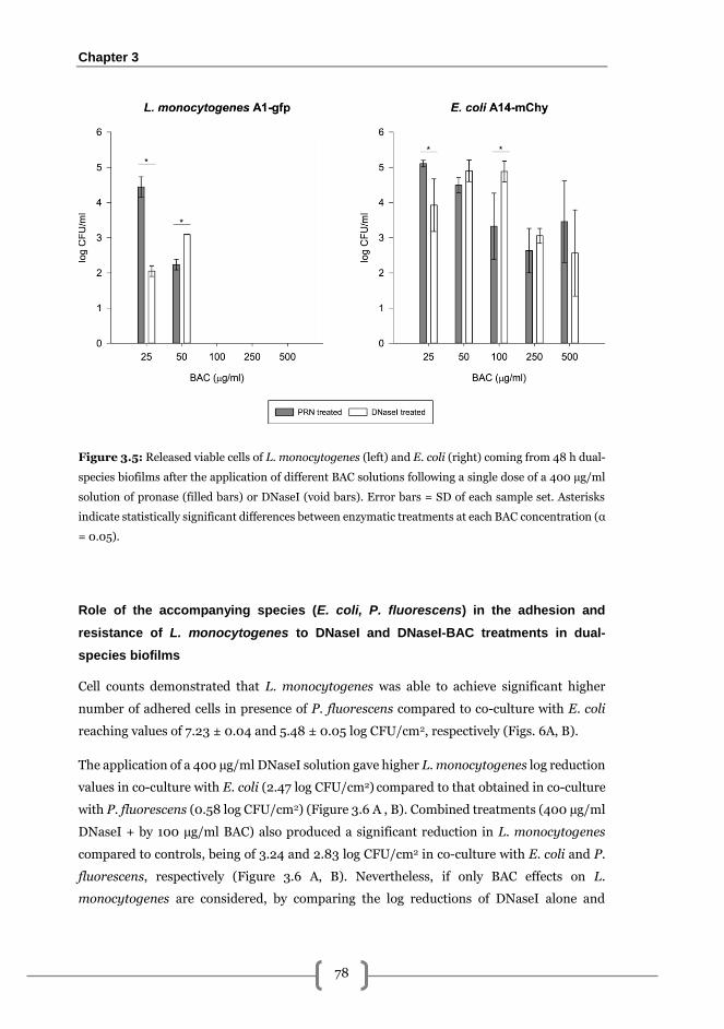
Chapter 3
78
Figure 3.5: Released viable cells of L. monocytogenes (left) and E. coli (right) coming from 48 h dual-
species biofilms after the application of different BAC solutions following a single dose of a 400 µg/ml
solution of pronase (filled bars) or DNaseI (void bars). Error bars = SD of each sample set. Asterisks
indicate statistically significant differences between enzymatic treatments at each BAC concentration (α
= 0.05).
Role of the accompanying species (E. coli, P. fluorescens) in the adhesion and
resistance of L. monocytogenes to DNaseI and DNaseI-BAC treatments in dual-
species biofilms
Cell counts demonstrated that L. monocytogenes was able to achieve significant higher
number of adhered cells in presence of P. fluorescens compared to co-culture with E. coli
reaching values of 7.23 ± 0.04 and 5.48 ± 0.05 log CFU/cm2, respectively (Figs. 6A, B).
The application of a 400 µg/ml DNaseI solution gave higher L. monocytogenes log reduction
values in co-culture with E. coli (2.47 log CFU/cm2) compared to that obtained in co-culture
with P. fluorescens (0.58 log CFU/cm2) (Figure 3.6 A , B). Combined treatments (400 µg/ml
DNaseI + by 100 µg/ml BAC) also produced a significant reduction in L. monocytogenes
compared to controls, being of 3.24 and 2.83 log CFU/cm2 in co-culture with E. coli and P.
fluorescens, respectively (Figure 3.6 A, B). Nevertheless, if only BAC effects on L.
monocytogenes are considered, by comparing the log reductions of DNaseI alone and
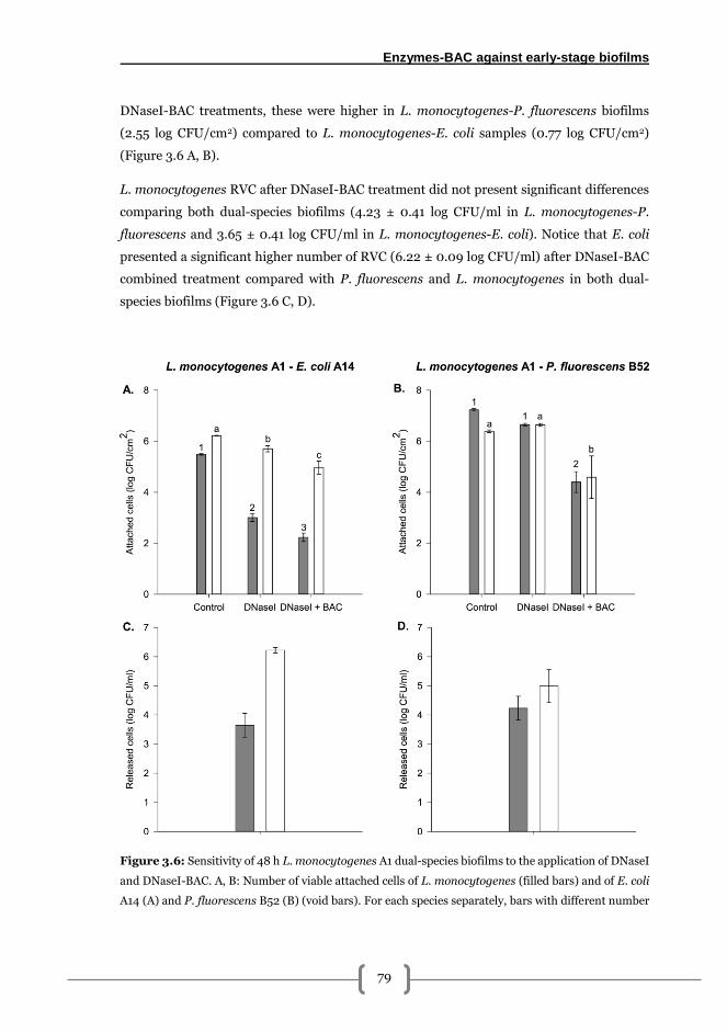
Enzymes-BAC against early-stage biofilms
79
DNaseI-BAC treatments, these were higher in L. monocytogenes-P. fluorescens biofilms
(2.55 log CFU/cm2) compared to L. monocytogenes-E. coli samples (0.77 log CFU/cm2)
(Figure 3.6 A, B).
L. monocytogenes RVC after DNaseI-BAC treatment did not present significant differences
comparing both dual-species biofilms (4.23 ± 0.41 log CFU/ml in L. monocytogenes-P.
fluorescens and 3.65 ± 0.41 log CFU/ml in L. monocytogenes-E. coli). Notice that E. coli
presented a significant higher number of RVC (6.22 ± 0.09 log CFU/ml) after DNaseI-BAC
combined treatment compared with P. fluorescens and L. monocytogenes in both dual-
species biofilms (Figure 3.6 C, D).
Figure 3.6: Sensitivity of 48 h L. monocytogenes A1 dual-species biofilms to the application of DNaseI
and DNaseI-BAC. A, B: Number of viable attached cells of L. monocytogenes (filled bars) and of E. coli
A14 (A) and P. fluorescens B52 (B) (void bars). For each species separately, bars with different number
Chapter 3
80
or letter indicate significant differences (α = 0.05). C, D: Number of viable released cells after the
DNaseI–BAC treatment. Error bars represent the standard deviation of each sample set (n = 3).
Microscopic analysis showed that both biofilms presented remarkable differences in their
2D-morphologies (Figure 3.7). While L. monocytogenes-E. coli biofilms showed a reticular
distribution in all biofilm, L. monocytogenes-P. fluorescens biofilms were characterised by
the presence of microcolonies surrounded by small cell groups. These microcolonies
presented a local accumulation of damaged cells compared to the rest of the sample, as
observed by a higher red signal in the central part of the microcolony. The same microcolony
formation tendency was also observed in our laboratory with other L. monocytogenes strains
when co-cultured with P. fluorescens B52 (data not shown).
Sequential DNaseI-BAC treatments produced a significant increase of the red cell signal
especially in L. monocytogenes-E. coli samples pointing out the BAC killing effects. Besides,
noticeable structural changes were observed in samples of both dual-species biofilms,
especially in L. monocytogenes-P. fluorescens biofilms in which the cellular groups
surrounding the microcolonies were substituted by sparsely distributed cells (Figure 3.7).
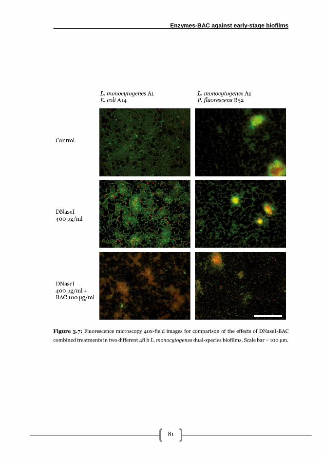
Enzymes-BAC against early-stage biofilms
81
Figure 3.7: Fluorescence microscopy 40x-field images for comparison of the effects of DNaseI-BAC
combined treatments in two different 48 h L. monocytogenes dual-species biofilms. Scale bar = 100 µm.
Chapter 3
82
Discussion
Biofilm kinetics of the L. monocytogenes A1-gfp-E. coli A14-chy biofilm showed a typical
biofilm fit-curve with minor fluctuations (Figure 3.1). E. coli viable counts were significantly
higher than L. monocytogenes at 24 and 100 h. Differences in AVC counts at 24 h could be
attributed to a better initial adhesion of E. coli compared with L. monocytogenes as
previously reported [283]. However, AVC values of both species were equilibrated at 36, 48
and 72 hours (Figure 3.1).
Microscopic images showed a uniform distribution of E. coli and L. monocytogenes over the
coupon despite the differences up to 3 log present between these species at 24 h (Figs. 1, 2).
Almeida et al. reported that in 48 h L. monocytogenes-E. coli biofilms grown on stainless
steel and plastic, species presented this sort of uniform distribution with E. coli being present
in higher numbers [146]. The fact that green fluorescence, corresponding to L.
monocytogenes cells, was similar to red despite viable counts (Figure 3.1), could have been
caused in part because a fraction of this green signal was emitted by cells in the viable but
non culturable (VBNC) state. Previous authors have observed that 24 h-old L.
monocytogenes biofilms present a part of VBNC [284]. In such condition, GFP remains
totally functional and fluoresces even though cells are not able to grow in solid media
[285,286].
Enzymes have been previously used as a biofilm removal strategy due to their specificity and
their low environmental impact [175,181,287]. In this work, comparison between the effects
of cellulase (CEL), DNaseI and pronase (PRN) demonstrated a maximum effect of a 400
µg/ml of DNaseI solution reducing about 2 log CFU/cm2 the number of AVC in L.
monocytogenes and E. coli (Figure 3.3). This reduction was followed by that produced by
PRN and CEL, despite no broad differences were observed between these two (Figure 3.3).
It has been reported that extracellular DNA (eDNA) is present in considerable amounts of
the extracellular matrix and considered as a requisite for biofilm formation in L.
monocytogenes [118] as well as in other Gram-positives [288,289]. Hence, DNaseI has been
proposed as an antibiofilm enzyme cleaving eDNA and thus interfering in biofilm
development. As an example, Harmsen et al. [118] observed that 100 µg/ml DNaseI solution
at 37 ºC, completely prevented L. monocytogenes EGDe biofilm formation if applied up to
24 h after strain inoculation and, from that point onwards, DNaseI antibiofilm capacity was
reduced. In other Gram-positives such as S. aureus, 1 h contact time at 37 ºC of a 100 µg/ml
DNaseI solution significantly reduced the biomass of 24 h biofilms grown on polystyrene
plates [290]. Despite this previously reported data, no complete removal with DNaseI was
achieved among the experiments performed in this work. This could be due to the application
Enzymes-BAC against early-stage biofilms
83
of a more realistic time of action (30 min) or to the biofilm age, which could affect DNaseI
biofilm removal activity [118]. Experimental data also showed an inverted effect of DNaseI
at concentrations higher that 400 µg/ml (i.e. higher doses produced a lower log reduction),
both in L. monocytogenes and E. coli (Figure 3.3). Nguyen and Burrows [117] demonstrated
a similar enzymatic stimulatory effect on planktonic L. monocytogenes cells in which the
more proteinase K present in the culture, the more stimulated its growth was. Focusing in
our experimental approach, these effects in the number of cells in the planktonic state, could
have had eventually provoked an upturn in the number of cells adhered to the biofilm
detected in AVC assays.
Proteases have also been proved to be effective in removing biofilms. In this line, Nguyen
and Burrows [117] demonstrated that the addition of 100 µg/ml of proteinase K for 24 h is
able to disperse 72 h L. monocytogenes biofilms grown on polystyrene up to undetectable
levels. In S. aureus it has been recently reported that active proteases remove biofilms
formed in polystyrene plates [208]. However, PRN effects against L. monocytogenes were
lower than expected compared with DNaseI considering the proteinaceous nature of L.
monocytogenes biofilm matrix [116,117] even though it has been demonstrated that teichoic
acids are also present [119].
Previous investigations have reported that interspecies interactions that take place within
multi-species biofilms significantly modify the matrix composition if compared with
monocultures [142,291]. This differential composition can affect, among others, the efficacy
of enzymes as well as several antimicrobial compounds [291]. In our particular case, the
dominance of E.coli in 24 h biofilms (Figure 3.1) could have given rise to a matrix with a
higher polysaccharide content as proposed for most Gram-negative bacteria [292].
Nevertheless, CEL showed the lowest effects against L. monocytogenes-E. coli biofilms
perhaps because polysaccharide constituents interacted among themselves and among other
molecules present thus concealing enzyme targets or they simply lack on glucose-glucose
bonds susceptible to cleavage by CEL specific ß (1→4) endoglucanase activity.
Considering the aforementioned results, it is logical to think that the use of dual-species
biofilms represents a more challenging environment for biofilm-degrading enzymes due to
a higher matrix complexity. Thus, the idea of a combination of enzymes would be an
interesting option to be considered for proper biofilm removal [44,181] especially when
dealing with Gram-negatives such as Pseudomonas sp. [208]. Efficacy of enzymatic mixtures
have been previously reported by Orgaz et al. [178] using proteinase, cellulase,
pectinesterase, pectin lyase and alginate lyase derived from fungal cultures against 24-hour-
Chapter 3
84
old P. fluorescens B52 biofilms on glass achieving removal values up to an 84 % of the total
biomass.
In any case, enzymatic solutions show only dispersing-but-not-killing effect as previously
reported [117]. As a consequence, enzyme based disinfection may need to be performed in
combination with biocides that are able to kill the cells avoiding the dispersion of live cells
released from the biofilm [181,208].
In food related premises, RVC could provoke a pathogen thus enhancing the formation of
new reservoirs and increasing the probability of product contamination. Also, pathogens
could be easily spread through rinse after disinfection via water or aerosols produced [293]
or by means of typical cleaning tools such as sponges or wipes [294]. Therefore, controlling
RVC after cleaning and disinfection treatments appears to be as an interesting topic to
consider for further investigation.
Enzyme-BAC combined treatments showed a differential effect on L. monocytogenes-E. coli
biofilms depending on the species. More specifically, BAC performed better against L.
monocytogenes when preceded by DNaseI whereas removal of E. coli from the coupon was
higher after PRN-BAC treatment (Table 3.2, Figure 3.4). In L. monocytogenes the lower BAC
RD90 values obtained after DNaseI treatment indicated that despite proteins are considered
the main fraction in L. monocytogenes biofilm matrix [116], eDNA degradation by DNaseI
provokes a higher decrease in L. monocytogenes AVC counts thus confirming the key role of
eDNA to maintain already formed biofilms [117,118]. This biofilm-dispersing capacity of
DNaseI to facilitate BAC access into the biofilm is especially relevant in L. monocytogenes-
carrying biofilms as this bacterium is usually located in the bottom layers [146] . In E. coli, a
better performance of BAC after enzymatic dwelling was also observed but to a lesser extent
(Figure 3.4). This can be attributed to its intrinsic higher resistance to QACs [201,295] and
also because of the possible presence of protective colanic acid capsules [296].
It is important to remark the fact that BAC effects against 48 h samples were different
depending on the species (Figure 3.4) whereas in 24 h biofilms DNaseI was the most efficient
enzyme in both species of the mixed biofilms (Figure 3.3). This points out that the biofilm
matrix varies its molecular composition along time. So, if proper enzyme-based biofilm
cleaning strategies are intended to be designed it is important to determine the constituents
(proteins, eDNA and polysaccharides) of the matrix of the target sessile community.
A release of live cells of both species is observed from biofilms after PRN-BAC or DNaseI-
BAC treatments, especially at low BAC concentrations (Figure 3.6). Pathogen dispersal after
sanitation is a factor to take into account in cleaning and disinfection methodologies
Enzymes-BAC against early-stage biofilms
85
[117,175]. This fact can be minimised by using appropriate effective concentrations of
disinfectants (e.g. BAC) after dispersing agents, enzymes in this particular case, to avoid
dissemination of live cells in adjacent areas after biofilm removal.
Another important issue to be assessed in dual-species biofilms of L. monocytogenes is the
role of the accompanying species. Significant differences were observed in the L.
monocytogenes AVC counts, as well as in the effect of the enzyme and enzyme-BAC
treatment depending on the accompanying bacterium (Figure 3.6). Regarding the first, a
higher number of L. monocytogenes A1 cells was attached to stainless steel after 48 h in
presence of P. fluorescens respecting to E. coli, probably due to an entrapping of the L.
monocytogenes into the polymeric matrix secreted by the P. fluorescens. Morphological
features agreed with previously reported data in which L. monocytogenes-E.coli biofilms
appeared as uniform layers [146] whereas L. monocytogenes-P. fluorescens were
characterised by local microcolony formation surrounded by smaller biofilm aggregates
randomly distributed (Figure 3.7) [144].
DNaseI produced a significant decrease of L. monocytogenes only in the mixed biofilm with
presence of E. coli, probably because matrix composition differently affected its diffusion
and effectiveness (Figure 3.6) [297]. Nevertheless, the application of BAC against L.
monocytogenes was more effective when co-cultured with P. fluorescens despite the latter is
considered a strong biofilm former (Figure 3.6) [157,268].
In summary, in this work the effectiveness of treatments with an enzyme solution alone and
combined with a BAC dose on L. monocytogenes dual-species biofilms was demonstrated.
In addition to this, results demonstrated that the removal efficacy of a combined enzyme-
BAC treatment against mixed biofilms depends not only on the enzyme chosen but also on
the biofilm species composition. Following this idea, for proper biofilm removal in food
related surfaces as well as in others capable of harbour bacterial biofilms, customised
treatments depending on the species composition should be considered when developing
new cleaning and disinfection methodologies. This would be intended not only to impede
biofilm formation but also to significantly remove already present structures while
minimising the amount of live cells released.

4
Pronase-Benzalkonium chloride combined
treatments against L. monocytogenes-
carrying late-stage biofilms

Pronase-BAC against late-stage biofilms
89
Introduction
This part of the thesis goes one step further in the assessment of the biofilm-removing
properties of combined treatments. Specifically, this study aimed to study the effectiveness
of combining PRN and BAC for removal of late-stage L. monocytogenes dual-species
biofilms grown on stainless steel, mimicking real industrial conditions where biofilms are
formed after long periods. PRN was selected since it has been reported that L.
monocytogenes biofilm matrix is mainly constituted by proteins [117,298] and gave the
highest maximum biofilm removal percentages in the assays performed in the previous
chapter (chapter 3) . Enzymes solutions were applied at room temperature to further
simulate realistic environmental conditions. The assessment of the effects was performed
combining microscopy and image analysis with classical microbiology methods.
Methods
Bacterial strains
Two different consortia were used. The first was formed by L. monocytogenes A1-E. coli A14,
both isolated from a fish processing plant in a previous survey [37]. The second one was
formed by a strain of L. monocytogenes G1, isolated from a cheese processing plant, kindly
provided by Dr. Luisa Brito [299] and Pseudomonas fluorescens B52, as one of the species
commonly isolated in dairy industry, was kindly provided by Dr. Carmen San José [268].
These consortia were chosen based on their relevance in fish and dairy industries, and their
capability to form dual-species biofilms. From now on consortia used will be referred as fish
industry and dairy industry consortia for A1-A14 and G1-B52 biofilms, respectively.
In all cases, stock cultures were maintained at -80 ºC in Brain-Heart infusion broth (BHI;
Biolife, Italy) containing 50% glycerol 1:1 (v/v) mixed. Work cultures were kept at -20 ºC in
Tripticase Soy Broth (TSB; Cultimed, Barcelona, Spain) containing 50% glycerol 1:1 (v/v)
mixed.
Setup of dual-species biofilms
100 µl of work cultures was cultured overnight in 5 ml sterile TSB at 37 ºC for L.
monocytogenes and E. coli and 25 ºC for P. fluorescens and subcultured once so as to ensure
a proper activation.
Chapter 4
90
Inocula preparation was performed as follows: briefly, Abs700 of cultures was adjusted to 0.1
±0.001 in sterile phosphate buffer saline (PBS), corresponding to a bacterial concentration
of about 108CFU/ml according to previous calibrations. Adjusted cultures were further
diluted in sterile mTSB (TSB (Cultimed, Barcelona, Spain) supplemented with 2.5 g/l glucose
(Vorquímica, S.L., Vigo, Spain) and 0.6 % yeast extract (Cultimed, Barcelona, Spain)) until
obtaining a final concentration of about 104 CFU/ml and 1:1 mixed.
Biofilms were grown on 10x10x1 mm AISI 316 stainless steel (SS) coupons (Comevisa, Vigo,
Spain). Pre-treatment of coupons included individual washing with industrial soap to
remove grease residues, thoroughly rinsing with tap water with a final rise with deionized
water and sterilized by autoclaving them at 121 ºC for 20 min. Coupons were individually
placed into a 24 flat-bottomed well plate and each well was inoculated with 1 ml of the
corresponding culture. Plates were incubated in a humidified atmosphere at 25 ºC statically
for 2 h for initial adhesion, and then in constant shaking at 100 rpm.
In all samplings, coupons were aseptically collected and briefly immersed in 1 ml sterile PBS
to remove loosely attached cells before any assay was performed.
Plate count assays
Attached viable cultivable cells (AVC) were harvested from coupons by scrapping using two
sterile cotton swabs moistened in sterile buffered peptone water (BPW). Swabs were then
suspended in 2 ml of BPW and vortexed vigorously for 1 min in order to release cells, serially
diluted in BPW and spread onto agar plates for number of AVC determination. In
reproducibility, repeatability and biofilm formation kinetics assays AVC values were
expressed in CFU/cm2 whereas in the PRN-BAC assays they were expressed in log CFU/cm2.
The number of released viable cells (RVC) into neutraliser was determined after treatments
performing direct serial dilution of the neutralising solution in BPW and spread onto
appropriate solid media. Outcomes were expressed in log CFU/ml.
In all cases, Listeria-PALCAM (Liofilchem, Italy) was used to select L. monocytogenes,
Chromogenic Escherichia coli agar (Cultimed, Barcelona, Spain) with a supplement of 5
mg/l of Vancomycin and Cefsulodine (Sigma- Aldrich) to isolate E. coli and Pseudomonas
agar base (PAB) with CFC supplement (Liofilchem, Italy) for P. fluorescens. Chromogenic
agar and PALCAM plates were incubated at 37 ºC whereas 25 ºC was preferred for PAB for
24-48 h.
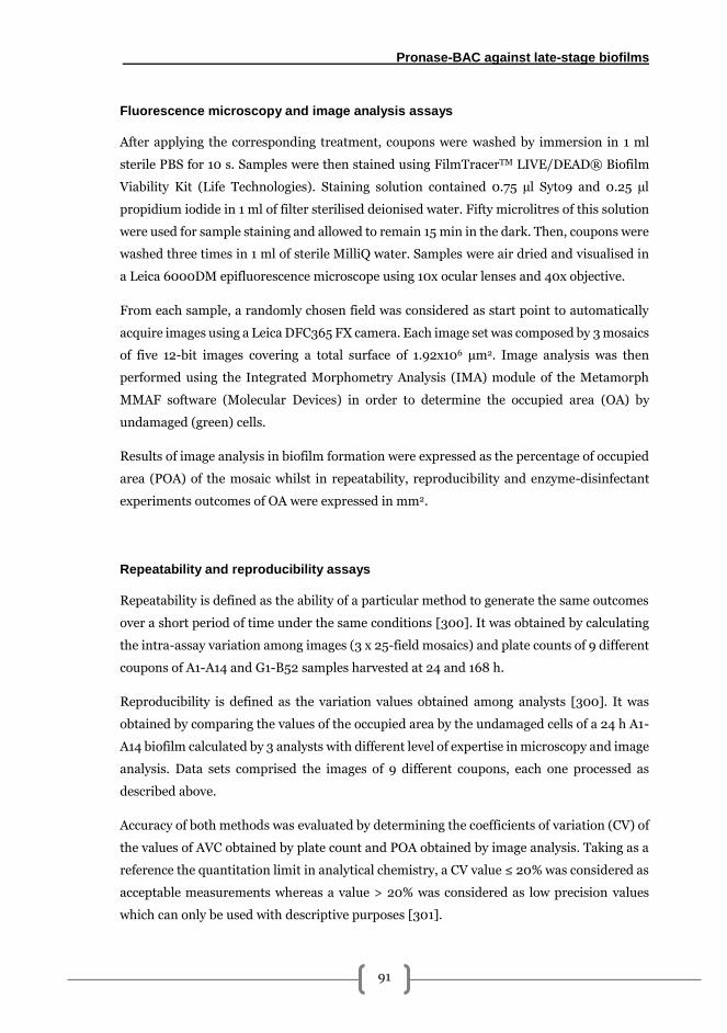
Pronase-BAC against late-stage biofilms
91
Fluorescence microscopy and image analysis assays
After applying the corresponding treatment, coupons were washed by immersion in 1 ml
sterile PBS for 10 s. Samples were then stained using FilmTracerTM LIVE/DEAD® Biofilm
Viability Kit (Life Technologies). Staining solution contained 0.75 µl Syto9 and 0.25 µl
propidium iodide in 1 ml of filter sterilised deionised water. Fifty microlitres of this solution
were used for sample staining and allowed to remain 15 min in the dark. Then, coupons were
washed three times in 1 ml of sterile MilliQ water. Samples were air dried and visualised in
a Leica 6000DM epifluorescence microscope using 10x ocular lenses and 40x objective.
From each sample, a randomly chosen field was considered as start point to automatically
acquire images using a Leica DFC365 FX camera. Each image set was composed by 3 mosaics
of five 12-bit images covering a total surface of 1.92x106 μm2. Image analysis was then
performed using the Integrated Morphometry Analysis (IMA) module of the Metamorph
MMAF software (Molecular Devices) in order to determine the occupied area (OA) by
undamaged (green) cells.
Results of image analysis in biofilm formation were expressed as the percentage of occupied
area (POA) of the mosaic whilst in repeatability, reproducibility and enzyme-disinfectant
experiments outcomes of OA were expressed in mm2.
Repeatability and reproducibility assays
Repeatability is defined as the ability of a particular method to generate the same outcomes
over a short period of time under the same conditions [300]. It was obtained by calculating
the intra-assay variation among images (3 x 25-field mosaics) and plate counts of 9 different
coupons of A1-A14 and G1-B52 samples harvested at 24 and 168 h.
Reproducibility is defined as the variation values obtained among analysts [300]. It was
obtained by comparing the values of the occupied area by the undamaged cells of a 24 h A1-
A14 biofilm calculated by 3 analysts with different level of expertise in microscopy and image
analysis. Data sets comprised the images of 9 different coupons, each one processed as
described above.
Accuracy of both methods was evaluated by determining the coefficients of variation (CV) of
the values of AVC obtained by plate count and POA obtained by image analysis. Taking as a
reference the quantitation limit in analytical chemistry, a CV value ≤ 20% was considered as
acceptable measurements whereas a value > 20% was considered as low precision values
which can only be used with descriptive purposes [301].
Chapter 4
92
Dual-species biofilm formation kinetics
Samples of both consortia were collected at 24, 48, 72, 96 and 168 h of incubation. In each
sampling time, 3 coupons were used for plate count and 3 more for microscopy analysis as
described above. For comparison of the accuracy of both techniques, the coefficients of
variation (CV) were also calculated dividing the standard deviation by the mean of the
sample set.
Effects of sequential pronase-benzalkonium chloride treatments on 168 h A1-A14
biofilms.
Preparation of the solutions
Pronase (PRN; from Streptomyces griseus, Roche) was prepared at concentrations listed in
Table 1 using 0.1 M Tris-HCl (Sigma Aldrich) buffer at pH = 7.5 ± 0.2 and then filter sterilised
through a 0.2 µm pore diameter syringe filter (Sartorius). Solutions were kept at -20 ºC until
use. Benzalkonium chloride (BAC; Guinama, Alboraya) was prepared at concentrations
listed in Table 1 dissolving the stock solution in sterile distilled water according to the
concentrations needed, and kept at 4 ºC until use.
Neutralising solution was prepared with following composition per litre: 10 ml of a 34 g/l
KH2PO4 solution adjusted to pH = 7.2 with NaOH(aq), 3 g soy lecithin, 5 g Na2S2O3, 1 g L-
histidine, 30 ml Tween 80 and deionised water [230]. This solution was sterilised by
autoclaving at 121 ºC for 20 min and kept at 4 ºC until use.
Experimental design
A first order factorial design [302,303] with 4 combinations of variables and 4 replicates in
the centre of the domain was carried out. The independent variables were the concentration
of PRN and the concentration of BAC. Natural and encoded values used are listed in Table
4.1. This sort of design permits to obtain information about various factors without
increasing the size of the assay. Besides, they are useful to quantify individual and synergistic
effects among different treatments in a given experimental ambit [302,303].
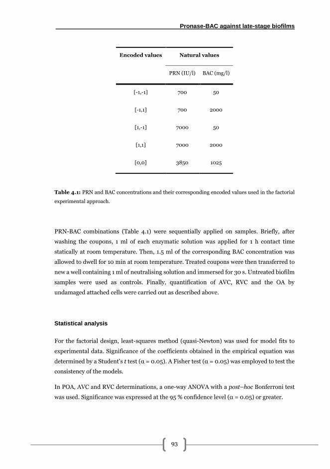
Pronase-BAC against late-stage biofilms
93
Encoded values Natural values
PRN (IU/l) BAC (mg/l)
[-1,-1] 700 50
[-1,1] 700 2000
[1,-1] 7000 50
[1,1] 7000 2000
[0,0] 3850 1025
Table 4.1: PRN and BAC concentrations and their corresponding encoded values used in the factorial
experimental approach.
PRN-BAC combinations (Table 4.1) were sequentially applied on samples. Briefly, after
washing the coupons, 1 ml of each enzymatic solution was applied for 1 h contact time
statically at room temperature. Then, 1.5 ml of the corresponding BAC concentration was
allowed to dwell for 10 min at room temperature. Treated coupons were then transferred to
new a well containing 1 ml of neutralising solution and immersed for 30 s. Untreated biofilm
samples were used as controls. Finally, quantification of AVC, RVC and the OA by
undamaged attached cells were carried out as described above.
Statistical analysis
For the factorial design, least-squares method (quasi-Newton) was used for model fits to
experimental data. Significance of the coefficients obtained in the empirical equation was
determined by a Student’s t test (α = 0.05). A Fisher test (α = 0.05) was employed to test the
consistency of the models.
In POA, AVC and RVC determinations, a one-way ANOVA with a post–hoc Bonferroni test
was used. Significance was expressed at the 95 % confidence level (α = 0.05) or greater.
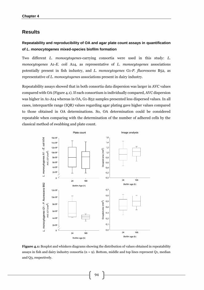
Chapter 4
94
Results
Repeatability and reproducibility of OA and agar plate count assays in quantification
of L. monocytogenes mixed-species biofilm formation
Two different L. monocytogenes-carrying consortia were used in this study: L.
monocytogenes A1-E. coli A14, as representative of L. monocytogenes associations
potentially present in fish industry, and L. monocytogenes G1-P. fluorescens B52, as
representative of L. monocytogenes associations present in dairy industry.
Repeatability assays showed that in both consortia data dispersion was larger in AVC values
compared with OA (Figure 4.1). If each consortium is individually compared, AVC dispersion
was higher in A1-A14 whereas in OA, G1-B52 samples presented less dispersed values. In all
cases, interquartile range (IQR) values regarding agar plating gave higher values compared
to those obtained in OA determinations. So, OA determination could be considered
repeatable when comparing with the determination of the number of adhered cells by the
classical method of swabbing and plate count.
Figure 4.1: Boxplot and whiskers diagrams showing the distribution of values obtained in repeatability
assays in fish and dairy industry consortia (n = 9). Bottom, middle and top lines represent Q1, median
and Q3, respectively.
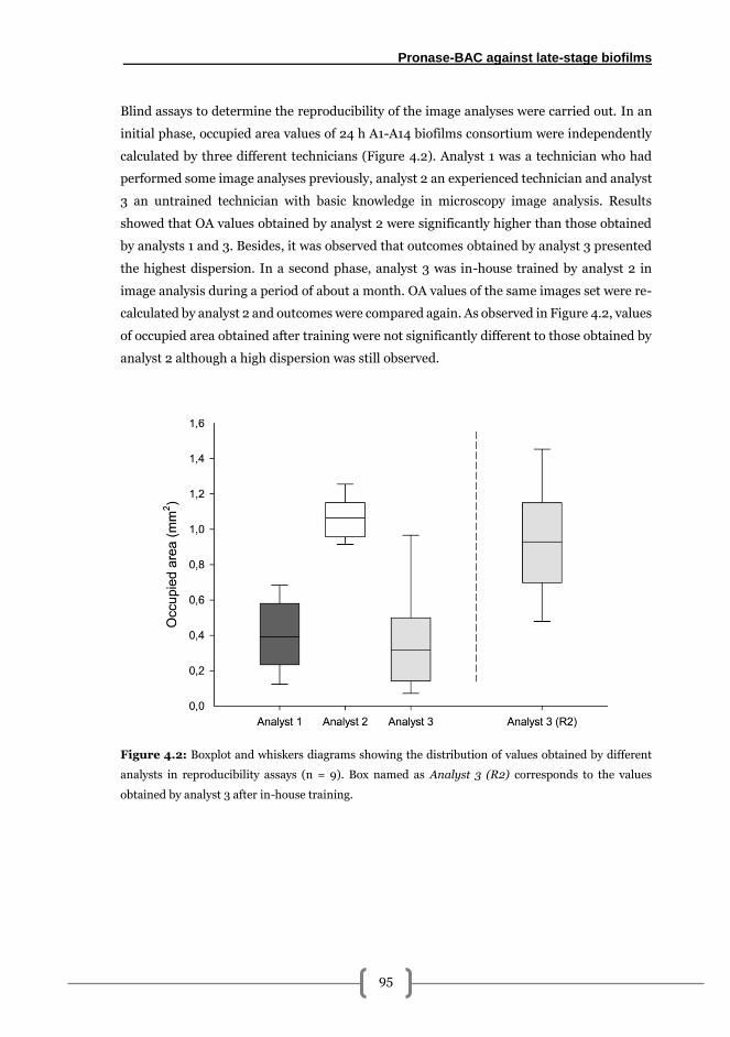
Pronase-BAC against late-stage biofilms
95
Blind assays to determine the reproducibility of the image analyses were carried out. In an
initial phase, occupied area values of 24 h A1-A14 biofilms consortium were independently
calculated by three different technicians (Figure 4.2). Analyst 1 was a technician who had
performed some image analyses previously, analyst 2 an experienced technician and analyst
3 an untrained technician with basic knowledge in microscopy image analysis. Results
showed that OA values obtained by analyst 2 were significantly higher than those obtained
by analysts 1 and 3. Besides, it was observed that outcomes obtained by analyst 3 presented
the highest dispersion. In a second phase, analyst 3 was in-house trained by analyst 2 in
image analysis during a period of about a month. OA values of the same images set were re-
calculated by analyst 2 and outcomes were compared again. As observed in Figure 4.2, values
of occupied area obtained after training were not significantly different to those obtained by
analyst 2 although a high dispersion was still observed.
Figure 4.2: Boxplot and whiskers diagrams showing the distribution of values obtained by different
analysts in reproducibility assays (n = 9). Box named as Analyst 3 (R2) corresponds to the values
obtained by analyst 3 after in-house training.
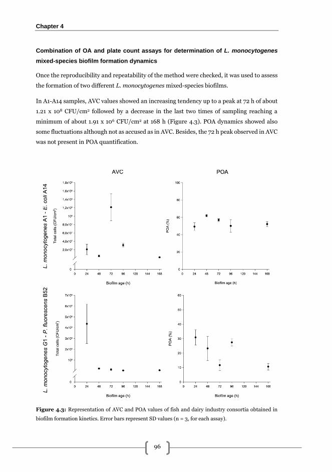
Chapter 4
96
Combination of OA and plate count assays for determination of L. monocytogenes
mixed-species biofilm formation dynamics
Once the reproducibility and repeatability of the method were checked, it was used to assess
the formation of two different L. monocytogenes mixed-species biofilms.
In A1-A14 samples, AVC values showed an increasing tendency up to a peak at 72 h of about
1.21 x 10⁸ CFU/cm2 followed by a decrease in the last two times of sampling reaching a
minimum of about 1.91 x 10⁶ CFU/cm2 at 168 h (Figure 4.3). POA dynamics showed also
some fluctuations although not as accused as in AVC. Besides, the 72 h peak observed in AVC
was not present in POA quantification.
Figure 4.3: Representation of AVC and POA values of fish and dairy industry consortia obtained in
biofilm formation kinetics. Error bars represent SD values (n = 3, for each assay).
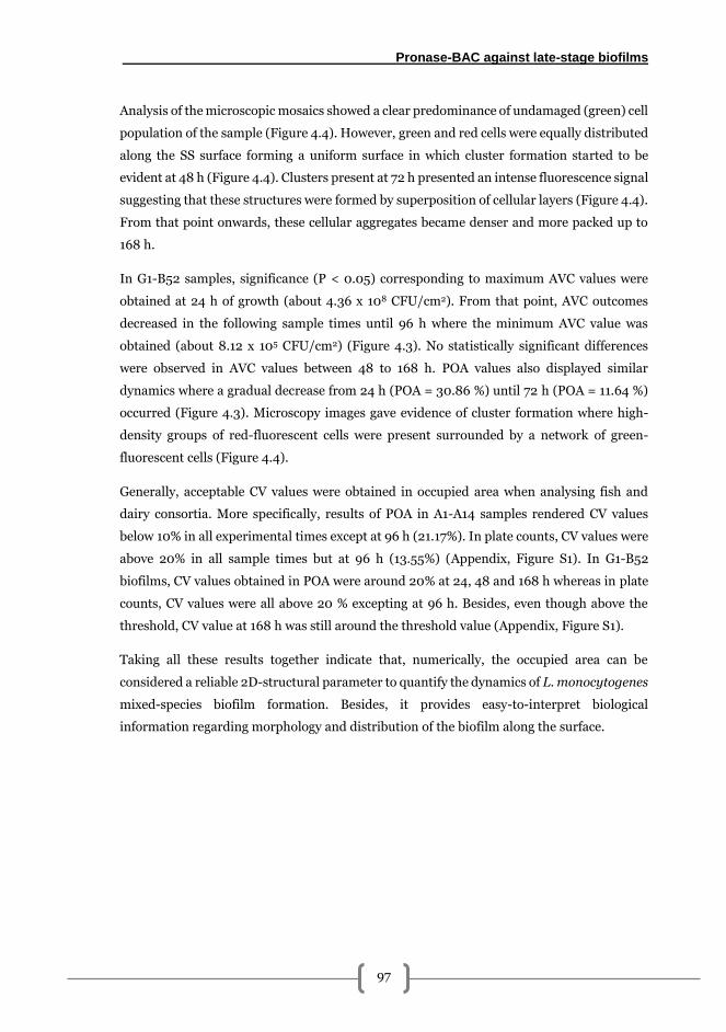
Pronase-BAC against late-stage biofilms
97
Analysis of the microscopic mosaics showed a clear predominance of undamaged (green) cell
population of the sample (Figure 4.4). However, green and red cells were equally distributed
along the SS surface forming a uniform surface in which cluster formation started to be
evident at 48 h (Figure 4.4). Clusters present at 72 h presented an intense fluorescence signal
suggesting that these structures were formed by superposition of cellular layers (Figure 4.4).
From that point onwards, these cellular aggregates became denser and more packed up to
168 h.
In G1-B52 samples, significance (P < 0.05) corresponding to maximum AVC values were
obtained at 24 h of growth (about 4.36 x 10⁸ CFU/cm2). From that point, AVC outcomes
decreased in the following sample times until 96 h where the minimum AVC value was
obtained (about 8.12 x 10⁵ CFU/cm2) (Figure 4.3). No statistically significant differences
were observed in AVC values between 48 to 168 h. POA values also displayed similar
dynamics where a gradual decrease from 24 h (POA = 30.86 %) until 72 h (POA = 11.64 %)
occurred (Figure 4.3). Microscopy images gave evidence of cluster formation where high-
density groups of red-fluorescent cells were present surrounded by a network of green-
fluorescent cells (Figure 4.4).
Generally, acceptable CV values were obtained in occupied area when analysing fish and
dairy consortia. More specifically, results of POA in A1-A14 samples rendered CV values
below 10% in all experimental times except at 96 h (21.17%). In plate counts, CV values were
above 20% in all sample times but at 96 h (13.55%) (Appendix, Figure S1). In G1-B52
biofilms, CV values obtained in POA were around 20% at 24, 48 and 168 h whereas in plate
counts, CV values were all above 20 % excepting at 96 h. Besides, even though above the
threshold, CV value at 168 h was still around the threshold value (Appendix, Figure S1).
Taking all these results together indicate that, numerically, the occupied area can be
considered a reliable 2D-structural parameter to quantify the dynamics of L. monocytogenes
mixed-species biofilm formation. Besides, it provides easy-to-interpret biological
information regarding morphology and distribution of the biofilm along the surface.
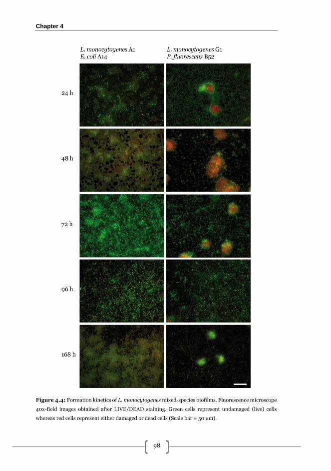
Chapter 4
98
Figure 4.4: Formation kinetics of L. monocytogenes mixed-species biofilms. Fluorescence microscope
40x-field images obtained after LIVE/DEAD staining. Green cells represent undamaged (live) cells
whereas red cells represent either damaged or dead cells (Scale bar = 50 µm).
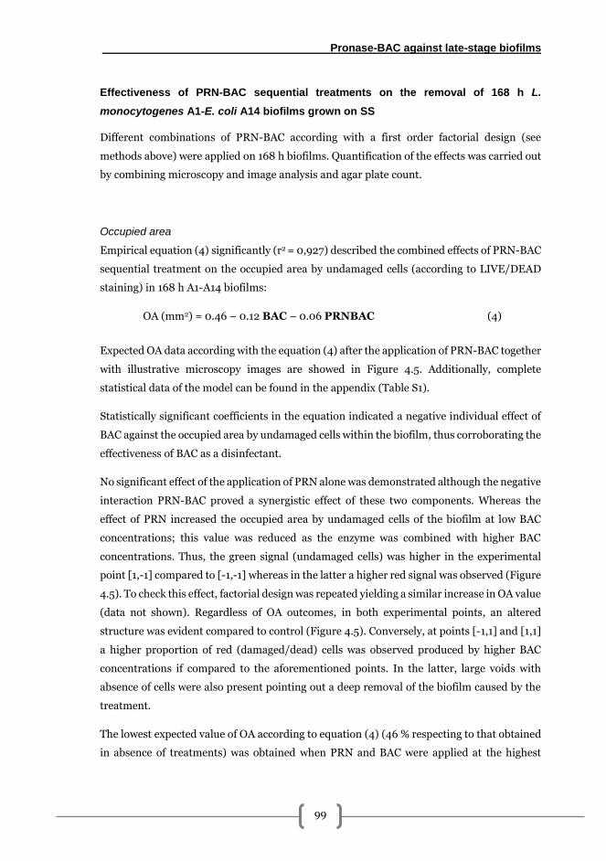
Pronase-BAC against late-stage biofilms
99
Effectiveness of PRN-BAC sequential treatments on the removal of 168 h L.
monocytogenes A1-E. coli A14 biofilms grown on SS
Different combinations of PRN-BAC according with a first order factorial design (see
methods above) were applied on 168 h biofilms. Quantification of the effects was carried out
by combining microscopy and image analysis and agar plate count.
Occupied area
Empirical equation (4) significantly (r2 = 0,927) described the combined effects of PRN-BAC
sequential treatment on the occupied area by undamaged cells (according to LIVE/DEAD
staining) in 168 h A1-A14 biofilms:
OA (mm2) = 0.46 – 0.12 BAC – 0.06 PRNBAC (4)
Expected OA data according with the equation (4) after the application of PRN-BAC together
with illustrative microscopy images are showed in Figure 4.5. Additionally, complete
statistical data of the model can be found in the appendix (Table S1).
Statistically significant coefficients in the equation indicated a negative individual effect of
BAC against the occupied area by undamaged cells within the biofilm, thus corroborating the
effectiveness of BAC as a disinfectant.
No significant effect of the application of PRN alone was demonstrated although the negative
interaction PRN-BAC proved a synergistic effect of these two components. Whereas the
effect of PRN increased the occupied area by undamaged cells of the biofilm at low BAC
concentrations; this value was reduced as the enzyme was combined with higher BAC
concentrations. Thus, the green signal (undamaged cells) was higher in the experimental
point [1,-1] compared to [-1,-1] whereas in the latter a higher red signal was observed (Figure
4.5). To check this effect, factorial design was repeated yielding a similar increase in OA value
(data not shown). Regardless of OA outcomes, in both experimental points, an altered
structure was evident compared to control (Figure 4.5). Conversely, at points [-1,1] and [1,1]
a higher proportion of red (damaged/dead) cells was observed produced by higher BAC
concentrations if compared to the aforementioned points. In the latter, large voids with
absence of cells were also present pointing out a deep removal of the biofilm caused by the
treatment.
The lowest expected value of OA according to equation (4) (46 % respecting to that obtained
in absence of treatments) was obtained when PRN and BAC were applied at the highest
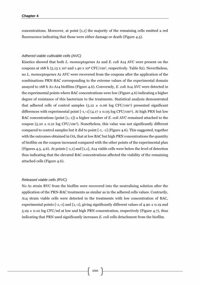
Chapter 4
100
concentrations. Moreover, at point [1,1] the majority of the remaining cells emitted a red
fluorescence indicating that those were either damage or death (Figure 4.5).
Adhered viable cultivable cells (AVC)
Kinetics showed that both L. monocytogenes A1 and E. coli A14 AVC were present on the
coupons at 168 h (5.13 x 10⁵ and 1.40 x 10⁶ CFU/cm2, respectively. Table S2). Nevertheless,
no L. monocytogenes A1 AVC were recovered from the coupons after the application of the
combinations PRN-BAC corresponding to the extreme values of the experimental domain
assayed to 168 h A1-A14 biofilms (Figure 4.6). Conversely, E. coli A14 AVC were detected in
the experimental points where BAC concentrations were low (Figure 4.6) indicating a higher
degree of resistance of this bacterium to the treatments. Statistical analysis demonstrated
that adhered cells of control samples (5.12 ± 0.06 log CFU/cm2) presented significant
differences with experimental point [-1,-1] (4.17 ± 0.05 log CFU/cm2). At high PRN but low
BAC concentrations (point [1,-1]) a higher number of E. coli AVC remained attached to the
coupon (5.10 ± 0.21 log CFU/cm2). Nonetheless, this value was not significantly different
compared to control samples but it did to point [-1, -1] (Figure 4.6). This suggested, together
with the outcomes obtained in OA, that at low BAC but high PRN concentrations the quantity
of biofilm on the coupon increased compared with the other points of the experimental plan
(Figures 4.5, 4.6). At points [-1,1] and [1,1], A14 viable cells were below the level of detection
thus indicating that the elevated BAC concentrations affected the viability of the remaining
attached cells (Figure 4.6).
Released viable cells (RVC)
No A1 strain RVC from the biofilm were recovered into the neutralising solution after the
application of the PRN-BAC treatments as similar as in the adhered cells values. Contrarily,
A14 strain viable cells were detected in the treatments with low concentration of BAC,
experimental points [-1,-1] and [1,-1], giving significantly different values of 4.90 ± 0.19 and
5.29 ± 0.10 log CFU/ml at low and high PRN concentration, respectively (Figure 4.7), thus
indicating that PRN used significantly increases E. coli cells detachment from the biofilm.
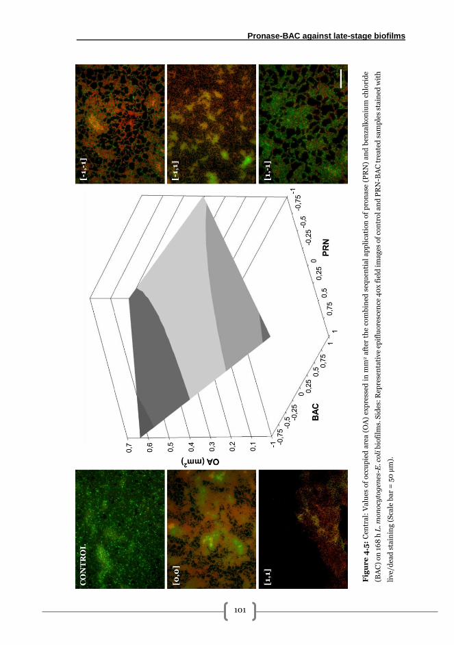
Pronase-BAC against late-stage biofilms
101
Fig
ur
e 4
.5:
Cen
tra
l: V
alu
es o
f o
ccu
pie
d a
rea
(O
A)
exp
ress
ed i
n m
m2 a
fter
th
e c
om
bin
ed s
equ
enti
al
ap
pli
cati
on
of
pro
na
se (
PR
N)
an
d b
enza
lko
niu
m c
hlo
rid
e
(BA
C)
on
16
8 h
L. m
on
ocy
tog
enes
-E. c
oli
bio
film
s. S
ides
: R
epre
sen
tati
ve
epif
luo
resc
ence
40
x f
ield
im
ag
es o
f co
ntr
ol
an
d P
RN
-BA
C t
rea
ted
sa
mp
les
sta
ined
wit
h
liv
e/d
ead
sta
inin
g (
Sca
le b
ar
= 5
0 µ
m).
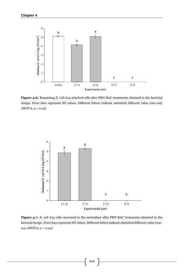
Chapter 4
102
Figure 4.6: Remaining E. coli A14 attached cells after PRN-BAC treatments obtained in the factorial
design. Error bars represent SD values. Different letters indicate statistical different value (one-way
ANOVA, α = 0.05)
Figure 4.7: E. coli A14 cells recovered in the neutraliser after PRN-BAC treatments obtained in the
factorial design. Error bars represent SD values. Different letters indicate statistical different value (one-
way ANOVA, α = 0.05)
Pronase-BAC against late-stage biofilms
103
Discussion
2D areal parameters have been considered as good biofilm descriptors giving biologically
meaningful information [152,153,157,160]. In the present study, accuracy of the OA for
quantifying biofilms was evaluated in terms of repeatability and reproducibility. This was
intended not to obtain the same results from both methods, but to evaluate their applicability
in biofilm studies from a numerical/statistical point of view.
Outcomes showed lower data dispersion and CV values in OA regarding those obtained by
the classical method (Appendix, Figure S1). Besides, it was demonstrated that after a period
of training the method is reproducible (Figure 4.2) making it suitable for assessment of
biofilm formation and to evaluate the biofilm removal effects of antimicrobials measuring
the ability to produce biofilm-free gaps on the surface after a given treatment.
In biofilm formation, fluorescence microscopy analysis provided information regarding 2D
structural features not detected by plate count. However, accuracy of results depends on the
uniformity of the structure, giving higher CV values in heterogeneous biofilms. This was of a
special relevance in G1-B52 samples in which a clear tendency to form microcolonies in most
cases was observed (Figure 4.4). These structures were formed by dense groups of red
(damaged/dead) cells surrounded by a network of undamaged (green) cells (Figure 4.4).
Bayles [304] pointed out the importance of dead cells as a biofilm support, anchoring the
structure to the surface, improving its stability.
The effects of PRN-BAC sequential treatments were assessed on late-stage L.
monocytogenes-E. coli biofilms using a first-order factorial design. Empirical equation (4)
demonstrated a significant individual effect of BAC and a synergistic effect between PRN and
BAC. Nevertheless, no individual effect of PRN on the occupied area by the mixed biofilm
was demonstrated in the experimental conditions used.
PRN is a mixture of various endo- and exo-peptidases [305]. However, although L.
monocytogenes biofilm matrix has a high protein content [117,298], in 168 h samples, L.
monocytogenes A1 population was about 2 log CFU/cm2 lower than E. coli A14 (data not
shown) thus the contribution for the final matrix composition of A1 strain may be
significantly lower if compared with A14 strain. Hence, the absence of individual PRN effect
could be related to the presence of soluble protective polysaccharides in the matrix secreted
by E. coli, becoming richer in sugar residues [292]. Besides, L. monocytogenes could have
also promoted this sugar-rich environment by secreting soluble polysaccharides such as
teichoic acids equal to those present in the cell membrane [119].
Chapter 4
104
So as to mimic the environmental conditions found in industrial premises, treatments were
applied at room temperature, below its optimal [306], that may have produced a lower
activity of the enzyme. Optimal temperatures have been used in previous biofilm-removal
studies involving PRN [307] as well as for other protein hydrolases [117,298]. Despite this,
Orgaz et al. [308] demonstrated the effectiveness of PRN at 25 ºC against P. fluorescens
biofilms, however, the concentration of PRN used were about 4 times more than the
maximum concentration used in this work. Nevertheless, among microscopy images it was
observed that, compared with controls, biofilm structure was affected by PRN-BAC
treatments regardless of BAC concentrations (Figure 4.5).
The unexpected increase of OA and the number of viable and attached cells of E. coli by PRN
in presence of low BAC concentrations can be explained by the dispersant effect of the
enzyme. Hence, the enzyme could provoke cell disaggregation in the biofilm, and the
released cells could subsequently re-adhere during the time of exposition. This hypothesis
would explain, by one hand, the observed increase in the occupied area by the re-adherence
and, on the other hand, the observed increase in the number of AVC and RVC detected as
cell aggregates give rise to less number of colonies in the plate counts.
BAC interacts with cell membranes promoting disruption of their integrity and cellular
content leakage [203,309]. According to results, it seems to be clear that PRN-BAC acted in
a synergic manner being this in agreement with a recent opinion of Meireles et al. [181], who
stated that a combination of enzymes and biocidal agents is desirable to obtain a good biofilm
biomass removal. Besides, PRN could have provoked a certain level of cell lysis as reported
for other hydrolytic enzymes [175,181,310] further enhancing the removal and biocidal
effects of the combined treatment at high BAC concentrations (Figure 4.5).
No adhered or released viable cells of L. monocytogenes or E. coli were detected in those
experimental points with the highest BAC concentrations (Figures 4.7, 4.8). However, values
of occupied area indicated the presence of undamaged cells on the coupon. Two main reasons
can explain the observed discrepancy. Firstly, the lower limit of detection of the microscopic
method (1 cell/field) respecting to the plate count method (1.70 log CFU/cm2), and secondly,
the presence of viable but non-cultivable cells (VBNC). In fact, considering that our
experimental system consisted of 168 h biofilms that have been exposed to PRN-BAC
treatments, it should be expected that in those biofilms the pool of VBNC cells will be
significant. In this state cells do not grow in solid media and microscopy assays are the only
alternative to detect them [284].
Moreover, it is becoming clear among microbiologists that microbial pathogens survive to
environmental stresses by entering into the VBNC state [284,311]. This status is reversible
Pronase-BAC against late-stage biofilms
105
under appropriate stimuli so, undetected pathogens can resuscitate from this dormant state
thus entailing several public health concerns [312]. In L. monocytogenes this process is
multifactorial [313]. Indeed, a recent study carried out in biofilms grown in tap water showed
that L. monocytogenes VBNC state depends not only on the nutrient availability but also on
the temperature [284].
Cell dispersion is an intrinsic process in the life-cycle of any biofilm [79]. Nevertheless, this
phenomenon can be accelerated if an antimicrobial treatment is applied due to an alteration
of the structural integrity. This has been considered as a topic of concern since it can facilitate
the dissemination of pathogens into the environment becoming a feasible cause of
contamination [181,314].
Enzymatic-based treatments have been used to sensitise biofilm so as to reduce the dose of
antibiotics [315] and antiseptics [211] needed for treatment of biofilm-colonised medical
devices. A recent study conducted by Stiefel et al., (2016) demonstrated that the use different
species-specific enzyme mixtures increased the efficacy of commercially available cleaners
to remove biofilms of Staphylococcus aureus and Pseudomonas aeruginosa potentially
present in endoscopes. Besides, once bacteria are removed from the biofilm, pathogenicity
factors may also be affected by enzymatic treatments. As an example, Longhi et al., (2008)
observed how the infectiveness of planktonic L. monocytogenes in Caco-2 cells was
significantly reduced after 24 h treatment with 200 U/ml of serratiopeptidase. This
phenomenon demonstrated that beside live cells are dispersed after enzyme contact, these
may represent a lower health threat.
In summary, it has been empirically demonstrated how PRN-BAC combined treatments can
synergically interact the biofilm removal of the system on L. monocytogenes-E. coli dual-
species biofilms grown on stainless steel. This approach performed in a straightforward
manner with a pre-treatment with PRN combined with a high dose of BAC. Besides, at high
BAC concentrations the quantity of RVC from the biofilm is also significantly diminished
thus avoiding potential pathogen spread into the environment. Moreover, it has been
demonstrated that microscopy 2D-image analysis combined with plate count may represent
a helpful tool in assays dealing with multispecies biofilms. It provides biologically
meaningful and easy-to-interpret data for quantification of biofilm development as well as
empirical determination of antimicrobial treatments’ effects.
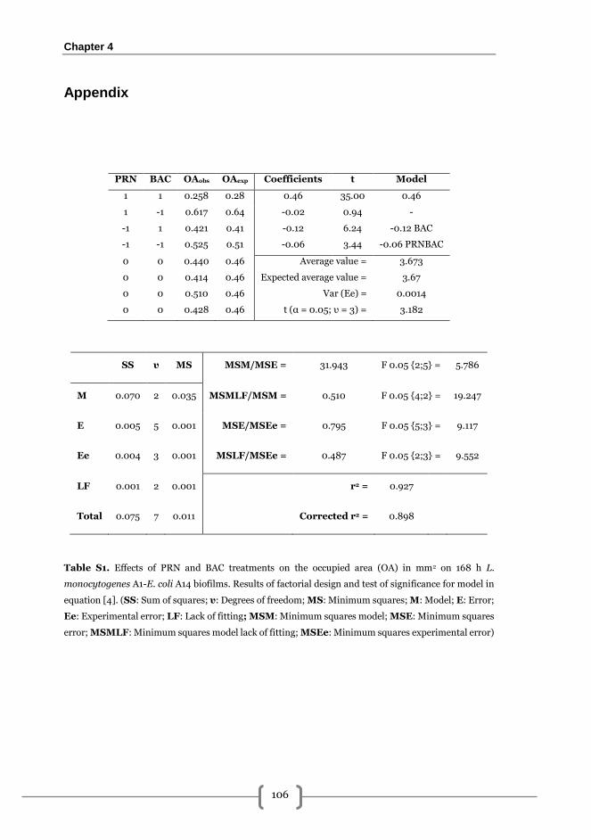
Chapter 4
106
Appendix
PRN BAC OAobs OAexp Coefficients t Model
1 1 0.258 0.28 0.46 35.00 0.46
1 -1 0.617 0.64 -0.02 0.94 -
-1 1 0.421 0.41 -0.12 6.24 -0.12 BAC
-1 -1 0.525 0.51 -0.06 3.44 -0.06 PRNBAC
0 0 0.440 0.46 Average value = 3.673
0 0 0.414 0.46 Expected average value = 3.67
0 0 0.510 0.46 Var (Ee) = 0.0014
0 0 0.428 0.46 t (α = 0.05; υ = 3) = 3.182
SS υ MS MSM/MSE = 31.943 F 0.05 {2;5} = 5.786
M 0.070 2 0.035 MSMLF/MSM = 0.510 F 0.05 {4;2} = 19.247
E 0.005 5 0.001 MSE/MSEe = 0.795 F 0.05 {5;3} = 9.117
Ee 0.004 3 0.001 MSLF/MSEe = 0.487 F 0.05 {2;3} = 9.552
LF 0.001 2 0.001 r2 = 0.927
Total 0.075 7 0.011 Corrected r2 = 0.898
Table S1. Effects of PRN and BAC treatments on the occupied area (OA) in mm2 on 168 h L.
monocytogenes A1-E. coli A14 biofilms. Results of factorial design and test of significance for model in
equation [4]. (SS: Sum of squares; υ: Degrees of freedom; MS: Minimum squares; M: Model; E: Error;
Ee: Experimental error; LF: Lack of fitting; MSM: Minimum squares model; MSE: Minimum squares
error; MSMLF: Minimum squares model lack of fitting; MSEe: Minimum squares experimental error)
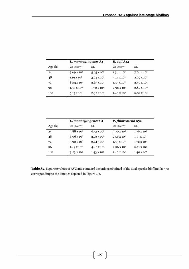
Pronase-BAC against late-stage biofilms
107
L. monocytogenes A1 E. coli A14
Age (h) CFU/cm2 SD CFU/cm2 SD
24 3.69 x 10⁶ 5.65 x 10⁵ 1.38 x 10⁷ 7.08 x 10⁶
48 1.19 x 10⁶ 3.24 x 10⁵ 4.14 x 10⁶ 2.29 x 10⁶
72 8.33 x 10⁵ 2.63 x 10⁵ 1.33 x 10⁸ 2.40 x 10⁷
96 1.50 x 10⁶ 1.70 x 10⁵ 2.96 x 10⁷ 2.82 x 10⁶
168 5.13 x 10⁵ 2.32 x 10⁵ 1.40 x 10⁶ 6.84 x 10⁵
L. monocytogenes G1 P. fluorescens B52
Age (h) CFU/cm2 SD CFU/cm2 SD
24 3.88 x 10⁷ 6.53 x 10⁶ 3.70 x 10⁸ 1.76 x 10⁸
48 6.06 x 10⁶ 2.73 x 10⁶ 2.56 x 10⁷ 1.13 x 10⁷
72 3.90 x 10⁶ 2.74 x 10⁶ 1.33 x 10⁸ 1.72 x 10⁷
96 1.49 x 10⁶ 4.46 x 10⁵ 2.96 x 10⁷ 6.71 x 10⁵
168 3.23 x 10⁵ 1.43 x 10⁵ 1.40 x 10⁶ 1.40 x 10⁶
Table S2. Separate values of AVC and standard deviations obtained of the dual-species biofilms (n = 3)
corresponding to the kinetics depicted in Figure 4.3.
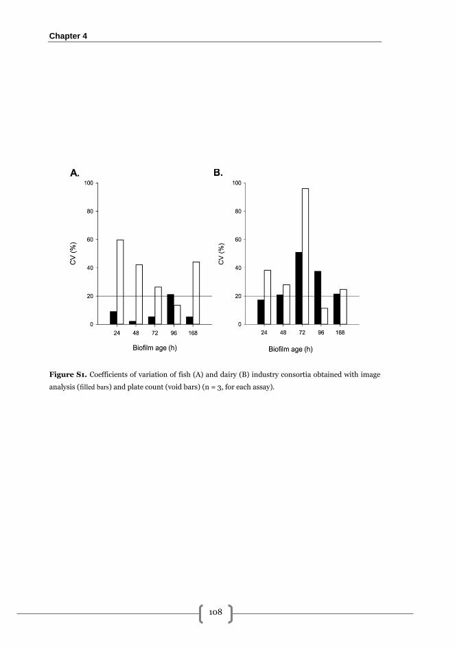
Chapter 4
108
Figure S1. Coefficients of variation of fish (A) and dairy (B) industry consortia obtained with image
analysis (filled bars) and plate count (void bars) (n = 3, for each assay).
5
Tolerance development to PRN-BAC
combined treatments in L. monocytogenes
dual-species biofilms

Tolerance development to combined treatments
111
Introduction
Tolerance development after sublethal exposure to BAC and other quaternary ammonium
compounds has extensively reported in planktonic cells of L. monocytogenes
[219,221,223,316] and E. coli [317,318]. However, a significant smaller number of studies
involving tolerance development after BAC sublethal exposure in mixed-species biofilms is
currently found in the scientific literature and, among them, no studies were found assessing
the tolerance to enzyme-BAC combined strategies. The only evidence found to BAC tolerance
development in mixed-species biofilms was the study performed by Machado et al. [206].
They demonstrated that sublethal exposure to BAC in Pseudomonas aeruginosa-E. coli 6-
day-old biofilms in polystyrene plates, presented a higher biomass compared with
unexposed samples despite the number of adhered cells was similar in both cases.
One of the main advantages of applying enzymes is that they do not suppose selective
pressure on bacteria [181]. However, if it is combined with biocides like BAC, it seems to be
important to consider both agents for a correct evaluation of tolerance development.
Therefore, the main aim of this las part of the present thesis was to assess the tolerance
development to PRN-BAC treatments after sublethal exposure in L. monocytogenes-E. coli
dual-species biofilms grown on SS coupons. The exposures were carried out following three
different approaches based on the concentrations of the antimicrobials used, the intervals of
dosage and the duration of the whole cycle.
Methods
Bacterial strains
Dual-species biofilms were formed by L. monocytogenes A1 and Escherichia coli A14, both
isolated in a previous survey performed among pre-sanitised surfaces in a fish processing
plant [37].
Stock cultures of all strains were maintained at – 80 ºC in brain-heart infusion broth (BHI;
Biolife, Italy) containing 50 % glycerol 1:1 (v/v) mixed. Work cultures were kept at -20 ºC in
Trypticase Soy Broth (TSB, Cultimed, Barcelona) containing 50 % glycerol 1:1 (v/v) mixed.
Chapter 5
112
Set-up of dual-species biofilms
100 µl of work cultures of L. monocytogenes and E. coli were cultured overnight in 5 ml
sterile TSB at 37 ºC and subcultured overnight in order to ensure a proper growth.
Inocula preparation was performed as described previously [145]. Briefly, Abs700 of each
culture was adjusted to 0.1 ± 0.001 in sterile phosphate buffer saline (PBS) using a 3000
Series scanning spectrophotometer (Cecil Instruments, Cambridge, England) corresponding
to a bacterial concentration of about 108 CFU/ml. Adjusted cultures were 1:104 diluted in
sterile mTSB (TSB supplemented with 2.5 g/l glucose (Vorquímica, S.L., Vigo, Spain) and
0.6 % yeast extract (Cultimed, Barcelona, Spain) to obtain a final concentration of about 104
CFU/ml. Then, equal volumes of these adjusted cultures were mixed to obtain the inoculum
for dual-species biofilms.
Biofilms were grown on 10x10x1 mm AISI 316 stainless steel (SS) coupons (Comevisa, Vigo,
Spain). Coupon pre-treatment included individual washing with industrial soap (Sutter
Wash, Sutter Ibérica, S.A., Madrid, Spain), rinsing with tap water, a final rinse with deionised
water and autoclaving at 121 ºC for 20 min. After this, coupons were individually placed into
a 24 flat-bottomed well plate and each well was inoculated with 1 ml of the inoculum. Plates
were incubated in a humidified atmosphere at 25 ºC statically for 2 h for initial adhesion and
then in constant shaking at 100 rpm.
Before any assay was performed, samples (SS coupons) were aseptically collected and briefly
immersed in 1 ml sterile PBS to remove loosely attached cells.
Adhered viable cultivable cells (AVC) quantification
After PBS washing, adhered viable cultivable cells (AVC), were collected from three different
coupons by swabbing using two sterile cotton swabs moistened in buffered peptone water
(BPW, Cultimed, Barcelona) per coupon. Swabs were resuspended in 2 ml of BPW and
vortexed vigorously for 1 min in order to release cells. Suspensions were serially diluted in
BPW and spread onto agar plates. Listeria-PALCAM (Liofilchem, Roseto degli Abruzzi, Italy)
was used for L. monocytogenes selection whereas HiCromeTM Coliform Agar (Sigma-
Aldrich, St Louis, MO, USA) supplemented with 5 µg/ml of vancomycin and cefsulodine
(Sigma-Aldrich) was used for E. coli. Plates were incubated at 37 ºC for 24-48 h and results
were expressed as the mean in log CFU/cm2 or log CFU/cm2 reduction depending on the
assay. The accepted limit of detection was 25 CFU in the plate of the lowest dilution
corresponding to 1.70 log CFU/cm2 [149].
Tolerance development to combined treatments
113
Microscopy assays
After washing, coupons were stained using LIVE/DEAD Bacterial viability kit (Life
Technologies) that allows distinguishing total cells with undamaged membranes (green
fluorescence) and damaged membranes (red fluorescence). Staining solution was prepared
mixing 0.25 µl of propidium iodide and 0.75 µl of Syto9 in 1 ml of filter sterilised distilled
water. 50-60 µl of this solution was poured onto each coupon for 15 min contact time.
Subsequently, staining solution was carefully removed and samples were washed three times
by immersion in 1 ml of sterile milliQ water for 30 s. Coupons were then air dried and
visualised under a Leica DM 6000 epifluorescence microscope (Leica, Wetzlar, Germany)
using a 40x objective and 10x ocular lenses. In each sample, a randomly chosen field was
considered as start point to acquire 12 bit images using a Leica DFC365 FX camera. Each
image set was composed by three 5 x 5 mosaics each one covering a total surface of 1.92 x 10⁶
μm2. Image analysis was then performed using the Integrated Morphometry Analysis (IMA)
module of Metamorph MMAF software (Molecular Devices, Sunnyvale, CA, USA) in order
to determine the occupied area (OA) by undamaged (green) cells. Results were expressed in
mm2.
Enzymes, BAC and neutralising solutions preparation
Pronase (PRN, from Streptomyces griseus, Roche, Mannheim, Germany) stock solutions
were prepared at concentrations 100, 1000 and 2000 µg/ml dissolved in 0.1 M Tris-HCl
(Sigma Aldrich) buffer at pH = 7.5 ± 0.2. After preparation, solutions were filter sterilised
through a 0.2 µm pore diameter syringe filter (Sartorius, Göttingen, Germany) and kept at -
20 ºC until use.
Benzalkonium chloride (BAC, Guinama, Alboraya, Spain) stock solutions were prepared at
concentrations 50, 2000 and 4000 µg/ml diluting the commercial solution in sterile distilled
water. Solutions were kept at 4 ºC until use.
Neutralising solution to stop BAC biocidal effects had the following composition per litre: 10
ml of a 34 g/l KH2PO4 solution adjusted to pH = 7.2 with NaOH(aq), 3 g soy lecithin, 5 g
Na2S2O3, 1 g L-histidine, 30 ml Tween 80 and adjusted with distilled water. This solution
was autoclaved at 121 ºC for 20 min and kept at 4 ºC until use.
Chapter 5
114
Experimental design
A two-phase experimental procedure was designed to evaluate the tolerance development
(TD) in dual-species L. monocytogenes-E. coli biofilms to the application of PRN-BAC
combined treatments.
Phase 1: sublethal expositions.
In this phase, 24 h L. monocytogenes-E. coli biofilms were exposed to a different number of
consecutive PRN-BAC sublethal treatments with or without medium renovation as
schematized in Figure 5.1. Sublethal concentrations were determined in previous assays
(data not shown). Both PRN and BAC stock solutions were diluted in mTSB at concentrations
for sublethal exposure (Figure 5.1). Additionally, a negative control experimental series in
which PRN and BAC solutions were substituted by equal volumes of sterile deionised water
was included in each experiment. Thus, the experimental approaches and concentrations
used were as follows:
i). Experimental approach 1 – Short term exposure: In this approach, two
consecutive expositions without medium renovation were carried out. At 24 h, the
bulk phase was carefully pipetted out and dual-species biofilms were exposed to 1
ml of 50 µg/ml PRN for 1 h contact time without agitation. Then, 1.5 ml of 25 µg/ml
BAC were added and plates were placed back at 25 ºC/100 rpm. At 48 h, bulk phase
was removed and dual-species biofilms were exposed to 1 ml of 100 µg/ml PRN, let
to dwell 1 h statically, followed by 1.5 ml of 50 µg/ml BAC, and placed back at 25
ºC/100 rpm until next step. In this approach, TD quantification in exposed biofilms
was determined after 72 h.
ii). Experimental approach 2 – Short term exposure with medium renewal: In this
second scheme, exposure to treatments was carried out as described for approach 1
but after every PRN-BAC exposition the bulk phase was pipetted out and
substituted by 1 ml of fresh mTSB (i.e. at 48 and 96 h), and incubated at 25 ºC/100
rpm for 24 h (Figure 5.1). In this case, final treatment and TD quantification to
PRN-BAC acquired resistance was carried out at 120 h (Figure 5.1).
iii). Experimental approach 3 – Long term exposure: In this last approach, a
discontinuous exposure to 1 ml 50 µg/ml PRN followed by 1.5 ml 25 µg/ml of BAC
was undertaken at 24, 48, 72, 96 and 168 h. At 190 h, final treatment and
quantification of the TD to PRN-BAC was carried out (Figure 5.1).
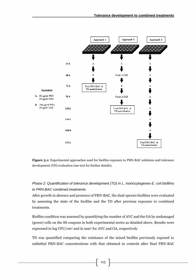
Tolerance development to combined treatments
115
Figure 5.1: Experimental approaches used for biofilm exposure to PRN-BAC solutions and tolerance
development (TD) evaluation (see text for further details).
Phase 2: Quantification of tolerance development (TD) in L. monocytogenes-E. coli biofilms
to PRN-BAC combined treatments
After growth in absence and presence of PRN-BAC, the dual-species biofilms were evaluated
by assessing the state of the biofilm and the TD after previous exposure to combined
treatments.
Biofilm condition was assessed by quantifying the number of AVC and the OA by undamaged
(green) cells on the SS coupons in both experimental series as detailed above. Results were
expressed in log CFU/cm2 and in mm2 for AVC and OA, respectively
TD was quantified comparing the resistance of the mixed biofilm previously exposed to
sublethal PRN-BAC concentrations with that obtained in controls after final PRN-BAC
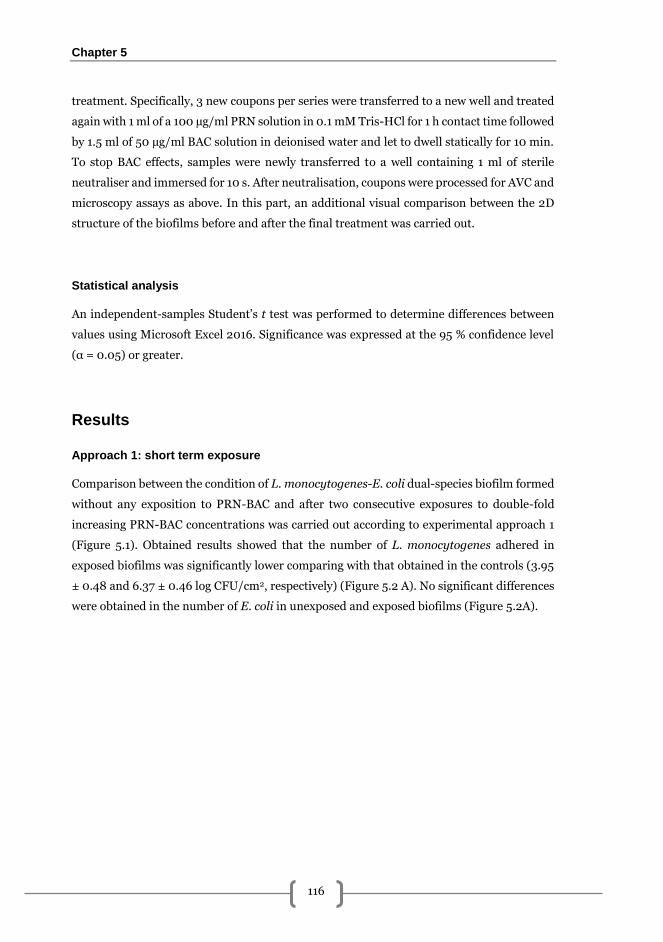
Chapter 5
116
treatment. Specifically, 3 new coupons per series were transferred to a new well and treated
again with 1 ml of a 100 µg/ml PRN solution in 0.1 mM Tris-HCl for 1 h contact time followed
by 1.5 ml of 50 µg/ml BAC solution in deionised water and let to dwell statically for 10 min.
To stop BAC effects, samples were newly transferred to a well containing 1 ml of sterile
neutraliser and immersed for 10 s. After neutralisation, coupons were processed for AVC and
microscopy assays as above. In this part, an additional visual comparison between the 2D
structure of the biofilms before and after the final treatment was carried out.
Statistical analysis
An independent-samples Student’s t test was performed to determine differences between
values using Microsoft Excel 2016. Significance was expressed at the 95 % confidence level
(α = 0.05) or greater.
Results
Approach 1: short term exposure
Comparison between the condition of L. monocytogenes-E. coli dual-species biofilm formed
without any exposition to PRN-BAC and after two consecutive exposures to double-fold
increasing PRN-BAC concentrations was carried out according to experimental approach 1
(Figure 5.1). Obtained results showed that the number of L. monocytogenes adhered in
exposed biofilms was significantly lower comparing with that obtained in the controls (3.95
± 0.48 and 6.37 ± 0.46 log CFU/cm2, respectively) (Figure 5.2 A). No significant differences
were obtained in the number of E. coli in unexposed and exposed biofilms (Figure 5.2A).
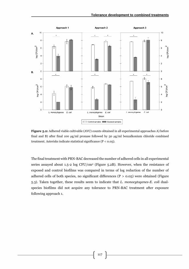
Tolerance development to combined treatments
117
Figure 5.2: Adhered viable cultivable (AVC) counts obtained in all experimental approaches A) before
final and B) after final 100 µg/ml pronase followed by 50 µg/ml benzalkonium chloride combined
treatment. Asterisks indicate statistical significance (P < 0.05).
The final treatment with PRN-BAC decreased the number of adhered cells in all experimental
series assayed about 1.5-2 log CFU/cm2 (Figure 5.2B). However, when the resistance of
exposed and control biofilms was compared in terms of log reduction of the number of
adhered cells of both species, no significant differences (P > 0.05) were obtained (Figure
5.3). Taken together, these results seem to indicate that L. monocytogenes-E. coli dual-
species biofilms did not acquire any tolerance to PRN-BAC treatment after exposure
following approach 1.
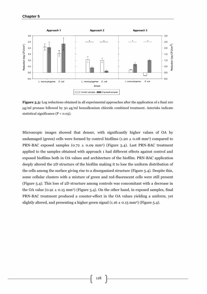
Chapter 5
118
Figure 5.3: Log reductions obtained in all experimental approaches after the application of a final 100
µg/ml pronase followed by 50 µg/ml benzalkonium chloride combined treatment. Asterisks indicate
statistical significance (P < 0.05).
Microscopic images showed that denser, with significantly higher values of OA by
undamaged (green) cells were formed by control biofilms (1.20 ± 0.08 mm2) compared to
PRN-BAC exposed samples (0.72 ± 0.09 mm2) (Figure 5.4). Last PRN-BAC treatment
applied to the samples obtained with approach 1 had different effects against control and
exposed biofilms both in OA values and architecture of the biofilm. PRN-BAC application
deeply altered the 2D structure of the biofilm making it to lose the uniform distribution of
the cells among the surface giving rise to a disorganised structure (Figure 5.4). Despite this,
some cellular clusters with a mixture of green and red-fluorescent cells were still present
(Figure 5.4). This loss of 2D structure among controls was concomitant with a decrease in
the OA value (0.91 ± 0.15 mm2) (Figure 5.4). On the other hand, in exposed samples, final
PRN-BAC treatment produced a counter-effect in the OA values yielding a uniform, yet
slightly altered, and presenting a higher green signal (1.16 ± 0.15 mm2) (Figure 5.4).
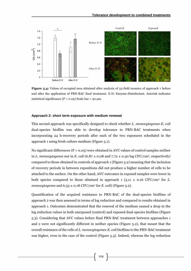
Tolerance development to combined treatments
119
Figure 5.4: Values of occupied area obtained after analysis of 25-field mosaics of approach 1 before
and after the application of PRN-BAC final treatment. E-D: Enzyme-Disinfectant. Asterisk indicates
statistical significance (P < 0.05) Scale bar = 50 µm.
Approach 2: short term exposure with medium renewal
This second approach was specifically designed to check whether L. monocytogenes-E. coli
dual-species biofilm was able to develop tolerance to PRN-BAC treatments when
incorporating 24 h-recovery periods after each of the two exposures scheduled in the
approach 1 using fresh culture medium (Figure 5.1).
No significant differences (P > 0.05) were obtained in AVC values of control samples neither
in L. monocytogenes nor in E. coli (6.87 ± 0.08 and 7.72 ± 0.30 log CFU/cm2, respectively)
compared to those obtained in controls of approach 1 (Figure 5.2) meaning that the inclusion
of recovery periods in between expositions did not produce a higher number of cells to be
attached to the surface. On the other hand, AVC outcomes in exposed samples were lower in
both species compared to those obtained in approach 1 (3.11 ± 0.16 CFU/cm2 for L.
monocytogenes and 6.53 ± 0.18 CFU/cm2 for E. coli) (Figure 5.2).
Quantification of the acquired resistance to PRN-BAC of the dual-species biofilms of
approach 2 was then assessed in terms of log reduction and compared to results obtained in
approach 1. Outcomes demonstrated that the renewal of the medium caused a drop in the
log reduction values in both unexposed (control) and exposed dual-species biofilms (Figure
5.3). Considering that AVC values before final PRN-BAC treatment between approaches 1
and 2 were not significantly different in neither species (Figure 5.2), that meant that the
overall resistance of the cells of L. monocytogenes-E. coli biofilms to the PRN-BAC treatment
was higher, even in the case of the control (Figure 5.3). Indeed, whereas the log reduction
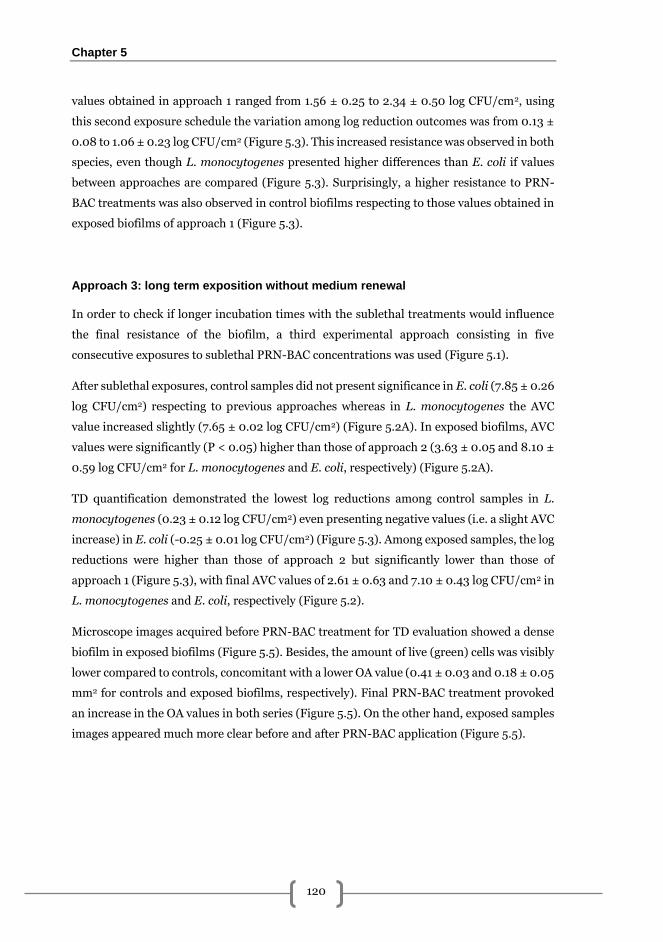
Chapter 5
120
values obtained in approach 1 ranged from 1.56 ± 0.25 to 2.34 ± 0.50 log CFU/cm2, using
this second exposure schedule the variation among log reduction outcomes was from 0.13 ±
0.08 to 1.06 ± 0.23 log CFU/cm2 (Figure 5.3). This increased resistance was observed in both
species, even though L. monocytogenes presented higher differences than E. coli if values
between approaches are compared (Figure 5.3). Surprisingly, a higher resistance to PRN-
BAC treatments was also observed in control biofilms respecting to those values obtained in
exposed biofilms of approach 1 (Figure 5.3).
Approach 3: long term exposition without medium renewal
In order to check if longer incubation times with the sublethal treatments would influence
the final resistance of the biofilm, a third experimental approach consisting in five
consecutive exposures to sublethal PRN-BAC concentrations was used (Figure 5.1).
After sublethal exposures, control samples did not present significance in E. coli (7.85 ± 0.26
log CFU/cm2) respecting to previous approaches whereas in L. monocytogenes the AVC
value increased slightly (7.65 ± 0.02 log CFU/cm2) (Figure 5.2A). In exposed biofilms, AVC
values were significantly (P < 0.05) higher than those of approach 2 (3.63 ± 0.05 and 8.10 ±
0.59 log CFU/cm2 for L. monocytogenes and E. coli, respectively) (Figure 5.2A).
TD quantification demonstrated the lowest log reductions among control samples in L.
monocytogenes (0.23 ± 0.12 log CFU/cm2) even presenting negative values (i.e. a slight AVC
increase) in E. coli (-0.25 ± 0.01 log CFU/cm2) (Figure 5.3). Among exposed samples, the log
reductions were higher than those of approach 2 but significantly lower than those of
approach 1 (Figure 5.3), with final AVC values of 2.61 ± 0.63 and 7.10 ± 0.43 log CFU/cm2 in
L. monocytogenes and E. coli, respectively (Figure 5.2).
Microscope images acquired before PRN-BAC treatment for TD evaluation showed a dense
biofilm in exposed biofilms (Figure 5.5). Besides, the amount of live (green) cells was visibly
lower compared to controls, concomitant with a lower OA value (0.41 ± 0.03 and 0.18 ± 0.05
mm2 for controls and exposed biofilms, respectively). Final PRN-BAC treatment provoked
an increase in the OA values in both series (Figure 5.5). On the other hand, exposed samples
images appeared much more clear before and after PRN-BAC application (Figure 5.5).
Tolerance development to combined treatments
121
Figure 5.5: Values of occupied area obtained after analysis of 25-field mosaics of approach 3 before
and after the application of PRN-BAC final treatment. E-D: Enzyme-Disinfectant. Asterisks indicate
statistical significance (P < 0.05) Scale bar = 50 µm.
Discussion
In the present study, the capability of L. monocytogenes-E. coli biofilms to grow and develop
tolerance to PRN-BAC combined treatments after sublethal exposure following three
different approaches was assessed.
Before any antimicrobial treatment can be applied, its efficacy against a particular must be
tested. Despite this, to date, no standardised methods for antibiofilm testing are available
[319] and none of them considers TD as a parameter, which is determinant to determine the
right dose to apply to avoid bacterial resistance especially when treatments are intended to
be used for long periods.
Results showed that the number of L. monocytogenes AVC decreased after sublethal
exposure to PRN-BAC regardless of the exposure scheme used (Figure 5.2A). Contrarily, in
E. coli the presence of PRN-BAC did not affect the number of AVC on SS coupons in
approaches 1 and 3 (Figure 5.2A). In approach 2, the number of AVC of E. coli in exposed
samples was slightly lower compared to control samples but the differences between series
were smaller than those obtained in L. monocytogenes (Figure 5.2A). These results were not
surprising considering that Gram-negatives are generally more resistant to QACs than
Gram-positives [201] and were in accordance with results obtained by Machado et al. [206]
who observed that the number of adhered cells in 6-day old dual-species biofilms of
Pseudomonas aeruginosa-E. coli was not affected by presence of 328.5 µg/ml BAC in the
culture medium.
Chapter 5
122
Obtained results showed that the acquired tolerance to PRN-BAC by L. monocytogenes-E.
coli dual-species biofilms is the result of the following two interdependent factors: i) the
effects produced by the cultural features of each experimental approach giving rise to a
different L. monocytogenes-E. coli dual-species biofilms, and ii) the specific physiological
effects provoked by the sublethal PRN-BAC expositions. Both effects can be
straightforwardly individually analysed comparing, in the first case, the acquired tolerance
in control biofilms after the application of antimicrobials in the three different experimental
approaches and, in the second case, comparing the tolerance developed by controls and by
exposed biofilms in each experimental approach.
Regarding the first comparison, significant higher values of log reduction were obtained in
the dual-species biofilms subjected to short-term approach (approach 1) compared to those
obtained when short-term with medium renewal and long-term exposures (approaches 2
and 3, respectively) were applied (Figure 5.3). In other words, biofilms grown in approach 1
were much less tolerant to PRN-BAC. This fact, together with the absence of significance
between AVC values in all the experimental approaches before and after the application of
the last PRN-BAC treatment, pointed out that longer incubation times (approaches 2 and 3)
and the incorporation of recovery periods (approach 2) gave rise to structures with higher
PRN-BAC tolerance.
The TD in terms of the number of AVC that resist the last PRN-BAC treatment after sublethal
exposure was only observed in approach 2, in which significant differences (P < 0.05) in the
log reduction between control and exposed dual-species biofilms were observed in both
strains.
In approach 1, microscopy images showed a deep alteration in the 2D structure especially in
control biofilms, not detected in AVC values (Figure 5.4). Contrarily, in approach 3 an
unexpected increase in OA values of control samples was observed (Figure 5.5) which could
be caused by the increment in the number of E. coli AVC (Figures 5.2A, B). Further
experimentation needs to be carried out, including additional parameters, in order to
determine the actual causes of such increase.
Bacterial recovery after sublethal injured has been reported to contribute to adaptation and
hardening of cells after stresses [320]. Among foodborne pathogens, this recovery period is
rather short; around 1 to 5 hours at 25-37 ºC in rich broth [321]. Thus, in biofilms, these
factors can promote the appearance of persistent strains in food-related environments [322].
Considering this, it is logical to think that the incorporation of recovery periods in approach
2, allowed injured bacteria to repair damaged structures, and develop stress-induced
strategies to prepare the cells for further external aggressions.
Tolerance development to combined treatments
123
Membrane alterations together with the expression of BAC-induced efflux pumps, have been
previously reported by several authors as the main responsible of BAC-sensitivity reduction
both in L. monocytogenes [219,223,323,324] and E. coli [317,318,325]. Following these
ideas, the recovery periods in approach 2 could have permitted the surviving subpopulation
after PRN-BAC exposure to undergo mechanisms for membrane damage repair, as
previously observed in L. monocytogenes after the application of sanitisers [326].
Additionally, these periods could have allowed cells in the bulk phase to have enough time
to repair possible damaged cellular structures and express chromosome and plasmid-
encoded efflux pumps, therefore causing the extrusion of BAC outside of the cell. With this
regard, Tamburro et al. [327] observed an alteration in the gene expression pattern
concomitant with a higher tolerance to BAC in L. monocytogenes after exposure to 10 µg/ml
BAC, highlighting a significant increase in the expression of mdrL (efflux pump) and sigB
(transcription factor) genes. Similar phenomena have also been reported in E. coli after BAC
sublethal exposure [318]. This increased tolerance is even more relevant if we consider that
these pumps can be effective against other molecules therefore promoting BAC-induced
multidrug resistance [323,328].
In addition to this, expression of genes related to physiological changes and metabolic
pathways, as a consequence of BAC exposure are also altered in both species. Bore et al. [318]
demonstrated an upregulation of the porin OmpC and the soxS gene, related with protection
against oxidative stress, in E. coli K-12 after BAC exposure. In L. monocytogenes it has been
observed that fatty acid composition, and therefore membrane fluidity and permeability, was
altered after BAC exposure which made cells to be less sensitive to BAC [223,324].
Nevertheless, the specific effect of the recovery periods has not been previously described in
biofilms. In planktonic cells of Pseudomonas spp. it was previously observed that the
adaptive resistance acquired after short term exposition to 200 µg/ml of BAC was rapidly
lost after overnight incubation in absence of the disinfectant [329]. Interestingly, the authors
also demonstrated that the level of tolerance of Pseudomonas sp. to the application of BAC
and BAC-EDTA was conditioned by the treatment applied during the sublethal exposition.
Besides, sublethal expositions to PRN-BAC would probably determine important specific
biological modifications in the final L. monocytogenes-E. coli dual-species biofilm. In fact,
previous studies have demonstrated that the presence of BAC during biofilm formation by
E. coli gives rise to a denser matrix richer in proteins and polysaccharides [206]. In this latter
case, the possible presence of high amounts of protective colanic acid would make the matrix
barrier to be very difficult to penetrate [120,330].
Chapter 5
124
The way that all these biological processes takes places in industrial settings, are directly
related with the way sanitation procedures are applied. Insufficient rinsing is one the most
common causes of presence of sublethal amounts among surfaces treated with QACs in the
food industry [224]. This fact together with the time in-between scheduled cleaning and
disinfection protocols applied in a processing plant, would permit bacteria to recover from
biocide injury eventually boosting their resistance to chemicals as demonstrated by the
results obtained.
Summarising, outcomes in this work have demonstrated that the manner in which L.
monocytogenes-E. coli biofilms are exposed to sublethal concentrations of PRN-BAC
influences the subsequent resistance to this combined treatment. Firstly, it was observed
that recovery periods lead to a selection of a resistant subpopulation compared to unexposed
samples. In addition to this, it was also observed that longer incubation times also influenced
the resistance of the biofilm. Therefore, for biofilm eradication, a thorough optimisation not
only of the right amounts of antimicrobial compounds utilised but also a proper time
scheduling would necessary prior to the application of any sanitation procedure in order to
obtain proper bactericidal effects while avoiding the selection of resistant variants.

General discussion

General discussion
127
Listeria monocytogenes is considered one of the pathogens of a major relevance in Europe
with an increasing incidence according to the EFSA [39].
The present thesis deals with the study of the effectiveness of the combined application of
enzymes and benzalkonium chloride (BAC) to remove L. monocytogenes-carrying biofilms
present in food-related industrial environments. Chapter 2 deals with the detection and
characterisation of L. monocytogenes-carrying consortia present in food industry surfaces.
Chapters 3 and 4 aimed to evaluate the effectiveness of the combined application of enzymes
and benzalkonium chloride against the removal of biofilms formed by the representative
bacterial associations characterized in chapter 2. Finally, in chapter 5, the development of
tolerance to the application of sublethal concentrations of enzymes and benzalkonium
chloride by one of the mixed biofilm was carried out.
In the present document, fluorescence microscopy together with image analysis were used
for visual characterisation of mixed species biofilms formation (chapters 3, 4 and 5) but also
for quantification of 2D structure related parameters of L. monocytogenes monospecies
biofilm formation using BIOFILMDIVER (chapter 2). Besides, the occupied area (OA) by the
biofilm determined by microscopy image analysis was successfully used to study the effects
of pronase (PRN)-BAC combined treatments (chapter 4). For that purpose, microscopic
studies were combined with agar plating to quantify the number of adhered viable culturable
cells (AVC) and the number of released viable cells (RVC). Biologically speaking, high
amounts of RVC after a sanitation treatment would indicate biofilm displacement, but not
bacterial killing. For this reason, RVC should be considered as a relevant
parameter when evaluating the efficacy of a particular hygienisation treatment.
2D areal parameters have been considered good biofilm descriptors giving biologically
meaningful information [152,153,157,160]. In the present study, it has been demonstrated
that microscopy 2D-image analysis together with plate count may represent a helpful tool in
assays dealing with multispecies biofilms. It provided biologically meaningful and easy-to-
interpret data for quantification of biofilm development as well as empirical determination
of antimicrobial treatments’ effectiveness.
Previous studies have demonstrated how different strains isolated from industrial
environments, are able to interact with L. monocytogenes forming multispecies biofilms
[129]. However, the number of studies dealing with the detection and characterisation of L.
monocytogenes-carrying present in food industry are still scarce.
Results in chapter 2 demonstrated an incidence of L. monocytogenes of a 4.44 % among
surfaces surveyed, with an heterogeneous distribution, remarking thus the capacity of this
General discussion
128
pathogen to adapt to different ecological niches [240,242,253]. Serogroup 1/2a–3a was the
most abundant among L. monocytogenes positive samples as previously reported [254–
256]. Despite this, no relationship between the sort of industry and the L. monocytogenes
serogroup was found.
The composition of L. monocytogenes-carrying communities present in food processing
plants keeps a direct relationship with the typical indigenous microbiota present in the
environment. Escherichia coli was found as the most frequent accompanying species in fish
industry, while Carnobacterium sp. was the most representative among meat industry
samples being in agreement with previous authors [242,245] and mostly related with the
microbiota present in raw products [240].
The species variety and the ubiquitous distribution of isolated consortia demonstrated in the
present thesis pointed out the capacity of ecological adaptation of these microorganisms and
the inefficacy of cleaning and disinfection treatments applied. In fact, one of the main
concerns in the food industry is the appearance of resistant bacteria derived from the biocide
misuse [73]. For this reason, the introduction of innovative sanitation strategies could be an
effective alternative to avoid, or at least reduce, the risk of biofilm formation and
antimicrobial resistance.
In this line, as an environmentally, worker-friendly alternative, several authors have
recommended the utilisation of enzymes breaking down the biofilm matrix [175] with
chemical biocidals [181], in order to improve the efficacy of the latter. With this regard, in
this thesis the effectiveness of the combined application of enzymes and BAC against early
and late-stage L. monocytogenes-carrying biofilms representative of the bacterial
associations present in the food industry was investigated.
In early-stage biofilms (chapter 3), comparison between the effects of cellulase (CEL),
DNaseI and PRN against L. monocytogenes-E. coli was assessed. Maximum effect (~ 2 log)
was obtained after the application of 400 µg/ml of DNaseI, followed by PRN and CEL
solutions on 24 h mixed biofilms. Previous authors have already proposed DNaseI as an
antibiofilm enzyme cleaving extracellular DNA (eDNA) that is present in the biofilm matrix
and considered as a requisite for biofilm formation in L. monocytogenes as well as in other
Gram-positives [288,289]. Proteases have been also proved to be effective in removing
biofilms with matrices of proteinaceous nature, like L. monocytogenes [117]. However, in
this particular case, the dominance of E. coli in 24 h biofilms (Figure 3.1) could have given
rise to a matrix with higher polysaccharide content [292].
General discussion
129
Although in 24 h biofilms, DNaseI was the most efficient enzyme in both species of the mixed
biofilm (Figure 3.3), BAC effects after 48 h samples varied depending on the species and the
enzyme applied. Thus, accordingly with the RD90 values showed in Table 3.2, in the case of
L. monocytogenes BAC performed better after DNaseI treatment, in E. coli obtained results
showed a higher effect of BAC after PRN treatment. These results reflected that the
composition of the biofilm matrix varies with time and also that the action of BAC is
conditioned by the enzymatic pre-treatment. In fact, subsequent studies carried out in late-
stage biofilms will demonstrate a synergic action between PRN and BAC against mixed L.
monocytogenes biofilms.
The role of the accompanying species on the effectiveness of the combined application of
DNaseI-BAC treatments against L. monocytogenes-E. coli and L. monocytogenes-P.
fluorescens mixed biofilms was also studied. A significant higher number of adhered cells of
L. monocytogenes was obtained in presence of P. fluorescens, probably due to entrapping of
L. monocytogenes into the polymeric matrix secreted by P. fluorescens. Regarding the effect
of the treatments, DNaseI produced a significant decrease of L. monocytogenes only in the
mixed biofilm with the presence of E. coli, probably because the matrix composition affected
its diffusion [297]. On the contrary, higher effectiveness of BAC against L. monocytogenes
was demonstrated in mixed biofilms with P. fluorescens. Additionally, microscopic analysis
demonstrated remarkable differences in their 2D structures. This variation in efficacy
derived from bacterial composition, pointed out the importance of considering
primary characterisation and specific target determination for sanitation
procedures design.
As mentioned above, when considering RD90 values, DNaseI was more effective against L.
monocytogenes-E. coli 48 h biofilms than PRN when combined with BAC (Figure 3.4).
However, PRN, a mixture of exo- and endo-peptidases [305], followed by BAC achieved
theoretical maximum biofilm removing values (K’) of 100% in Listeria monocytogenes
(Table 3.2), whereas 94% was reached in presence of DNaseI. So, PRN was next combined
with BAC to assess biofilm removal of late stage mixed L. monocytogenes biofilms. The
effects of the treatments against biofilm removal were evaluated in terms of the occupied
area (OA) by the mixed biofilm, the number of remaining adhered cells and the number of
released viable cells of both species after the treatment.
The effects of the combined treatment against the OA by the mixed biofilm were quantified
following a factorial experimental design. A significant empirical equation that describes the
individual and combined effects of PRN and BAC was obtained (Equation 4). Contrary to the
effects obtained against 24 and 48 h L. monocytogenes-E. coli biofilms (chapter 3), no
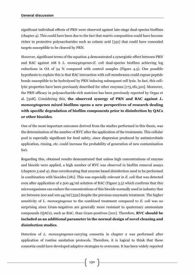
General discussion
130
significant individual effects of PRN were observed against late-stage dual-species biofilms
(chapter 4). This could have been due to the fact that matrix composition could have become
richer in protective polysaccharides such as colanic acid [331] that could have concealed
targets susceptible to be cleaved by PRN.
However, significant terms of the equation 4 demonstrated a synergistic effect between PRN
and BAC against 168 h L. monocytogenes-E. coli dual-species biofilms achieving log
reductions in OA of 54 % compared with control samples (Figure 4.5). One possible
hypothesis to explain this is that BAC interaction with cell membranes could expose peptide
bonds susceptible to be hydrolysed by PRN inducing subsequent cell lysis. In fact, this cell-
lytic properties have been previously described for other enzymes [175,181,310]. Moreover,
the PRN efficacy in polysaccharide-rich matrices has been previously reported by Orgaz et
al. [308]. Considering this, the observed synergy of PRN and BAC against L.
monocytogenes mixed biofilms opens a new perspectives of research dealing
with specific degradation of biofilm components prior to disinfection by QACs
or other biocides.
One of the most important outcomes derived from the studies performed in this thesis, was
the determination of the number of RVC after the application of the treatments. This cellular
pool is especially significant for food safety, since dispersion produced by antimicrobials
application, rinsing, etc. could increase the probability of generation of new contamination
foci.
Regarding this, obtained results demonstrated that unless high concentrations of enzyme
and biocide were applied, a high number of RVC was observed in biofilm removal assays
(chapters 3 and 4), thus corroborating that enzyme based disinfection need to be performed
in combination with biocides [181]. This was especially relevant in E. coli that was detected
even after application of a 500 µg/ml solution of BAC (Figure 3.5) which confirms that this
microorganisms can endure the concentrations of this biocide normally used in industry that
are between 200 and 100 µg/ml [332] despite the previous enzymatic treatment. The higher
sensitivity of L. monocytogenes to the combined treatment compared to E. coli was no
surprising since Gram-negatives are generally more resistant to quaternary ammonium
compounds (QACs), such as BAC, than Gram-positives [201]. Therefore, RVC should be
included as an additional parameter in the normal design of novel cleaning and
disinfection studies.
Detection of L. monocytogenes-carrying consortia in chapter 2 was performed after
application of routine sanitation protocols. Therefore, it is logical to think that these
consortia could have developed adaptive strategies to overcome. It has been widely reported
General discussion
131
that in food industry, constant exposure of bacteria to sublethal concentrations of biocides
due to either their misuse [73] or to residues left after rinsing [333,334] are among the main
causes of resistance generation. One of the main purposes for including enzymes in
hygienisation systems is that, it could improve the efficacy of disinfectants and, theoretically,
bacteria are unable to develop resistance to such molecules. The present thesis presents the
first evaluation of tolerance development in L. monocytogenes-E. coli dual-species biofilms
to PRN-BAC combined treatments. Outcomes obtained demonstrated how, in exposed
biofilms, the periods in-between sublethal exposures significantly decreases (P < 0.05) the
level of log reduction obtained after PRN-BAC treatments (Figure 5.3). These periods could
have permitted injured bacteria to repair cellular damage while synthesising structures, such
as efflux pumps, to prepare the cell for possible subsequent aggressions as reported in L.
monocytogenes [223,323,324] and E. coli [317,318]. Contrarily, in unexposed samples, an
effect of time was observed achieving lower log reduction values presumably because the
matureness, and therefore the level of resistance of the dual-species biofilms was higher.
Taken together, results obtained in chapter 5 indicate that tolerance development in L.
monocytogenes-E. coli biofilms is of a multifactorial nature. Additionally, they suggest
that the development of resistances should be incorporated to the tests
performed in investigations regarding novel sanitation systems.


Conclusions

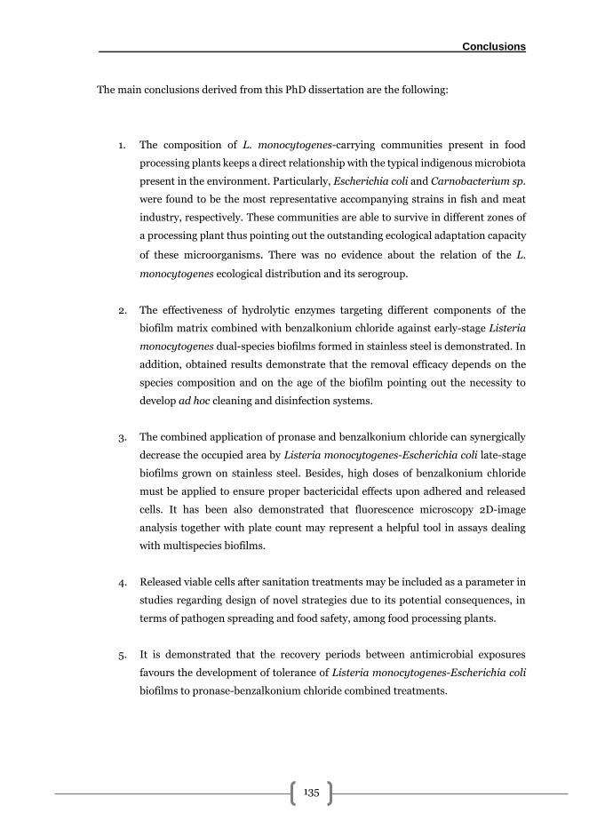
Conclusions
135
The main conclusions derived from this PhD dissertation are the following:
1. The composition of L. monocytogenes-carrying communities present in food
processing plants keeps a direct relationship with the typical indigenous microbiota
present in the environment. Particularly, Escherichia coli and Carnobacterium sp.
were found to be the most representative accompanying strains in fish and meat
industry, respectively. These communities are able to survive in different zones of
a processing plant thus pointing out the outstanding ecological adaptation capacity
of these microorganisms. There was no evidence about the relation of the L.
monocytogenes ecological distribution and its serogroup.
2. The effectiveness of hydrolytic enzymes targeting different components of the
biofilm matrix combined with benzalkonium chloride against early-stage Listeria
monocytogenes dual-species biofilms formed in stainless steel is demonstrated. In
addition, obtained results demonstrate that the removal efficacy depends on the
species composition and on the age of the biofilm pointing out the necessity to
develop ad hoc cleaning and disinfection systems.
3. The combined application of pronase and benzalkonium chloride can synergically
decrease the occupied area by Listeria monocytogenes-Escherichia coli late-stage
biofilms grown on stainless steel. Besides, high doses of benzalkonium chloride
must be applied to ensure proper bactericidal effects upon adhered and released
cells. It has been also demonstrated that fluorescence microscopy 2D-image
analysis together with plate count may represent a helpful tool in assays dealing
with multispecies biofilms.
4. Released viable cells after sanitation treatments may be included as a parameter in
studies regarding design of novel strategies due to its potential consequences, in
terms of pathogen spreading and food safety, among food processing plants.
5. It is demonstrated that the recovery periods between antimicrobial exposures
favours the development of tolerance of Listeria monocytogenes-Escherichia coli
biofilms to pronase-benzalkonium chloride combined treatments.

Conclusions
136
The experimental design regarding the combination of enzymes and benzalkonium chloride,
and its efficacy against L. monocytogenes dual-species biofilms isolated from food
processing environments detailed in this PhD dissertation, open new perspectives in the
design of novel sanitation strategies for pathogen control in the food industry.

Reference list

References
139
1. McLauchlin J, Rees CED, Dodd CER. Listeria monocytogenes and the genus Listeria.
The Prokaryotes: Firmicutes and Tenericutes. 2014. pp. 241–259. doi:10.1007/978-
3-642-30120-9
2. Weller D, Andrus A, Wiedmann M, den Bakker HC. Listeria booriae sp. nov. and
Listeria newyorkensis sp. nov., from food processing environments in the USA. Int
J Syst Evol Microbiol. 2015;65: 286–292. doi:10.1099/ijs.0.070839-0
3. Orsi RH, Wiedmann M. Characteristics and distribution of Listeria spp., including
Listeria species newly described since 2009. Appl Microbiol Biotechnol. Applied
Microbiology and Biotechnology; 2016;100: 5273–5287. doi:10.1007/s00253-016-
7552-2
4. den Bakker HC, Warchocki S, Wright EM, Allred AF, Ahlstrom C, Manuel CS, et al.
Listeria floridensis sp. nov., Listeria aquatica sp. nov., Listeria cornellensis sp. nov.,
Listeria riparia sp. nov. and Listeria grandensis sp. nov., from agricultural and
natural environments. Int J Syst Evol Microbiol. 2014;64: 1882–1889.
doi:10.1099/ijs.0.052720-0
5. Murray EGD, Webb RA, Swann HBR. A disease of rabbits characterized by a large
mononuclear leucocytosis caused by a hitherto undescribed bacillus Bacterium
monocytogenes (n.sp.). J Pathol Bacteriol. 1926;29: 407–439.
doi:10.1002/path.1700290409
6. Pirie JHH. Listeria: Change of Name for a Genus Bacteria. Nature. 1940;145: 264.
doi:10.1038/145264a0
7. Seeliger HPR, Jones D. Genus Listeria. In: Sneath PHA, Mair NS, Sharpe ME, Holt
JG, editors. Bergey’s manual of systematic bacteriology, vol 2. The Williams and
Wilkins Co.; 1986. pp. 1235–1245.
8. Paterson JS. The antigenic structure of organisms of the genus Listerella. J Pathol
Bacteriol. 1940;51: 427–436.
9. Seeliger HPR, Höhne K. Chapter II Serotyping of Listeria monocytogenes and
Related Species. Methods Microbiol. 1979;13: 31–49. doi:10.1016/S0580-
9517(08)70372-6
10. Liu D. Identification, subtyping and virulence determination of Listeria
monocytogenes, an important foodborne pathogen. Journal of Medical
Microbiology. 2006. pp. 645–659. doi:10.1099/jmm.0.46495-0
References
140
11. Doumith M, Buchrieser C, Glaser P, Jacquet C, Martin P. Differentiation of the major
Listeria monocytogenes serovars by multiplex PCR. J Clin Microbiol. 2004;42:
3819–22. doi:10.1128/JCM.42.8.3819-3822.2004
12. Wiedmann M, Bruce JL, Knorr R, Bodis M, Cole EM, McDowell CI, et al. Ribotype
diversity of Listeria monocytogenes strains associated with outbreaks of listeriosis
in ruminants. J Clin Microbiol. American Society for Microbiology (ASM); 1996;34:
1086–1090.
13. Jacquet C, Gouin E, Jeannel D, Cossart P, Rocourt J. Expression of ActA, Ami, InlB,
and Listeriolysin O in Listeria monocytogenes of human and food origin. Appl
Environ Microbiol. American Society for Microbiology (ASM); 2002;68: 616–622.
doi:10.1128/AEM.68.2.616-622.2002
14. Wiedmann M, Bruce JL, Keating C, Johnson AE, McDonough PL, Batt CA. Ribotypes
and virulence gene polymorphisms suggest three distinct Listeria monocytogenes
lineages with differences in pathogenic potential. Infect Immun. American Society
for Microbiology; 1997;65: 2707–2716.
15. Nadon CA, Woodward DL, Young C, Rodgers FG, Wiedmann M. Correlations
between molecular subtyping and serotyping of Listeria monocytogenes. J Clin
Microbiol. 2001;39: 2704–2707. doi:10.1128/JCM.39.7.2704-2707.2001
16. Van Der Veen S, Moezelaar R, Abee T, Wells-Bennik MHJ. The growth limits of a
large number of Listeria monocytogenes strains at combinations of stresses show
serotype- and niche-specific traits. J Appl Microbiol. Blackwell Publishing Ltd;
2008;105: 1246–1258. doi:10.1111/j.1365-2672.2008.03873.x
17. Blackman IC, Frank JF. Growth of Listeria monocytogenes as a Biofilm on Various
food-processing surfaces. J Food Prot. 1996;59: 827–831.
18. Gandhi M, Chikindas ML. Listeria: A foodborne pathogen that knows how to survive.
Int J Food Microbiol. 2007;113: 1–15. doi:10.1016/j.ijfoodmicro.2006.07.008
19. Moltz AG, Martin SE. Formation of biofilms by Listeria monocytogenes under
various growth conditions. J Food Prot. 2005;68: 92–97.
20. Vivant A-L, Garmyn D, Piveteau P. Listeria monocytogenes, a down-to-earth
pathogen. Front Cell Infect Microbiol. Frontiers Media SA; 2013;3: 87.
doi:10.3389/fcimb.2013.00087
References
141
21. Dowe M, Jackson E, Mori J, Colin B. Listeria monocytogenes Survival in Soil and
Incidence in Agricultural Soils. J Food Prot. International Association for Food
Protection; 1997;10: 1158–1286.
22. Linke K, Rückerl I, Brugger K, Karpiskova R, Walland J, Muri-Klinger S, et al.
Reservoirs of Listeria species in three environmental ecosystems. Appl Environ
Microbiol. American Society for Microbiology; 2014;80: 5583–5592.
doi:10.1128/AEM.01018-14
23. Locatelli A, Spor A, Jolivet C, Piveteau P, Hartmann A. Biotic and Abiotic Soil
Properties Influence Survival of Listeria monocytogenes in Soil. Freitag NE, editor.
PLoS One. Public Library of Science; 2013;8: e75969.
doi:10.1371/journal.pone.0075969
24. Orsi RH, den Bakker HC, Wiedmann M. Listeria monocytogenes lineages:
Genomics, evolution, ecology, and phenotypic characteristics. Int J Med Microbiol.
Elsevier GmbH.; 2011;301: 79–96. doi:10.1016/j.ijmm.2010.05.002
25. Lomonaco S, Nucera D, Filipello V. The evolution and epidemiology of Listeria
monocytogenes in Europe and the United States. Infect Genet Evol. 2015;35: 172–
183. doi:10.1016/j.meegid.2015.08.008
26. Söderqvist K, Thisted Lambertz S, Vågsholm I, Boqvist S. Foodborne Bacterial
Pathogens in Retail Prepacked Ready-to-Eat Mixed Ingredient Salads. J Food Prot.
2016;79: 978–985. doi:10.4315/0362-028X.JFP-15-515
27. Gombas DE, Chen Y, Clavero RS, Scott VN. Survey of Listeria monocytogenes in
ready-to-eat foods. J Food Prot. 2003;66: 559–69.
28. Pagadala S, Parveen S, Rippen T, Luchansky JB, Call JE, Tamplin ML, et al.
Prevalence, characterization and sources of Listeria monocytogenes in blue crab
(Callinectus sapidus) meat and blue crab processing plants. Food Microbiol. Elsevier
Ltd; 2012;31: 263–70. doi:10.1016/j.fm.2012.03.015
29. Goh SG, Leili A-H, Kuan CH, Loo YY, Lye YL, Chang WS, et al. Transmission of
Listeria monocytogenes from raw chicken meat to cooked chicken meat through
cutting boards. Food Control. Elsevier Ltd; 2014;37: 51–55.
doi:10.1016/j.foodcont.2013.08.030
30. Chasseignaux E, Toquin M-T, Ragimbeau C, Salvat G, Colin P, Ermel G. Molecular
epidemiology of Listeria monocytogenes isolates collected from the environment,
References
142
raw meat and raw products in two poultry- and pork-processing plants. J Appl
Microbiol. 2001;91: 888–899. doi:10.1046/j.1365-2672.2001.01445.x
31. Thévenot D, Dernburg a, Vernozy-Rozand C. An updated review of Listeria
monocytogenes in the pork meat industry and its products. J Appl Microbiol.
2006;101: 7–17. doi:10.1111/j.1365-2672.2006.02962.x
32. Ajayeoba TA, Atanda OO, Obadina AO, Bankole MO, Adelowo OO. The incidence
and distribution of Listeria monocytogenes in ready-to-eat vegetables in South-
Western Nigeria. Food Sci Nutr. 2016;4: 59–66. doi:10.1002/fsn3.263
33. Oliveira M, Viñas I, Colàs P, Anguera M, Usall J, Abadias M. Effectiveness of a
bacteriophage in reducing Listeria monocytogenes on fresh-cut fruits and fruit
juices. Food Microbiol. Elsevier Ltd; 2014;38: 137–42.
doi:10.1016/j.fm.2013.08.018
34. Viswanath P, Murugesan L, Knabel SJ, Verghese B, Chikthimmah N, Laborde LF.
Incidence of Listeria monocytogenes and Listeria spp. in a small-scale mushroom
production facility. J Food Prot. 2013;76: 608–15. doi:10.4315/0362-028X.JFP-12-
292
35. Carpentier B, Cerf O. Review - Persistence of Listeria monocytogenes in food
industry equipment and premises. Int J Food Microbiol. Elsevier B.V.; 2011;145: 1–
8. doi:10.1016/j.ijfoodmicro.2011.01.005
36. Ferreira V, Wiedmann M, Teixeira P, Stasiewicz MJ. Listeria monocytogenes
persistence in food-associated environments: epidemiology, strain characteristics,
and implications for public health. J Food Prot. 2014;77: 150–70. doi:10.4315/0362-
028X.JFP-13-150
37. Rodríguez-López P, Saá-Ibusquiza P, Mosquera-Fernández M, López-Cabo M.
Listeria monocytogenes – carrying consortia in food industry. Composition,
subtyping and numerical characterisation of mono-species biofilm dynamics on
stainless steel. Int J Food Microbiol. Elsevier B.V.; 2015;206: 84–95.
doi:10.1016/j.ijfoodmicro.2015.05.003
38. Scallan E, Hoekstra RM, Angulo FJ, Tauxe R V., Widdowson MA, Roy SL, et al.
Foodborne illness acquired in the United States-Major pathogens. Emerg Infect Dis.
2011;17: 7–15. doi:10.3201/eid1701.P11101
39. EFSA. The European Union summary report on trends and sources of zoonoses,
References
143
zoonotic agents and food-borne outbreaks in 2014. EFSA J. 2015;13: 4329.
doi:10.2903/j.efsa.2015.4329
40. Vázquez-Boland JA, Kuhn M, Berche P, Chakraborty T, Domi G, González-zorn B, et
al. Listeria Pathogenesis and Molecular Virulence Determinants Listeria
Pathogenesis and Molecular Virulence Determinants. Clin Microbiol Rev. 2001;14:
584–640. doi:10.1128/CMR.14.3.584
41. Vázquez-Boland JA, Domínguez-Bernal G, González-Zorn B, Kreft J, Goebel W.
Pathogenicity islands and virulence evolution in Listeria. Microbes Infect. 2001;3:
571–584. doi:10.1016/S1286-4579(01)01413-7
42. Brouwer MC, van de Beek D, Heckenberg SGB, Spanjaard L, de Gans J. Community-
acquired Listeria monocytogenes meningitis in adults. Clin Infect Dis. Oxford
University Press; 2006;43: 1233–8. doi:10.1086/508462
43. Farber JM, Peterkin PI. Listeria monocytogenes, a food-borne pathogen. Microbiol
Rev. 1991;55: 476–511. doi:10.1104/pp.110.161547
44. Simões M, Simões LC, Vieira MJ. A review of current and emergent biofilm control
strategies. LWT - Food Sci Technol. 2010;43: 573–583.
doi:10.1016/j.lwt.2009.12.008
45. Bille J, Blanc DS, Schmid H, Boubaker K, Baumgartner A, Siegrist HH, et al.
Outbreak of human listeriosis associated with tomme cheese in northwest
Switzerland, 2005. Euro Surveill. 2006;11: 91–93.
46. Vít M, Olejník R, Karpíšková R, Částková J, Príkazský V, Príkazská M, et al. Outbreak
of listeriosis in the Czech Republic, late 2006 – preliminary report. Euro Surveill.
2007;12: pii=3132.
47. Koch J, Dworak R, Prager R, Becker B, Brockmann S, Wicke A, et al. Large listeriosis
outbreak linked to cheese made from pasteurized milk, Germany, 2006-2007.
Foodborne Pathog Dis. Mary Ann Liebert, Inc. 140 Huguenot Street, 3rd Floor New
Rochelle, NY 10801 USA; 2010;7: 1581–1584. doi:10.1089/fpd.2010.0631
48. CDC. Outbreak of Listeria monocytogenes Infections Associated with Pasteurized
Milk from a Local Dairy --- Massachusetts, 2007. Morb Mortal Wkly Rep. 2008;57:
1097–1100.
49. Johnsen BO, Lingaas E, Torfoss D, Strøm EH, Nordøy I. A large outbreak of Listeria
References
144
monocytogenes infection with short incubation period in a tertiary care hospital. J
Infect. 2010;61: 465–470. doi:10.1016/j.jinf.2010.08.007
50. Canada G of. Report of the Independent Investigator into the 2008 Listeriosis
Outbreak. 2009. pp. 1–95. doi:10.1017/CBO9781107415324.004
51. Fretz R, Sagel U, Ruppitsch W, Pietzka A, Stoger A, Huhulescu S, et al. Listeriosis
outbreak caused by acid curd cheese Quargel , Austria and Germany 2009. Euro
Surveill. 2010;15.
52. Magalhães R, Almeida G, Ferreira V, Santos I, Silva J, Mendes MM, et al. Cheese-
related listeriosis outbreak, Portugal, March 2009 to February 2012. Euro Surveill.
2015;20: pii=21104. doi:10.2807/1560-7917.ES2015.20.17.21104
53. CDC. Multistate outbreak of listeriosis associated with Jensen Farms Cantaloupe --
- United States, August--September 2011. Morbidity and Mortality Weekly Report.
2011. pp. 1357–1358.
54. CDC. Listeria outbreaks [Internet]. [cited 8 Nov 2016]. Available:
https://www.cdc.gov/listeria/outbreaks/
55. Ruppitsch W, Prager R, Halbedel S, Hyden P, Pietzka A, Huhulescu S, et al. Ongoing
outbreak of invasive listeriosis, Germany, 2012 to 2015. Euro Surveill. 2015;20:
pii=30094. doi:10.2807/1560-7917.ES.2015.20.50.30094
56. Pérez-Trallero E, Zigorraga C, Artieda J, Alkorta M, Marimón JM. Two outbreaks of
listeria monocytogenes infection, Northern Spain. Emerg Infect Dis. 2014;20: 2155–
2157. doi:10.3201/eid2012.140993
57. Anonymous. Listeria. In: Wingstrand A, Sorensen A, Helwigh B, editors. Annual
Report on Zoonoses in Denmark 2014. National Food Institute, Technical University
of Denmark; 2015. pp. 14–17. doi:10.1128/AEM.02991-09
58. Swaminathan B, Gerner-Smidt P. The epidemiology of human listeriosis. Microbes
and Infection. 2007. pp. 1236–1243. doi:10.1016/j.micinf.2007.05.011
59. Schlech IWF, Lavigne PM, Bortolussi RA. Epidemic listeriosis - Evidence for
transmission by food. N Engl J Med. Massachusetts Medical Society; 1983;308:
203–206. doi:10.1056/NEJM198301273080407
60. Gilbert RJ, McLauchlin J, Velani SK. The contamination of paté by Listeria
monocytogenes in England and Wales in 1989 and 1990. Epidemiol Infect.
References
145
Cambridge University Press; 1993;110: 543–51. doi:10.1017/S0950268800050962
61. Salamina G, Dalle Donne E, Niccolini A, Poda G, Cesaroni D, Bucci M, et al. A
foodborne outbreak of gastroenteritis involving Listeria monocytogenes. Epidemiol
Infect. 1996;117: 429–436. doi:10.1017/S0950268800059082
62. Dalton CB, Austin CC, Sobel J, Hayes PS, Bibb WF, Graves LM, et al. An outbreak of
gastroenteritis and fever due to Listeria monocytogenes in milk. N Engl J Med.
Massachusetts Medical Society; 1997;336: 100–5.
doi:10.1056/NEJM199701093360204
63. Dinu LD, Delaquis P, Bach S. Nonculturable response of animal enteropathogens in
the agricultural environment and implications for food safety. Journal of Food
Protection. 2009. pp. 1342–1354.
64. Dreux N, Albagnac C, Federighi M, Carlin F, Morris CE, Nguyen-the C. Viable but
non-culturable Listeria monocytogenes on parsley leaves and absence of recovery to
a culturable state. J Appl Microbiol. 2007;103: 1272–81. doi:10.1111/j.1365-
2672.2007.03351.x
65. Campdepadrós M, Stchigel AM, Romeu M, Quilez J, Solà R. Effectiveness of two
sanitation procedures for decreasing the microbial contamination levels (including
Listeria monocytogenes) on food contact and non-food contact surfaces in a dessert-
processing factory. Food Control. Elsevier Ltd; 2012;23: 26–31.
doi:10.1016/j.foodcont.2011.05.017
66. Chavant P, Gaillard-Martinie B, Hébraud M. Antimicrobial effects of sanitizers
against planktonic and sessile Listeria monocytogenes cells according to the growth
phase. FEMS Microbiol Lett. 2004;236: 241–8. doi:10.1016/j.femsle.2004.05.040
67. Saá Ibusquiza P, Herrera JJR, Cabo ML. Resistance to benzalkonium chloride,
peracetic acid and nisin during formation of mature biofilms by Listeria
monocytogenes. Food Microbiol. 2011;28: 418–25. doi:10.1016/j.fm.2010.09.014
68. Pan Y, Breidt F, Kathariou S. Resistance of Listeria monocytogenes biofilms to
sanitizing agents in a simulated food processing environment. Appl Environ
Microbiol. 2006;72: 7711–7. doi:10.1128/AEM.01065-06
69. Mack WN, Mack JP, Ackerson AO. Microbial film development in a trickling filter.
Microb Ecol. 1975;2: 215–226. doi:10.1007/BF02010441
References
146
70. Donlan RM, Costerton JW. Biofilms: Survival mechanisms of clinically relevant
microorganisms. Clinical Microbiology Reviews. American Society for Microbiology;
2002. pp. 167–193. doi:10.1128/CMR.15.2.167-193.2002
71. Costerton JW, Lewandowski Z, Caldwell DE, Korber DR, Lappin-Scott HM.
Microbial biofilms. Annual Review of Microbiology. 1995. pp. 711–745.
72. Winkelströter LK, Teixeira FB dos R, Silva EP, Alves VF, De Martinis ECP.
Unraveling Microbial Biofilms of Importance for Food Microbiology. Microb Ecol.
Springer New York LLC; 2013;68: 35–46. doi:10.1007/s00248-013-0347-4
73. Abdallah M, Benoliel C, Drider D, Dhulster P, Chihib NE. Biofilm formation and
persistence on abiotic surfaces in the context of food and medical environments.
Arch Microbiol. 2014;196: 453–472. doi:10.1007/s00203-014-0983-1
74. Vlamakis H. The world of biofilms. Virulence. 2011;2: 431–434.
doi:10.4161/viru.2.5.17902
75. Watnick P, Kolter R. Biofilm, city of microbes. Journal of Bacteriology. American
Society for Microbiology; 2000. pp. 2675–2679. doi:10.1128/JB.182.10.2675-
2679.2000
76. Stewart PS, Franklin MJ. Physiological heterogeneity in biofilms. Nat Rev Microbiol.
Nature Publishing Group; 2008;6: 199–210. doi:10.1038/nrmicro1838
77. Bjarnsholt T, Alhede M, Alhede M, Eickhardt-Sørensen SR, Moser C, Kühl M, et al.
The in vivo biofilm. Trends Microbiol. 2013;21: 466–74.
doi:10.1016/j.tim.2013.06.002
78. Hall-Stoodley L, Costerton J, Stoodley P. Bacterial biofilms: from the natural
environment to infectious diseases. Nat Rev Microbiol. 2004;2: 95–108.
doi:10.1038/nrmicro821
79. Petrova OE, Sauer K. Escaping the biofilm in more than one way: Desorption,
detachment or dispersion. Current Opinion in Microbiology. 2016. pp. 67–78.
doi:10.1016/j.mib.2016.01.004
80. Rutherford ST, Bassler BL. Bacterial quorum sensing: its role in virulence and
possibilities for its control. Cold Spring Harb Perspect Med. 2012;2: 1–25.
doi:10.1101/cshperspect.a012427
81. Nadell CD, Xavier JB, Levin SA, Foster KR. The evolution of quorum sensing in
References
147
bacterial biofilms. Moran NA, editor. PLoS Biol. Public Library of Science; 2008;6:
e14. doi:10.1371/journal.pbio.0060014
82. Xavier KB, Bassler BL. Interference with AI-2-mediated bacterial cell-cell
communication. Nature. Nature Publishing Group; 2005;437: 750–3.
doi:10.1038/nature03960
83. Garmyn D, Gal L, Lemaitre JP, Hartmann A, Piveteau P. Communication and
autoinduction in the species Listeria monocytogenes: A central role for the agr
system. Communicative and Integrative Biology. 2009. pp. 371–374.
doi:10.4161/cib.2.4.8610
84. Van Der Veen S, Abee T. Importance of SigB for listeria monocytogenes static and
continuous-flow biofilm formation and disinfectant resistance. Appl Environ
Microbiol. 2010;76: 7854–7860. doi:10.1128/AEM.01519-10
85. Dickson JS, Koohmaraie M, Hruska RL. Cell Surface Charge Characteristics and
Their Relationship to Bacterial Attachment to Meat Surfaces. Appl Environ
Microbiol. 1989;55: 832–836.
86. Palmer J, Flint S, Brooks J. Bacterial cell attachment, the beginning of a biofilm. J
Ind Microbiol Biotechnol. 2007;34: 577–588. doi:10.1007/s10295-007-0234-4
87. Davies DG. Physiological events in biofilm formation. In: Allison D, Gilbert P,
Lappin-Scott HM, Wilson M, editors. Community structure and co-operation in
biofilms. 2000. pp. 37–51.
88. Dunne WM. Bacterial adhesion: Seen any good biofilms lately? Clin Microbiol Rev.
American Society for Microbiology; 2002;15: 155–166. doi:10.1128/CMR.15.2.155-
166.2002
89. Schwab U, Hu Y, Wiedmann M, Boor KJ. Alternative Sigma Factor σB Listeria
monocytogenes Is Not Essential for Surface Attachment. J Food Prot. 2005;68: 311–
317.
90. Herald PJ, Zottola EA. Attachment of Listeria monocytogenes to Stainless Steel
Surfaces at Various Temperatures and pH Values. J Food Sci. 1988;53: 1549–1562.
doi:10.1111/j.1365-2621.1988.tb09321.x
91. Poimenidou S, Belessi CA, Giaouris ED, Gounadaki AS, Nychas GJE, Skandamis PN.
Listeria monocytogenes attachment to and detachment from stainless steel surfaces
References
148
in a simulated dairy processing environment. Appl Environ Microbiol. 2009;75:
7182–7188. doi:10.1128/AEM.01359-09
92. Briandet R, Meylheuc T, Maher C, Bellon-Fontaine MN. Listeria monocytogenes
Scott A: cell surface charge, hydrophobicity, and electron donor and acceptor
characteristics under different environmental growth conditions. Appl Environ
Microbiol. 1999;65: 5328–5333.
93. Peel M, Donachie W, Shaw A. Temperature-dependent expression of flagella of
Listeria monocytogenes studied by electron microscopy, SDS-PAGE and western
blotting. J Gen Microbiol. 1988;134: 2171–2178. doi:10.1099/00221287-134-8-2171
94. Mai TL, Conner DE. Effect of temperature and growth media on the attachment of
Listeria monocytogenes to stainless steel. Int J Food Microbiol. 2007;120: 282–286.
doi:10.1016/j.ijfoodmicro.2007.09.006
95. Camper AK, Hayes JT, Sturman PJ, Jones WL, Cunningham AB. Effects of motility
and adsorption rate coefficient on transport of bacteria through saturated porous
media. Appl Environ Microbiol. 1993;59: 3455–3462.
96. Kim KY, Frank JF. Effect of Nutrients on Biofilm Formation by Listeria
monocytogenes on Stainless Steel. J Food Prot. 1995;58: 24–28.
97. Guilbaud M, Piveteau P, Desvaux M, Brisse S, Briandet R. Exploring the Diversity of
Listeria monocytogenes Biofilm Architecture by High-Throughput Confocal Laser
Scanning Microscopy and the Predominance of the Honeycomb-Like Morphotype.
Appl Environ Microbiol. 2015;81: 1813–1819. doi:10.1128/AEM.03173-14
98. Jaradat ZW, Bhunia AK. Glucose and nutrient concentrations affect the expression
of a 104-kilodalton Listeria adhesion protein in Listeria monocytogenes. Appl
Environ Microbiol. American Society for Microbiology; 2002;68: 4876–83.
doi:10.1128/AEM.68.10.4876-4883.2002
99. Kyoui D, Hirokawa E, Takahashi H, Kuda T, Kimura B. Effect of glucose on Listeria
monocytogenes biofilm formation, and assessment of the biofilm’s sanitation
tolerance. Biofouling. Taylor & Francis; 2016;32: 815–826.
doi:10.1080/08927014.2016.1198953
100. Borucki MK, Peppin JD, White D, Loge F, Call DR. Variation in biofilm formation
among strains of Listeria monocytogenes. Appl Environ Microbiol. 2003;69: 7336–
7342. doi:10.1128/AEM.69.12.7336
References
149
101. Mosquera-Fernández M, Rodríguez-López P, Cabo ML, Balsa-Canto E. Numerical
spatio-temporal characterization of Listeria monocytogenes biofilms. Int J Food
Microbiol. Elsevier B.V.; 2014;182–183: 26–36.
doi:10.1016/j.ijfoodmicro.2014.05.005
102. Saá P, Cabo ML, Rodríguez JJ. Effects of mussel processing soils on the adherence
of Listeria monocytogenes to polypropylene and stainless steel. J Food Prot.
2009;72: 1885–90.
103. Silva S, Teixeira P, Oliveira R, Azeredo J. Adhesion to and viability of Listeria
monocytogenes on food contact surfaces. J Food Prot. 2008;71: 1379–1385.
104. Lemon KP, Higgins DE, Kolter R. Flagellar motility is critical for Listeria
monocytogenes biofilm formation. J Bacteriol. American Society for Microbiology;
2007;189: 4418–4424. doi:10.1128/JB.01967-06
105. Guerrieri E, de Niederhäusern S, Messi P, Sabia C, Iseppi R, Anacarso I, et al. Use of
lactic acid bacteria (LAB) biofilms for the control of Listeria monocytogenes in a
small-scale model. Food Control. 2009;20: 861–865.
doi:10.1016/j.foodcont.2008.11.001
106. Tresse O, Lebret V, Benezech T, Faille C. Comparative evaluation of adhesion,
surface properties, and surface protein composition of Listeria monocytogenes
strains after cultivation at constant pH of 5 and 7. J Appl Microbiol. Blackwell
Publishing Ltd; 2006;101: 53–62. doi:10.1111/j.1365-2672.2006.02968.x
107. Vatanyoopaisarn S, Nazli A, Dodd CER, Rees CED, Waites WM. Effect of flagella on
initial attachment of Listeria monocytogenes to stainless steel. Appl Environ
Microbiol. 2000;66: 860–863. doi:10.1128/AEM.66.2.860-863.2000
108. Todhanakasem T, Young GM. Loss of flagellum-based motility by Listeria
monocytogenes results in formation of hyperbiofilms. J Bacteriol. 2008;190: 6030–
6034. doi:10.1128/JB.00155-08
109. Takhistov P, George B. Linearized kinetic model of Listeria monocytogenes biofilm
growth. Bioprocess Biosyst Eng. 2004;26: 259–270. doi:10.1007/s00449-004-
0357-1
110. Flemming HC, Neu TR, Wozniak DJ. The EPS matrix: The “House of Biofilm Cells.”
J Bacteriol. 2007;189: 7945–7947. doi:10.1128/JB.00858-07
References
150
111. Stewart PS. Diffusion in biofilms. Journal of Bacteriology. American Society for
Microbiology; 2003. pp. 1485–1491. doi:10.1128/JB.185.5.1485-1491.2003
112. Stepanović S, Ćirković I, Ranin L, Švabić-Vlahović M. Biofilm formation by
Salmonella spp. and Listeria monocytogenes on plastic surface. Lett Appl Microbiol.
2004;38: 428–432. doi:10.1111/j.1472-765X.2004.01513.x
113. Gilbert P, Allison DG, McBain AJ. Biofilms in vitro and in vivo: do singular
mechanisms imply cross-resistance? J Appl Microbiol. Blackwell Science Ltd.;
2002;92: 98S–110S. doi:10.1046/j.1365-2672.92.5s1.5.x
114. Chen LH, Köseoğlu VK, Güvener ZT, Myers-Morales T, Reed JM, D’Orazio SEF, et
al. Cyclic di-GMP-dependent Signaling Pathways in the Pathogenic Firmicute
Listeria monocytogenes. PLoS Pathog. 2014;10: e1004301.
doi:10.1371/journal.ppat.1004301
115. Köseoğlu VK, Heiss C, Azadi P, Topchiy E, Güvener ZT, Lehmann TE, et al. Listeria
monocytogenes exopolysaccharide: Origin, structure, biosynthetic machinery and c-
di-GMP-dependent regulation. Mol Microbiol. 2015;96: 728–743.
doi:10.1111/mmi.12966
116. Combrouse T, Sadovskaya I, Faille C, Kol O, Guérardel Y, Midelet-Bourdin G.
Quantification of the extracellular matrix of the Listeria monocytogenes biofilms of
different phylogenic lineages with optimization of culture conditions. J Appl
Microbiol. 2013;114: 1120–31. doi:10.1111/jam.12127
117. Nguyen UT, Burrows LL. DNase I and proteinase K impair Listeria monocytogenes
biofilm formation and induce dispersal of pre-existing biofilms. Int J Food
Microbiol. Elsevier B.V.; 2014;187: 26–32. doi:10.1016/j.ijfoodmicro.2014.06.025
118. Harmsen M, Lappann M, Knøchel S, Molin S. Role of extracellular DNA during
biofilm formation by Listeria monocytogenes. Appl Environ Microbiol. 2010;76:
2271–9. doi:10.1128/AEM.02361-09
119. Brauge T, Sadovskaya I, Faille C, Benezech T, Maes E, Guerardel Y, et al. Teichoic
acid is the major polysaccharide present in the Listeria monocytogenes biofilm
matrix. FEMS Microbiol Lett. The Oxford University Press; 2016;363: fnv229.
doi:10.1093/femsle/fnv229
120. Flemming HC, Wingender J. The biofilm matrix. Nat Rev Microbiol. Nature
Publishing Group; 2010;8: 623–33. doi:10.1038/nrmicro2415
References
151
121. Neu TR, Lawrence JR. Lectin-binding analysis in biofilm systems. Methods
Enzymol. 1999;310: 145–152. doi:10.1016/S0076-6879(99)10012-0
122. Petrova OE, Sauer K. Sticky situations: key components that control bacterial
surface attachment. J Bacteriol. 2012;194: 2413–25. doi:10.1128/JB.00003-12
123. Kaplan JB. Biofilm dispersal: mechanisms, clinical implications, and potential
therapeutic uses. J Dent Res. 2010;89: 205–218. doi:10.1177/0022034509359403
124. Rice SA, Koh KS, Queck SY, Labbate M, Lam KW, Kjelleberg S. Biofilm formation
and sloughing in Serratia marcescens are controlled by quorum sensing and nutrient
cues. J Bacteriol. American Society for Microbiology; 2005;187: 3477–3485.
doi:10.1128/JB.187.10.3477-3485.2005
125. Boles BR, Thoendel M, Singh PK. Rhamnolipids mediate detachment of
Pseudomonas aeruginosa from biofilms. Mol Microbiol. Blackwell Science Ltd;
2005;57: 1210–1223. doi:10.1111/j.1365-2958.2005.04743.x
126. Liu D, Lawrence ML, Ainsworth AJ, Austin FW. Toward an improved laboratory
definition of Listeria monocytogenes virulence. International Journal of Food
Microbiology. 2007. pp. 101–115. doi:10.1016/j.ijfoodmicro.2007.07.045
127. Davey ME, O’toole GA. Microbial biofilms: from ecology to molecular genetics.
Microbiol Mol Biol Rev. 2000;64: 847–67. doi:10.1128/MMBR.64.4.847-867.2000
128. Kalmokoff ML, Austin JW, Wan XD, Sanders G, Banerjee S, Farber JM. Adsorption,
attachment and biofilm formation among isolates of listeria monocytogenes using
model conditions. Journal of Applied Microbiology. Blackwell Science Ltd; 2001. pp.
725–734. doi:10.1046/j.1365-2672.2001.01419.x
129. Carpentier B, Chassaing D. Interactions in biofilms between Listeria monocytogenes
and resident microorganisms from food industry premises. Int J Food Microbiol.
2004;97: 111–22. doi:10.1016/j.ijfoodmicro.2004.03.031
130. Daneshvar Alavi HE, Truelstrup Hansen L. Kinetics of biofilm formation and
desiccation survival of Listeria monocytogenes in single and dual species biofilms
with Pseudomonas fluorescens, Serratia proteamaculans or Shewanella baltica on
food-grade stainless steel surfaces. Biofouling. 2013;29: 1253–68.
doi:10.1080/08927014.2013.835805
131. Rieu A, Briandet R, Habimana O, Garmyn D, Guzzo J, Piveteau P. Listeria
References
152
monocytogenes EGD-e biofilms: no mushrooms but a network of knitted chains.
Appl Environ Microbiol. 2008;74: 4491–7. doi:10.1128/AEM.00255-08
132. van der Veen S, Abee T. Mixed species biofilms of Listeria monocytogenes and
Lactobacillus plantarum show enhanced resistance to benzalkonium chloride and
peracetic acid. Int J Food Microbiol. Elsevier B.V.; 2011;144: 421–31.
doi:10.1016/j.ijfoodmicro.2010.10.029
133. Leriche V, Carpentier B. Limitation of adhesion and growth of Listeria
monocytogenes on stainless steel surfaces by Staphylococcus sciuri biofilms. J Appl
Microbiol. 2000;88: 594–605. doi:10.1046/j.1365-2672.2000.01000.x
134. Giaouris E, Chorianopoulos N, Doulgeraki A, Nychas G-J. Co-culture with Listeria
monocytogenes within a dual-species biofilm community strongly increases
resistance of Pseudomonas putida to benzalkonium chloride. PLoS One. 2013;8:
e77276. doi:10.1371/journal.pone.0077276
135. Kolenbrander PE. Oral microbial communities: biofilms, interactions, and genetic
systems. Annual review of microbiology. 2000. pp. 413–437.
doi:10.1146/annurev.micro.54.1.413
136. Benítez-Páez A, Belda-Ferre P, Simón-Soro A, Mira A. Microbiota diversity and gene
expression dynamics in human oral biofilms. BMC Genomics. 2014;15: 311.
doi:10.1186/1471-2164-15-311
137. Hall-Stoodley L, Stoodley P, Kathju S, Høiby N, Moser C, William Costerton J, et al.
Towards diagnostic guidelines for biofilm-associated infections. FEMS Immunol
Med Microbiol. 2012;65: 127–145. doi:10.1111/j.1574-695X.2012.00968.x
138. Yang L, Liu Y, Wu H, Song Z, H??iby N, Molin S, et al. Combating biofilms. FEMS
Immunol Med Microbiol. 2012;65: 146–157. doi:10.1111/j.1574-695X.2011.00858.x
139. Yang L, Liu Y, Wu H, Hóiby N, Molin S, Song Z. Current understanding of multi-
species biofilms. Int J Oral Sci. 2011;3: 74–81. doi:10.4248/IJOS11027
140. Chasseignaux E, Gérault P, Toquin MT, Salvat G, Colin P, Ermel G. Ecology of
Listeria monocytogenes in the environment of raw poultry meat and raw pork meat
processing plants. FEMS Microbiol Lett. 2002;210: 271–275. doi:10.1016/S0378-
1097(02)00637-7
141. Bridier A, Sanchez-Vizuete P, Guilbaud M, Piard JC, Naïtali M, Briandet R. Biofilm-
References
153
associated persistence of food-borne pathogens. Food Microbiol. Elsevier Ltd;
2015;45: 167–178. doi:10.1016/j.fm.2014.04.015
142. Giaouris E, Heir E, Desvaux M, Hébraud M, Møretrø T, Langsrud S, et al. Intra- and
inter-species interactions within biofilms of important foodborne bacterial
pathogens. Front Microbiol. 2015;6: 1–26. doi:10.3389/fmicb.2015.00841
143. Røder HL, Raghupathi PK, Herschend J, Brejnrod A, Knøchel S, Sørensen SJ, et al.
Interspecies interactions result in enhanced biofilm formation by co-cultures of
bacteria isolated from a food processing environment. Food Microbiol. Elsevier Ltd;
2015;51: 18–24. doi:10.1016/j.fm.2015.04.008
144. Puga CH, SanJose C, Orgaz B. Spatial distribution of Listeria monocytogenes and
Pseudomonas fluorescens in mixed biofilms. In: Hambrick EC, editor. Listeria
monocytogenes: Food Sources, Prevalence and Management Strategies. New York,
NY: Nova Publishers; 2014. pp. 115–132.
145. Rodríguez-López P, Carballo-Justo A, Draper LA, Cabo ML. Removal of Listeria
monocytogenes dual-species biofilms using enzyme-benzalkonium chloride
combined treatments. Biofouling. 2017;33: 45–58.
doi:10.1080/08927014.2016.1261847
146. Almeida C, Azevedo NF, Santos S, Keevil CW, Vieira MJ. Discriminating multi-
species populations in biofilms with peptide nucleic acid fluorescence in situ
hybridization (PNA FISH). PLoS One. 2011;6: e14786.
doi:10.1371/journal.pone.0014786
147. Puga CH, Orgaz B, SanJose C. Listeria monocytogenes Impact on Mature or Old
Pseudomonas fluorescens Biofilms During Growth at 4 and 20°C. Front Microbiol.
2016;7: 1–9. doi:10.3389/fmicb.2016.00134
148. Puga CH, SanJose C, Orgaz B. Biofilm development at low temperatures enhances
Listeria monocytogenes resistance to chitosan. Food Control. 2016;65: 143–151.
doi:10.1016/j.foodcont.2016.01.012
149. Sutton S. Accuracy of plate counts. J Valid Technol. 2011;17: 42–46.
150. Liu J, Dazzo FB, Glagoleva O, Yu B, Jain AK. CMEIAS: A Computer-Aided System
for the Image Analysis of Bacterial Morphotypes in Microbial Communities. Microb
Ecol. 2001;41: 173–194. doi:10.1007/s002480000004
References
154
151. Heydorn A, Nielsen AT, Hentzer M, Sternberg C, Givskov M, Ersboll BK, et al.
Quantification of biofilm structures by the novel computer program COMSTAT.
Microbiology. 2000;146: 2395–2407.
152. Yang X, Beyenal H, Harkin G, Lewandowski Z. Quantifying biofilm structure using
image analysis. J Microbiol Methods. 2000;39: 109–119. doi:10.1016/S0167-
7012(99)00097-4
153. Beyenal H, Lewandowski Z, Harkin G. Quantifying biofilm structure: facts and
fiction. Biofouling. 2004;20: 1–23. doi:10.1080/0892701042000191628
154. de Carvalho C, da Fonseca MM. Assessment of three-dimensional biofilm structure
using an optical microscope. Biotechniques. 2007;42: 616–620.
doi:10.2144/000112403
155. Bridier a, Dubois-Brissonnet F, Boubetra a, Thomas V, Briandet R. The biofilm
architecture of sixty opportunistic pathogens deciphered using a high throughput
CLSM method. J Microbiol Methods. Elsevier B.V.; 2010;82: 64–70.
doi:10.1016/j.mimet.2010.04.006
156. Dusane DH, Rajput JK, Kumar AR, Nancharaiah Y V, Venugopalan VP, Zinjarde SS.
Disruption of fungal and bacterial biofilms by lauroyl glucose. Lett Appl Microbiol.
2008;47: 374–9. doi:10.1111/j.1472-765X.2008.02440.x
157. Jackson G, Beyenal H, Rees WM, Lewandowski Z. Growing reproducible biofilms
with respect to structure and viable cell counts. J Microbiol Methods. 2001;47: 1–
10. doi:10.1016/S0167-7012(01)00280-9
158. Lewandowski Z. Structure and function of biofilms. In: Evans L V, editor. Biofilms:
Recent advances in their study and control. Harwood Academic Publishers; 2000.
pp. 1–17.
159. Lewandowski Z, Webb D, Hamilton M, Harkin G. Quantifying biofilm structure.
Water Sci Technol. 1999;39: 71–76. doi:10.1016/S0273-1223(99)00152-3
160. Christensen BE, Ertesvag H, Beyenal H, Lewandowski Z. Resistance of biofilms
containing alginate-producing bacteria to disintegration by an alginate degrading
enzyme (Algl). Biofouling. 2001;17: 203–210. doi:10.1080/08927010109378479
161. Fox E, Hunt K, O’Brien M, Jordan K. Listeria monocytogenes in Irish Farmhouse
cheese processing environments. Int J Food Microbiol. Elsevier B.V.; 2011;145
References
155
Suppl: S39-45. doi:10.1016/j.ijfoodmicro.2010.10.012
162. Tompkin RB. Control of Listeria monocytogenes in the food-processing
environment. J Food Prot. 2002;65: 709–725.
163. Vongkamjan K, Roof S, Stasiewicz MJ, Wiedmann M. Persistent Listeria
monocytogenes subtypes isolated from a smoked fish processing facility included
both phage susceptible and resistant isolates. Food Microbiol. Elsevier Ltd; 2013;35:
38–48. doi:10.1016/j.fm.2013.02.012
164. Wulff G, Gram L, Ahrens P, Vogel BF. One group of genetically similar Listeria
monocytogenes strains frequently dominates and persists in several fish slaughter-
and smokehouses. Appl Environ Microbiol. 2006;72: 4313–22.
doi:10.1128/AEM.02288-05
165. Unnerstad H, Bannerman E, Bille J, Danielsson-Tham ML, Waak E, Tham W.
Prolonged contamination of a dairy with Listeria monocytogenes. Netherlands Milk
Dairy J. 1996;50: 493–499.
166. Morganti M, Scaltriti E, Cozzolino P, Bolzoni L, Casadei G, Pierantoni M, et al.
Processing-dependent and clonal contamination patterns of Listeria monocytogenes
in the cured ham food chain revealed by genetic analysis. Appl Environ Microbiol.
2015;82: 822–831. doi:10.1128/AEM.03103-15
167. Stasiewicz MJ, Oliver HF, Wiedmann M, den Bakker HC. Whole-genome sequencing
allows for improved identification of persistent Listeria monocytogenes in food-
associated environments. Appl Environ Microbiol. 2015;81: 6024–6037.
doi:10.1128/AEM.01049-15
168. Kornacki JL, Gurtler JB. Incidence and Control of Listeria in Food Processing
Facilities. In: Ryser ET, Marth EH, editors. Listeria, Listeriosis and Food Safety. 3rd
ed. CRC Press, Taylor and Francis Group, Boca Raton, FL; 2007.
169. Muhterem-Uyar M, Dalmasso M, Bolocan AS, Hernandez M, Kapetanakou AE,
Kuchta T, et al. Environmental sampling for Listeria monocytogenes control in food
processing facilities reveals three contamination scenarios. Food Control. 2015;51:
94–107. doi:10.1016/j.foodcont.2014.10.042
170. Miettinen MK, Björkroth KJ, Korkeala HJ. Characterization of Listeria
monocytogenes from an ice cream plant by serotyping and pulsed-field gel
electrophoresis. Int J Food Microbiol. 1999;46: 187–192. doi:10.1016/S0168-
References
156
1605(98)00185-8
171. Autio T, Hielm S, Miettinen M, Aarnisalo K, Björkroth J, Mattila-Sandholm T, et al.
Sources of Listeria monocytogenes Contamination in a Cold-Smoked Rainbow Trout
Processing Plant Detected by Pulsed-Field Gel Electrophoresis Typing Sources of
Listeria monocytogenes Contamination in a Cold-Smoked Rainbow Trout
Processing Plant Detected by. Appl Environ Microbiol. 1999;65: 150–155.
172. Peran i Sala RM, de Balabarca VC, Etoundi JM, Odame-Darkwah J, Oppong-Otoo J,
Hinson DCT, et al. Establishment of good hygiene practice-based microbiological
criteria in food industries: Guidelines using an example for meat preparations. Food
Control. 2015;58: 7–11. doi:10.1016/j.foodcont.2014.10.041
173. Rocourt J, Cossart P. Listeria monocytogenes. In: Doyle MP, Beuchat LR, Montville
TJ, editors. Food Microbiology Fundamentals and Frontiers. Washington, DC:
American Society for Microbiology; 1997. pp. 337–352.
174. Bonnet M, Montville TJ. Acid-tolerant Listeria monocytogenes persist in a model
food system fermented with nisin-producing bacteria. Lett Appl Microbiol.
Blackwell Science Ltd; 2005;40: 237–242. doi:10.1111/j.1472-765X.2005.01661.x
175. Cordeiro AL, Werner C. Enzymes for Antifouling Strategies. J Adhes Sci Technol.
2011;25: 2317–2344. doi:10.1163/016942411X574961
176. Lequette Y, Boels G, Clarisse M, Faille C. Using enzymes to remove biofilms of
bacterial isolates sampled in the food-industry. Biofouling. 2010;26: 421–31.
doi:10.1080/08927011003699535
177. Johansen C, Falholt P, Gram L. Enzymatic removal and disinfection of bacterial
biofilms. Appl Environ Microbiol. 1997;63: 3724–3728.
178. Orgaz B, Kives J, Pedregosa AM, Monistrol IF, Laborda F, SanJose C. Bacterial
biofilm removal using fungal enzymes. Enzyme Microb Technol. 2006;40: 51–56.
doi:10.1016/j.enzmictec.2005.10.037
179. Kaplan JB. Biofilm matrix-degrading enzymes. Methods Mol Biol. Humana Press
Inc.; 2014;1147: 203–13. doi:10.1007/978-1-4939-0467-9_14
180. Giaouris E, Heir E, Hébraud M, Chorianopoulos N, Langsrud S, Møretrø T, et al.
Attachment and biofilm formation by foodborne bacteria in meat processing
environments: Causes, implications, role of bacterial interactions and control by
References
157
alternative novel methods. Meat Sci. Elsevier Ltd; 2014;97: 289–309.
doi:10.1016/j.meatsci.2013.05.023
181. Meireles A, Borges A, Giaouris E, Simões M. The current knowledge on the
application of anti-biofilm enzymes in the food industry. Food Res Int. 2016;86:
140–146. doi:10.1016/j.foodres.2016.06.006
182. Bakkali F, Averbeck S, Averbeck D, Idaomar M. Biological effects of essential oils –
A review. Food Chem Toxicol. 2008;46: 446–475. doi:10.1016/j.fct.2007.09.106
183. Burt S. Essential oils: their antibacterial properties and potential applications in
foods - a review. Int J Food Microbiol. 2004;94: 223–253.
184. Perricone M, Arace E, Corbo MR, Sinigaglia M, Bevilacqua A. Bioactivity of essential
oils: A review on their interaction with food components. Frontiers in Microbiology.
Frontiers Media SA; 2015. p. 76. doi:10.3389/fmicb.2015.00076
185. Viuda-Martos M, Ruiz Navajas Y, Sánchez Zapata E, Fernández-López J, Pérez-
Álvarez JA. Antioxidant activity of essential oils of fi ve spice plants widely used in a
Mediterranean diet. Flavour Fragr J. 2010;25: 13–19. doi:10.1002/ffj .1951
186. Hyldgaard M, Mygind T, Meyer RL. Essential oils in food preservation: Mode of
action, synergies, and interactions with food matrix components. Front Microbiol.
Frontiers; 2012;3: 12. doi:10.3389/fmicb.2012.00012
187. Cotter PD, Ross RP, Hill C. Bacteriocins - a viable alternative to antibiotics? Nat Rev
Microbiol. 2013;11: 95–105. doi:10.1038/nrmicro2937
188. Bolocan AS, Pennone V, Connor PMO, Coffey A, Jordan K, Food T. Inhibition of
Listeria monocytogenes biofilms by bacteriocin producing bacteria isolated from
mushroom compost Corresponding author : J Appl Microbiol. 2016; 1–18.
doi:10.1111/jam.13337
189. Pérez-Ibarreche M, Castellano P, Leclercq A, Vignolo G. Control of Listeria
monocytogenes biofilms on industrial surfaces by the bacteriocin-producing
Lactobacillus sakei CRL1862. FEMS Microbiology Letters. 2016. pp. 1–6.
doi:10.1093/femsle/fnw118
190. Camargo AC, de Paula OAL, Todorov SD, Nero LA. In Vitro Evaluation of
Bacteriocins Activity Against Listeria monocytogenes Biofilm Formation. Appl
Biochem Biotechnol. 2016;178: 1239–1251. doi:10.1007/s12010-015-1941-3
References
158
191. Loc-Carrillo C, Abedon ST. Pros and cons of phage therapy. Bacteriophage. 2011;1:
111–114. doi:10.4161/bact.1.2.14590
192. Garcia P, Martinez B, Obeso JM, Rodriguez A. Bacteriophages and their application
in food safety. Lett Appl Microbiol. 2008;47: 479–485. doi:10.1111/j.1472-
765X.2008.02458.x
193. Sillankorva SM, Oliveira H, Azeredo J. Bacteriophages and their role in food safety.
Int J Microbiol. 2012; 863945. doi:10.1155/2012/863945
194. Motlagh AM, Bhattacharjee AS, Goel R. Biofilm control with natural and genetically-
modified phages. World J Microbiol Biotechnol. Springer Netherlands; 2016;32: 1–
10. doi:10.1007/s11274-016-2009-4
195. Hagens S, Loessner MJ. Phages of Listeria offer novel tools for diagnostics and
biocontrol. Front Microbiol. 2014;5: 159. doi:10.3389/fmicb.2014.00159
196. Klumpp J, Loessner MJ. Listeria phages: Genomes, evolution, and application.
Bacteriophage. Taylor & Francis; 2013;3: e26861. doi:10.4161/bact.26861
197. Soni K a, Nannapaneni R. Removal of Listeria monocytogenes biofilms with
bacteriophage P100. J Food Prot. 2010;73: 1519–1524.
198. Montañez-Izquierdo VY, Salas-Vázquez DI, Rodríguez-Jerez JJ. Use of
epifluorescence microscopy to assess the effectiveness of phage P100 in controlling
Listeria monocytogenes biofilms on stainless steel surfaces. Food Control. Elsevier
Ltd; 2012;23: 470–477. doi:10.1016/j.foodcont.2011.08.016
199. EFSA. The use and mode of action of bacteriophages in food production -- Scientific
Opinion of the Panel on Biological Hazards. EFSA J. 2009;10: 1–9.
doi:10.2903/j.efsa.2015.3991
200. Marriott N, Gravani RB. Principles of food sanitation. 5th ed. New York, NY:
Springer; 2006.
201. Mcdonnell G, Russell AD. Antiseptics and disinfectants: Activity, action, and
resistance. Clin Microbiol Rev. 1999;12: 147–179. doi:10.1007/s13398-014-0173-7.2
202. Gerba CP. Quaternary Ammonium Biocides: Efficacy in Application. Appl Environ
Microbiol. 2015;81: 464–469. doi:10.1128/AEM.02633-14
203. Tezel U, Pavlostathis SG. Quaternary ammonium disinfectants: microbial
References
159
adaptation, degradation and ecology. Curr Opin Biotechnol. Elsevier Ltd; 2015;33:
296–304. doi:10.1016/j.copbio.2015.03.018
204. Nakamura H, Takakura K-I, Sone Y, Itano Y, Nishikawa Y. Biofilm formation and
resistance to benzalkonium chloride in Listeria monocytogenes isolated from a fish
processing plant. J Food Prot. 2013;76: 1179–86. doi:10.4315/0362-028X.JFP-12-
225
205. Houari A, Di Martino P. Effect of chlorhexidine and benzalkonium chloride on
bacterial biofilm formation. Lett Appl Microbiol. 2007;45: 652–6.
doi:10.1111/j.1472-765X.2007.02249.x
206. Machado I, Lopes SP, Sousa AM, Pereira MO. Adaptive response of single and binary
Pseudomonas aeruginosa and Escherichia coli biofilms to benzalkonium chloride. J
Basic Microbiol. 2012;52: 43–52. doi:10.1002/jobm.201100137
207. Phillips CA. Bacterial biofilms in food processing environments: a review of recent
developments in chemical and biological control. Int J Food Sci Technol. 2016;51:
1731–1743. doi:10.1111/ijfs.13159
208. Stiefel P, Mauerhofer S, Schneider J, Maniura-Weber K, Rosenberg U, Ren Q.
Enzymes enhance biofilm removal efficiency of cleaners. Antimicrob Agents
Chemother. 2016;60: 3647–3652. doi:10.1128/AAC.00400-16
209. Vickery K, Pajkos A, Cossart Y. Removal of biofilm from endoscopes: Evaluation of
detergent efficiency. Am J Infect Control. 2004;32: 170–176.
doi:10.1016/j.ajic.2003.10.009
210. Parkar SG, Flint SH, Brooks JD. Evaluation of the effect of cleaning regimes on
biofilms of thermophilic bacilli on stainless steel. J Appl Microbiol. 2004;96: 110–
116. doi:10.1046/j.1365-2672.2003.02136.x
211. Kaplan JB. Therapeutic potential of biofilm-dispersing enzymes. Int J Artif Organs.
2009;32: 545–554.
212. Denyer SP, Stewart GSAB. Mechanisms of action of disinfectants. Int Biodeterior
Biodegrad. 1998;41: 261–268. doi:10.1016/S0964-8305(98)00023-7
213. Maillard JY. Bacterial target sites for biocide action. J Appl Microbiol Symp Suppl.
2002;92: 16S–27S.
214. Rahman S, Khan I, Oh DH. Electrolyzed Water as a Novel Sanitizer in the Food
References
160
Industry: Current Trends and Future Perspectives. Compr Rev Food Sci Food Saf.
2016;15: 471–490. doi:10.1111/1541-4337.12200
215. Ayebah B, Hung Y-C, Kim C, Frank JF. Efficacy of electrolyzed water in the
inactivation of planktonic and biofilm Listeria monocytogenes in the presence of
organic matter. J Food Prot. 2006;69: 2143–2150.
216. Luo K, Oh DH. Inactivation kinetics of Listeria monocytogenes and Salmonella
enterica serovar Typhimurium on fresh-cut bell pepper treated with slightly acidic
electrolyzed water combined with ultrasound and mild heat. Food Microbiol.
2016;53: 165–171. doi:10.1016/j.fm.2015.09.014
217. Du S, Zhang Z, Xiao L, Lou Y, Pan Y, Zhao Y. Acidic electrolyzed water as a novel
transmitting medium for high hydrostatic pressure reduction of bacterial loads on
shelled fresh shrimp. Front Microbiol. 2016;7: 305. doi:10.3389/fmicb.2016.00305
218. Wirtanen G, Salo S. Disinfection in food processing – efficacy testing of
disinfectants. Rev Environ Sci Biotechnol. 2003;2: 293–306.
doi:10.1023/B:RESB.0000040471.15700.03
219. Romanova NA, Wolffs PFG, Brovko LY. Role of Efflux Pumps in Adaptation and
Resistance of Listeria monocytogenes to Benzalkonium Chloride Role of Efflux
Pumps in Adaptation and Resistance of Listeria monocytogenes to Benzalkonium
Chloride. Appl Environ Microbiol. 2006;72: 3498–3503.
doi:10.1128/AEM.72.5.3498-3503.2006
220. Kovacevic J, Ziegler J, Walecka-Zacharska E, Reimer A, Kitts DD, Gilmour MW.
Tolerance of Listeria monocytogenes to quaternary ammonium sanitizers is
mediated by a novel efflux pump encoded by emrE. Appl Environ Microbiol.
2015;82: 939–953. doi:10.1128/AEM.03741-15
221. Jiang X, Yu T, Liang Y, Ji S, Guo X, Ma J, et al. Efflux pump-mediated benzalkonium
chloride resistance in Listeria monocytogenes isolated from retail food. Int J Food
Microbiol. 2016;217: 141–145. doi:10.1016/j.ijfoodmicro.2015.10.022
222. Martinez-Suarez J V., Ortiz S, López-Alonso V. Potential impact of the resistance to
quaternary ammonium disinfectants on the persistence of Listeria monocytogenes
in food processing environments. Front Microbiol. Frontiers Media SA; 2016;7: 1–
8. doi:10.3389/fmicb.2016.00638
223. To MS, Favrin S, Romanova N, Griffiths MW. Postadaptational resistance to
References
161
benzalkonium chloride and subsequent physicochemical modifications of Listeria
monocytogenes. Appl Environ Microbiol. 2002;68: 5258–5264.
doi:10.1128/AEM.68.11.5258-5264.2002
224. Møretrø T, Schirmer BCT, Heir E, Fagerlund A, Hjemli P, Langsrud S. Tolerance to
quaternary ammonium compound disinfectants may enhance growth of Listeria
monocytogenes in the food industry. Int J Food Microbiol. 2017;241: 215–224.
doi:10.1016/j.ijfoodmicro.2016.10.025
225. Costerton JW, Stewart PS, Geenberg EP. Bacterial biofilms: a common cause of
persistent infections. Science (80- ). 1999;284: 1318–1322.
226. Fatemi P, Frank JF. Inactivation of Listeria monocytogenes/Pseudomonas biofilms
by peracid sanitizers. J Food Prot. International Association for Food Protection;
1999;62: 761–5.
227. Campanac C, Pineau L, Payard A, Baziard-Mouysset G, Roques C. Interactions
between biocide cationic agents and bacterial biofilms. Antimicrob Agents
Chemother. 2002;46: 1469–1474. doi:10.1128/AAC.46.5.1469-1474.2002
228. Mah T-FC, O’Toole G a. Mechanisms of biofilm resistance to antimicrobial agents.
Trends Microbiol. 2001;9: 34–39. doi:10.1016/S0966-842X(00)01913-2
229. Corona F, Martinez J. Phenotypic Resistance to Antibiotics. Antibiotics. 2013;2:
237–255. doi:10.3390/antibiotics2020237
230. Saá Ibusquiza P, Herrera JJR, Vázquez-Sánchez D, Cabo ML. Adherence kinetics,
resistance to benzalkonium chloride and microscopic analysis of mixed biofilms
formed by Listeria monocytogenes and Pseudomonas putida. Food Control.
2012;25: 202–210. doi:10.1016/j.foodcont.2011.10.002
231. Kostaki M, Chorianopoulos N, Braxou E, Nychas G-J, Giaouris E. Differential
biofilm formation and chemical disinfection resistance of sessile cells of listeria
monocytogenes strains under monospecies and dual-species (with Salmonella
enterica) conditions. Appl Environ Microbiol. 2012;78: 2586–95.
doi:10.1128/AEM.07099-11
232. Fox EM, Leonard N, Jordan K. Molecular Diversity of Listeria monocytogenes
Isolated from Irish Dairy Farms. 2011;8.
233. Weisburg WG, Barns SM, Pelletier DA, Lane DJ. 16S ribosomal DNA amplification
References
162
for phylogenetic study. J Bacteriol. 1991;173: 697–703.
234. Van Belkum A, Kluytmans J, Van Leeuwen W, Bax R, Quint W, Fluit A, et al.
Multicenter evaluation of arbitrarily primed PCR for typing of Staphylococcus
aureus strains . Multicenter Evaluation of Arbitrarily Primed PCR for Typing of
Staphylococcus aureus Strains. J Clin Microbiol. 1995;33: 1537–1547.
235. Williams JG, Kubelik AR, Livak KJ, Rafalski JA, Tingey S V. DNA polymorphisms
amplified by arbitrary primers are useful as genetic markers. Nucleic Acids Res.
1990;18: 6531–6535. doi:10.1093/nar/18.22.6531
236. Halpin JL, Garrett NM, Ribot EM, Graves LM, Cooper KL. Re-evaluation,
optimization, and multilaboratory validation of the PulseNet-standardized pulsed-
field gel electrophoresis protocol for Listeria monocytogenes. Foodborne Pathog Dis.
2010;7: 293–298. doi:10.1089/fpd.2009.0394
237. Vázquez-Sánchez D, López-Cabo M, Saá-Ibusquiza P, Rodríguez-Herrera JJ.
Incidence and characterization of Staphylococcus aureus in fishery products
marketed in Galicia (Northwest Spain). Int J Food Microbiol. Elsevier B.V.;
2012;157: 286–296. doi:10.1016/j.ijfoodmicro.2012.05.021
238. Hunter PR, Gaston MA. Numerical Index of the Discriminatory Ability of Typing
Systems : an Application of Simpson â€TM s Index of Diversity. 1988;26: 2465–2466.
239. Tenover FC, Arbeit RD, Goering R V, Mickelsen PA, Murray BE, Persing DH, et al.
Interpreting chromosomal DNA restriction patterns produced by pulsed- field gel
electrophoresis: Criteria for bacterial strain typing. J Clin Microbiol. 1995;33: 2233–
2239. doi:0095-1137/
240. Bagge-Ravn D, Ng Y, Hjelm M, Christiansen JN, Johansen C, Gram L. The microbial
ecology of processing equipment in different fish industries—analysis of the
microflora during processing and following cleaning and disinfection. Int J Food
Microbiol. 2003;87: 239–250. doi:10.1016/S0168-1605(03)00067-9
241. Gram L, Dalgaard P. Fish spoilage bacteria – problems and solutions. Curr Opin
Biotechnol. 2002;13: 262–266. doi:10.1016/S0958-1669(02)00309-9
242. Ercolini D, Russo F, Nasi A, Ferranti P, Villani F. Mesophilic and Psychrotrophic
Bacteria from Meat and Their Spoilage Potential In Vitro and in Beef. Appl Environ
Microbiol. 2009;75: 1990–2001. doi:10.1128/AEM.02762-08
References
163
243. Furet J-P, Firmesse O, Gourmelon M, Bridonneau C, Tap J, Mondot S, et al.
Comparative assessment of human and farm animal faecal microbiota using real-
time quantitative PCR. FEMS Microbiol Ecol. 2009;68: 351–362. doi:10.1111/j.1574-
6941.2009.00671.x
244. Walk ST, Alm EW, Calhoun LM, Mladonicky JM, Whittam TS. Genetic diversity and
population structure of Escherichia coli isolated from freshwater beaches. Environ
Microbiol. 2007;9: 2274–2288. doi:10.1111/j.1462-2920.2007.01341.x
245. Costa RA. Escherichia coli in seafood : A brief overview. 2013;2013: 450–454.
246. Gómez D, Ariño A, Carramiñana JJ, Rota C, Yangüela J. Comparison of sampling
procedures for recovery of Listeria monocytogenes from stainless steel food contact
surfaces. J Food Prot. 2012;75: 1077–82. doi:10.4315/0362-028X.JFP-11-421
247. Gudmundsdóttir S, Gudbjörnsdóttir B, Lauzon HL, Einarsson H, Kristinsson KG,
Kristjánsson M. Tracing Listeria monocytogenes isolates from cold-smoked salmon
and its processing environment in Iceland using pulsed-field gel electrophoresis. Int
J Food Microbiol. 2005;101: 41–51. doi:10.1016/j.ijfoodmicro.2004.08.023
248. Chitlapilly Dass S, Abu-Ghannam N, Antony-Babu S, J. Cummins E. Ecology and
molecular typing of L. monocytogenes in a processing plant for cold-smoked salmon
in the Republic of Ireland. Food Res Int. Elsevier Ltd; 2010;43: 1529–1536.
doi:10.1016/j.foodres.2010.04.030
249. Rivoal K, Fablet A, Courtillon C, Bougeard S, Chemaly M, Protais J. Detection of
Listeria spp. in liquid egg products and in the egg breaking plants environment and
tracking of Listeria monocytogenes by PFGE. Int J Food Microbiol. 2013;166: 109–
116. doi:10.1016/j.ijfoodmicro.2013.06.014
250. Thévenot D, Delignette-Muller ML, Christieans S, Vernozy-Rozand C. Prevalence of
Listeria monocytogenes in 13 dried sausage processing plants and their products. Int
J Food Microbiol. 2005;102: 85–94. doi:10.1016/j.ijfoodmicro.2004.12.008
251. Kovačević J, McIntyre LF, Henderson SB, Kosatsky T. Occurrence and distribution
of listeria species in facilities producing ready-to-eat foods in British Columbia,
Canada. J Food Prot. 2012;75: 216–24. doi:10.4315/0362-028X.JFP-11-300
252. Serraino a, Giacometti F, Piva S, Florio D, Pizzamiglio V, Zanoni RG. Isolation of
glucosidase and phospholipase positive Bacillus circulans on ALOA medium. Lett
Appl Microbiol. 2011;53: 244–6. doi:10.1111/j.1472-765X.2011.03084.x
References
164
253. Valderrama WB, Cutter CN. An ecological perspective of Listeria monocytogenes
biofilms in food processing facilities. Crit Rev Food Sci Nutr. 2013;53: 801–17.
doi:10.1080/10408398.2011.561378
254. Gianfranceschi M, Gattuso A, Tartaro S, Aureli P. Incidence of Listeria
monocytogenes in food and environmental samples in Italy between 1990 and 1999:
Serotype distribution in food, environmental and clinical samples. Eur J Epidemiol.
2003;18: 1001–1006. doi:10.1023/A:1025849532417
255. Lambertz ST, Ivarsson S, Lopez-Valladares G, Sidstedt M, Lindqvist R. Subtyping of
Listeria monocytogenes isolates recovered from retail ready-to-eat foods, processing
plants and listeriosis patients in Sweden 2010. Int J Food Microbiol. 2013;166: 186–
192. doi:10.1016/j.ijfoodmicro.2013.06.008
256. Nucera D, Lomonaco S, Bianchi DM, Decastelli L, Grassi MA, Bottero MT, et al. A
five year surveillance report on PFGE types of Listeria monocytogenes isolated in
Italy from food and food related environments. Int J Food Microbiol. 2010;140: 271–
276. doi:10.1016/j.ijfoodmicro.2010.04.016
257. Fox E, Leonard N, Jordan K. Physiological and transcriptional characterization of
persistent and nonpersistent Listeria monocytogenes isolates. Appl Environ
Microbiol. 2011;77: 6559–69. doi:10.1128/AEM.05529-11
258. Verghese B, Lok M, Wen J, Alessandria V, Chen Y, Kathariou S, et al. comK prophage
junction fragments as markers for Listeria monocytogenes genotypes unique to
individual meat and poultry processing plants and a model for rapid niche-specific
adaptation, biofilm formation, and persistence. Appl Environ Microbiol. 2011;77:
3279–92. doi:10.1128/AEM.00546-11
259. Gram L, Ravn L, Rasch M, Bruhn JB, Christensen AB, Givskov M. Food spoilage—
interactions between food spoilage bacteria. Int J Food Microbiol. 2002;78: 79–97.
doi:10.1016/S0168-1605(02)00233-7
260. Talon R, Lebert I, Lebert a, Leroy S, Garriga M, Aymerich T, et al. Traditional dry
fermented sausages produced in small-scale processing units in Mediterranean
countries and Slovakia. 1: Microbial ecosystems of processing environments. Meat
Sci. 2007;77: 570–9. doi:10.1016/j.meatsci.2007.05.006
261. Schirmer BCT, Heir E, Møretrø T, Skaar I, Langsrud S. Microbial background flora
in small-scale cheese production facilities does not inhibit growth and surface
References
165
attachment of Listeria monocytogenes. J Dairy Sci. 2013;96: 6161–6171.
doi:10.3168/jds.2012-6395
262. Campos C a., Mazzotta AS, Montville TJ. Inhibition of Listeria Monocytogenes By
Carnobacterium Piscicola in Vacuum-Packaged Cooked Chicken At Refrigeration
Temperatures. J Food Saf. 1997;17: 151–160. doi:10.1111/j.1745-
4565.1997.tb00184.x
263. Brillet a, Pilet M-F, Prevost H, Bouttefroy a, Leroi F. Biodiversity of Listeria
monocytogenes sensitivity to bacteriocin-producing Carnobacterium strains and
application in sterile cold-smoked salmon. J Appl Microbiol. 2004;97: 1029–37.
doi:10.1111/j.1365-2672.2004.02383.x
264. Lourenço A, Machado H, Brito L. Biofilms of Listeria monocytogenes produced at 12
°C either in pure culture or in co-culture with Pseudomonas aeruginosa showed
reduced susceptibility to sanitizers. J Food Sci. 2011;76: M143-8. doi:10.1111/j.1750-
3841.2010.02011.x
265. EFSA. The European Union summary report on trends and sources of zoonoses,
zoonotic agents and food-borne outbreaks in 2013. EFSA J 2015. 2015;13: 3991.
266. Giaouris E, Chorianopoulos N, Nychas G-J. Impact of acid adaptation on attachment
of Listeria monocytogenes to stainless steel during long-term incubation under low
or moderate temperature conditions and on subsequent recalcitrance of attached
cells to lethal acid treatments. Int J Food Microbiol. 2014;171: 1–7.
doi:10.1016/j.ijfoodmicro.2013.11.013
267. Norwood DE, Gilmour A. The growth and resistance to sodium hypochlorite of
Listeria monocytogenes in a steady-state multispecies biofilm. J Appl Microbiol.
2000;88: 512–520. doi:10.1046/j.1365-2672.2000.00990.x
268. Allison DG, Ruiz B, Sanjose C, Jaspe A, Gilbert P. Extracellular products as
mediators of the formation and detachment of Pseudomonas fluorescens biofilms.
FEMS Microbiol Lett. 1998;167: 179–184. doi:10.1016/S0378-1097(98)00386-3
269. Fortineau N, Trieu-Cuot P, Gaillot O, Pellegrini E, Berche P, Gaillard J-L.
Optimization of green fluorescent protein expression vectors for in vitro and in vivo
detection of Listeria monocytogenes. Res Microbiol. 2000;151: 353–360.
doi:10.1016/S0923-2508(00)00158-3
270. Lauer P, Yin M, Chow N, Loessner MJ, Portnoy A, Calendar R, et al. Construction ,
References
166
Characterization , and Use of Two Listeria monocytogenes Site-Specific Phage
Integration Vectors Construction , Characterization , and Use of Two Listeria
monocytogenes Site-Specific Phage Integration Vectors. J Bacteriol. 2002;184:
4177–4186. doi:10.1128/JB.184.15.4177
271. Monk IR, Gahan CGM, Hill C. Tools for functional postgenomic analysis of listeria
monocytogenes. Appl Environ Microbiol. 2008;74: 3921–34.
doi:10.1128/AEM.00314-08
272. Bron PA, Monk IR, Corr SC, Hill C, Gahan CGM. Novel luciferase reporter system
for in vitro and organ-specific monitoring of differential gene expression in Listeria
monocytogenes. Appl Environ Microbiol. 2006;72: 2876–84.
doi:10.1128/AEM.72.4.2876-2884.2006
273. Hillyar CRT. Genetic recombination in bacteriophage lambda. Biosci Horizons.
2012;5: 1–7.
274. Serra-Moreno R, Acosta S, Hernalsteens JP, Jofre J, Muniesa M. Use of the lambda
Red recombinase system to produce recombinant prophages carrying antibiotic
resistance genes. BMC Mol Biol. 2006;7: 31. doi:10.1186/1471-2199-7-31
275. Gonzales MF, Brooks T, Pukatzki SU, Provenzano D. Rapid protocol for preparation
of electrocompetent Escherichia coli and Vibrio cholerae. J Vis Exp. 2013;50684:
e50684. doi:10.3791/50684
276. Chaveroche MK, Ghigo JM, d’Enfert C. A rapid method for efficient gene
replacement in the filamentous fungus Aspergillus nidulans. Nucleic Acids Res.
2000;28: E97.
277. Sutcliffe JG. Nucleotide sequence of the ampicillin resistance gene of Escherichia
coli plasmid pBR322. Proc Natl Acad Sci. 1978;75: 3737–3741.
doi:10.1073/pnas.75.8.3737
278. Lagendijk EL, Validov S, Lamers GEM, de Weert S, Bloemberg G V. Genetic tools for
tagging Gram-negative bacteria with mCherry for visualization in vitro and in
natural habitats, biofilm and pathogenicity studies. FEMS Microbiol Lett. 2010;305:
81–90. doi:10.1111/j.1574-6968.2010.01916.x
279. Cabo ML, Murado MA, Gonzalez MP, Pastoriza L. A method for bacteriocin
quantification. J Appl Microbiol. 1999;87: 907–914. doi:10.1046/j.1365-
2672.1999.00942.x
References
167
280. Knight KP, McKellar RC. Influence of cinnamon and clove essential oils on the D-
and z-values of Escherichia coli O157:H7 in apple cider. J Food Prot. 2007;70: 2089–
94.
281. Murado MA, Vázquez JA. The notion of hormesis and the dose-response theory: A
unified approach. J Theor Biol. 2007;244: 489–499. doi:10.1016/j.jtbi.2006.09.002
282. Murado MA, González MP, Vázquez JA. Dose–response relationships: an overview,
a generative model and its application to the verification of descriptive models.
Enzyme Microb Technol. 2002;31: 439–455. doi:10.1016/S0141-0229(02)00109-6
283. Azevedo AS, Almeida C, Melo LF, Azevedo NF. Interaction between atypical
microorganisms and E. coli in catheter-associated urinary tract biofilms. Biofouling.
Taylor & Francis; 2014;30: 893–902. doi:10.1080/08927014.2014.944173
284. Gião MS, Keevil CW. Listeria monocytogenes can form biofilms in tap water and
enter into the viable but non-cultivable state. Microb Ecol. 2014;67: 603–11.
doi:10.1007/s00248-013-0364-3
285. Lowder M, Unge A, Maraha N, Jansson JK, Swiggett J, Oliver JD. Effect of
Starvation and the Viable-but-Nonculturable State on Green Fluorescent Protein
(GFP) Fluorescence in GFP-Tagged Pseudomonas fluorescens A506. Appl Environ
Microbiol. 2000;66: 3160–3165. doi:10.1128/AEM.66.8.3160-3165.2000
286. Cho J-C, Kim S-J. Viable, but non-culturable, state of a green fluorescence protein-
tagged environmental isolate of Salmonella typhi in groundwater and pond water.
FEMS Microbiol Lett. The Oxford University Press; 1999;170: 257–264.
doi:10.1111/j.1574-6968.1999.tb13382.x
287. Thallinger B, Prasetyo EN, Nyanhongo GS, Guebitz GM. Antimicrobial enzymes: an
emerging strategy to fight microbes and microbial biofilms. Biotechnol J. 2013;8:
97–109. doi:10.1002/biot.201200313
288. Vilain S, Pretorius JM, Theron J, Brözel VS. DNA as an adhesin: Bacillus cereus
requires extracellular DNA to form biofilms. Appl Environ Microbiol. 2009;75:
2861–8. doi:10.1128/AEM.01317-08
289. Qin Z, Ou Y, Yang L, Zhu Y, Tolker-Nielsen T, Molin S, et al. Role of autolysin-
mediated DNA release in biofilm formation of Staphylococcus epidermidis.
Microbiology. 2007;153: 2083–2092. doi:10.1099/mic.0.2007/006031-0
References
168
290. Izano EA, Amarante MA, Kher WB, Kaplan JB. Differential roles of poly-N-
acetylglucosamine surface polysaccharide and extracellular DNA in Staphylococcus
aureus and Staphylococcus epidermidis biofilms. Appl Environ Microbiol. 2008;74:
470–476. doi:10.1128/AEM.02073-07
291. Sanchez-Vizuete P, Orgaz B, Aymerich S, Le Coq D, Briandet R. Pathogens
protection against the action of disinfectants in multispecies biofilms. Front
Microbiol. 2015;6: 705. doi:10.3389/fmicb.2015.00705
292. Sutherland IW. Microbial polysaccharides from Gram-negative bacteria. Int Dairy J.
2001;11: 663–674. doi:10.1016/S0958-6946(01)00112-1
293. Todd ECD, Greig JD, Bartleson CA, Michaels BS. Outbreaks Where Food Workers
Have Been Implicated in the Spread of Foodborne Disease. Part 6. Survival of
Pathogens in the Food Processing and Preparation environment. J Food Prot.
2009;72: 202–219.
294. Kusumaningrum HD, Riboldi G, Hazeleger WC, Beumer RR. Survival of foodborne
pathogens on stainless steel surfaces and cross-contamination to foods. Int J Food
Microbiol. 2003;85: 227–236. doi:10.1016/S0168-1605(02)00540-8
295. Augustin M, Ali-Vehmas T, Atroshi F. Assessment of enzymatic cleaning agents and
disinfectants against bacterial biofilms. J Pharm Pharm Sci. 2004;7: 55–64.
296. Miajlovic H, Smith SG. Bacterial self-defence: How Escherichia coli evades serum
killing. FEMS Microbiology Letters. 2014. pp. 1–9. doi:10.1111/1574-6968.12419
297. Allison DG. The biofilm matrix. Biofouling. 2003;19: 139–50.
doi:10.1080/0892701031000072190
298. Longhi C, Scoarughi GL, Poggiali F, Cellini A, Carpentieri A, Seganti L, et al. Protease
treatment affects both invasion ability and biofilm formation in Listeria
monocytogenes. Microb Pathog. 2008;45: 45–52.
doi:10.1016/j.micpath.2008.01.007
299. Leite P, Rodrigues R, Ferreira M, Ribeiro G, Jacquet C, Martin P, et al. Comparative
characterization of Listeria monocytogenes isolated from Portuguese farmhouse
ewe’s cheese and from humans. Int J Food Microbiol. 2006;106: 111–21.
doi:10.1016/j.ijfoodmicro.2005.05.017
300. Synder LR, Kirkland JJ, Doland JW. Method Validation. Introduction to Modern
References
169
Liquid Chromatography, Third edition. 2010. pp. 531–567.
doi:10.1002/9780470508183.ch12
301. Mark Green J. A practical guide to analytical method validation. Anal Chem.
1996;68.
302. Box GEP. Multi-Factor Designs of First Order. Biometrika. 1952;39: 49–57.
doi:10.1093/biomet/39.1-2.49
303. Box GEP, Hunter WG, Hunter JS. Diseños factoriales y transformación de datos.
Estadística para investigadores Diseño, innovación y descubrimiento (2nd Edition).
2nd ed. Ed. Reverté; 2008.
304. Bayles KW. The biological role of death and lysis in biofilm development. Nat Rev
Microbiol. 2007;5: 721–6. doi:10.1038/nrmicro1743
305. Narahashi Y. Pronase. Methods Enzymol. 1970;19: 651–664. doi:10.1016/0076-
6879(70)19052-5
306. Kumar ABV, Gowda LR, Tharanathan RN. Non-specific depolymerization of
chitosan by pronase and characterization of the resultant products. Eur J Biochem.
2004;271: 713–723. doi:10.1111/j.1432-1033.2003.03975.x
307. Inoue T, Shingaki R, Sogawa N, Sogawa CA, Asaumi J, Kokeguchi S, et al. Biofilm
formation by a fimbriae-deficient mutant of Actinobacillus actinomycetemcomitans.
Microbiol Immunol. 2003;47: 877–881. doi:10.1111/j.1348-0421.2003.tb03454.x
308. Orgaz B, Neufeld RJ, SanJose C. Single-step biofilm removal with delayed release
encapsulated Pronase mixed with soluble enzymes. Enzyme Microb Technol.
2007;40: 1045–1051. doi:10.1016/j.enzmictec.2006.08.003
309. Buffet-Bataillon S, Tattevin P, Bonnaure-Mallet M, Jolivet-Gougeon A. Emergence
of resistance to antibacterial agents: The role of quaternary ammonium compounds
- A critical review. International Journal of Antimicrobial Agents. 2012. pp. 381–
389. doi:10.1016/j.ijantimicag.2012.01.011
310. Loeffler JM, Nelson D, Fischetti VA. Rapid killing of Streptococcus pneumoniae with
a bacteriophage cell wall hydrolase. Science (80- ). American Association for the
Advancement of Science; 2001;294: 2170–2172. doi:10.1126/science.1066869
311. Oliver JD. The viable but nonculturable state in bacteria. J Microbiol. 2005;43: 93–
100.
References
170
312. Li L, Mendis N, Trigui H, Oliver JD, Faucher SP. The importance of the viable but
non-culturable state in human bacterial pathogens. Front Microbiol. 2014;5: 1–1.
doi:10.3389/fmicb.2014.00258
313. Besnard V, Federighi M, Declerq E, Jugiau F, Cappelier JM. Environmental and
physico-chemical factors induce VBNC state in Listeria monocytogenes. Vet Res.
2002;33: 359–370.
314. Shen Y, Huang C, Monroy GL, Janjaroen D, Derlon N, Lin J, et al. Response of
Simulated Drinking Water Biofilm Mechanical and Structural Properties to Long-
Term Disinfectant Exposure. Environ Sci Technol. 2016;50: 1779–1787.
doi:10.1021/acs.est.5b04653
315. Selan L, Berlutti F, Passariello C, Berlutii F, Comodi-ballanti MR, Thaller MC.
Proteolytic enzymes : a new treatment strategy for prosthetic infections ? Proteolytic
Enzymes : a New Treatment Strategy for Prosthetic Infections ? Antimicrob Agents
Chemother. 1993;37: 2618–2621. doi:10.1128/AAC.37.12.2618.Updated
316. Saá Ibusquiza P, Herrera JJR, Vázquez-Sánchez D, Parada A, Cabo ML. A new and
efficient method to obtain benzalkonium chloride adapted cells of Listeria
monocytogenes. J Microbiol Methods. Elsevier B.V.; 2012;91: 57–61.
doi:10.1016/j.mimet.2012.07.009
317. Moen B, Rudi K, Bore E, Langsrud S. Subminimal inhibitory concentrations of the
disinfectant benzalkonium chloride select for a tolerant subpopulation of
Escherichia coli with inheritable characteristics. Int J Mol Sci. 2012;13: 4101–4123.
doi:10.3390/ijms13044101
318. Bore E, Hébraud M, Chafsey I, Chambon C, Skjæret C, Moen B, et al. Adapted
tolerance to benzalkonium chloride in Escherichia coli K-12 studied by
transcriptome and proteome analyses. Microbiology. 2007;153: 935–946.
doi:10.1099/mic.0.29288-0
319. Malone M, Goeres DM, Gosbell I, Vickery K, Jensen S, Stoodley P. Approaches to
biofilm-associated infections: the need for standardized and relevant biofilm
methods for clinical applications. Expert Rev Anti Infect Ther. 2016;
doi:10.1080/14787210.2017.1262257
320. Wesche AM, Gurtler JB, Marks BP, Ryser ET. Stress, sublethal injury, resuscitation,
and virulence of bacterial foodborne pathogens. J Food Prot. 2009;72: 1121–1138.
References
171
321. Wu VCH. A review of microbial injury and recovery methods in food. Food
Microbiology. 2008. pp. 735–744. doi:10.1016/j.fm.2008.04.011
322. Simões M, Simões LC, Machado I, Pereira MO, Vieira MJ. Control of Flow-
Generated Biofilms with Surfactants. Food Bioprod Process. 2006;84: 338–345.
doi:10.1205/fbp06022
323. Aase B, Sundheim G, Langsrud S, Rørvik LM. Occurrence of and a possible
mechanism for resistance to a quaternary ammonium compound in Listeria
monocytogenes. Int J Food Microbiol. 2000;62: 57–63. doi:10.1016/S0168-
1605(00)00357-3
324. Bisbiroulas P, Psylou M, Iliopoulou I, Diakogiannis I, Berberi A, Mastronicolis SK.
Adaptational changes in cellular phospholipids and fatty acid composition of the
food pathogen Listeria monocytogenes as a stress response to disinfectant sanitizer
benzalkonium chloride. Lett Appl Microbiol. 2011;52: 275–280. doi:10.1111/j.1472-
765X.2010.02995.x
325. Langsrud S, Sundheim G, Holck AL. Cross-resistance to antibiotics of Escherichia
coli adapted to benzalkonium chloride or exposed to stress-inducers. J Appl
Microbiol. 2004;96: 201–208. doi:10.1046/j.1365-2672.2003.02140.x
326. Donnelly CW. Detection and isolation of Listeria monocytogenes from food samples:
Implications of sublethal injury. J AOAC Int. 2002;85: 495–500.
327. Tamburro M, Ripabelli G, Vitullo M, Dallman TJ, Pontello M, Amar CFL, et al. Gene
expression in Listeria monocytogenes exposed to sublethal concentration of
benzalkonium chloride. Comp Immunol Microbiol Infect Dis. 2015;40: 31–39.
doi:10.1016/j.cimid.2015.03.004
328. Rakic-Martinez M, Drevets DA, Dutta V, Katic V, Kathariou S. Listeria
monocytogenes strains selected on ciprofloxacin or the disinfectant benzalkonium
chloride exhibit reduced susceptibility to ciprofloxacin, gentamicin, benzalkonium
chloride, and other toxic compounds. Appl Environ Microbiol. 2011;77: 8714–8721.
doi:10.1128/AEM.05941-11
329. Langsrud S, Sundheim G. Factors contributing to the survival of poultry associated
Pseudomonas spp. exposed to a quaternary ammonium compound. J Appl
Microbiol. Blackwell Science Ltd; 1997;82: 705–712. doi:10.1046/j.1365-
2672.1997.00186.x
References
172
330. Sutherland IW. Biofilm exopolysaccharides: A strong and sticky framework.
Microbiology. 2001;147: 3–9. doi:10.1051/lait:2001108
331. Hanna A, Berg M, Stout V, Razatos A. Role of capsular colanic acid in adhesion of
uropathogenic Escherichia coli. Appl Environ Microbiol. 2003;69: 4474–4481.
doi:10.1128/AEM.69.8.4474-4481.2003
332. Gaulin C, Lê M, Shum M, Fong D. Disinfectants and sanitizers for use on food
contact surfaces. Natl Collab Cent Environ Heal. 2011; 1–15.
333. Soumet C, Méheust D, Pissavin C, Le Grandois P, Frémaux B, Feurer C, et al.
Reduced susceptibilities to biocides and resistance to antibiotics in food-associated
bacteria following exposure to quaternary ammonium compounds. J Appl
Microbiol. 2016;121: 1275–1281. doi:10.1111/jam.13247
334. Lundén J, Autio T, Markkula A, Hellström S, Korkeala H. Adaptive and cross-
adaptive responses of persistent and non-persistent Listeria monocytogenes strains
to disinfectants. Int J Food Microbiol. 2003;82: 265–272. doi:10.1016/S0168-
1605(02)00312-4

List of original publications

Original publications
175
The present thesis is based on the following articles:
Chapter 2
Rodríguez-López P, Saá-Ibusquiza P, Mosquera-Fernández M, López-Cabo M. Listeria
monocytogenes-carrying consortia in food industry. Composition, subtyping and numerical
characterisation of mono-species biofilms dynamics on stainless steel. Int. J. Food Micro.
2015; 206:84-95 doi:10.1016/j.ijfoodmicro.2015.05.003
Chapter 3
Rodríguez-López P, Carballo-Justo A, Draper L A, Cabo M L. Removal of Listeria
monocytogenes dual-species biofilms using enzyme-benzalkonium chloride combined
treatments. Biofouling. 2017; 33(1):45-58 doi:10.1080/08927014.2016.1261847
Chapter 4
Rodríguez-López P, Puga C H, Orgaz B, Cabo M L. Quantifying the combined effects of
pronase and benzalkonium chloride to remove late-stage Listeria monocytogenes dual-
species biofilms. Submitted to Biofouling.
Chapter 5
Rodríguez-López P, Cabo M L. Assessment of tolerance development in Listeria
monocytogenes-Escherichia coli mixed biofilms after pronase-benzalkonium chloride
exposure. Submitted to Food Microbiology.


Acknowledgements


Acknowledgements
179
El desarrollo de esta tesis doctoral no hubiese sido posible sin el apoyo, en primer lugar, de
la Dra. Marta López Cabo. Muchas gracias por confiar en mí y darme la oportunidad de
formarme como investigador en el Instituto de Investigaciones Marinas.
Al Dr. Juan José Rodríguez Herrera, por sus comentarios críticos (y cítricos en ocasiones)
que siempre me han resultado de tanta utilidad.
Ós meus compañeiros do grupo de Microbioloxía e Tecnoloxía de Produtos Mariños: Alberto,
Eva, Graci e Marta pero, en especial, ás miñas ladies: Ana Belén, Sonia, Teresa, Vanessa. Sen
olvidar a Elsi do grupo EsMaBa e as nosas incontables charlas.
Thank you very much to Dr. Colin Hill for hosting me in his laboratory. Also, thanks to Dr.
Lorraine Draper and all the crew of 337 laboratory for their invaluable her help during my
stay in Cork… well for this and for all the sweets!
A tot l’equip de tecnòlegs de Corbion, en especial al Sr. Joan Miquel Roig. Gràcies per fer-me
recuperar la confiança en mi mateix i ensenyar-me a ser disciplinat al laboratori. Vaig
aprendre molt, molt de tu.
A mis amigos de Vigo, Valencia, Sabadell, Italia, Inglaterra… y en general a toda mi gente
distribuida por lo ancho y largo del globo que siempre me ha apoyado (y alojado en sus casas
en vacaciones) durante mi etapa predoctoral.
Y como no, destacar el apoyo incondicional que siempre he recibido de mi familia: Álex,
Chari, Cris, Fran, María y Sonia. Gracias por darme energías para siempre dar otro paso más,
a pesar del agotamiento.
A mi abuela. Un ejemplo a seguir.
A mi padre. Tú que me diste todos los recursos necesarios para llegar donde estoy hoy espero
que, estés donde estés, puedas ver el resultado.
A mi madre. Gran parte de este trabajo es mérito tuyo, por proporcionarme siempre la ayuda
y los consejos necesarios en el momento justo, incluso en las épocas en las que los ánimos no
acompañaban. Por tu fortaleza y sabiduría, siempre vas a ser un referente en mi vida.
A tod@s nuevamente, gracias.
Vigo, diciembre de 2016