Embed Size (px)
Citation preview

Sveriges lantbruksuniversitet Fakulteten för Veterinärmedicin och husdjursvetenskap Institutionen för Biomedicin och veterinär folkhälsovetenskap
African swine fever in Uganda- description of a recent
outbreak and studies of possible differential diagnoses
Malin Andersson
Uppsala
2010
Examensarbete inom veterinärprogrammet
ISSN 1652-8697
Examensarbete 2011:34

SLU Sveriges lantbruksuniversitet
African swine fever in Uganda- description of a recent
outbreak and studies of possible differential diagnoses
Malin Andersson
Handledare: Mikael Berg, Institutionen för Biomedicin och veterinär folkhälsovetenskap
Examinator: Sandor Belak, Institutionen för Biomedicin och veterinär folkhälsovetenskap
Examensarbete inom veterinärprogrammet, Uppsala 2010
Fakulteten för Veterinärmedicin och husdjursvetenskap
Institutionen för Biomedicin och veterinär folkhälsovetenskap
Kurskod: EX0234, Nivå X, 30hp
Nyckelord: African swine fever, Classical swine fever, Porcine Reproductive and Respiratory Syndrome, Uganda
Online publication of this work: http://epsilon.slu.se ISSN 1652-8697
Examensarbete 2011:34

CONTENTS
Abstract .................................................................................................................... 1
Sammanfattning ....................................................................................................... 1
Introduction .............................................................................................................. 2
Aims ..................................................................................................................... 2
Background .......................................................................................................... 2
Uganda ................................................................................................................. 2
African Swine Fever ................................................................................................ 3
Global distribution of African swine fever ...................................................... 4
Hosts ................................................................................................................ 5
Clinical symptoms ........................................................................................... 5
Epidemiology ................................................................................................... 6
Immunity .......................................................................................................... 7
Diagnostic methods .......................................................................................... 7
Differential diagnosis ....................................................................................... 7
Material and Methods ............................................................................................ 10
Study region and population .............................................................................. 10
Sample and data collection ................................................................................ 12
Laboratory analyses ........................................................................................... 13
Results .................................................................................................................... 14
Chronological development of the outbreak in Mityana ............................... 14
Presence of CSF and PRRS ........................................................................... 20
Discussion .............................................................................................................. 21
Conclusions ........................................................................................................ 23
Acknowledgement ................................................................................................. 24
References .............................................................................................................. 25
Appendices ............................................................................................................. 27

1
ABSTRACT
This study had two different aims. The main aim was to investigate the dynamics
and impact of African swine fever (ASF) on a farm in Uganda during a recent
outbreak through a case study. The second aim was to estimate the presence of
two important differential diagnoses of ASF: Classical swine fever (CSF) and
Porcine Reproduction and Respiratory syndrome (PRRS).
The field and laboratory based case study of the farm level dynamics of ASF virus
during a recent outbreak (October-December 2010) on a farm in the district of
Mityana, Uganda, was conducted, using interviews, ELISA and RT-PCR. The
financial impact on the farm was also estimated. The impact of the outbreak was
profound. The farmer lost approximately over half of the population of pigs;
mainly adults and newborn piglets were affected. Weaners and older piglets
survived to a relatively larger extent. The outbreak spread between pens and units
probably via direct and indirect contact. The source of the infection was difficult
to identify since there were several suspected sources.
A pilot study of presence CSF and PRRS in Uganda was conducted using ELISA
and RT-PCR in a cross-sectional study on 239 samples from the district of Rakai
in southern Uganda and 80 samples from reported outbreaks of mortality in pigs
where ASF virus had not been confirmed as the cause. All samples were negative
for CSF and only one sample was seropositive for PRRS. The one positive sample
for PRRS was suspected to be a false positive.
SAMMANFATTNING
Studien hade två olika syften. Det huvudsakliga syftet var att undersöka
dynamiken och effekterna av Afrikansk svinpest (ASF) på en gård i Uganda under
ett utbrott genom en fallstudie. Det andra målet var att uppskatta förekomsten av
två viktiga differentialdiagnoser av ASF: Klassisk svinpest (CSF) och Porcine
Reproduction and Respiratory syndrome (PRRS).
Den fält och laboratoriebaserade fallstudien av dynamiken på gårdsnivå av ASF
virus genomfördes med hjälp av intervjuer, ELISA och RT-PCR, under ett
pågående utbrott (Oktober-December 2010) på en gård i distriktet Mityana,
Uganda. Den ekonomiska effekten av utbrottet på gården uppskattades. Effekterna
av utbrottet var djupgående. Gårdsägaren förlorade ungefär hälften av
populationen av grisar, främst vuxna och nyfödda grisar. Avvanda grisar och äldre
smågrisar överlevde i en relativt sett större utsträckning. Utbrottet spreds mellan
boxar och enheter via direkt och indirekt kontakt. Källan till infektionen var svår
att identifiera eftersom det fanns flera misstänkta källor av introduktion av smittan
till gården.
En pilotstudie av förekomst av CSF och PRRS i Uganda utfördes med ELISA och
RT-PCR i en cross-sectional studie med 239 prover från distriktet Rakai i södra
Uganda och 80 prover från rapporterade utbrott av dödlighet hos svin där ASF
virus inte bekräftats som orsaken. Alla prover var negativa för CSF och endast ett
prov var seropositivt för PRRS. Det enda positiva provet för PRRS misstänktes
dock vara falskt positivt.

2
INTRODUCTION
Aims
The main aim of this study was to investigate the dynamics and impact of ASF on
a farm in Uganda during a recent outbreak through a case study.
The secondary aim was to investigate the presence of CSF and PRRS in Uganda.
These diseases had never been reported or studied in the country. In the study
samples from the district of Rakai and samples from suspected ASF outbreaks in
Uganda from 2010 where ASF had not been confirmed were analysed for
antibodies and virus of CSF and PRRS.
Background
The project was a Minor Field Study (MFS) and part of a larger project on ASF
epidemiology in Uganda. I was one of three students from the veterinary
programme of the Swedish University of Agricultural Sciences (SLU) who
conducted a MFS as my master thesis. The three of us focused on different parts
in the epidemiology of ASF. The larger project, that this study was a small part of,
was a collaboration between SLU, Makerere University in Kampala, Uganda, the
Ministry of Agriculture Animal Industries and Fisheries of Uganda (MAAIF),
Uganda Wildlife Authority (UWA) and International Livestock Research Institute
(ILRI). The study was financed by the Faculty of Veterinary medicine and animal
science and the Swedish International Development Cooperation Agency (Sida)
and took place mainly in Uganda except for some preparations in Sweden.
Livestock in developing countries
Over the last twenty years the World Bank has spent over US$400 million dollars
on agricultural education and training in developing countries to increase the
income of farmers, where smallholder farms are the most common way of
sustaining the household (The World Bank, 2010). “The Livestock Revolution” is
the term used to describe the increased consumption of meat and other livestock
products during the last decades. This has occurred especially in developing
countries like Uganda. Because of the increased demand for meat and meat
products there is an opportunity for poor farmers to raise themselves from poverty
(The Livestock revolution, FAO, 2010). Livestock contributes to the livelihoods
of an estimated 70% of the world’s rural poor by providing a small but steady
stream of food and income (PPLI project, FAO, 2010).
Uganda
Uganda is located in East Africa and is crossed by the equator. The country
borders to Sudan, Kenya, Rwanda, the Democratic Republic of Congo, and
Tanzania (Briggs, Roberts 2010). The population is 33,7 million persons (UNDP,
2010). The major exports are coffee, fish, tea and tobacco. Uganda was a colony
of Great Britain before the country became independent in 1962. The ruling party
is the NRM (National resistance movement) with President Yoweri Museveni.
NRM and Museveni took charge of Uganda in 1986. Uganda has been coloured of
several internal conflicts and wars, the most infamous ruler was Idi Amin (Briggs,
Roberts 2010). The most recent conflict was between the Lord’s Resistance Army
(LRA) and the government. LRA was active mainly in northern parts of the
country and affected the civilians’ financial and security status negatively during

3
the two last decades. In 2005 LRA and Museveni came to a peace agreement and
since then LRA has drawn back their main forces to the Democratic Republic of
Congo. The northern Uganda that was part of LRA territory is still slowly
recovering from the guerrilla wars (IRIN, 2010).
In the Global Human Development Report 2010 Uganda is rated as number 143
of 177 countries, with 51,3 % of the population living below $1,25 per day
(UNDP, 2010).
Pig production in Uganda
As most other developing countries the civilian economy of Uganda depends on
smaller farms. Smallholder pig production is a good way to raise money quickly
due to the fast rate at which pigs can be produced and the good return of the
investment. Pig production is increasing rapidly in Uganda, between 2000 and
2008 with approximately 600,000 pigs, an increase of 39% (FAO statistics
division, 2010; see table 1). The pig producers of Uganda vary in scale of
production and range in knowledge of diseases and biosecurity. During this study
both large, modern pig producing farms with hundreds of pigs, and the more
common rural pig producers that own only one to a few free-ranging or tethered
pigs for their own consumption, were visited.
Table 1. Number of pigs (in thousands) in Uganda 2000-2008 (FAOSTAT, 2010)
Year 2000 2001 2002 2003 2004 2005 2006 2007 2008
Nr of
pigs
1573 1644 1709,8 1778 1940 2000 2060 2122 2186
Legislation on epizootic diseases in Uganda
Uganda closely follows the OIE guidelines for disease reporting and control,
albeit with many challenges. Despite these challenges, there are some laws that
are supposed to prevent the spread of epidemic diseases. For example, any person
who have an animal in his or her care or possession and/or suspect an animal
being sick with an epizootic disease like ASF should prevent further spread of
disease, isolate the animal and contact the local veterinary officer. The veterinary
officer has the right to examine and sample any animal he or she suspects to be
infected with an epizootic disease. When the existence of the epizootic disease is
confirmed, the veterinary officer has the obligation to report this to the
commissioner of livestock health and entomology. When the outbreak is
confirmed, the commissioner will inform the farmers in the neighbourhood of the
outbreak. The veterinary officers have the right to slaughter any affected animals,
animals suspected of carrying the disease or have been in contact with other
infected animals (Animal Diseases Act, Cp 38, 1918).
AFRICAN SWINE FEVER
ASF is a highly infectious and lethal disease of swine caused by a large DNA
virus of the Asfarviridae family, genus Asfivirus. The ASF virus has only one
serotype, but there are more than 20 genotypes and numerous subtypes of ASF
virus of varied virulence (Penrith et al, 2009).
4
The ASF virus is highly resistant in a protein rich and moist environment. It can
survive in chilled meat or carcasses for up to six months, and at 4 ºC for two
years. It remains infective in smoked and salted pork. It is also highly resistant to
putrefaction; it can remain in faeces for at least 11 days and in decomposed serum
for 15 weeks (Penrith et al, 2009; Radostitis et al, 2007; Epiwebb 2010). The virus
is inactivated at 60 ºC for 20 minutes, and can survive in pH ranging from 3,9-
11,5 (OIE ASF, 2010). The virus is sensitive in the environment and is rapidly
inactivated by sunlight and desiccation. In pig sties in tropical countries, even in
absence of cleaning and disinfection, the virus do not remain infective for more
than three to four days (Penrith et al, 2009).
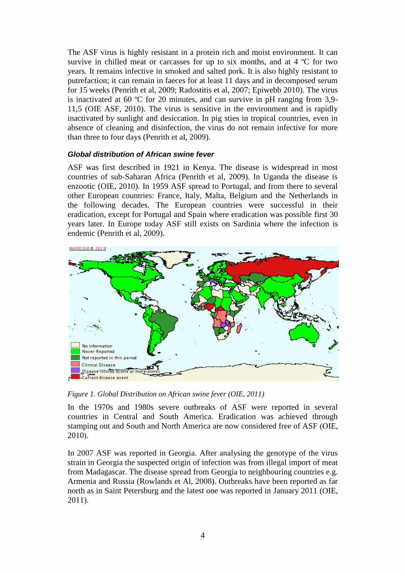
Global distribution of African swine fever
ASF was first described in 1921 in Kenya. The disease is widespread in most
countries of sub-Saharan Africa (Penrith et al, 2009). In Uganda the disease is
enzootic (OIE, 2010). In 1959 ASF spread to Portugal, and from there to several
other European countries: France, Italy, Malta, Belgium and the Netherlands in
the following decades. The European countries were successful in their
eradication, except for Portugal and Spain where eradication was possible first 30
years later. In Europe today ASF still exists on Sardinia where the infection is
endemic (Penrith et al, 2009).
Figure 1. Global Distribution on African swine fever (OIE, 2011)
In the 1970s and 1980s severe outbreaks of ASF were reported in several
countries in Central and South America. Eradication was achieved through
stamping out and South and North America are now considered free of ASF (OIE,
2010).
In 2007 ASF was reported in Georgia. After analysing the genotype of the virus
strain in Georgia the suspected origin of infection was from illegal import of meat
from Madagascar. The disease spread from Georgia to neighbouring countries e.g.
Armenia and Russia (Rowlands et Al, 2008). Outbreaks have been reported as far
north as in Saint Petersburg and the latest one was reported in January 2011 (OIE,
2011).

5
The risk of spreading ASF to the rest of the European countries is considered to
be a big threat to the large pig producing countries within the EU. The risk factors
to be considered are a large population of wild boars in these parts of Europe and
the rather unstable political situation in some of the affected countries, which
complicate efficient control measures and reporting (Costard et al, 2009).
Hosts
ASF virus infects only species from the pig family (Suidae). Both wild and
domestic pigs can be infected but domestic pigs are the most sensitive of
infection, regardless of gender and age (Penrith et al, 2009).
Clinical symptoms
The course of the diseases is often acute or peracute with a mortality rate up to
100 %. The incubation time is 5-15 days, acute cases have a much faster course
and incubation is often 3-4 days. Chronic and subacute cases may occur. These
cases have a longer duration but inevitable end in death. The chronic cases may
have been infected with a less virulent virus subtype then the acute and peracute
cases (Penrith et al, 2009).
Peracute
The peracutely infected pigs are usually found dead without premonitory signs.
Common signs before death, but not always seen because of the rapid course of
the disease are: lethargy, high fever, reddened skin of the abdomen and legs (can
be seen in white pigs), shade seeking, huddling together and shallow breathing
(Penrith et al, 2009).
Acute
The acute course is slower than the peracute, and death normally occurs after two
to seven days. Clinical signs that are often seen include high fever, huddling
together, lethargy, anorexia, seeking shade and water, reluctance to move,
reddened or cyanotic skin particularly on the ears, lower legs and ventral abdomen
(seen in white pigs), abdominal pain and mucopurulent ocular and nasal
discharges. Vomiting is common as well as constipation or bloody diarrhoea. As a
result of the high fever, pregnant sows abort in any stage of the pregnancy. In the
final stages lung oedema evolves resulting in clinical signs such as difficulty of
breathing, bloody froth from mouth and nostrils. Usually the lung oedema is the
primary cause of death but if the pigs survive longer they may evolve nervous
signs because of viral encephalitis/vasculitis or of terminal nature. If the pig
recovers from the acute course it is usually asymptomatic (Penrith et al, 2009).
Subacute
Pigs with subacute ASF can survive from weeks to several months, since they are
often infected with a less virulent strain of the virus. Depending on if they develop
a chronic form of ASF or not, they die or recover. The subacute form occurs
mainly in enzootic areas (Epiwebb, 2010). Clinical signs consist of fluctuant
fever, the pigs usually grow thin with swollen and painful joints, cardiac damage
and a moist cough and difficulty of breathing. Secondary bacterial infections such
as interstitial pneumonia are common (Penrith et al, 2009).

6
Chronic
Common signs of a chronic infected pig are emancipation, inhibition of growth
and a long, dull hair coat. Respiratory signs, lameness, sores and ulcers over bony
parts of the pigs can also be seen. Chronically infected pigs are susceptible to
secondary bacterial infections. A chronically infected pig may survive for many
months but recovery is unlikely (Penrith et al, 2009).
Epidemiology
The mortality rate of acute ASF may reach 100 %. In central Africa and in certain
local breed populations of pigs higher survival rates has been observed, even in
outbreaks with virulent strains of ASF virus. The theory behind this observation is
that either some kind of inherent resistance may be evolving through natural
selection in the exposed pig population (Penrith et al, 2009) or a decrease of the
virulence of the virus occurs with time in enzootic areas (Radostitis et al, 2007).
During the decades when ASF existed in Spain and Portugal chronic and
persistent infected pigs were observed. The affected pigs had decreased mortality
rates and a wide range of clinical symptoms, this made it harder to diagnose the
disease. The spreading of the infection through introduction of infected pigs,
either during the incubation period or by persistently infected pigs was identified
as one of most important transmission routes (Wilkinson, 1984).
The virus is spread through two different cycles: a sylvatic and a domestic. The
sylvatic cycle involves wild species of swine spreading the virus by soft ticks of
the family Ornithodoros (Penrith et al, 2009). In Africa the major host for the
ASF virus is the warthog but all wild species of swine in Africa can be silent
carriers. The ticks live in the burrows of the warthogs (Penrith et al, 2009). The
Ornithodoros ticks can survive for a long time and can harbour the virus for
several years with only a gradual decrease of infectivity (Radostitis et al, 2007).
The bushpig and giant forest hog can become infected by ASF virus but their roles
in the epidemiology has not been fully determined (Jori and Bastos, 2009). In
commercial farms it is unlikely for the domestic pigs to come in contact with wild
pigs and their ticks, but this is considered more common in traditional free-
ranging systems (Wilkinson, 1984).
The domestic cycle involves domestic pigs spreading the virus to other domestic
pigs through direct or indirect contact. The virus infects the pig through the
oronasal route. In the infected domestic pig the virus is shed in enormous amounts
in all bodily secretions and excretions, tissues and blood 24 to 48 hours before
clinical symptoms are shown. If the pig survives the acute disease it will remain
infected for a couple of months but only shed virus for approximately one month.
The virus has been found in lymphoid tissues in domestic pigs for up to three to
four months after infection (Penrith et al, 2009). When the ASF virus existed in
Spain in the 1960s reactivation of virus by stress factors like transportation, in
recovered pigs was suspected to be a source of transmission. A study where
recovered pigs which carried the ASF virus was administrated corticosteroids
during 9 to 31 weeks showed that corticosteroids can reactive the virus replication
up to six months after infection. The reactivated pigs did not produce sufficient
levels of viremia to transmit the disease by direct contact to other susceptible pigs
nor did the pigs show any symptoms of the disease. The theory was that virus
levels might have been too low to be transmittable through direct contact but

7
might be high enough to transmit the disease through shedding in blood and
through ingestion of tissues (Wilkinson, 1984).
Transmission of ASF virus through indirect contact are via fomites (equipment,
vehicles, people, clothing) or swill feeding with infected meat (Penrith et al,
2009). Undercooked swill feeding has been identified as the major source of
spread of ASF to former free areas and the source if infection can almost always
be traced back to airports or harbours (Epiwebb, 2010). Domestic pigs can also
sustain the infection in the population through ticks but without involvement of
the wild pig species (Penrith et al, 2009; OIE ASF, 2010).
Immunity
The pigs that survive the peracute and acute phase of ASF have detectable levels
of antibodies in serum against the ASF virus after 7-12 days after the first clinical
symptoms. Both in warthogs and domestic pigs the antibodies persist for long
time, probably for life. In domestic pigs the antibodies do not protect fully against
further infection with ASF virus, but a certain degree of protection against
infection with homologous strains of the virus has been reported. Sows that are
serologically positive transmit antibodies to their piglets through the colostrums.
In subacutely and chronically infected cases the virus replication continues
regardless the presence of antibodies. There is no vaccine available against ASF
virus (Penrith et al, 2009).
Diagnostic methods
ASF cannot be distinguished from Classical swine fever (CSF) by symptoms or
macroscopic lesions. Therefore CSF is the most important differential diagnosis
of ASF. Diagnosis can be confirmed by virus isolation, antigen detection e.g.
ELISA, direct immunofluorescence, histopathology with immunohistochemistry,
virus nucleic acids detection with PCR, or antibody detection with ELISA
(antibodies can be analysed after seven to twelve days after infection and are
lifelong). Blood or infected tissues for example lung, spleen, liver, kidney, tonsils
and lymphnodes can be used in diagnostic methods (Penrith et al, 2009; Radostits
et al, 2007).
To learn more of the histopathological lesions and immunohistochemistry of ASF
see the thesis written by Justine Ganowiak.
Differential diagnosis
CSF and ASF cannot, as mentioned earlier, be separated only on symptoms and
macroscopic findings and therefore CSF is considered to be one of the most
important differential diagnoses to ASF. Other diseases to consider are Porcine
Reproductive and Respiratory Syndrome (PRRS), Porcine dermatitis/nephropathy
syndrome (PDNS), Pasteurellosis, Salmonellosis and Erypesipelasis (Epiwebb,
2010; Penrith et al, 2009).
Classical Swine Fever
CSF or hog cholera is a contagious, febrile and lethal disease of pigs (Merck Vet
Manual, 2008). The disease is caused by a small, enveloped RNA virus of the
family Flaviviridae, genus Pestivirus. Only one serotype has been found but
antigenic differences have been defined between different viral strains. CSF virus
8
is related to other Pestiviruses for example Bovine virus diarrhoea virus (CFSPH,
2009; Radostits 2007). Hog cholera was first described in the early 19th
century in
the USA. A condition in Europe called swine fever was later recognized to be the
same disease. Today the disease is called CSF to distinguish it from ASF (Merck
Vet Manual, 2008).
CSF is widespread in Asia, some Caribbean islands, Madagascar, Mauritius and
South and Central America (CFSPH, 2009). CSF is endemic in wild boar in parts
of Europe. Hungary, Serbia and Russia have had outbreaks in 2010. South Africa
had one outbreak in august 2007 but since then there have not been any reported
cases. Uganda has never reported a case (OIE, 2010). In 1997-98 the Netherlands
had an outbreak that involved 429 herds and over 12 million pigs were stamped
out to eradicate the disease (Merck Vet Manual, 2008).
The incubation time of CSF is between 2-15 days depending on the dose and
virulence of the virus strain and the age and susceptibility of the pig. Less virulent
strains often cause subclinical, subacute and chronic cases and more virulent
strains cause acute and peracute cases. Younger pigs are more likely to show more
severe symptoms than older pigs (CFSPH, 2009). In the most severe form of CSF,
acute CSF, pigs often die within 1-3 weeks. The incubation time is often shorter
because of more aggressive course of the disease, 2-6 days (Merck Vet Manual,
2008). Symptoms of acute CSF include high fever, lethargy, anorexia,
constipation followed by diarrhoea, conjunctivitis, nervous symptoms like
unsteadiness, staggering gate, convulsions which progress to posterior paresis,
vomiting, respiratory signs, reddened to cyanotic skin on ears, ventral abdomen,
legs (seen in white pigs). The course of the subacute form is similar to the acute
form but the symptoms are less severe, since it is caused by moderate virulent
strains of CSF virus. Survival rates differ, some will survive others die within a
month (CFSPH, 2009).
Chronic cases are caused by less virulent strains of CSF virus and infected pigs
often survive over 30 days but the course of the disease is almost always fatal.
Symptoms resemble the acute and subacute course in the initial stages with the
difference that the affected pigs often improve after a couple of weeks. However,
chronically pigs develop recurrent symptoms like intermittent fever, anorexia,
periods of constipation or diarrhoea, wasting or slowed growth, alopecia and skin
lesions. They often develop secondary bacterial infections (CFSPH, 2009).
In subclinical cases the only symptoms can be poor reproductive performance and
this makes the disease difficult to diagnose on clinical symptoms only. Subclinical
infected pigs are infected with low virulence strains of the CSF virus. Other
symptoms of subclinical infected sows are abortion or birth of stillborn,
mummified, malformed or weak piglets. Newborn piglets can have congenital
tremors or congenital malformations of visceral organs and the central nervous
system. The unborn piglets can become persistently infected in utero if they
survive. These animals are persistently viremic and can spread the virus before
they become clinically ill themselves. The persistently infected pigs often stay
asymptomatic for several months before they get symptoms like lethargy,
depression, slowed growth, dermatitis, diarrhoea, conjunctivitis, ataxia and

9
posterior paresis. Affected pigs often survive six months or longer but die within a
year (CFSPH, 2009).
Transmission route of CSF is usually direct or indirect contact between pigs or
with infected material, like pork, from pigs by oral or oronasal route.
Transmission can also occur through mucus membranes, conjunctiva and skin
abrasion. Infected pigs can spread the virus through blood, all body secretions and
excretions and tissues. Virus shedding can begin before clinical signs are visible
and continues throughout the acute and subclinical course. Chronically infected
pigs can shed the virus continuously or intermittently for a long time (months)
(CFSPH, 2009). Meat that has not been properly cooked can also be a way of
transmission since the virus is partially resistant to heat (inactivated at 56 ºC) and
a wide range of pH values (OIE, Classical Swine Fever, 2010). Furthermore the
virus can remain infectious up to three months in refrigerated meat and more than
four years in frozen meat. The CSF virus does not become inactivated by smoking
or salt curing; survival range from 17 to more than 180 days. The virus can be
spread through mating and artificial insemination. Other routes of transmission
are through fomites, mechanically by insects, birds and other animals. Airborne
transmission seems to be possible but the maximum distance of spread is unclear
(CFSPH, 2009).
Diagnostic methods are similar to the ones used to diagnose ASF: virus isolation,
antigen detection with ELISA, direct immunofluorenscense, serological analysis
with ELISA (antibodies are produced two to three weeks after infection and
persist for life) and virus’ nucleic acids detection with PCR. Congenitally infected
pigs are immunotolerant and therefore negative on antibody detection tests
(CFSPH, 2009).
Porcine reproduction and respiratory syndrome
PRRS is caused by a small enveloped virus from the family Arteriviridae, genus
Arterivirus. PRRS is also known under the names: Mystery Swine Disease, Blue
Ear Disease, Porcine Endemic Abortion and Respiratory Syndrome (PEARS) and
Swine Infertility Respiratory Syndrome (SIRS) (Beltran-Alcrudo et al, 2007). The
virus is very stable under cold/freezing conditions. It can retain infectivity for 4
months at -70 ºC but is inactivated at temperatures above 56 ºC (15-20 min)
(Merck Vet Manual, 2008).
PRRS is an important contagious swine disease and was first identified in USA in
1987 and later found the Netherlands in 1990. Today the virus is spread
worldwide and is found in all larger pig producing areas of the world. It is
endemic in parts of Asia e.g. China and Vietnam. In Africa the disease situation is
unknown, and South Africa is the only country that have reported outbreaks, in
January 2005 and in April 2008 (OIE 2010; Beltran-Alcrudo et al, 2007). In 1992
two genotypes of PRRS virus were identified: type I representing a European
strain and type II a Northern American strain. The different strains of PRRS virus
differ greatly in their pathogenicities. Vaccination has been used as an appropriate
strategy for prevention of PRRS but it has been suggested that several newly
identified virulent PRRS virus isolates have their source in PRRS virus-derived
inactivated vaccines (Tian et al, 2007).

10
The pig is the only species known to be susceptible to the PRRS virus. The
incubation period has experimentally been four to eight days but it can vary from
3-37 days (Beltran-Alcruid et al, 2007). In outbreaks of PRRS in China, 2006, the
course of the disease varied between 5-20 days and the infected pigs spread the
disease to the entire herd in three to five days. The virus can remain in lymphoid
tissues for up to 150 days after exposure, even if it is cleared from the blood (Tian
et al, 2007).
The clinical symptoms can vary between different herds but in general PRRS is
characterized by reproductive failure of sows and respiratory distress of piglets
and growing pigs. Reproductive symptoms can be: infertility, foetal
mummification, abortions, agalactia, stillbirths and weak piglets that usually die
shortly after birth due to secondary bacterial respiratory infection. Young piglets
have the highest mortality and losses can reach 60-70 % in the peak of an
outbreak, but 30-50 % is more common. The respiratory symptoms are mostly
seen in weaners and porkers: anorexia, depression, cutaneous hyperaemia,
dyspnea, rough hair coat, slowed growth and increased susceptibility and
mortality to secondary bacterial infections. Finishing pigs, sows and boars have
more often a subclinical course of disease (Beltran-Alcrudo et al, 2007).
As mentioned before, China had outbreaks of PRRS in the summer of 2006. The
infected pigs had the following symptoms: reddened skin, petechiae,
erythematous blanching raches and pimples often observed in ears, mouth, noses,
back and inner thigh. Furthermore high fever, lethargy, anorexia, cough,
lameness, shivering, diarrhoea was also observed. Many adult pigs died during
this epidemic period, which is uncharacteristic for PRRS. ASF or CSF was
suspected in the beginning but investigators found that the cause of the epidemic
was a highly virulent strain of PRRS (Tian et al, 2007).
The diagnosis of PRRS can be difficult, mainly because virus isolation is made
from the alveolar macrophages which need to be harvested from specific pathogen
free pigs under 6-8 weeks of age. Otherwise serological test like ELISA and virus
detection by PCR can be used for diagnose (OIE, PRRS, 2010).
MATERIAL AND METHODS
Study region and population
The main part of the study was conducted on a farm in Mityana district, Uganda
during three months (October-December) 2010. During three visits to the farm
and close phone contact with the manager of the farm the development of the
outbreak could be monitored. No contact was ever made with the owner of the
farm; all communication was made through the manager.

11
Figure 2. Map of Mityana district, Uganda (Google Earth, 2011)
The farm
The farm was founded in 1995 and has an area of approximately 200 acres. The
manager had been working at the farm since 2008, and during his two years of
work no outbreaks of enzootic diseases like ASF or other diseases with high
mortality rate had been reported. The farm had an integrated pig production and
also kept cattle. The farm consisted of five units A, B, C, D and E. Unit A and B
held pregnant sows, sows with piglets and, weaners (the piglets were weaned at
around eight weeks of age) and three boars. Unit C held gilts and porkers and Unit
D had two pens with porkers (for schematic drawing see Appendix 1). The fifth
unit, unit E the isolation unit, a hut approximately two kilometres away from the
rest of the farm, up on a hill.
The farm had a contract with a meat product company to whom they sold their
porkers. The porkers were slaughtered on the farm at about nine to twelve months
of age and the meat was then transported with the farms’ own transport vehicle to
the meat company. Old sows and other pigs in the production were sold to smaller
slaughter houses in Kampala. The biosecurity measures of the farm consisted of
two footbaths, one in front of unit A and one in front of unit D and C. To limit the
access to the farm there was a gate in the entrance of the farm and there were also
plans to fence in the farm.
The farm had eight workers employed. Three of the workers were responsible
only for the pig production units. One worker was responsible for unit C and D
and the other two for unit A respective unit B. The workers were not supposed to
go between the units. The isolation unit was handled by the worker handling the
tractor, he did not come in contact with the rest of the units. Three of the
remaining workers were diggers. One of the diggers also mixed the feed for all the
pigs. The remaining worker handled the cattle. The workers lived in the
neighbourhood of the farm area and some of the workers kept pigs of local type.
One of the workers who took care of the pig production had had pigs at home but
there was no record if he still kept pigs. Additional animals, apart from the pigs

12
and cattle, which were kept on the farm were some dogs and a cat. The cattle,
dogs and cat were roaming freely over the farm area.
Feed for the pigs were bought from a local market in Kisenyi. The feed consisted
of maize brand, wheat brans, fish and snails shells, no swill feeding occurred on
the farm. The vehicle for transport of the feed was hired but otherwise the entry to
the farm was minimized.
Waste from the pig production was put away in a waste ditch. The waste was used
in the garden as manure, but only on the farm.
There was also a well on the farm, which people from the nearby villages used.
The villagers had to pass through the farm to access this well.
There were wild pigs, bushpigs, in the forest around the farm but they did as far as
it is known not come in contact with the domestic pigs. There were other domestic
free-ranging pigs in the neighbourhood, which might have been sick. Some of the
workers’ pigs had died but there were no information on whose since all pigs were
free-ranging. No new pigs had been introduced to the farm during the last six
months, and the last time any animals were sold was in August 2010 when nine
sows were sold for slaughter because of lack of reproductive out-put.
The outbreak
The manager suspected that the source of infection originated from somewhere
outside the farm. A possible source of infection was pig carcasses from a
neighbouring lady who had pigs that had died before the outbreak started on the
farm. The carcasses were dropped on the farm area and the dogs belonging to the
farm had eaten from the remains. The lady had nine pigs from the beginning, then
a few were sold and the rest died.
The clinical symptoms that had been observed were firstly loss of appetite then,
one to two days later, the affected pigs started to show reddened areas on the ears
and the abdomen and an increased rate of breath. Later the pigs started shivering
and shortly after they died. The manager tried to report the outbreak to the local
district veterinary officer (DVO) but had not been able to get in contact with him.
Rakai
The other part of the study consisted of a screening for CSF and PRRS in the
district of Rakai in south Uganda and in 80 samples from reported outbreaks of
mortality in pigs where ASFV was not confirmed as the cause. In Rakai, eight
sub-counties were included, and in each, five villages were selected. In each
village two pig farmers were chosen and asked if they wanted to be part of the
study. On each farm three pigs were chosen for sampling. The DVOs chose the
farms for the sampling. As compensation for participation in study to the farmers,
each of the sampled pigs was dewormed with Ivermectin.
Sample and data collection
Blood samples were taken from the pigs in the study, one serum and one EDTA
tube from each pig. The pigs were captured with a pig catcher, or if they were
piglets laid on their back, and blood was collected using vacutainer from the
jugular vein.

13
Data on sex, age and breed of the pig was collected and each sample from the pig
was labelled. The breeds were classified as: improved (white pig), local (black
pig) or mixed (black and white pig). The age was estimated and the different
categories were defined as: 0-2 months, 3-5 months, 6-9 months, 9-12 months,
and adults (>12 months).
Figure 4. Sampling of piglet with acute ASF in the farm, Mityana district (picture J.
Ganowiak)
Laboratory analyses
All laboratory analyses were performed at the molecular biology laboratory of the
Makerere University Institute of Environment and Natural Resources at Makerere
University, Microbiology lab, Kampala.
ELISA
The samples from the farm in Mityana were tested for antibodies against ASF
virus using a commercially available ELISA kit (Ingenasa, Madrid, Spain) in
accordance with the instructions from the manufacturer. The samples were diluted
1:2 by diluting 50 µl of sample with 50 µl of sample diluents. The samples were
not tested in duplicates. The wells in the test were washed with approximately 300
µl washing solution and then emptied manually by turning the plate upside down
and forcefully hit the plate against a towel. The samples were measured at 450
nm. The test was considered valid when the optic density (OD) of the negative
control was, at least, four times higher than the OD of the positive control. The
calculation for the positive and negative cut-offs were:
Positive cut off: CN – ((CN-CP) x 0.5)
Negative cut off: CN – ((CN-CP) x 0.4)
(CN= control negative, CP= control positive)

14
The sample was considered positive if the OD was smaller than the positive cut-
off and negative if the OD was greater than the negative cut-off. If the sample had
an OD in-between the two cut-offs it was considered to be in the grey zone.
Samples from Rakai and the 80 samples of undiagnosed mortality in pigs were
tested for antibodies against CSF and PRRS (PRRS Antibody X3 Herdcheck kit
and CSF Antibody test kit, IDEXX Laboratories Inc., Maine, USA). The test
procedure followed the IDEXX Herdcheck manual that came with the test kit. The
samples was diluted 1:40 for the PRRS test by diluting 5µl with 95 µl in a clean
96-well diluting plate and then taken 50 µl of that mix and dilute further another
50 µl of sample diluents. The PRRS x3 Herdcheck had a specificity of 99.9% and
sensitivity of 98.8% (IDEXX, 2010). The CSF samples were diluted 1:2 by
diluting 50 µl of sample with 50 µl of sample diluents. The samples were not
tested in duplicates, with exception for the positive samples that were rerun in
duplicates for confirmation. The wells in both the PRRS and CSF tests were
washed with approximately 300 µl washing solution and then emptied manually
by turning the plate upside down and forcefully hit the plate against a towel. The
absorbance of the CSF samples were measured at 450 nm and the PRRS samples
was measured at 630 nm instead of 650 nm. The positive cut-off in the PRRS test
was OD 0.40.
PCR
The samples from Mityana were tested for presence of ASF virus nucleic acids.
DNeasy Blood and tissue kit from Qiagen was used for extraction of viral DNA
from EDTA blood samples. The samples from the district of Bundibugyo were
tested for presence of CSF and PRRS nucleic acids. The PRRS kits detected both
European and the American strains of the virus. The RNeasy Mini Kit from
Qiagen was used to extract viral RNA. The extracted RNA samples were kept on
freezer blocks when handled for the PCR analysis. The RT-PCR was performed
with the Cepheid Smartcycler v3. For ASF, PRRS and CSF the Tetracore ASF
resp. CSF kits were used and test procedure was performed according to the
manual that came with the kit.
The result of the RT-PCR was considered positive when the samples accumulated
enough fluorescent signal to cross a defined threshold. The unit used, CT (cycle
threshold), was defined as the number of cycles required for the sample to
accumulate enough fluorescence to cross the threshold. The threshold for a
positive CT was programmed in the smartcycler according to the test manual. The
positive control that came with the test kit had an average positive CT, for the
ASF test normally around CT 28-30. The positive CT values of the samples were
compared to the positive control. The sample was considered to be highly positive
for virus if the positive CT was lower than the positive control. If the sample had
a higher positive CT than the positive control it was interpreted as a positive result
but with a lower amount of virus.
RESULTS
Chronological development of the outbreak in Mityana
7th
of October 2010 one sow, mother of nine piglets approximately two months of
age, was found dead by the workers in the morning in unit B, block 1, pen 5. The
nine piglets and piglets from neighbouring unit B, block 1, pen B11 were able go

15
between the pens. The manger believed that the cause of death was that the sow
had been over suckled and therefore exhausted. The nine piglets were moved to
unit A, block 2, pen 2 together with some weaners.
8th
of October one of the nine piglets died suddenly. The remaining eight piglets
were moved back to unit B, block 1, pen 5 and the passage to pen 11 was blocked.
12th
of October another three piglets of the litter of nine died. In unit B, block 1,
pen 11 the sow with eleven piglets was getting sick and two of her piglets died.
The sow and remaining piglets of pen 11 were put in the isolation unit.
13th
of October one piglet of unit B, block 1, pen 5 and three piglets in the
isolation unit died (former unit B, block 1, pen 11).
14th
of October, the first investigation was performed. Three other piglets of unit
B, block 1, pen 5 had died during the night/morning and the one remaining piglet
was very sick with symptoms like shivering, reddened ears and legs. It died later
during the night.
Figure 5. Piglet from pen B5 with acute ASF, note the reddened ears and legs (picture M.
Andersson)
In the isolation hut, there was one dead piglet and the remaining piglets and the
sow were feverish and weak. The sow died the next day and her piglets died one
by one the following days.
Necropsies were performed on three pigs of unit B, block1, pen 5 that had died
earlier that day. The sick piglet from pen 5 and three others from the isolation unit
were sampled (org. pen 11). RT-PCR analysis 15th
of October confirmed ASF as
diagnosis. The piglet from pen 5 and two from the isolation unit tested strongly
positive for ASF. The outbreak was reported to the commissioner of livestock and
entomology and the DVO.

16
Figure 6. Results from real-time PCR assay. First visit at the farm, 14th of October.
18th
of October a sow, mother of three, in unit B, block 1, pen 6 died. Her piglets
were put in the isolation unit. The following days after the sows´ death her piglets
started dying. On the second visit, the 23rd
of October, only one of these piglets
remained alive.
20th
of October pregnant gilt in unit B, block 1, pen 7 died.
23rd
of October, the second visit. The night before the visit on 23rd
of October
2010 one of the sows in unit B had given birth to 5 mummified foetuses and eight
live piglets, no other reproduction problems had been reported on the farm. Minor
problems with lameness in piglets and weak born piglets had been observed, but
similar problems had occurred before the outbreak as well. At the visit 18 pigs
from five different pens were chosen for sampling (see Appendix 2). All pigs that
were sampled were also earmarked so they could be identified later. The RT-PCR
results of the samples showed that all pigs sampled in block A, unit 2, pen 2 were
positive except for one. In unit B, block 1, pen 13 one pig was also positive but
with a higher CT of 39,6. The rest of the samples were negative on the RT-PCR.
Figure 7. Results from RT-PCR assay. The farm unit A block 2 pen 2.
All samples were also tested for antibodies of ASF virus with ELISA. All samples
except for two pigs in unit B, block 1, pen 12 were serologically negative.
25th
of October, one of the gilts in unit C aborted. One day after the gilt was found
dead. After that the manager decided to send all gilts in that pen to slaughter to
prevent further transmission. The gilts had direct contact with the porkers in the
17
neighbouring pen through a gate, but none of porkers had shown any clinical signs
around the time when the gilt died. The manager suspected that the infection had
spread from unit A and B to unit C through the workers. According to him it was
difficult to enforce the workers to follow the new directions of strict hygiene rules
between units and pens. After the gilts were sent to slaughter fences were put up
around every unit to minimize spreading between units. Separate rubber-boots
was introduced for each unit. The workers were allowed only to use the unit
specific rubber-boots in their unit.
The weaners in unit A, block 2, pen 2 started to die around this date. They were
put in the isolation unit but all died during the following days.
Shortly after the visit on 23rd
of October 2010 the two sows in unit B, block 1, pen
13 started to show clinical signs (loss of appetite) and therefore they were sold for
slaughter. The sows were mothers of piglets in unit B, block 1, pen 6 and piglets
in unit B, block 1, pen 12. Two of the piglets in unit B, block 1, pen 12 were
serologically positive for ASF virus on the visit the 23rd
of October but without
any clinical signs.
27th
of October seven sows from unit A, block 1, and 2 (Block 1; Pen 1, 3 and 4,
Block 2; Pen 1 and 4) started to show clinical signs like loss of appetite. The sows
were sold for slaughter to a small slaughterhouse in Kampala. One of the sows
from unit A, block 1, pen 2 did not have any clinical signs like the others and
because of that she was put in unit B, block 1, pen 7 separated from the rest of the
pigs in that block. The manager did not want the sow to be placed in the isolation
unit because the sow was pregnant, approximately 3 months. If she was to be
placed in the isolation unit and she did not develop any symptoms she would not
be able to go back to the rest of the pigs since the isolation unit was considered
contaminated. The sow was supposed to be in “quarantine” until the sign of
disease developed or she delivered. At the third visit on 12th
of November, 2010
the sow had no symptoms of ASF.
A sow with three piglets in unit B, block 2, pen 3 was put in isolation unit after
getting clinical signs, loss of appetite. In the isolation she got worse and was sold
for slaughter. Her three piglets died in isolation during the following days.
12th
of November, the third visit. Following pigs had clinical symptoms at the
visit:
Unit A, block 1, pen 2 one sow had loss of appetite. The sow had aborted
seven foetuses and had delivered six live piglets four days ago.
Unit A, block 2, pen 3 and 5, two sows in each pen had symptoms like loss
of appetite and lethargy.
Unit B, block 2, pen 5 a sow had aborted a few days ago but was still
without any clinical symptoms at the visit, she was only having problems
moving.
One of the boars, in unit B, block 1, pen 8, had reddened skin on its hind
legs and abdomen. The worker started the cleaning of the block in the pen
of the boar and then continued cleaning all the rest of pens in that block. If
the boar was infected the disease had spread to the whole block since the
workers do not change rubber boots or cleaning equipment between the

18
pens, expect for the “quarantine” sow in unit B, block 1, pen 7 which had
its own equipment.
The three weak born piglets that were in unit B, block 1, pen 10 at the last visit
were now moved to unit B, block 2, pen 4 but they showed no clinical signs. No
other pigs had been moved.
15th
of November 2010, information was gathered through phone contact with the
manager. The manager had sold the boar in unit B, block 1, pen 8 for slaughter as
a preventive measurement. Two sows of the four sows with loss of appetite in unit
A, block 2, pen 3 and 5 had died and the remaining two sows had been sold for
slaughter. The sow with newly born piglets in unit A, block 1, pen 2 had died,
both sow and piglets were buried on the farm. The sow in the “quarantine” in unit
B, block 1, pen 7 had been sold to slaughter because it had developed clinical
signs.
29th
of November, information was gathered through phone contact with the
manager. In unit A all sows were dead or slaughtered, there were only a few
piglets left in that unit. In unit B there were remaining five sows and one boar.
There had been no more spread of the infection since the boar from unit B, block
1, pen 8 was sold for slaughter.
13th
of December, information was gathered through phone contact with the
manager. On 3rd
of December, four of the porkers in unit C started to show
clinical signs and where sold for slaughter on the 3rd
of December and the 5th
of
December. The remaining 16 porkers were sold as a preventive measure to the
pork abattoir in Kampala on the 10th
of December. No more pigs with clinical
symptoms had been seen in unit B.
Impact of the outbreak on the farmer
Since ASF is endemic in Uganda the farmers do not get any compensation for
their losses. The outbreak of ASF was very devastating for the economy of the
farm. During the visits to the farm and the conversations with the manager a sense
of hopelessness was felt. The manager was more and more concerned about the
possibility of ever eradicate the infection and about how many pigs that would
die.
When looking at the numbers of pigs lost either to clinical disease or to slaughter
over half of the population disappeared because of the ASF outbreak (see table 2).

19
Table 2. Summary of the impact of the number of pigs on a commercial pig farm during
an outbreak of ASF in Uganda 2010
Date 23/10 13/12 Difference between
23/10 and 3/12
Sows 41 5 -39
Boars 3 1 -2
Porkers 87 - -87
Gilts 11 - -11
Weaners 89 70 -19
Piglets 109 75 -34*
Total 340 151 -192
*The loss of piglets was probably higher since several have been born during this time period.
To estimate the losses the following numbers are presented:
The porkers were sold for 4200 UGX/kg at dressed weight ~50 kg (live
weight ~80-100 kg) in total 210 000 UGX/porker to the meat company the
farm had a contract with.
The price for buying a sow was 450-500 000 UGX.
According to the manager, the biggest boar was bought for 500 000 UGX
and the two smaller ones for 260 000 UGX each.
The value of pregnant gilt was >500 000 UGX and a nonpregnant gilt was
worth around 400 000 UGX.
The mean value of piglets per litter was estimated to 6 piglets/litter from
the number of piglets per litter at the first visit. Assuming that the
production of porkers are equal to the amount the piglets/litter minus
expenses for feed etc the profit of each litter was estimated to 600 000
UGX/litter (210 000 x 6 – 50%).
The future production of litters of gilts and sows was estimated from
numbers from Swedish pig production. In average a sow in Sweden
produces three litters before she is slaughtered (Fellström, 2009). No
statistics was found on the number of litters a gilt produces so this was
estimated as higher than the sows and therefore the number six was used.
The estimated losses are presented in Table 3 and 4.

20
Table 3. Estimated losses because of deceased animals
Category of animals Number of deceased
animals
Estimated loss (million
UGX)
Gilt 1 0,5
Future litters of gilt* 6 x 1 3,6
Sows 25 11,25-12,5
Future litters of sows** 3 x25 45
Piglets 34 3,4
Weaners 19 1,9
Total 79 65,65-66,9
*Estimated production of litters is six, lost profit of litter is estimated to 600 000 UGX/litter
**Estimated remaining litters to produce is three, lost profit of litter is estimated to 600 000
UGX/litter
Table 4. Estimated losses because of sanitary slaughter
Category of animals Numbers of sanitary
slaughtered animals
Estimated loss (in million
UGX)
Gilts (pregnant) 1)
10 1,35
Future litters of gilts* 6 x 10 36
Sows 2)
14 0,7
Future litters of sows** 3 x 14 25,2
Total 23 63,25
1) Loss by slaughtering a pregnant gilt was estimated to 135,000UGX/gilt
2)Loss by slaughtering sows was estimated to 50,000 UGX/sow
* Estimated production of litters is six, lost profit of litter is estimated to 600,000 UGX/litter
** Estimated remaining litters to produce is three, lost profit of litter is estimated to 600,000
UGX/litter
Total loss was estimated to: 128,9-130,15 million UGX (so far) = 362,000-
365,500 SEK1.
There was no information on the price for the boars and the porkers were sold to
the smaller slaughterhouses, but one can assume that they were also sold for a
lower price than normal. Another factor that affects the farm was the boars which
also were important for the future production and loss of genetic material.
Presence of CSF and PRRS
In total 319 samples were analysed for antibodies for both CSF virus and PRRS
virus. All 239 samples from Rakai were negative. Of the 80 samples from
outbreaks in which ASF had not been confirmed, one tested positive for PRRS, all
the rest were negative for CSF and PRRS. The positive sample was rerun in
duplicates to confirm the result and both duplicates were still positive. The
positive sample measured OD 0.43 first time and OD 0.47 and 0.46 in the rerun.
According to test manual the sample is positive if the OD measures over 0.40.
1 0,0028089 2011-02-03 http://omvandlare.com/valuta
21
The samples in Rakai were 239 instead of 240 because in one farm the owner did
only have two suitable pigs for sampling instead of three.
DISCUSSION
ASF has existed and been enzootic in Uganda for a long time. There seem to be
several factors that contribute to the presence of the disease in the country. The
sylvatic cycle of ASF exists in Uganda (based on the results of this study) and this
is one reason that can possibly explain why the disease is maintained in the area.
Also in comparison to the other problems in Uganda, poverty and human disease
like HIV and malaria, the control of ASF is probably not prioritized. In Uganda
there are many other important animal diseases, like Foot-and-mouth disease and
Contagious bovine pleuropneumonia. Diseases affecting cattle may be more
prioritized since in comparison with pigs, cattle are considered more valuable.
Another factor are the Veterinary Officers (VOs) who have an important role
during an outbreak and also for the control and diagnostics of ASF. From my
observations in Uganda it seemed like the VOs lacked resources in terms of
diagnostic methods and transportation (running costs in general). The VOs
sometimes lack access to cars and are forced to ride motorbikes, often lacking
enough fuel. The farms were also generally difficult to reach because many of the
roads were in poor condition. Another contributing factor is probably the structure
of husbandry and the knowledge of ASF amongst the farmers in Uganda. During
the field trips it was noted that in many of the smaller farms the pigs were often
free-ranging or tethered. This may create a greater risk for the pigs of coming in
contact with infected ticks and/or pigs, wild or domestic. The knowledge of the
spreading of the ASF virus did not seem to be sufficient amongst several of the
farmers that were visited. Out of my observations during the study it seemed like
farmers rarely report an outbreak of disease with high mortality to their DVO, for
example the neighbouring lady of the Mityana farm. If pigs in a smaller farm start
to acutely die, the normal thing seemed to be either to sell the pigs that were still
alive or slaughter them and sell the meat. This increases the risk of spreading ASF
since movements of infected pigs and pork are the most important routes of
spreading. The question is why are not all outbreaks reported? It may be that the
farmers do not have enough tradition or knowledge of the importance of reporting
an outbreak and/or do not gain anything on reporting. One example was the farm
in Mityana where both the manager and personnel from the project reported the
outbreak but no one from the authorities contacted the farm. Regardless, since
most outbreaks are not reported it is difficult to have a good control of the spread
and make preventive actions. More reported and monitored outbreaks would make
it possible to get a better overlook of the spread of the disease, a better knowledge
of which areas are more exposed and maybe aid the affected farmers in a better
way.
On the farm level, the farm in Mityana, the impact of the ASF outbreak was
profound. From the first visit and the continuous contact with the manager it was
possible to follow how the infection spread from pen to pen and unit to unit. The
source of infection probably originated from somewhere outside the farm. It is
difficult to point out the exact route from where the infection entered the farm
since in the interviews with the manager he pointed out several possible routes of
infection. The most likely source of infection was the lady with the neighbouring

22
farm, but it is difficult to definitely say since no investigation was carried out to
trace the infection. Another possible route was the workers, if they had pigs
themselves that were infected and in turn brought the infection with them to the
farm. The spread of disease in the farm between units and pens were more clearly
visible with the direct and indirect contact between pigs. The infection seemed to
have started in unit B block 1 and then spread through the movement of pigs, the
workers and the equipment to unit A and then further to units C and D. The
workers seemed to be an important route of spreading and are probably the reason
for the spread to units C and D.
During the early phase of the outbreak the pigs with clinical disease seemed to be
acutely or peracutely affected, indicating the possibility of a strain of virus with
high virulence. The outbreak lasted three months and during the later phase the
course of the disease went more slowly which indicate a more chronic state. One
interesting observation of the infection during the outbreak was that mostly the
older pigs showed clinical signs and died. The weaners and older piglets did not
die in the same numbers as the adults and newborn piglets. One example of that
was the weaners from unit B, block 1, pen 12 that are, presumably, still alive and
did not shown any clinical signs at the visits of the farm. The results from the
sampling showed that one of them was positive on the serology and one was in
the gray zone. According to the literature domestic pigs are susceptible of the
infection irrespective of gender and age but some pigs are more likely to develop
a subclinical to chronic infection or they might inherit a bigger resistance to the
infection (Penrith et al, 2009). Since the outbreak is still going on and not all pigs
have been sampled it is not possible to evaluate the status of all pigs fully. The
weaners of pen 12 seemed to have been exposed to the virus since at least one of
them developed antibodies against ASF. Antibodies are detectable 7-12 days after
the first clinical symptoms are shown and in infected pigs the virus can be shed
for up to one month after being infected (Penrith et al, 2009). None of the weaners
of pen 12 were positive on the RT-PCR. The infection might have come in to the
population much earlier than suspected and the weaners have therefore stopped
shedding the virus, which then is not detectable in the blood but might still be
present in other tissues like lymphoid tissues. The weaners and older piglets can
also be more likely to develop a chronic state than the older pigs. Another
possible theory is that they have been able to eliminate the virus because they
have a bigger resistance to the infection. These theories may explain the larger
survival rate of the weaners and older piglets, but for more information about the
status of the pigs it is needed to sample all remaining pigs and if possible take
samples from lymphoid tissues.
As earlier discussed, there are several reasons for the disease existence in Uganda
of the disease amongst the famers is a contributing factor. In the farm in Mityana
the biosecurity measures were probably one reason why the infection was spread
to the farm. At the first visit, the farm had a few footbaths and they seemed to not
be changed regularly. The farm had a gate but since people from the villages
nearby walked through the farm to get to a well the gate did not help to limit the
access to the farm. There were also little knowledge of the workers and if they
owned any sick pigs. During the outbreak the manager improved the level of
biosecurity with better fencing, even around every unit, better footbaths and
separate equipment for each unit. He tried to limit the workers’ movements

23
between and in the units so they would not spread the disease further. He started
to slaughter all pigs with clinical symptoms to prevent further spreading. The
biosecurity measures taken to stop the spreading might have saved some pigs in
the population but most likely all pigs have already encountered the disease but
has different levels of resistance to the ASF virus and therefore a different rate of
survival. In unit B block 2 almost no deaths or pigs with clinical infection had
occurred and it is not known if the spreading of the disease was prevented there or
those pigs have not shown any clinical signs yet. No pigs were sampled in that
block so the virus and serology status are unknown. It would be interesting to
sample all remaining pigs in the population in a couple of months, if some
survive, to see if they have been able to eradicate the infection or if they are
chronically infected but with no clinical symptoms. This will be done in February
2011.
In the screening for PRRS and CSF in Rakai and the undiagnosed samples there
was only one positive sample for PRRS, from the district of Bundibugyo. It was
the only positive sample from that region and outbreak. The result turned out to be
a very weak positive result. Therefore the most likely interpretation of the result
was that it was a false positive. To confirm the result the sample need to be
analysed with an alternative test procedure and the best would be to take another
sample from the animal and from the other pigs in that population. In the study
there was no opportunity to test the sample in another procedure nor take another
sample of the pig, but it is something that can be done in future studies. CSF was
not found in the samples that were analysed. But since this is a smaller study one
should consider that further studies are needed to clearly show that Uganda is free
from CSF and PRRS.
Conclusions
ASF has existed a long time in Uganda and there are several reasons why it is
maintained in the country. The lacking of reports and follow-ups of reported
outbreak makes the controlling of the disease difficult.
ASF is an important disease for the individual farmer but also in a global view.
For the individual farmer, as in the farm in Mityana, the impact of an outbreak of
ASF can devastate the whole production. In a global view the disease causes
economical losses in both developed and developing countries.
Lacking strong biosecurity, knowledge and resources to eradicate the infection is
the main reason of the spreading to the farm in Mityana. Both the manager and the
representatives from the lab have reported the outbreak to the authorities but no
one has answered or given any directions to the manager.
The serologically positive sample for PRRS was probably a false positive. To
evaluate it further the animal should be re-sampled and run in another test
procedure. Since this was a small study the negative result of CSF and suspected
negative result of PRRS cannot rule out that the diseases are present in Uganda.

24
ACKNOWLEDGEMENT
I want to thank everyone that made the project possible.
My supervisors in Kampala, Uganda: Dr Karl Ståhl and Dr Charles Masembe. My
supervisor in Uppsala, Sweden: Dr Mikael Berg.
Everyone that has been involved in the field trips and lab analyses in Uganda.
Sida and the Faculty of Veterinary medicine and animal science, SLU, for funding
the project.

25
REFERENCES
Agriculture Education and training, The World Bank (online) Available: http://web.worldbank.org/WBSITE/EXTERNAL/TOPICS/EXTARD/0,,contentMDK:20445369
~isCURL:Y~menuPK:1308455~pagePK:148956~piPK:216618~theSitePK:336682,00.html)
2010-11-08
Animal Diseases Act 1918, Chapter 38 (online) Available:
http://www.ulii.org/ug/legis/consol_act/ada1918138/ (2010-11-21)
Beltran-Alcrudo et al, Focus on Porcine Reproductive and Respiratory Syndrome
(regional awareness), FAO Empres, No2 2007
Briggs P, Roberts A. Brandt travel guides: Uganda (1994), 6th ed 2010, Brandt Travel
guides, ISBN-13 978 184162309 2
the Centre for food security and public health (CFSPH), Institute for international
cooperation in animal biologics, Iowa State University; OIE, Classical Swine Fever,
Last updated 16 September 2009 Available:
http://www.cfsph.iastate.edu/DiseaseInfo/disease.php?name=classical-swine-
fever&lang=en (2011-02-03)
Costard et al, African swine fever: how can global spread be prevented? 2009,The Royal
Society
Epiwebb, Afrikansk Svinpest (online) Available:
http://epiwebb.se/02_afrikansk_svinpest/index.shtml (2010-12-04)
Fellström C, Griskompendiet, version 7, Instutionen för kliniska vetenskaper, SLU, 2009
FAO statistics division 2010 (online) Available:
http://faostat.fao.org/site/573/DesktopDefault.aspx?PageID=573#ancor (2010-12-04)
Google Maps (online) Available:
http://maps.google.se/maps?hl=sv&rlz=1T4SVEC_svSE380SE383&q=Mityana%20u
ganda&um=1&ie=UTF-8&sa=N&tab=wl (2011-02-03)
Human Development Report, UNDP (online) Available:
http://hdrstats.undp.org/en/countries/profiles/UGA.html (2010-12-21)
Jori F. et Bastos A., Role of the wild suids in the epidemiology of African Swine Fever,
International Association of ecology and health (Ecohealth), 2009
NAADS (online) Available: http://www.naads.or.ug/naads.php (2010-11-08)
Penrith et al, Preparation of African Swine fever contingency plan, FAO, 2009, ISBN
978-92-5-106426-9
Pro-poor livestock policy initiative, FAO (online) Available:
http://www.fao.org/ag/againfo/programmes/en/pplpi/about.html (2010-11-08)
Radostitis O. et al, Veterinary Medicine, Elsevier Limitied 2007 10th ed, Chapter 21: Hog
Cholera s 1157-1167, African Swine Fever s1167-1172 ISBN 10: 7020 2777 4 ISBN:
13: 978 0702 07772
Rowlands et Al, 2008, African swine fever virus isolate, Georgia, 2007, Emerging
Infectious Diseases, 2008 December; 14(12): 1870–1874.
Söderling Liselotte, Personal Note, 2010-12-08
Tian et al, Emergence of Fatal PRRSV Variants: Unparalleled Outbreaks of Atypical
PRRS in China and Molecular Dissection of the Unique Hallmark , PLos One 2007
June 13.
The Livestock revolution, FAO (online) Available: http://www.fao.org/WAIRDOCS/LEAD/X6115E/x6115e03.htm#TopOfPage (2010-11-08)

26
The Merck Veterinary Manual 2008,
Classical Swine Fever (online)Available: http://www.merckvetmanual.com/mvm/index.jsp?cfile=htm/bc/53400.htm&word=classical%2
cswine%2cfever (2010-12-21)
PRRS (online) Available:
http://www.merckvetmanual.com/mvm/index.jsp?cfile=htm/bc/54100.htm&word=PRRS (2010-
12-21)
Uganda: government halts forced IDP repatriation, IRIN (online) Available:
http://www.irinnews.org/Report.aspx?ReportId=86958 (2010-11-08)
WAHID Interface, World organisation of animal health, OIE
African Swine fever (online) Available: http://www.oie.int/wahis/public.php?page=disease_outbreak_map&disease_type=Terrestrial
&disease_id=12 (2010-11-21)
Available: http://web.oie.int/wahis/public.php (2011-02-03)
African Swine fever distribution (online) Available:
http://www.oie.int/wahis/public.php?page=disease_status_map (2011-01-25)
Classical Swine Fever, (online) Available: http://www.oie.int/wahis/public.php?page=disease_immediate_summary&disease_type=Terr
estrial&disease_id=13 (2010-12-04)
PRRS (online) Available: http://www.oie.int/wahis/public.php (2010-11-21)
Wilkinson P.J, The persistence of African swine fever in Africa and the Mediterranean,
Preventive Veterinary Medicine, 2 (1984) 71-82 Elsevier Science Publishers B.V
Amsterdam.
World Organisation of animal health, OIE, African Swine Fever updated 22/04/2002
(online) Available: http://www.oie.int/eng/maladies/fiches/a_a120.htm#5 (2010-12-04)
World organisation of animal health, OIE, Classical Swine Fever (online) Available:
http://www.oie.int/eng/maladies/fiches/a_a130.htm (2010-12-04)
World organisation of animal health, OIE, PRRS (online) Available:
http://www.oie.int/esp/normes/mmanual/a_00099.htm (2010-12-04)

27
APPENDICES
Appendix 1

28
Appendix 2
Naming of pens sampled and numbers of pigs in each pen that
were sampled 23/10-10. Arrows indicate sewer draining.


![n fostera prrs Z002 web-画像 - maff.go.jp · [1] Yoshii M, Okinaga T, Miyazaki A, et al : Genetic polymorphism of the nsp2 gene in North American type- Porcine reproductive and](https://img.pdfslide.tips/doc/110x75/5b83fb6b7f8b9a934f8e7012/n-fostera-prrs-z002-web-maffgojp-1-yoshii-m-okinaga-t-miyazaki.jpg)