Embed Size (px)
Citation preview
Pesq. Vet. Bras. 38(1):147-153, janeiro 2018
147
RESUMO.- [Desenvolvimento do sistema nervoso cen-tral de bovinos.] Os estudos que descrevem o desenvol-vimento do sistema nervoso central (SNC) são de suma
importância para compreensão da organogênese e iden-tificação dos prováveis eventos que resultam em malfor-mações congênitas. Estes dados podem subsidiar novas
Bovine central nervous system development1
Amanda O. Ferreira2*, Bruno G. Vasconcelos3, Phelipe O. Favaron2, Amilton C. Santos2, Rafael M. Leandro2, Flávia T.V. Pereira4, Durvanei A. Maria2 and Maria Angélica Miglino2
ABSTRACT.- Ferreira A.O., Vasconcelos B.G., Favaron P.O., Santos A.C., Leandro R.M., Pereira F.T.V., Maria D.A. & Miglino M.A. 2018. Bovine central nervous system development. Pesqui-sa Veterinária Brasileira 38(1):147-153. Setor de Anatomia dos Animais Domésticos e Silves-tres, Faculdade de Medicina Veterinária e Zootecnia, Universidade de São Paulo, Av. Prof. Dr. Orlando Marques de Paiva 87, São Paulo, SP 05508-030, Brasil. E-mail: [email protected]
Central nervous system (CNS) development researches are extremely important to the most common congenital disorders and organogenesis comprehension. However, few studies show the entire developmental process during the critical period. Present research can provide data to new researches related to normal development and abnormalities and changes that occur along the CNS organogenesis, especially nowadays with the need for preliminary studies in animal models, which could be used for experimental research on the influence of viruses, such as the influence of Zika virus on the development of the neural system and its correlation with microcephaly in human newborns. Then, present study describes CNS organogenesis in cattle according to microscopic and macroscopic aspects, identifying structures and correla-ting to gestational period. Fourteen embryos and nine bovine fetuses at different ages were collected and analyzed. All individuals were measured in order to detect the gestational period. Bovine embryo at 17 days age has its neural tube, cranial neuropore, caudal neuropore and somites developed. After 24 days of development, were observed in cranial part of neural tube five encephalic vesicles denominated: telencephalon, diencephalon, mesencephalon, metence-phalon and myelencephalon. In addition, the caudal part of neural tube was identified with the primitive spinal cord. The primordial CNS differentiation occurred from 90 to 110 days. The five encephalic vesicles, primordial spinal cord and the cavities: third ventricule, mesencephalic aqueduct, fourth ventricle and central canal in spinal cord were observed. With 90 days, the main structures were identified: (1) cerebral hemispheres, corpus callosum and fornix, of the telencephalon; (2) interthalamic adhesion, thalamus, hypothalamus and epythalamus (glandu-la pinealis), of the diencephalon; (3) cerebral peduncles and quadruplets bodies, of the mesen-cephalon; (4) pons and cerebellum, of the metencephalon; (5) medulla oblongata or bulb, of the myelencephalon; and (6) spinal cord, of the primitive spinal cord. After 110 days of gestation, the five encephalic vesicles and its structures were completely developed. It was noted the pre-sence of the spinal cord with the cervicothoracic and lumbossacral intumescences. In summary, the results describes the formation of the neural tube from the neural plate of the ectoderm, the encephalic vesicles derived from the neural tube and subsequent structural and cavities subdi-visions, thus representing the complete embryology of the central nervous system.INDEX TERMS: Bovine central nervous system, encephalon, encephalic vesicles, CNS, ontogeny, spinal cord.
1 Received on April 24, 2016.Accepted for publication on November 18, 2016.
2 Faculdade de Medicina Veterinária e Zootecnia, Universidade de São Paulo, Av. Prof. Dr. Orlando Marques de Paiva 87, São Paulo, SP 05508-270, Brazil. E-mails: [email protected], [email protected], [email protected], [email protected]; [email protected]; *Cor-responding author: [email protected]
3 Instituto de Ciências Agrárias, Universidade Federal dos Vales Jequiti-nhonha e Mucuri, Av. Vereador João Narciso 1380, Bairro Cachoeira, Unaí, MG 38610-000, Brazil. E-mail: [email protected]
4 Universidade Estadual Paulista “Júlio de Mesquita Filho” (Unesp), Campus Dracena, Rodovia Comandante João Ribeiro de Barros Km 651, Bairro das Antas, Dracena, SP 17900-000, Brazil. E-mail: [email protected]
Pesq. Vet. Bras. 38(1):147-153, janeiro 2018
148 Amanda O. Ferreira et al.
pesquisas relacionadas ao desenvolvimento normal, e in-terpretação de malformações e alterações que ocorrem ao longo da organogênese do SNC, considerando neste mo-mento a necessidade de estudos preliminares em modelos animais, os quais poderiam ser utilizados para pesquisas experimentais sobre a influência de agentes infecciosos como o Zika vírus, no desenvolvimento do sistema nervo-so e suas relações com a microcefalia em humanos recém--nascidos. O presente estudo teve como objetivo descrever os aspectos morfológicos macro e microscópicos da orga-nogênese do SNC de bovinos, buscando correlacionar os achados morfológicos com a idade gestacional. Todos os animais foram mensurados para detectar o período gesta-cional. Foram coletados e analisados 14 embriões e nove fetos de bovinos de diferentes idades gestacionais. No em-brião bovino a partir do décimo sétimo dia de gestação, en-contra-se a formação do tubo neural, o neuroporo cranial e neuroporo caudal, e formação dos somitos. Após 24 dias de desenvolvimento, são observadas na parte cranial do tubo neural cinco vesículas encefálicas denominadas: telencéfa-lo, diencéfalo, mesencéfalo, metencéfalo e mielencéfalo; e na parte caudal do tubo neural, encontra-se a medula espi-nhal primitiva. Entre 90 a 110 dias de gestação, observa-se a total diferenciação das cinco vesículas do SNC. Com 90 dias, são identificas as principais estruturas: (1) do telencé-falo, os hemisférios cerebrais, corpo caloso e fórnix; (2) do diencéfalo, a aderência intertalâmica, tálamo, hipotálamo e epitálamo (glândula pineal); (3) do mesencéfalo, os pedún-culos cerebrais e os corpos quadrigêmios; (4) do meten-céfalo, a ponte e o cerebelo; (5) do mielencéfalo, a medula oblonga (ou bulbo); e (6) da medula espinhal primitiva, a medula espinhal. Após 110 dias, as cinco vesículas encefá-licas e as suas subdivisões se encontram completamente desenvolvidas. Notou-se a presença da medula espinhal com as intumescências cervicotorácica e lombossacral. Em resumo, os resultados demonstram a formação do tubo neural a partir da placa neural do ectoderma, as vesículas encefálicas provenientes do tubo neural e posteriormente as subdivisões das estruturas e das cavidades, que repre-sentam a completa embriologia do sistema nervoso central.TERMOS DE INDEXAÇÃO: Sistema nervoso central, bovinos, encé-falo, medula espinhal, ontogenia, SNC, vesículas encefálicas.
INTRODUCTIONThe largest exporters of beef in the world are Brazil, In-dia, Australia and United States of America (USA). Since 1995, Brazil has increased beef production and its global market participation. Between 1995 and 2012, beef global exports increased 47% while the Brazilian exports increa-sed 568%. However, from 1995 to 2002 exporters of beef faced a difficult period. Bovine spongiform encephalopathy or “mad cow” crisis caused the reduction of exports in the same period (Melz et al. 2014).
Rissi et al. (2010) found that the main diseases of the central nervous system (CNS) diagnosed in cattle in Brazil were rabies, hepatic encephalopathy due to liver failure by ingestion of Senecio spp., bovine herpesvirus meningoen-cephalitis, cerebral babesiosis, Solanum fastigiatum poiso-ning, bluetongue and the polioencephalomalacia.
In addition, in early third of the gestation embryonic mortality ratio in cattle is high. In the embryonic period (from 15th to 45th day of gestation) occurs the formation of the main tissues, organs and systems, setting out the main external characteristics of the body. The fetal period runs from the day 45, which is the period that the forma-tion of the external features of the body occurs (Maddox--Hyttel et al. 2007).
To improve the quality and efficiency of the production of cattle and, in particular, to avoid losing by neurological diseases, it becomes necessary greater knowledge related to CNS and its embryonic development, which is the period that makes it possible to raise important results that support the diagnosis of diseases and congenital abnormalities. In the CNS ontogeny process, the neurulation involves multiple morphological coordinated events, resulting in the conver-sion of neural plate in the neural tube and later its subdivi-sions. Therefore, as results it generates neural tube defects and severe CNS abnormalities (Greene & Copp 2009).
Embryonic CNS development studies are extremely re-levant to the organogenesis and its congenital abnormali-ties probable causes comprehension (Cagnoto et al. 2009). In addition, the greatest susceptibility to teratogens is the embryonic period (Evans & Sack 1973), the stage in which the primitive germ layers form the major organs. Every or-gan has a critical stage of development, featuring an orde-red sequence of biochemical processes involved, which are integrated by several genes, which lead to growth and its differentiation (Arthur 1979, Moore & Persaud 2004, Syno-watz 2010, Silva et al. 2016).
CNS researches are extremely important to the most common congenital disorders and organogenesis com-prehension (Silva et al. 2016). However, few studies show the entire developmental process during the critical period. Present research can provide data to new researches related to normal development and abnormalities and changes that occur along the CNS organogenesis, especially nowadays with the need for preliminary studies in animal models, which could be used for experimental research on the in-fluence of viruses, such as the influence of Zika virus (Cugo-la et al. 2016) on the development of the neural system and its correlation with microcephaly in human newborns.
Whereas the insufficiency of studies completely cove-ring the CNS organogenesis during its critical developmen-tal period, the aim of the present study was to describe ma-croscopically and microscopically the CNS organogenesis, identifying the structures and correlating with gestational age in order to provide data for better understanding yours development and for future pathological and pharmacolo-gical studies.
MATERIALS AND METHODSFor this study 23 uterus of pregnant Bos indicus cattle in diffe-rent gestational ages from slaughterhouses in Andradina SP and Inhumas GO were collected. All laboratory procedures followed the precepts of the Brazilian College of Animal Experimentation (COBEA).
Immediately after collection of uterus, the fetuses and em-bryos were extracted. Then, a total of 14 embryos and nine fetuses were collected.
Pesq. Vet. Bras. 38(1):147-153, janeiro 2018
149Bovine central nervous system development
Embryos were fixed by injection of 10% formaldehyde solu-tion. For fetuses, formaldehyde was channeled by the umbilical vein and umbilical arteries ligation. Subsequently it was perfor-med new ligature in umbilical vein and umbilical arteries and the pieces were stored in the same solution for 48 hours.
To verify the animals macroscopic analysis it was performed the Crown-rump (CR) measurement that is the distance of the nu-chal crest until the last sacral vertebra. The CR measurement was taken by using digital MITUTOYO® pachymeter. Photography of embryo and fetuses were taken with Nikon digital camera model D40x for further description.
Embryos gestational age was estimated in days according to the previous studies (Winters et al. 1942, Evans & Sacks 1973, Noden & Lahunta 1990). In fetuses over 1.5 months of gestation it was used the formula X=M (M+2), where X is the CR measurement in centimeters and M the gestational age in months (Rexroad et al. 1974).
Microstructural analysis of the CNS, embryos and fetuses were processed in increasing ethanol series (70 to 100%) for dehydration and stained in xylene, by using conventional proce-dure for subsequent inclusion in paraffin. So, tissue sections were obtained with 5 µm of thickness in semi-automatic microtome. Then paraffin was removed from the samples, stained in hemato-xylin and eosin (HE), analyzed and microphotography were taken (Olympus BX4160 Microscope coupled to Axio camera CAM HRc) with Zeiss KS400 software.
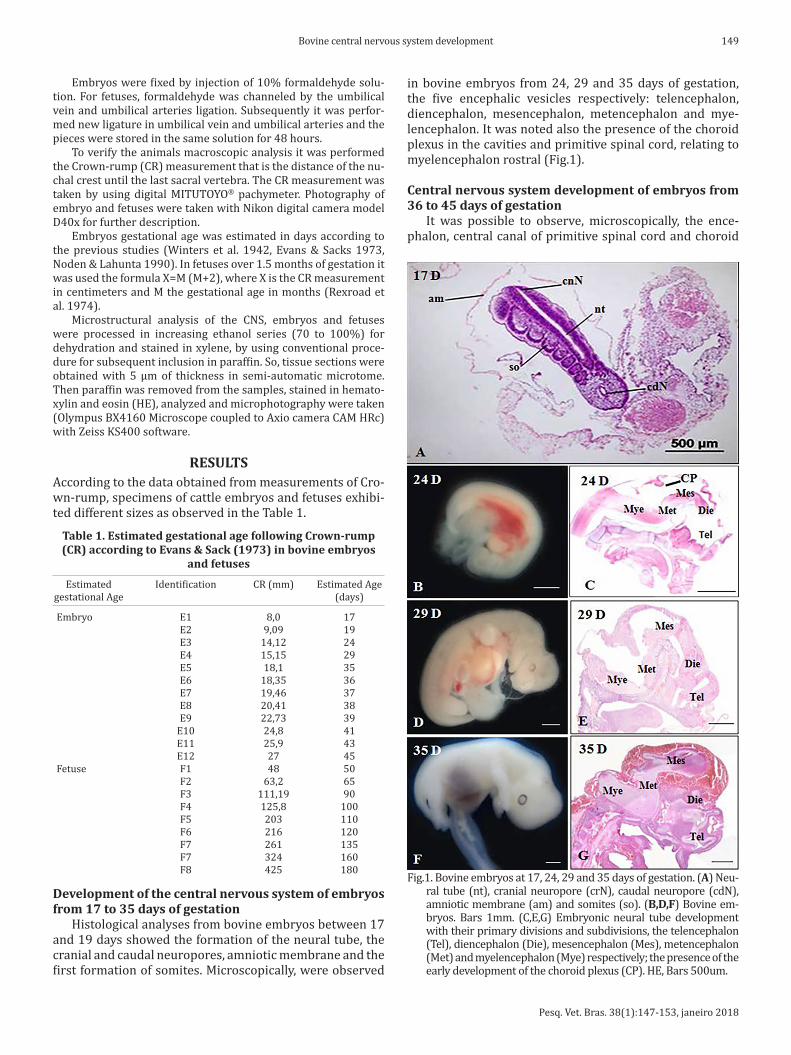
RESULTSAccording to the data obtained from measurements of Cro-wn-rump, specimens of cattle embryos and fetuses exhibi-ted different sizes as observed in the Table 1.
in bovine embryos from 24, 29 and 35 days of gestation, the five encephalic vesicles respectively: telencephalon, diencephalon, mesencephalon, metencephalon and mye-lencephalon. It was noted also the presence of the choroid plexus in the cavities and primitive spinal cord, relating to myelencephalon rostral (Fig.1).
Central nervous system development of embryos from 36 to 45 days of gestation
It was possible to observe, microscopically, the ence-phalon, central canal of primitive spinal cord and choroid
Table 1. Estimated gestational age following Crown-rump (CR) according to Evans & Sack (1973) in bovine embryos
and fetuses
Estimated Identification CR (mm) Estimated Age gestational Age (days)
Embryo E1 8,0 17 E2 9,09 19 E3 14,12 24 E4 15,15 29 E5 18,1 35 E6 18,35 36 E7 19,46 37 E8 20,41 38 E9 22,73 39 E10 24,8 41 E11 25,9 43 E12 27 45 Fetuse F1 48 50 F2 63,2 65 F3 111,19 90 F4 125,8 100 F5 203 110 F6 216 120 F7 261 135 F7 324 160 F8 425 180
Development of the central nervous system of embryos from 17 to 35 days of gestation
Histological analyses from bovine embryos between 17 and 19 days showed the formation of the neural tube, the cranial and caudal neuropores, amniotic membrane and the first formation of somites. Microscopically, were observed
Fig.1. Bovine embryos at 17, 24, 29 and 35 days of gestation. (A) Neu-ral tube (nt), cranial neuropore (crN), caudal neuropore (cdN), amniotic membrane (am) and somites (so). (B,D,F) Bovine em-bryos. Bars 1mm. (C,E,G) Embryonic neural tube development with their primary divisions and subdivisions, the telencephalon (Tel), diencephalon (Die), mesencephalon (Mes), metencephalon (Met) and myelencephalon (Mye) respectively; the presence of the early development of the choroid plexus (CP). HE, Bars 500um.
Pesq. Vet. Bras. 38(1):147-153, janeiro 2018
150 Amanda O. Ferreira et al.
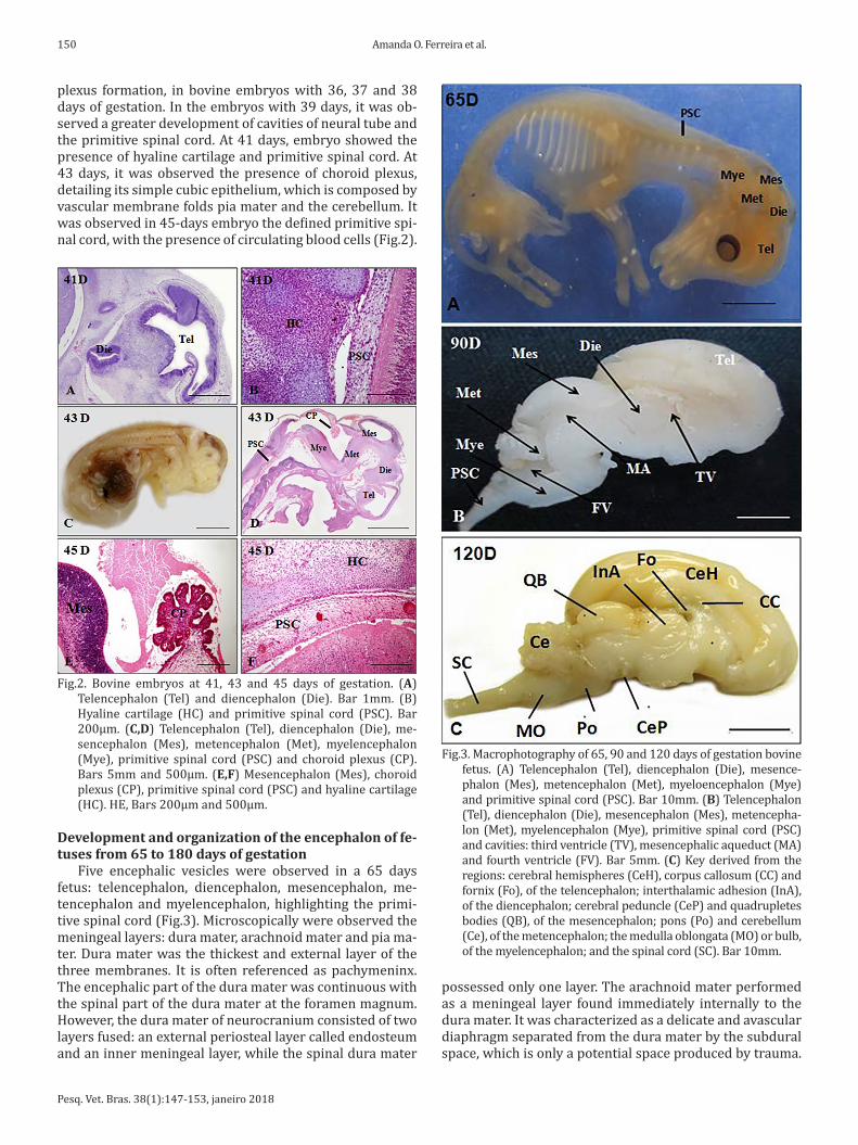
plexus formation, in bovine embryos with 36, 37 and 38 days of gestation. In the embryos with 39 days, it was ob-served a greater development of cavities of neural tube and the primitive spinal cord. At 41 days, embryo showed the presence of hyaline cartilage and primitive spinal cord. At 43 days, it was observed the presence of choroid plexus, detailing its simple cubic epithelium, which is composed by vascular membrane folds pia mater and the cerebellum. It was observed in 45-days embryo the defined primitive spi-nal cord, with the presence of circulating blood cells (Fig.2).
possessed only one layer. The arachnoid mater performed as a meningeal layer found immediately internally to the dura mater. It was characterized as a delicate and avascular diaphragm separated from the dura mater by the subdural space, which is only a potential space produced by trauma.
Fig.2. Bovine embryos at 41, 43 and 45 days of gestation. (A) Telencephalon (Tel) and diencephalon (Die). Bar 1mm. (B) Hyaline cartilage (HC) and primitive spinal cord (PSC). Bar 200µm. (C,D) Telencephalon (Tel), diencephalon (Die), me-sencephalon (Mes), metencephalon (Met), myelencephalon (Mye), primitive spinal cord (PSC) and choroid plexus (CP). Bars 5mm and 500µm. (E,F) Mesencephalon (Mes), choroid plexus (CP), primitive spinal cord (PSC) and hyaline cartilage (HC). HE, Bars 200µm and 500µm.
Development and organization of the encephalon of fe-tuses from 65 to 180 days of gestation
Five encephalic vesicles were observed in a 65 days fetus: telencephalon, diencephalon, mesencephalon, me-tencephalon and myelencephalon, highlighting the primi-tive spinal cord (Fig.3). Microscopically were observed the meningeal layers: dura mater, arachnoid mater and pia ma-ter. Dura mater was the thickest and external layer of the three membranes. It is often referenced as pachymeninx. The encephalic part of the dura mater was continuous with the spinal part of the dura mater at the foramen magnum. However, the dura mater of neurocranium consisted of two layers fused: an external periosteal layer called endosteum and an inner meningeal layer, while the spinal dura mater
Fig.3. Macrophotography of 65, 90 and 120 days of gestation bovine fetus. (A) Telencephalon (Tel), diencephalon (Die), mesence-phalon (Mes), metencephalon (Met), myeloencephalon (Mye) and primitive spinal cord (PSC). Bar 10mm. (B) Telencephalon (Tel), diencephalon (Die), mesencephalon (Mes), metencepha-lon (Met), myelencephalon (Mye), primitive spinal cord (PSC) and cavities: third ventricle (TV), mesencephalic aqueduct (MA) and fourth ventricle (FV). Bar 5mm. (C) Key derived from the regions: cerebral hemispheres (CeH), corpus callosum (CC) and fornix (Fo), of the telencephalon; interthalamic adhesion (InA), of the diencephalon; cerebral peduncle (CeP) and quadrupletes bodies (QB), of the mesencephalon; pons (Po) and cerebellum (Ce), of the metencephalon; the medulla oblongata (MO) or bulb, of the myelencephalon; and the spinal cord (SC). Bar 10mm.
Pesq. Vet. Bras. 38(1):147-153, janeiro 2018
151Bovine central nervous system development
The arachnoid mater was separated from the innermost layer of the meninges, the pia mater, subarachnoid space that contains cerebrospinal fluid (CSF). The two innermost meninges are embryologically originates from a single layer, the leptomeninx. In the fetus of 90 days there was a differentiation of primary CNS regions in which resembles the morphology of its main structures, but was not yet pos-sible to tell them apart in this period.
In 90-days bovine fetuses, it was observed that there was a differentiation of primary CNS regions in which re-sembled the morphology of its major subdivisions, but it was not possible to distinguish them. Thus, viewed the te-lencephalon, diencephalon, mesencephalon, metencepha-lon, myelencephalon, primitive spinal cord and encephalic cavities: lateral ventricles, third ventricle, mesencephalic aqueduct and fourth ventricle. The main structures were: (1) of the cerebral hemispheres, corpus callosum and for-nix, of the telencephalon; (2) of the interthalamic adhesion, thalamus, hypothalamus and epythalamus (glandula pine-alis), of the diencephalon; (3) cerebral peduncles and qua-druplets bodies, of the mesencephalon; (4) pons and cere-bellum, of the metencephalon; (5) the medulla oblongata or bulb, of the myelencephalon; and (6) the spinal cord, of the primitive spinal cord. In this period also showed the gestational five encephalic vesicles, but more developed than those found in fetuses at 65 days (Fig.4).
As observed, shortly after the 43 days of gestation, there was the last distinction of encephalic vesicles, the telence-phalon, diencephalon, mesencephalon, metencephalon and myelencephalon. Later, from the 90th day the formation of its main structures was observed.
Spinal Cord development in embryos and fetuses from 43 to 180 days
In fetuses with respective ages 110, 120 and 160 days of gestation, the total development of five cavities and their subdivisions was observed. It also was observed the presence of an equal and continuous spinal cord with the
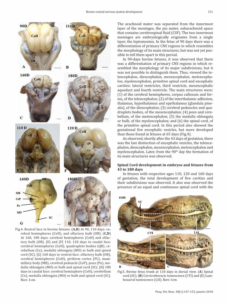
Fig.5. Bovine fetus trunk at 110 days in dorsal view. (A) Spinal cord (SC). (B) Cervicothoracic tumescence (CTI) and (C) Lum-bosacral tumescence (LSI). Bars 1cm.
Fig.4. Rostral face in bovine fetuses. (A,B) At 90, 110 days: ce-rebral hemispheres (CeH), and olfactory bulb (OB). (C,D) At 160, 180 days: cerebral hemispheres (CeH) and olfac-tory bulb (OB). [E] and [F] 110, 120 days in caudal face: cerebral hemispheres (CeH), quadruplets bodies (QB), ce-rebellum (Ce), medulla oblongata (MO) or bulb and spinal cord (SC). [G] 160 days in ventral face: olfactory bulb (OB), cerebral hemispheres (CeH), piriform cortex (PC), mam-millary body (MB), cerebral peduncle (CeP), pons (Po), me-dulla oblongata (MO) or bulb and spinal cord (SC). [H] 180 days in caudal face: cerebral hemisphere (CeH), cerebellum (Ce), medulla oblongata (MO) or bulb and spinal cord (SC). Bars 1cm.
Pesq. Vet. Bras. 38(1):147-153, janeiro 2018
152 Amanda O. Ferreira et al.
cervicothoracic and lumbosacral tumescence different pe-riods of pregnancy. No difference to the disposition of the spinal cord for each bovine fetus was observed . The ori-gins of the spinal nerves were perpendicular (90°) of spinal cord (Fig.5).
The primitive spinal cord originated from the caudal portion of the neural tube was composed of a thick neu-roepithelium. Under the neuroepithelium, a layer of loose connective tissue was observed. We were able to notice the mesenchyme lining the neural tube, which compose the primitive meninges; a thick membrane which forms the dura mater, and its thin internal surface, which remains and constitutes the leptomeninx composed by the arach-noid mater and pia mater.
DISCUSSIONThe nervous system is particularly interesting for the mor-phologist for being the most complex of the body, even if it is conservative regarding to changes. Comparative and de-velopmental studies enable morphologist to build sketches of phylogenetic, especially of this system. From the nervous system, it is possible to determine accurately, for example, the general habits of an animal (in cases where the infor-mation is suitable). Thus, by adding knowledge about the macroscopy, microscopy and ultrastructure of this system that the morphologist is able to give its biggest contribu-tion in applied sciences. The nervous system is a very acti-ve field of research (Hildebrand & Goslow 2006, Silva et al. 2016). Then, in present studies, detailed data were found and might be useful for future researches on the CNS.
By 18 days of gestation, the bovine embryos are present in the gastrula, with formation of amnion and primitive line, which is characterized by converting the bilaminar germ disc in three layers: ectoderm, mesoderm and endo-derm. In our study, we observe the somites and the neural tube from the 17 days of pregnancy. Evans & Sack (1973) described only at 19 days occurs the first somites of the folding of the neural plate. Already Maddox-Hyttel et al. (2007) describe that 21 days embryos, feature a primitive line extended towards the cranial pole, however, without neural tube development.
According to Winters et al. (1942), at 22 days and 16 hours of gestation, bovine embryos exhibit the neural canal completely closed and 19 pairs of somites and the cephalic region set. Until the 24th day of gestation, before implan-tation period, the embryo undergoes significant growth and cellular differentiation, following the uterine tubes (fallopian tubes) to the uterus for implantation. The author describes that from 24th to 26th, three encephalic vesicles become visible, but in our study after 24 days of gestation have been observed the five encephalic vesicles.
The neurological system in the cattle embryo found in our study follows the pattern of differentiation that found in Buffalo, where Morini (2009) describes the CNS arises from the neural plate and subsequently the neural tube of ectodermal origin. With the growth of the embryo, during pregnancy, there are three encephalic vesicles, called: pro-sencephalon, mesencephalon and rhombencephalon (Sino-watz 2010, Franciolli et al. 2011).
The subdivisions of the neural tube and later of five en-cephalic vesicles found in our study follows the one descri-bed for animals domestic mammals described by Sinowatz (2010) and also those described in humans (Moore & Per-saud 2004). Table 2 illustrates these events of differentia-tion and derivation of the neural tube and the encephalic vesicles.
The present study found detailed results in CNS em-bryology in cattle from 17 days, until late pregnancy. As demonstrated, the CNS has a complex system of anatomi-cal and tissue differentiation. Therefore, subdivisions of embryonic neural plate and neural tube that will form the CNS require a deeper understanding that can describe the molecular and cellular mechanisms that determine the suc-cessive developments and tissue derivation, giving rise to a complex system of encephalic vesicles. The detailed em-bryology of the CNS was described in this study, thus other studies can further search for teratogenic factors (Evans & Sack 1973), which affect the CNS in cattle during pregnancy.
As follows, the thorough anatomical knowledge and rai-sed tissue in this study, if associated with molecular biology techniques may assist in the discovery of the mechanisms involved in CNS abnormalities, also it can provide support to pharmaceuticals formulation and prophylaxis of diffe-rent neurological pathologies and dysfunctions that affect cattle herds and generate economic losses as described by Rissi et al. (2010) and Melz et al. (2014). Furthermore, currently, studies about embryology of the CNS have recei-ved more attention due to microcephaly outbreak, which is characterized by malformation of the central nervous sys-tem, caused by Zika virus (Cugola et al. 2016).
However, this study showed that a 17 days bovine em-bryo presents neural tube formation, cranial neuropore, caudal neuropore and somites training. After 24 days it was observed the five encephalic vesicles: telencephalon, diencephalon, mesencephalon, metencephalon and mye-lencephalon; and the neural tube is continued caudally by primitive spinal cord. The 90 days is the differentiation of primary CNS regions. It can be observed the cavity of pri-
Table 2. Tertiary division of the neural tube and the relationship of the their main structures and lumen, in
fetuses at 90 days
Division Main Structures Lumen
Telencephalon Cerebral Hemispheres Corpus Callosum Fornix Lateral Ventricules Interthalamic Adhesion Thalamus Diencephalon Hypothalamus Third Ventricle Epithalamus (Glandula pinealis) Mesencephalon Cerebral peduncles Quadruplets bodies Mesencephalic Aqueduct Pons Metencephalon Cerebellum Rostral part of the Fourth Ventricule Myelencephalon Medulla oblongata Caudal part of the Fourth (or bulb) Ventricle Primitive Spinal Cord Spinal cord Central Canal of Spinal(Remaining Neural Tube) Cord

Pesq. Vet. Bras. 38(1):147-153, janeiro 2018
153Bovine central nervous system development
mitive spinal cord, the central canal of spinal cord, and the ventricular system (lateral ventricles, third ventricle, me-sencephalic aqueduct, fourth ventricle and central canal of the spinal cord). The main structures were identified: (1) cerebral hemispheres, corpus callosum and fornix, of the telencephalon; (2) interthalamic adhesion, thalamus, hy-pothalamus and epythalamus (glandula pinealis), of the diencephalon; (3) cerebral peduncles and quadruplets bo-dies, of the mesencephalon; (4) pons and cerebellum, of the metencephalon; (5) medulla oblongata or bulb, of the myelencephalon; and (6) spinal cord, of the primitive spi-nal cord. After 110 days, until the end of the pregnancy, the five encephalic vesicles and its structures are completely developed. Also was noted the presence of the spinal cord with the cervicothoracic and lumbosacral intumescences. In addition, the origins of the spinal nerves were perpendi-cular (90°) of spinal cord, showing the symmetric growth of nervous system and vertebral column (Prada 2014).
CONCLUSIONSThe present study described the formation of the neural
tube from the neural plate of the ectoderm and the encepha-lic vesicles derived from the neural tube and, subsequently, the structures formed of each of these vesicles, further the primitive spinal cord, thus constituting the complete em-bryology of the central nervous system.
These data provide new knowledge related to a normal development and may be useful for CNS abnormalities stu-dies.
Acknowledgements.- Thanks are due to CAPES (Coordenação de Aper-feiçoamento de Pessoal de Nível Superior) for funding the project.
REFERENCESArthur G.H. 1979. Reprodução e Obstetrícia em Veterinária. 4ª ed. Ganaba-
ra Koogan, Rio de Janeiro.Cagnoto D.G., Guerra R.R., Alberto M.V., Ambrósio C.E., Santos E.J.M. & Mi-
glino M.A. 2009. Morfologia e desenvolvimento ultraestrutural do siste-ma renal de embriões bovinos com idade gestacional entre 10 e 50 dias. Ciência Rural 39:2154-2161.
Cugola F.R., Fernandes I.R., Russo F.B., Freitas B.C., Dias J.L.M., Guimarães K.P., Benazzato C., Almeida N., Pignatari G.C., Romero S., Polonio C.M., Cunha I., Freitas C.L., Brandão W.N., Rossato C., Andrade D., Faria D.P.,
Garcez A.T., Buchpigel C.A., Braconi C.T., Mendes E., Sall A.A., Zanot-to P.M.A., Peron J.P.S., Muotri A.S. & Beltrão-Braga P.C.B. 2016. Nature 534:267-271.
Evans H.E. & Sack W.O. 1973. Prenatal development of domestic and lab-oratory mammals: growth curves, external features and selected refer-ences. Anat. Histol. Embriol. 2:11-45.
Franciolli A.L.R., Ambrósio C.E., Oliveira M.F., Morini A.C., Favaron P.O., Ma-chado M.R.F. & Miglino M.A. 2011. Os histricomorfos sul-americanos: uma análise comparativa do desenvolvimento embriológico. Pesq. Vet. Bras. 31:441-446.
Greene N.D. & Copp A.J. 2009. O desenvolvimento do sistema nervoso central dos vertebrados: a formação do tubo neural. Prenatal Diagnosis 29:303-311.
Hildebrand M. & Gowlow G.E. 2006. Análise da Estrutura dos Vertebrados. 2ª ed. Atheneu, São Paulo.
Maddox-Hyttel P., Wolf X.A., Rasmussen M.A. & Schauser K. 2007. Embry-onic stem cells in pig and cattle: Derivation, culture and potential appli-cations. Acta Sci. Vet. 35:823-830.
Melz L.J., Marion-Filho P.J., Bender-Filho R. & Gastardelo T.A.R. 2014. De-terminantes da Demanda Internacional de Carne Bovina Brasileira: evidências de quebras estruturais. Revta Economia Sociologia Rural 52(4):743-760.
Moore K.L. & Persaud T.V.N. 2004. Embriologia Clínica. 6ª ed. Guanabara Koogan, Rio de Janeiro.
Morini A.C. 2009. Desenvolvimento embrionário em búfalo (Bubalus bu-balis Linnaeus, 1758). Dissertação de Mestrado em Anatomia dos Ani-mais, Faculdade de Medicina Veterinária e Zootecnia, Universidade de São Paulo, São Paulo.
Noden D.M. & De Lahunta A. 1990. Embriologia de los animales domésti-cos. Acríbia, Zaragoza.
Prada I. 2014. Neuroanatomia Funcional em Medicina Veterinária com Correlações Clínicas. Editora Terra Molhada, Jaboticabal.
Rexroad C.E., Casida L.E. & Tyler W.J.M. 1974. Crown-rump length of fetus-es in purebred Holstein-Friesian cows. J. Dairy Sci. 57:346-347.
Rissi D.R., Pierezan F., Oliveira-Filho J.C., Lucena R.B., Carmo P.M.S. & Bar-ros C.S.L. 2010. Abordagem diagnóstica das principais doenças do siste-ma nervoso de ruminantes e equinos no Brasil. Pesq. Vet. Bras. 30:958-967.
Silva F.M.O., Alcântara D., Carvalho R.C., Favaron P.O., Santos A.C., Viana D.C. & Miglino M.A. 2016. Development of the central nervous system in guinea pig (Cavia porcellus, Rodentia, Caviidae). Pesq. Vet. Bras. 36:753-760.
Sinowatz F. 2010. Development of the central and peripheral nervous sys-tem. In: Hyttel P., Sinowatz F. & Vejlsted M. (Eds), Essential of Domestic Animal Embriology. Elsevier, China.
Winters L.M., Green W.W. & Comstock R.E. 1942. Prenatal development of the bovine. Agric. Exp. Station Tech. Bull. 151:1-50.