Embed Size (px)
Citation preview

Mariana Filipa Mestre Pina Amaro Licenciatura em Bioquímica
Development of a theranostic system for central
nervous system based on superparamagnetic
nanoparticles
Dissertação para obtenção do Grau de Mestre em
Bioquímica
Orientador: Professor Doutor Miguel Castanho, IMM FM/UL
Co – orientadores: Professor Doutor João Paulo Borges, DCM-
FCT/UNL
Dr.ª Vera Luísa Santos Neves, IMM FM/UL
Jurí:
Presidente: Professor Doutor José Ricardo Ramos Franco Tavares Arguente: Doutora Maria Manuela de Jesus Guilherme Gaspar Vogais: Doutora Vera Luísa Santos Neves
October 2016


Mariana Filipa Mestre Pina Amaro
Licenciatura em Bioquímica
Development of a theranostic systems for central nervous
systems based on superparamagnetic nanoparticles
Dissertação para obtenção do Grau de Mestre em Bioquímica
Orientador: Professor Doutor Miguel Castanho, IMM FM, UL Co-orientador: Professor Doutor João Paulo Borges, DCM-FCT/UNL
Doutora Vera Luísa Santos Neves, IMM FM, UL
Jurí:
Presidente: Professor Doutor José Ricardo Ramos Franco Tavares Arguente: Doutora Maria Manuela de Jesus Guilherme Gaspar Vogais: Doutora Vera Luísa Santos Neves, IMM FM, UL
October 2016


Development of a theranostic systems for central nervous
systems based on superparamagnetic nanoparticles
Copyright Mariana Filipa Mestre Pina Amaro, FCT/UNL, UNL
A Faculdade de Ciências e Tecnologia e a Universidade Nova de Lisboa têm o direito, perpétuo e sem
limites geográficos, de arquivar e publicar esta dissertação através de exemplares impressos
reproduzidos em papel ou de forma digital, ou por qualquer outro meio conhecido ou que venha a ser
inventado, e de a divulgar através de repositórios científicos e de admitir a sua cópia e distribuição com
objetivos educacionais ou de investigação, não comerciais, desde que seja dado crédito ao autor e editor.


“Never give up on a dream just because of the time it will take to
accomplish it. The time will pass anyway.”
Earl Nightingale


Acknowledgements
vii
Acknowledgements
Quando pensei em fazer o mestrado em bioquímica não pensei que as coisas fossem sempre fáceis,
mas houve alturas em que foram particularmente mais difíceis e não me querendo alongar tenho de
agradecer a todos (ou quase) que de um modo ou de outro me ajudaram. É uma tarefa difícil pois
encontrei bons colegas e, sem dúvida, bons amigos!
Em primeiro lugar, quero agradecer aos meus orientadores: Prof. Miguel Castanho, pela
oportunidade em fazer parte do seu grupo de investigação, ao Prof. João Paulo Borges por ter acreditado
em mim e por me ter dito algumas palavras sábias que me encorajavam e me ajudavam a não desistir.
À Dr.ª Vera Neves, pelo incansável apoio, suporte e orientação ao longo de todo o trabalho. Foi um
prazer poder estar ao teu lado. Obrigada!
À Dr.ª Paula Soares, que mesmo não estando envolvida no meu trabalho nunca me faltou a tua ajuda,
o teu apoio, a tua amizade e quando as coisas nem sempre pareciam ser fáceis tu ajudavas a que parecesse
mais simples!
Ao Dr. Cesar Laia, pela ajuda que sempre se prontificou a dar, pela sua partilha sábia de
conhecimento e pelo apoio!
Ao Prof. Jorge Silva, por se ter prontificado a ajudar-me com a microscopia de fluorescência.
À Coro, Susete e Ana Baptista, obrigada pelo vosso apoio e pela vossa boa energia! É um prazer
estar com vocês!
À Tânia Vieira, obrigada colega! Tu estiveste sempre ao meu lado e daqui fica a nossa amizade para
a vida!
Aos meus colegas de laboratório da FCT-UNL: Ana Almeida, João, Carlos, Gabi, Ana e Jaime!
Vocês foram uns verdadeiros colegas de trabalho e boa disposição.
À Augusta, que alegrava os nossos dias com a sua boa disposição e tinha sempre uma palavra de
amiga para dar. Obrigada!
Maïssa, you are a good friend. Thank you very much for all your help and your support from the
begging! I will always wait for your visit!
Aos meus colegas de laboratório do IMM: Susana, Iris e Marco obrigada por terem recebido e sempre
me ter sentido “em casa” na vossa companhia. Nuno, Filipa e Tiago, obrigada por me de uma maneira
ou de outra me terem ajudado quando foi preciso.
À Rute, obrigada pela ajuda e pela partilha de conhecimento e de tarefas.
À Liliana, por toda a ajuda e partilha de conhecimento no laboratório.
À Sara, mais do que partilha de trabalho e conhecimento, foi partilha de muitos momentos que nem
sempre foram bons, mas a tua ajuda e tua boa disposição tornava as coisas fáceis. Fica a nossa amizade
para a vida! Obrigada!
Aos meus amigos, à Fi, ao Nani, à Mary e à Marta! Vocês são os maiores e melhores do mundo!!

Acknowledgements
viii
À minha família (aos meus pais, à minha irmã, meu “irmão” João e à minha Tia Carolina), por sempre
me terem encorajado, me terem apoiado incondicionalmente e por perceberem mesmo quando a minha
ausência se fazia sentir e mesmo assim compreenderem. Ao meu Salvador, que é pequenino e quando
as saudades são muitas tenta lidar com elas à maneira dele! És o melhor do meu mundo! Estas palavras
seguramente não chegam para agradecer e dizer o quanto foram e são importantes.
A minha nova família Travassos Leandro, que sempre me trataram com muito amor e foram um
apoio muito importante! Obrigada!
E por fim, mas não menos importante, ao meu namorado, marido, amigo, companheiro João.
Obrigada, obrigada por sempre me teres apoiado, ajudado, compreendido e teres sido sempre a melhor
pessoa com quem se pode partilhar o mundo, a vida!
O trabalho apresentado está no âmbito do projeto TRANSDRUG (PTDC/BBBNAN/1578/2014)
financiado pela Fundação para a Ciência e Tecnologia (FCT).
This work is funded by FEDER funds through the COMPETE 2020 Program and National Funds
through FCT - Portuguese Foundation for Science and Technology under the project number POCI-01-
0145-FEDER-007688, Reference UID/CTM/50025.
Resumo
ix
Resumo
A nanotecnologia tem revelado um enorme potencial na área da biomedicina com uma contribuição
fundamental na área dos agentes de contraste para ressonância magnética de imagem ou no
desenvolvimento de sistemas de libertação controlada de fármacos. Diversos estudos têm demonstrado
que o efeito de alguns fármacos pode ser potenciado quando ligados a nanopartículas (NPs). Uma
abordagem semelhante poderá ser relevante para o tratamento de doenças do sistema nervoso central
(CNS).
Nos últimos anos, o conhecimento cérebro tem sido aprofundado e o consequente avanço da
tecnologia tem levado a diversas aplicações biomédicas para o tratamento de várias doenças. No entanto,
doenças como a de Alzheimer, Parkinson ou cancro continuam a ter uma taxa de mortalidade elevada e
poucos tratamentos disponíveis. Na realidade, existem muitas moléculas com fins terapêuticos para estas
doenças, mas mais de 98% das pequenas moléculas, e ~100% de proteínas, não conseguem atravessar a
barreira hematoencefálica (BBB) e chegar ao CNS. A BBB é constituída por células endoteliais que
estão alinhadas com os capilares de modo a prevenir a passagens de substâncias indesejadas do sangue
para o tecido nervoso. Uma abordagem para ultrapassar esta dificuldade seria encontrar um sistema de
libertação controlada capaz de levar o fármaco até à área afetada. Contudo, sistemas como estes podem
afetar o correto funcionamento da BBB. Assim, o desenvolvimento de péptidos com capacidade de
translocação pode ser um método alternativo. Estes péptidos translocadores de membrana (CPP) têm a
capacidade de atravessar para o interior das células com várias moléculas a eles ligadas, por exemplo,
fármacos com baixo peso molecular, lipossomas, anticorpos e NPs. Estes péptidos são degradados em
compostos não tóxicos, têm baixo potencial para interações fármaco-fármaco e baixa probabilidade para
causar reações imunológicas, quando comparados com proteínas.
A doença de Alzheimer é caracterizada por uma acumulação de proteínas insolúveis, como -
amiloides (Aplacas senis (SP) ou novelos neurofibrilares (NFT). Esta acumulação, leva à perda de
sinapses e resulta em neurodegeneração, conduzindo a uma perda de memória e declínio cognitivo.
No presente trabalho, foram utilizadas nanopartículas de óxido de ferro duplamente funcionalizadas
com pequenos CPP e anticorpo contra AO sistema tem a vantagem de poder funcionar como um
sistema de teragnóstico, usando como mais-valia as propriedades magnéticas das NPs.
As NPs foram caracterizadas por várias técnicas em diferentes fases do trabalho e do processo de
funcionalização. Foram obtidas partículas com estrutura cubica cristalina simples caracterizada por
difração de raio-X, um tamanho médio de 9 nm obtido por microscopia eletrónica de transmissão, e um
tamanho hidrodinâmico de 32.4 ± 2.1 nm para as SPIONs revestidas por acido dimercaptosuccínico
(DMSA), obtido por dispersão dinâmica da luz. De modo a simular a BBB, foram utilizados sistemas
transwell in vitro para estudar a transmigração celular das NPs funcionalizadas. Foi possível observar
após 6 h, 23.7 ± 3.7% das SPIONs funcionalizas com o péptido foram capazes de translocar a BBB. As

Resumo
x
NPs funcionalizadas com anticorpo e o péptido CPP são capazes de diminuir a agregação dos péptidos
Aquando comparado com um inibidor conhecido (morin).
Palavras-chave: Nanopartículas, barreira hematoencefálica, péptidos translocadores de membrana,
anticorpos, inibidor de -amiloide, doença de Alzheimer

Abstract
xi
Abstract
Nanotechnology has revealed its potential in the biomedicine area with fundamental contributions to
the imaging contrast agents field or to drug delivery systems development. Several studies have shown
that the effect of drugs could be improved when linked to nanoparticles (NPs). A similar approach may
be relevant to the treatment of central nervous system (CNS) diseases. In the last decades the knowledge
of the brain has increased and the advance in technology has led to several biomedical applications to
treat human disorders. However, diseases such as Alzheimer’s, Parkinson or cancer continue to be
devastating and poorly treatable. There are several therapeutic molecules likely to treat these disorders,
but more than 98% of all small molecules drugs, and ~100% of all large proteins are not able to cross
the blood-brain-barrier (BBB) and get to the CNS.
The BBB is formed by endothelial cells that are aligned with the capillaries to prevent unwanted
substances crossing from blood to nervous tissue. One approach to overtake this difficulty is to find a
controlled delivery system able to supply the drug to the affected tissue. However, such systems have
the potential to affect the correct BBB behavior. An alternative method is the use of peptides with
translocation capacity. These cell penetrating peptides (CPPs) have capacity to translocate various types
of cargo molecules to the cells interior, e.g. low molecular weight drugs, liposomes, antibodies and NPs.
CPPs are degraded in non-toxic compounds, they have low potential to drug-drug interactions and low
probability to cause immunologic reactions when compared with larger proteins.
Alzheimer’s disease is characterized by an accumulation of insoluble protein as -amyloid (A),
senile plaques (SP) and neurofibrillary tangles (NFT). The accumulation of these aggregates leads to a
loss of synapses and neurodegeneration resulting in memory impairment and cognitive decline.
In the present work we have used iron oxide nanoparticles a dual functionalization, with a small
peptide with translocation capacity and with therapeutic antibody against -amyloid peptides. The
system has the ability to function as a theranostic system by taking advantage of the magnetic properties
of the nanoparticles. The nanoparticles were characterized by several techniques at different phases of
the functionalization process.
The iron oxide nanoparticles revealed a simple cubic crystalline structure (by powder x-ray
diffraction), a size of 9 nm (by transmission electron microscopy) and a hydrodynamic size of 32.4 ±
2.1 nm for the coated nanoparticles with dimercaptosuccinic acid (DMSA) (by dynamic light scattering).
To mimic the BBB, a transwell in vitro system was used to study the translocation of functionalized
nanoparticles across this barrier. After 6 h, 23.7 ± 3.7 % of functionalized nanoparticles were able to
cross the BBB. In addition, to assess if the developed nanoparticle is able decrease or stop -amyloid
aggregation, the SensoLyte Thioflavin-T Beta-Amyloid (1-42) aggregation assay was used. It was found
that functionalized iron oxide nanoparticles were able to inhibit -amyloid aggregation when compared
to a known inhibitor (morin).

Abstract
xii
Keywords: Nanoparticles, theranostic system, blood-brain-barrier, cell penetrating peptides, antibodies,
-amyloid inhibition, Alzheimer disease
Table of contents
xiii
TABLE OF CONTENTS
Acknowledgements ............................................................................................................... vii
Resumo ................................................................................................................................... ix
Abstract .................................................................................................................................. xi
List of Figures ....................................................................................................................... xv
List of Tables ....................................................................................................................... xvi
Abbreviations ...................................................................................................................... xvii
Part I
General introduction .................................................................................................................. 1
1. Alzheimer’s disease ................................................................................................................... 3
1.2. - Amyloid Hypothesis ..................................................................................................... 3
1.3. Diagnosis of Alzheimer’s Disease..................................................................................... 5
1.4. Therapies ........................................................................................................................... 6
1.4.1. Immunotherapy ………………………………………………………………….. 8
1.5. Blood Brain Barrier ........................................................................................................... 8
1.6. Cell penetrating peptide .................................................................................................. 10
1.6.1. CPP from dengue virus capside protein ………………………………………... 10
2. Magnetic nanoparticles............................................................................................................ 11
2.1. Superparamagnetic Nanoparticles ................................................................................... 11
2.2. Synthesis of Iron Oxide Nanoparticles………………………………………………… 11
2.3. Surface Modifications of SPIONs ................................................................................... 13
2.4. Biomedical applications of SPIONs ................................................................................ 13
2.5. Drug delivery and a theranostic system........................................................................... 14
Part II
Aims of the study....................................................................................................................... 17
Part III
Matherials and Methods .......................................................................................................... 21
3.1. Synthesis of superparamagnetic iron oxide nanoparticles............................................... 23
Table of cotents
xiv
3.1.1. Spectrophotometric determination of iron by UV-Vis …………………………. 23
3.1.2. SPIONs oxidation and Surface modification with DMSA .................................... 23
3.2. SPIONs functionalization ................................................................................................ 24
3.3. Characterization............................................................................................................... 25
3.3.1. Transmission Electron Microscopy ....................................................................... 25
3.3.2. X-Ray Diffraction .................................................................................................. 25
3.3.3. Fourier Transform Infrared Spectroscopy ............................................................. 25
3.3.4. Fluorescence microscopy ...................................................................................... 26
3.3.5. Dynamic Light Scattering...................................................................................... 26
3.4. Protein Expression and purification ................................................................................ 27
3.4.1. Protein expression ................................................................................................. 27
3.5. In vitro BBB MODEL ........................................................................................................ 29
3.6. Thioflavin-T fluorescence assay...................................................................................... 32
Part IV
Results and Discussion ….......................................................................................................... 35
4.1. Synthesis of SPIONs ....................................................................................................... 38
4.2. Surface modification with DMSA ................................................................................... 40
4.3. Spions Functionalization ................................................................................................. 44
4.4. Cell-based assays ............................................................................................................. 46
4.4.1. integrity of the BBB model ................................................................................... 46
4.4.2. Translocation across the BBB model .................................................................... 47
4.4.3. Inhibition of -amyloid aggregation by conjugated SPIONs ................................ 50
Part V
Concluding Remarks and Future Perspectives....................................................................... 53
References.................................................................................................................................. 59
Appendix.................................................................................................................................... 67
Appendix I ..…………………………………………………………………………... 70
Appendix II ………………………………………………………………………….... 71
Appendix III ……………………………………………………………………...….... 74

List of Figures
xv
LIST OF FIGURES
Figure 1.1. A possible timeline for progression and clinical symptoms in Alzheimer’s disease (AD). .. 3
Figure 1.2. The amyloid hypothesis. ....................................................................................................... 4
Figure 1.3. Amyloid precursor protein processing (APP). ...................................................................... 4
Figure 1.4. Targeting of the principal roots in Alzheimer’s disease (AD) pathogenesis. ....................... 7
Figure 1.5. Antibodies to recognize different A aggregate species. ...................................................... 8
Figure 1.6. The Blood Brain Barrier. ...................................................................................................... 9
Figure 1.7. Application of an external magnetic field.. ......................................................................... 12
Figure 1.8. Multi functionalization of magnetic nanoparticles. ............................................................ 14
Figure 3.1. Particles are illuminated by a laser, and they will scatter the light in all directions. .......... 27
Figure 3.2. Scheme of SDS-PAGE system. .......................................................................................... 29
Figure 3.3. In vitro BBB model.. ........................................................................................................... 30
Figure 3.4. Western Blotting scheme. ................................................................................................... 31
Figure 3.5. Thioflavin assay.. ................................................................................................................ 32
Figure 4.1. XRD of SPIONs oxidized by different conditions. ............................................................. 39
Figure 4.2. Dimercaptosuccinic acid (DMSA). ..................................................................................... 40
Figure 4.3. FTIR of naked SPIONs, SPIONs after surface modification with 0.3 M DMSA and
functionalized SPIONs-sdAb-pepA. ..................................................................................................... 41
Figure 4.4. Images of SPIONs obtainded by TEM and respective size destribution. ........................... 43
Figure 4.5. sdAb-pepA sequence. ......................................................................................................... 44
Figure 4.6. Functionalization reaction with EDC and Sulfo-NHS crosslinkers. ................................... 44
Figure 4.7. Images of SPIONs-GFP-pepA obtained by fluorescence microscopy................................ 45
Figure 4.8. SDS-PAGE of GFP-pepA, DMSA coated SPIONs and SPIONs-GFP-pepA. ................... 46
Figure 4.9. Determination of BBB integrity in the presence of conjugated SPIONs at different
incubation times. ................................................................................................................................... 47
Figure 4.10. Translocation of SPIONs-GFP-pepA in the bEnd.3 transwell BB model. ....................... 48
Figure 4.11. Translocation of SPIONs-GFP-pepA and SPIONs-sdAb-pepA in the bEnd.3 transwell
BBB evaluated by western blot ............................................................................................................. 49
Figure 4.12. Inhibition assay. ................................................................................................................ 50
Figure 5.1. Calibration curve of iron salt. ............................................................................................. 71
Figure 5.2. Iron oxide species report obtained from the condition I by Match software. ..................... 72
Figure 5.3. Iron oxide species report obtained from the condition II by Match software ..................... 73
Figure 5.4. Coated SPIONs with 0.3M DMSA report obtained by Match software. ............................ 74
Figure 5.5. Interaction with the cells membrane. .................................................................................. 75

List of tables
xvi
LIST OF TABLES
Table 4.1. Characterization of DMSA coated SPIONs by DLS. ........................................................... 41
Table 4.2. The pH effect in the zeta potential of the DMSA coated SPIONs. SPIONs in solution at
different pH, coated with 0.3 M DMSA. ............................................................................................... 42
Table 5.1. Concentrations of iron (μg/ml) and respective absorbances. ............................................... 71
Abbreviations
xvii
ABBREVIATIONS
Aβ – Amyloid beta
AD – Alzheimer disease
APP – -Amyloid precursor protein
ATR – Attenuated total reflectance
BBB – Blood Brain Barrier
BC – Bottom Chamber
CNS – Central Nervous System
CPP – Cell-penetrating peptides
DENV – Dengue virus capsid
DEN2C – Dengue virus type 2 capsid protein
dH2O – Deionized water
DLS – Dynamic Light Scattering
DMEM – Dulbecco's Modified Eagle's Medium
DMSA – Dimercaptosuccinic acid
DMSO – Dimethyl sulfoxide
ECs Endothelial Cells
EDC – 1-ethyl-3-(-3-dimethylaminopropyl) carbodiimide hydrochloride
EMA – European Medical Agency
FBS – Fetal Bovine Serum
FDA – Food and Drug Administration
FTIR – Fourier Transform Infrared Spectroscopy
GFP – Green Fluorescent Protein
IMAC – Immobilized Metal-Affinity Chromatography
IPTG – Isopropyl β-D-thiogalactoside
IONs – Iron oxide nanoparticles
MNPs – Magnetic nanoparticles
MRI – Magnetic Resonance Image
NHS – N-hydroxysuccinimide
NFT – Neurofibrillary tangle
NPs – Nanoparticles

Abreviations
xviii
OD – Optical Density
PET – Positron Emission Tomography
SP – Senile Plaques
SPIONs – Superparamagnetic Iron Oxide Nanoparticles
TEM – Transmission electron microscopy
TJs – Tight Junctions
UC – Upper Chamber
XRD – X-ray diffraction

Part I
General Introduction

General Introduction
3
1. ALZHEIMER’S DISEASE
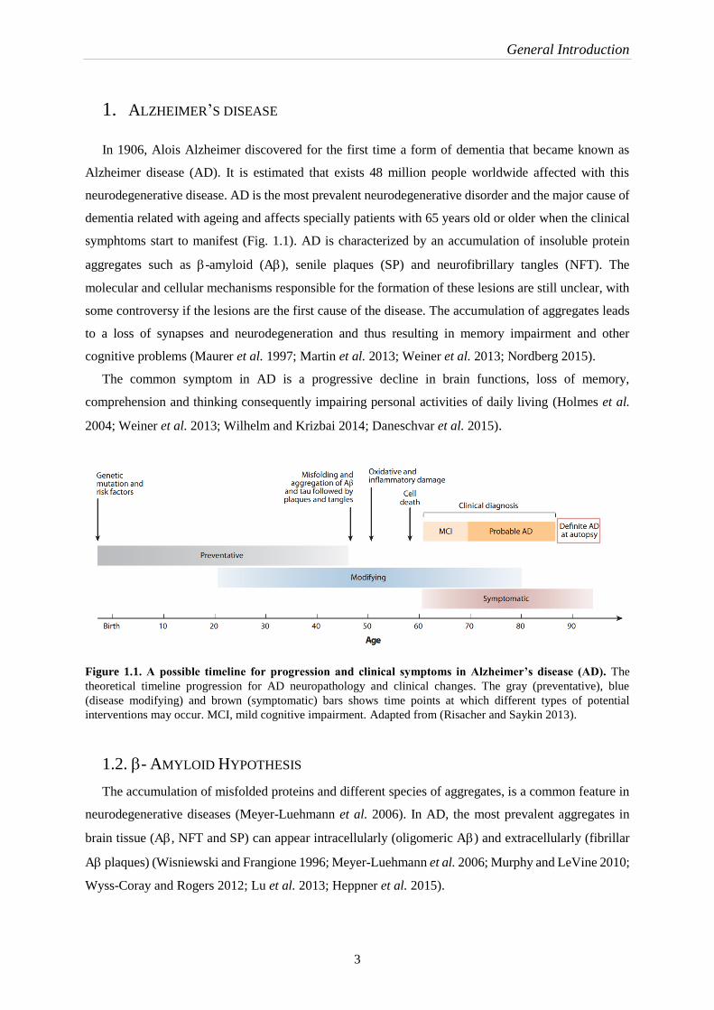
In 1906, Alois Alzheimer discovered for the first time a form of dementia that became known as
Alzheimer disease (AD). It is estimated that exists 48 million people worldwide affected with this
neurodegenerative disease. AD is the most prevalent neurodegenerative disorder and the major cause of
dementia related with ageing and affects specially patients with 65 years old or older when the clinical
symphtoms start to manifest (Fig. 1.1). AD is characterized by an accumulation of insoluble protein
aggregates such as -amyloid (A), senile plaques (SP) and neurofibrillary tangles (NFT). The
molecular and cellular mechanisms responsible for the formation of these lesions are still unclear, with
some controversy if the lesions are the first cause of the disease. The accumulation of aggregates leads
to a loss of synapses and neurodegeneration and thus resulting in memory impairment and other
cognitive problems (Maurer et al. 1997; Martin et al. 2013; Weiner et al. 2013; Nordberg 2015).
The common symptom in AD is a progressive decline in brain functions, loss of memory,
comprehension and thinking consequently impairing personal activities of daily living (Holmes et al.
2004; Weiner et al. 2013; Wilhelm and Krizbai 2014; Daneschvar et al. 2015).
Figure 1.1. A possible timeline for progression and clinical symptoms in Alzheimer’s disease (AD). The
theoretical timeline progression for AD neuropathology and clinical changes. The gray (preventative), blue
(disease modifying) and brown (symptomatic) bars shows time points at which different types of potential
interventions may occur. MCI, mild cognitive impairment. Adapted from (Risacher and Saykin 2013).
1.2. - AMYLOID HYPOTHESIS
The accumulation of misfolded proteins and different species of aggregates, is a common feature in
neurodegenerative diseases (Meyer-Luehmann et al. 2006). In AD, the most prevalent aggregates in
brain tissue (, NFT and SP) can appear intracellularly (oligomeric A) and extracellularly (fibrillar
A plaques) (Wisniewski and Frangione 1996; Meyer-Luehmann et al. 2006; Murphy and LeVine 2010;
Wyss-Coray and Rogers 2012; Lu et al. 2013; Heppner et al. 2015).
General Introduction
4
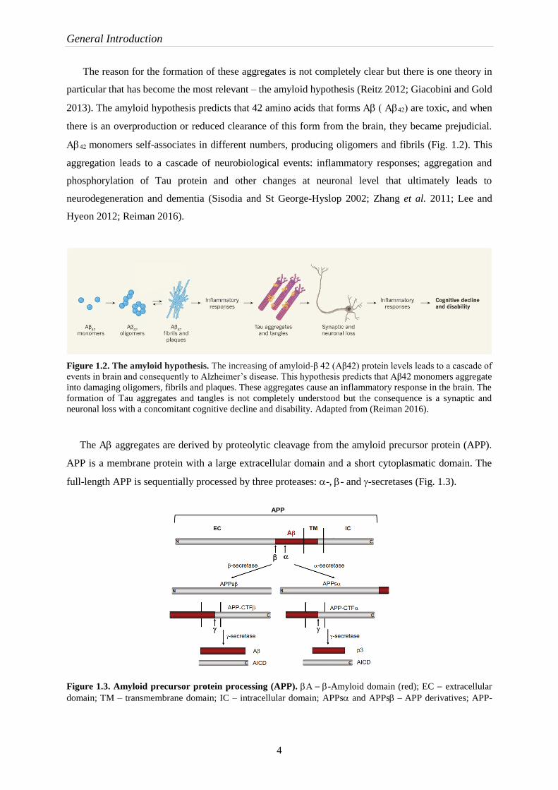
The reason for the formation of these aggregates is not completely clear but there is one theory in
particular that has become the most relevant – the amyloid hypothesis (Reitz 2012; Giacobini and Gold
2013). The amyloid hypothesis predicts that 42 amino acids that forms are toxic, and when
there is an overproduction or reduced clearance of this form from the brain, they became prejudicial.
monomers self-associates in different numbers, producing oligomers and fibrils (Fig. 1.2). This
aggregation leads to a cascade of neurobiological events: inflammatory responses; aggregation and
phosphorylation of Tau protein and other changes at neuronal level that ultimately leads to
neurodegeneration and dementia (Sisodia and St George-Hyslop 2002; Zhang et al. 2011; Lee and
Hyeon 2012; Reiman 2016).
Figure 1.2. The amyloid hypothesis. The increasing of amyloid-β 42 (Aβ42) protein levels leads to a cascade of
events in brain and consequently to Alzheimer’s disease. This hypothesis predicts that Aβ42 monomers aggregate
into damaging oligomers, fibrils and plaques. These aggregates cause an inflammatory response in the brain. The
formation of Tau aggregates and tangles is not completely understood but the consequence is a synaptic and
neuronal loss with a concomitant cognitive decline and disability. Adapted from (Reiman 2016).
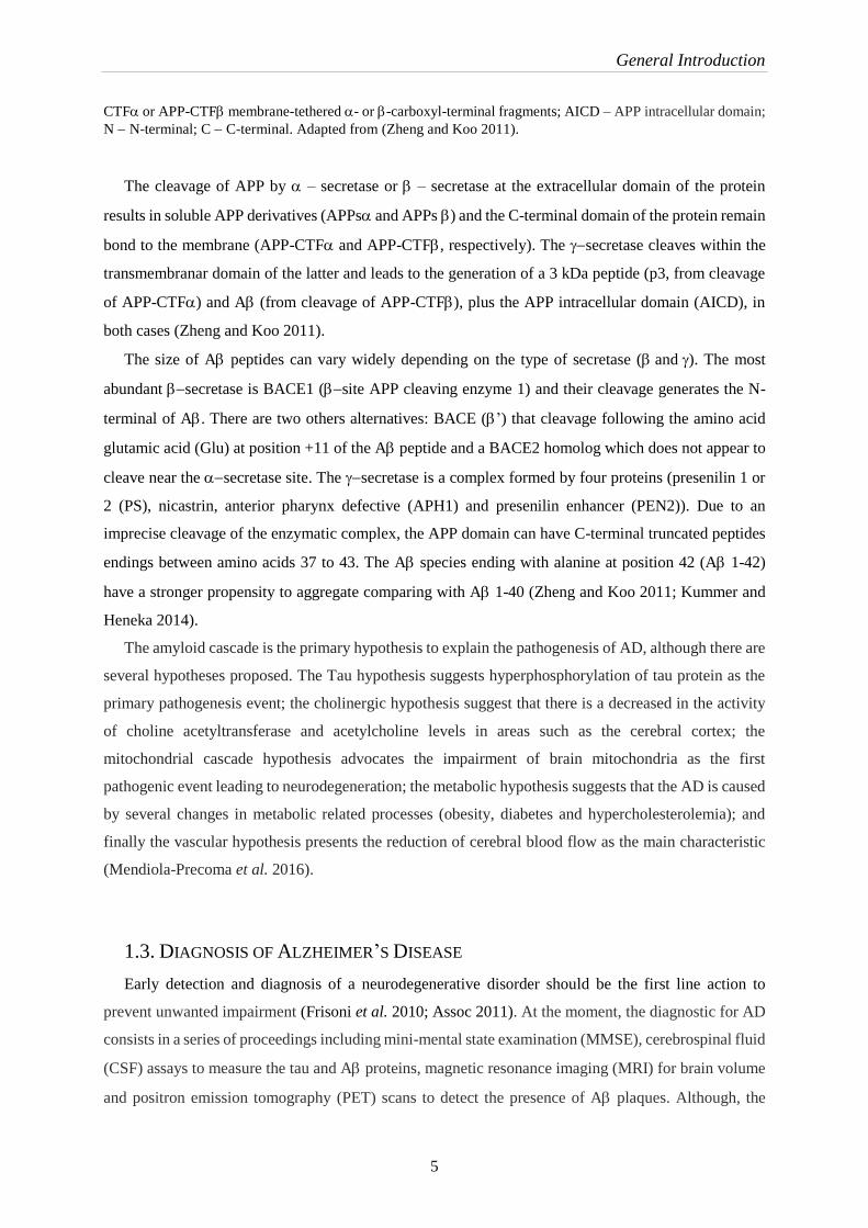
The A aggregates are derived by proteolytic cleavage from the amyloid precursor protein (APP).
APP is a membrane protein with a large extracellular domain and a short cytoplasmatic domain. The
full-length APP is sequentially processed by three proteases: -, - and -secretases (Fig. 1.3).
Figure 1.3. Amyloid precursor protein processing (APP). -Amyloid domain (red); EC extracellular
domain; TM – transmembrane domain; IC – intracellular domain; APPs and APPsAPP derivatives; APP-
APP
General Introduction
5
CTF or APP-CTF membrane-tethered - or-carboxyl-terminal fragments; AICD – APP intracellular domain;
N N-terminal; C C-terminal. Adapted from (Zheng and Koo 2011).
The cleavage of APP by – secretase or – secretase at the extracellular domain of the protein
results in soluble APP derivatives (APPs and APPs) and the C-terminal domain of the protein remain
bond to the membrane (APP-CTF and APP-CTF, respectively). The secretase cleaves within the
transmembranar domain of the latter and leads to the generation of a 3 kDa peptide (p3, from cleavage
of APP-CTF) and A (from cleavage of APP-CTF), plus the APP intracellular domain (AICD), in
both cases (Zheng and Koo 2011).
The size of A peptides can vary widely depending on the type of secretase (and). The most
abundant secretase is BACE1 (site APP cleaving enzyme 1) and their cleavage generates the N-
terminal of A. There are two others alternatives: BACE (’) that cleavage following the amino acid
glutamic acid (Glu) at position +11 of the A peptide and a BACE2 homolog which does not appear to
cleave near the secretase site. The secretase is a complex formed by four proteins (presenilin 1 or
2 (PS), nicastrin, anterior pharynx defective (APH1) and presenilin enhancer (PEN2)). Due to an
imprecise cleavage of the enzymatic complex, the APP domain can have C-terminal truncated peptides
endings between amino acids 37 to 43. The A species ending with alanine at position 42 (A 1-42)
have a stronger propensity to aggregate comparing with A 1-40 (Zheng and Koo 2011; Kummer and
Heneka 2014).
The amyloid cascade is the primary hypothesis to explain the pathogenesis of AD, although there are
several hypotheses proposed. The Tau hypothesis suggests hyperphosphorylation of tau protein as the
primary pathogenesis event; the cholinergic hypothesis suggest that there is a decreased in the activity
of choline acetyltransferase and acetylcholine levels in areas such as the cerebral cortex; the
mitochondrial cascade hypothesis advocates the impairment of brain mitochondria as the first
pathogenic event leading to neurodegeneration; the metabolic hypothesis suggests that the AD is caused
by several changes in metabolic related processes (obesity, diabetes and hypercholesterolemia); and
finally the vascular hypothesis presents the reduction of cerebral blood flow as the main characteristic
(Mendiola-Precoma et al. 2016).
1.3. DIAGNOSIS OF ALZHEIMER’S DISEASE
Early detection and diagnosis of a neurodegenerative disorder should be the first line action to
prevent unwanted impairment (Frisoni et al. 2010; Assoc 2011). At the moment, the diagnostic for AD
consists in a series of proceedings including mini-mental state examination (MMSE), cerebrospinal fluid
(CSF) assays to measure the tau and A proteins, magnetic resonance imaging (MRI) for brain volume
and positron emission tomography (PET) scans to detect the presence of A plaques. Although, the
General Introduction
6
definitive diagnosis still needs histopathological confirmation, the inaccessibility of the brain leads to
the imaging has a major player in diagnosis of CNS diseases (Herholz et al. 2007; Johnson et al. 2012)
(Johnson et al. 2012).
There are several contrast agents for PET approved by Food and Drug Administration (FDA) and
European Medical Agency (EMA) although these agents are radioactive. Therefore there are an
emergency to develop a novel imaging approach or contrast agent sensitive to imaging of A peptide or
tau protein that could change the landscape for diagnosis of CNS related diseases by imaging (Albert et
al. ; Yang et al. 2012; Nordberg 2015).
The urge to diagnose AD in early stages of the disease using a non-invasive approach has been a
relevant point for researchers, and consequently MRI and PET are the most investigated approaches.
There are several studies with different contrast agents to MRI, in particular the use of
superparamagnetic iron oxide nanoparticles and the incorporation of gadolinium (Gd3+) into peptides
that bind to A using a protector of ligands (Johnson et al. 2012; Matharu et al. 2015).
Comparing PET with MRI it is possible to highlight some disadvantages of PET such as, some
individual plaques cannot be seen due to the relatively low resolution of PET because of the
radionucleotides short half-life and occasionally some false-positives are assumed. In contrast, MRI has
higher resolution and can resolve individual plaques and the used contrast agents are not radioactive (Lu
et al. 2013; Matharu et al. 2015).
1.4. THERAPIES
The current therapies available to AD do not prevent or reverse the disease. These drugs are focused
on the reduction of symptoms (Pérez et al. 2016; Sala Frigerio and De Strooper 2016). Most of them are
associated with A cascade hypothesis to restore Ahomeostasis in the brain that is disturbed by
alterations in the production and clearance of A. Based on this hypothesis, four strategies have been
investigated to reduce the levels of Ain the brain. Prevention or reduction of Aformation, by
targeting proteolytic enzymes that mediate processing of APP (and-secretase); removal of existing
amyloid deposits through immunotherapy; prevention or reduction of A aggregation; and the
enhancement of Aclearance (Fig.1.4) (Giacobini and Gold 2013; Sala Frigerio and De Strooper 2016).
The efficiency of all therapies depends on the ability of the drug to cross the BBB. Despite the large
number of drugs that had been developed or are in clinical trials, ~99.6% of them do not get to the CNS
probably due to the permeability issues at the BBB (Cummings et al. 2014).
General Introduction
7
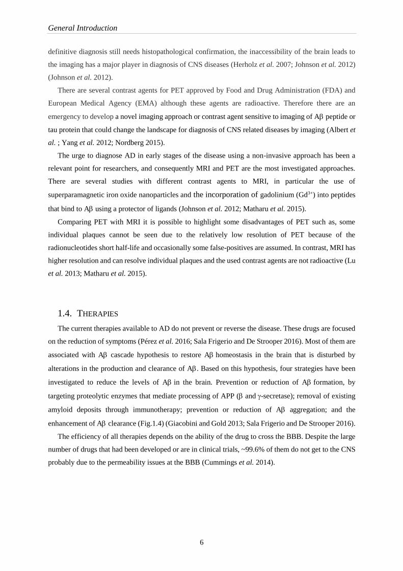
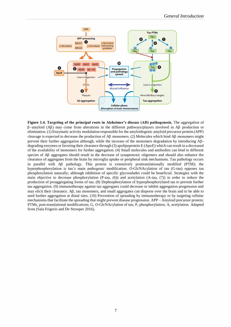
Figure 1.4. Targeting of the principal roots in Alzheimer’s disease (AD) pathogenesis. The aggregation of
amyloid (A) may come from alterations in the different pathways/players involved in A production or
elimination. (1) Enzymatic activity modulation responsible for the amyloidogenic amyloid precursor protein (APP)
cleavage is expected to decrease the production of Amonomers. (2) Molecules which bind Amonomers might
prevent their further aggregation although, while the increase of the monomers degradation by introducing A-
degrading enzymes or favoring their clearance through (3) apolipoprotein E (ApoE) which can result in a decreased
of the availability of monomers for further aggregation. (4) Small molecules and antibodies can bind to different
species of A aggregates should result in the decrease of synaptotoxic oligomers and should also enhance the
clearance of aggregates from the brain by microglia uptake or peripheral sink mechanisms. Tau pathology occurs
in parallel with A pathology. This protein is extensively posttranslationally modified (PTM); the
hyperphosphorylation is tau’s main pathogenic modification. O-GlcNAcylation of tau (G-tau) opposes tau
phosphorylation naturally; although inhibition of specific glycosidades could be beneficial. Strategies with the
main objective to decrease phosphorylation (P-tau, (6)) and acetylation (A-tau, (7)) in order to reduce the
production of proaggregating forms of tau. (8) Dephosphorylation of hyperphosphorylated tau to prevent further
tau aggregation. (9) immunotherapy against tau aggregates could decrease or inhibit aggregation progression and
may elicit their clearance. A, tau monomers, and small aggregates can disperse over the brain and to be able to
seed further aggregation at distal sites. (10) Prevention of spreading by immunotherapy or by targeting cellular
mechanisms that facilitate the spreading that might prevent disease progression. APP – Amyloid precursor protein;
PTMs, post-translational modifications; G, O-GlcNAcylation of tau; P, phosphorylation; A, acetylation. Adapted
from (Sala Frigerio and De Strooper 2016).
(Disruption of brain homeostasis)

General Introduction
8
1.4.1. IMMUNOTHERAPY
AD immunotherapy targeting A has been recognized as one of the most promising approaches to
treat AD. Antibodies have shown capacity as therapeutic agents, being highly selective and having the
ability to treat many diseases, although their use in the brain is limited by the BBB (Liu et al. 2016;
Neves et al. 2016).
Actually, it is possible to modify and/or design the antibodies using engineering to improve their
therapeutic applications and translocation of the BBB using the natural entrances of the brain. In the
past decades, extensive research in immunotherapy using monoclonal antibodies has revealed a great
potential in inhibit or clearing the formation of A species. This approach can break the amyloid
cascade, halt neurodegeneration progress and prevent further reduction in cognitive and physical
function (Giacobini and Gold 2013; Liu et al. 2016; Neves et al. 2016).
New strategies are being developed using monoclonal antibodies with conformation-specific anti-
A to recognize epitopes related to the conformational structure of A aggregates, as well as epitopes
based on the primary sequence of A peptides (Fig. 1.5) (Liu et al. 2016).
Figure 1.5. Antibodies to recognize different A aggregate species. The solid line under each conformation
represents the corresponding A aggregate that the antibody is able to recognize. A – amyloid-; mAb
monoclonal antibody. Adapted from (Liu et al. 2016).
A recent study using an antibody (aducanumab) which binds to the A aggregates has demonstrated
to reduce brain levels of A and it is already in phase III clinical program (Sevigny et al. 2016).
1.5. BLOOD BRAIN BARRIER
The main problem of treating CNS disorders is not due to the lack of molecules abbe to treat the
disease but their ability to cross the BBB. From all developed molecules, ~98% of small molecules and
~100% of large molecules are not capable to get into CNS. For this reason, the development of drug
Antibodies (mAb) binding to different
species

General Introduction
9
delivery system able to cross the BBB and that is be able to perform its function is an emergent
target(Pardridge 2005; de Boer and Gaillard 2007).
The BBB is a multicellular vascular structure that separates the CNS from the peripheral blood
circulation. This barrier is formed by endothelial cells (ECs) that are tightly connected to each other by
tight junctions (TJ), astrocytes and pericytes (Fig. 1.6). At the molecular level, there are transporters and
receptors which regulate what is able to across the BBB (Wolburg and Lippoldt 2002; Cecchelli et al.
2007; Banks and Erickson 2010; Obermeier et al. 2013; Neves et al. 2016).
Figure 1.6. The Blood Brain Barrier. A, The BBB is formed by endothelial cells that interact with perivascular
elements (basal lamina, closely associated astrocytic end-feet processes, perivascular neurons (represented by
interneuron) and pericytes) to form a functional BBB. B, Cerebral endothelial cells form tight junctions produced
by the interactions of several transmembrane proteins that effectively seal the paracellular pathway. These complex
make the brain protect and practically inaccessible. Adapted from (Cecchelli et al. 2007).
The main function of this barrier is to maintain brain homeostasis, regulation of influx and efflux and
transport and protection from any harmful substances. BBB is known as a dual role: first, it is a barrier
for cells and solutes, and on the other hand, is selective to transport various substances, essential
nutrients to the brain and potentially harmful metabolic products to the blood (Deguchi 2002).
In the last years, some strategies to cross the BBB have been developed to delivery therapeutic
molecules to the brain but the ideal method should be controlled and should not damage the integrity of
the BBB. Molecules such as glucose, amino acids and nucleosides can be transported into the brain
through the endogenous BBB transporters, receptor-mediated transport (RMT). To find a new system
capable to overcome the BBB is necessary to develop a drug delivery system able to translocate the
BBB via endogenous BBB transporters (carrier-mediated transport (CMT), active efflux transport
(AET) or receptor-mediated transport (RMT)) or as an alternative they can use the adsorptive-mediated
transcytosis (AMT).
Brain Astrocyte end-foot
General Introduction
10
CMT and AET are responsible for the transport of small molecules (such as glucose, amino acids
and nucleosides) between blood and the brain and the RMT is responsible for the transport across the
BBB of specific endogenous large molecules (such as insulin, low-density lipoprotein (LDL) or
transferrin). AMT is usually induced by electrostatic interaction of cationic proteins with endothelial
cells (Neves et al. 2016).
1.6. CELL PENETRATING PEPTIDE (CPP)
Peptides are responsible for the regulation of many physiological processes. They are being used as
a therapeutic agent in areas such as neurology, endocrinology and hematology (Santos et al. 2012).
The cell penetrating peptides (CPP) are short peptides (5 – 40 amino acids in length) with ability to
achieve the access to the cell interior. The uptake mechanisms by which CPP enter in the cells are not
completely understood but there are two principal uptake mechanisms, endocytosis and direct
penetration. These CPP have the capacity to carry cargos such as peptides, protein, oligonucleotides and
nanoparticles. They are positively-charged at physiological pH, usually, rich in lysine or arginine and
due to their charges they are able to interact with the negatively-charged plasma membrane and the
positively charged carrier (Sarko et al. 2010; Trabulo et al. 2010; Madani et al. 2011; Jafari et al. 2015).
The incessant search for effective and safe ways to transport compounds from the blood to the brain
is imperative but it is extremely difficult not to interfere with BBB functions (Zou et al. 2013; Prades et
al. 2015).
1.6.1. CPP FROM DENGUE VIRUS CAPSID PROTEIN
Dengue virus (DENV) is responsible for a variable spectrum of disease that ranges from an
undifferentiated fever to dengue fever with potentially fatal dengue shock syndrome. DENV has a great
capacity to delivery their DNA into the cells, resulting in an effective interaction with the cell
membranes. The capsids of the DENV have been used to develop a CPPs using the capsids as a template
(DENV C). The DENV C has four -helical segments (1, 2, 3 and 4) (Mairuhu et al. 2004; Freire
et al. 2014).
The CPPs that adopt a specific secondary structure have higher stability with the membranes and
promote their cellular uptake. Considering this fact, a CPP from the capsid protein of Dengue virus type
2 (DEN2C) from 3 domain (pepH3 or pepA), was developed in order to be used as a shuttle in and
out of the brain. A study revealed a great ability to cross the BBB returning to the blood circulation to
be eliminated (Mairuhu et al. 2004; Eiriksdottir et al. 2010; Freire et al. 2014; Castanho et al. 2015).
General Introduction
11
2. MAGNETIC NANOPARTICLES
In the last decades, nanomaterials have been intensively investigated, with particular interest, in
magnetic nanoparticles (MNPs). In particular, nanoparticles with 1 – 100 nm have been used in
biomedicine as contrast agents in magnetic resonance imaging, cancer therapy, hyperthermia, tissue
repair and drug delivery (Gupta and Gupta 2005; Lim et al. 2013b; Ruiz et al. 2014).
MNPs can be divided in paramagnetic, ferromagnetic and superparamagnetic particles. The impact
of superparamagnetic and ferromagnetic particles on magnetic resonance imaging is similar. The
composition of paramagnetic particles is different from the composition of superparamagnetic and
ferromagnetic particles. Superparamagnetic and ferromagnetic particles have a magnetic core and a
surface coating. The paramagnetic particles do not have an explicit magnetic core. Thus, their behavior
on MRI is rather different from superparamagnetic and ferromagnetic particles (Rümenapp et al. 2012).
MNPs are usually formed by a mineral core of a magnetic mineral that could be iron, nickel, cobalt
and their oxides, with an organic coating. From all iron oxide NPs, the most common biocompatible
magnetic nanomaterials are pure oxides, magnetite (Fe3O4) and maghemite (-Fe2O3) (Gupta and Gupta
2005; Lim et al. 2013b; Ling and Hyeon 2013; Calero et al. 2014; Soares et al. 2014).
2.1. SUPERPARAMAGNETIC NANOPARTICLES
In last decades, superparamagnetic iron oxide nanoparticles (SPIONs) have gained a significant
relevance. Iron oxide nanoparticles are designated by SPIONs when they are smaller than the critical
size (< 15 nm) and consists of a single magnetic domain. In other words, these NPs exhibit a remarkably
high MRI contrast effect, they are easily controlled by a magnetic external field and display a strong
magnetic response even if subjected to a low magnetic field. For this reason, their movement in the
blood can be controlled with external magnetic field and immobilized close to the targeted area (Fig.
1.7 ) (Faiyas et al. 2010; Busquets et al. 2014; Hola et al. 2015). SPIONs are commonly composed of
magnetite (Fe3O4) and maghemite (Fe2O3) cores (Laurent et al. 2008).

General Introduction
12
Figure 1.7. Application of an external magnetic field. A, No magnetic field applied and the iron oxide
nanoparticles are in the blood circulation. B, External magnetic field is applied and the nanoparticles are
immobilized to the target. Adapted from (Wu et al. 2015).
Despite the NPs being commonly used due to their natural elimination or their simple synthesis, there
are some adverse reactions to avoid. When nanomaterials are exposed to biological systems, many
undesirable reactions may occur. The generation of reactive oxygen species (ROS) upon exposure of
cells is generally considered as a major contributor to the toxicity of naked iron oxide nanoparticles. For
this reason, coating must be used to avoid this side effect (Tartaj et al. 2011; Ling and Hyeon 2013; Ruiz
et al. 2014).
2.2. SYNTHESIS OF IRON OXIDE NANOPARTICLES
The synthesis methods can influence the NPs properties, such as size, dispersion of the particle-size
and morphology. The most commonly precipitation method is chemical co-precipitation due to their
extremely flexible method, and the surface properties controlled changing the experimental parameters
(pH value, temperature and ionic strength). Using this option, the NPs are synthetized in water, which
becomes an advantage for biomedical applications. There are several other options to synthetize NPs
including microemulsions, sol-gel synthesis, hydrothermal reaction, hydrolysis and thermolysis of
precursors (Hadj Farhat and Joubert 1984; Costa et al. 1994; López-Pérez et al. 1997; Wu et al. 2015).
Co-precipitation of ferric and ferrous ions in solution using a strong base leads to the formation of
magnetite or maghemite (nucleation phase), depending up on the iron concentration, pH and the ratio of
ferrous and ferric ions (Gnanaprakash et al. 2007).
External magnetic field
General Introduction
13
2.3. SURFACE MODIFICATIONS OF SPIONS
The surface of SPIONs can be modified through the creation of atomic layers of some organic or
inorganic molecules, which allows further functionalization by binding various bioactive molecules.
However, more than the functionalization, it is the contribution of the coating to the NPs colloidal
stability (Gupta and Gupta 2005).
Despite the fact that naked NPs are stable in high- and low- pH suspensions, but not in neutral pH,
they must be coated for several reasons: increase the stability and protect SPIONs against aggregation,
to protect the magnetic core against oxidation, to provide a reactive surface to allow their
functionalization and, finally, to protect NPs against reticuloendothelial system (RES) uptake and
elimination in order to enhance the blood circulation time and internalization efficiency (Laurent et al.
2008).
From all different polymers/molecules which can be used for coating NPs, meso-2,3-
dimercaptosuccinic acid (DMSA) presents a great potential for target-drug delivery. This molecule binds
to the nanoparticle through the carboxylate groups which establishes a negative surface charge of the
particle and the thiol groups generates a “cage” of disulfide-cross-linked DMSA around the nanoparticle
(Zhang et al. 2016).
Coated SPIONs with DMSA have been tested in vitro and in vivo, in particular their cell interaction
process, pharmacokinetics and biodistribution in animal models with the main objective of drug
delivery, NMR imaging and hyperthermia. (Ruiz et al. 2014).
2.4. BIOMEDICAL APPLICATIONS OF SPIONS
When the physicochemical properties of SPIONs (size, composition and morphology) are combined
with superparamagnetism, makes them highly promising candidates for many applications, such as drug
delivery system, contrast agent in MRI and hyperthermia to cancer therapy. Due to their magnetic
properties, they are commonly used as contrast agent in MRI (Laurent et al. 2008; Teja and Koh 2009;
Ge et al. 2013).
Contrast agents are used to enhance the image obtained from MR. Some images obtained using a
common contrast agent by magnetic resonance of soft tissues have a relatively high quality, although
there are some cases where it is difficult to acquire sufficient image of contrast to diagnose the pathology
of interest. In this case, SPIONs can be used to overcome this difficult. Actually, paramagnetic
substances, such as gadolinium (Gd), have enhanced the magnetic resonance signal but the toxicity
associated with them has led to a new focus of research in SPIONs (Li et al. 2013; Busquets et al. 2014;
Hola et al. 2015).
General Introduction
14
A promisor strategy to treat AD has been studied to allow an early diagnosis, before clinical
symptoms of the disease are manifested. The strategy involved the use of functionalized SPIONs with
an antibody against A1-42 to detect in vivo amyloid deposition by MRI (Andrieux and Couvreur 2013).
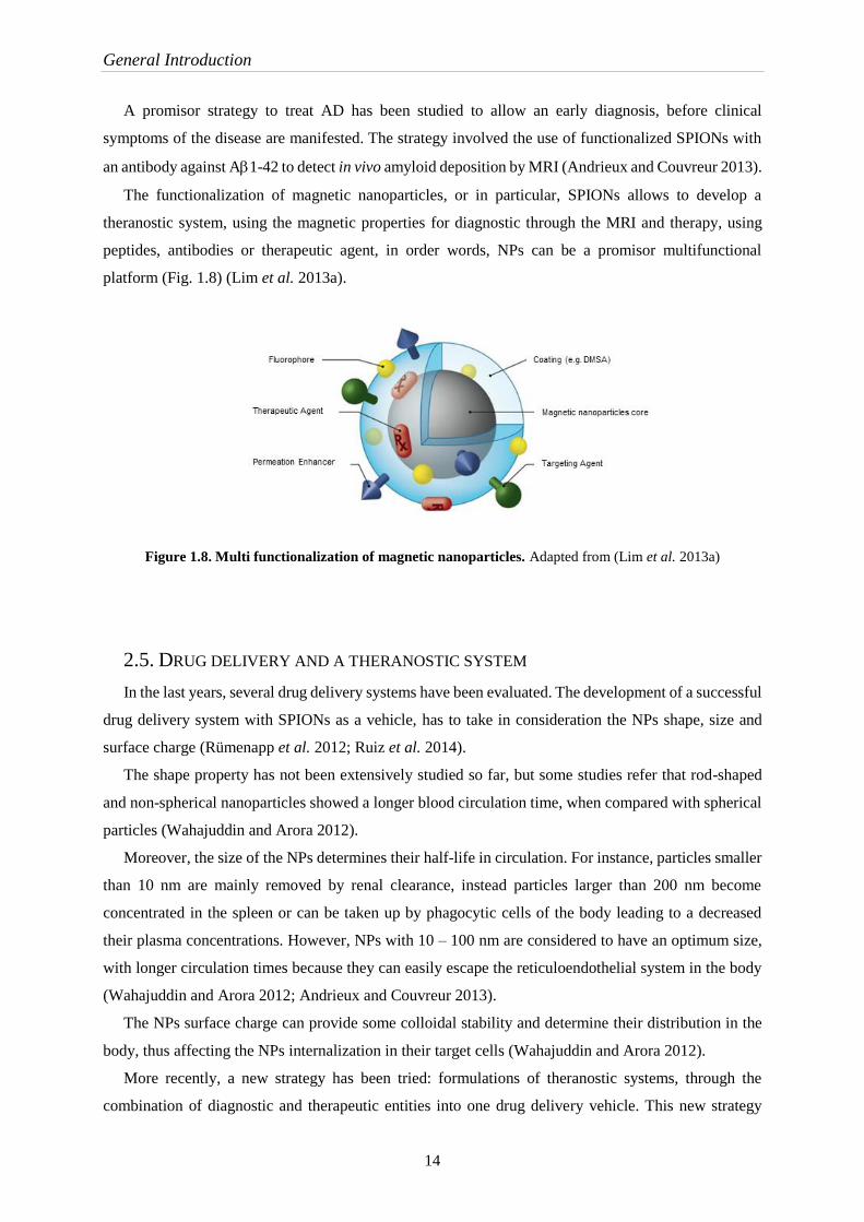
The functionalization of magnetic nanoparticles, or in particular, SPIONs allows to develop a
theranostic system, using the magnetic properties for diagnostic through the MRI and therapy, using
peptides, antibodies or therapeutic agent, in order words, NPs can be a promisor multifunctional
platform (Fig. 1.8) (Lim et al. 2013a).
Figure 1.8. Multi functionalization of magnetic nanoparticles. Adapted from (Lim et al. 2013a)
2.5. DRUG DELIVERY AND A THERANOSTIC SYSTEM
In the last years, several drug delivery systems have been evaluated. The development of a successful
drug delivery system with SPIONs as a vehicle, has to take in consideration the NPs shape, size and
surface charge (Rümenapp et al. 2012; Ruiz et al. 2014).
The shape property has not been extensively studied so far, but some studies refer that rod-shaped
and non-spherical nanoparticles showed a longer blood circulation time, when compared with spherical
particles (Wahajuddin and Arora 2012).
Moreover, the size of the NPs determines their half-life in circulation. For instance, particles smaller
than 10 nm are mainly removed by renal clearance, instead particles larger than 200 nm become
concentrated in the spleen or can be taken up by phagocytic cells of the body leading to a decreased
their plasma concentrations. However, NPs with 10 – 100 nm are considered to have an optimum size,
with longer circulation times because they can easily escape the reticuloendothelial system in the body
(Wahajuddin and Arora 2012; Andrieux and Couvreur 2013).
The NPs surface charge can provide some colloidal stability and determine their distribution in the
body, thus affecting the NPs internalization in their target cells (Wahajuddin and Arora 2012).
More recently, a new strategy has been tried: formulations of theranostic systems, through the
combination of diagnostic and therapeutic entities into one drug delivery vehicle. This new strategy

General Introduction
15
could prevent, diagnose and treat some diseases and may be possible the development of a personalized
medicine (Ruiz et al. 2014; Ryu et al. 2014).
A promising approach is the use of SPIONs to detect and treat lesions in neurodegenerative disease
as AD, since nanoparticles are able to cross the BBB (Mejias et al. 2011).


Part II
Aims of the Study

Aims of the study
19
Alzheimer’s disease (AD) is an age related neurodegenerative disease affecting, approximately, 48
million people worldwide (2015). AD is an incurable disease and the approved therapies are only able
to treat symptomatic symptoms that do not mitigate the course of the disease. Therefore, there is an
urgent need to find new therapies to treat AD. Immunotherapy targeting amyloid-Ais one of the
most promising therapies to treat AD. The main obstacles are the requirement of the antibody to cross
the blood-brain-barrier (BBB), in order to reach the Adeposits in the affected brain tissue The
immunotherapy combined with magnetic resonance imaging (MRI) could provide more effective
treatment of AD.
The main goal of this project was the development of a theranostic (diagnostic and therapy) system
combining: the therapeutic properties of A antibody; with the translocation properties of a cell
penetrating peptide (CPP) and; coupling with iron oxide nanoparticles as a contrast agent in MRI.
The work was divided in five main aims:
1. Synthesis of superparamagnetic iron oxide nanoparticles (SPIONs);
2. Surface modification to obtain stable SPIONs and to provide the chemistry for bio-conjugation;
3. Functionalization of SPIONs with a CPP and A antibody;
4. Evaluation of the translocation capacity of the system in a cell based BBB model; and
5. Proof-of-concept: antibody functionalized nanoparticle in inhibition of Ab aggregation


Materials and Methods
Part III

Materials and Methods
23
3.1. SYNTHESIS OF SUPERPARAMAGNETIC IRON OXIDE NANOPARTICLES
(SPIONS)
Iron oxide nanoparticles were synthesized using an adapted method from Soares et al. based on
chemical co-precipitation of the iron species (Soares et al. 2014). Briefly, ferrous and ferric chlorides
FeCl3.6H2O and FeCl2.4H2O (Sigma-Aldrich, USA) were dissolved in 10 and 2.5 mL of deionized water
(dH2O), respectively, in order to obtain 1 and 2 M solutions. 50 mL of dH2O was added to the iron
mixture to achieve a final molar ratio of 1:2 (Fe2+:Fe3+). The final solution was deaerated with bubbling
N2. Furthermore, 10 mL of NH4OH 25% (Sigma-Aldrich, USA) was rapidly added under vigorous
stirring during 5 min. The reaction was stopped with 60 mL of dH2O and the superparamagnetic iron
oxide nanoparticles (SPIONs) were washed five times with water.
3.1.1. SPECTROPHOTOMETRIC DETERMINATION OF IRON BY UV-VIS
Iron content in the SPIONs was measured using -phenantroline colorimetric method (Talelli et
al. 2009; Soares et al. 2014). Briefly, 40 L of diluted SPIONs suspension were added to 20 L HCl
37% and incubated 1 h at room temperature. Afterwards, 100 L of hydroxylamine (100 mg.mL-1)
(Sigma-Aldrich, USA) (previously prepared in 0.01 M HCl), 500 L of 1,10-phenantroline monohydrate
(3 mg/mL) (Sigma-Aldrich, USA) (previously prepared in 0.01 M HCl) and 1140 L of 500 mM
ammonium acetate buffer solution (Panreac, Spain) were added. All the samples were made in
triplicates, and the absorbance was measured at 510 nm using a (PG Instruments model T90+)
spectrophotometer.
The calibration curve (Appendix I) was prepared using ammonium iron (II) sulfate hexahydrate
(Sigma-Aldrich, USA) to prepare a stock solution of 360 g/mL of iron. Different concentrations of the
standard were prepared (0.7, 1.4, 2.8, 7.1, 10.6, 14.2, 35.6, 71.2, 106.8, 142.4, 178.0 and 213.6 g/mL)
and analyzed following the procedure described above.
3.1.2. SPIONS OXIDATION AND SURFACE MODIFICATION WITH DMSA
Dimercaptosuccinic acid (DMSA) was selected as a coating since it allows high stability of the
nanoparticles in aqueous media and free ligand groups for further biomolecule conjugation (Mejias et
al. 2010). The surface modification with DMSA was adapted from Fauconnier et al. and Xie et al.
(Fauconnier et al. 1997; Xie et al. 2011). A set of pilot experiments were designed in order to find stable
NPs with sizes smaller than 200 nm. The first step was the generation of oxidized SPIONs as they are
more stable in aqueous solutions.
Materials and Methods
24
Before coating the SPIONs with DMSA, the pH of the suspension was adjusted to pH 3 with nitric
acid 65% (Merck, Germany) with agitation to oxidize the nanoparticles. The oxidized SPIONs were
kept at room temperature. The obtained SPIONs were stable but they were difficult to separate using a
magnet or by centrifugation, hampering the subsequent coating and functionalization steps. For this
reason, another method to oxidize SPIONs was tried by increasing the temperature.
In other approach, after synthesis the SPIONs solution was heated to 60 ºC during 30 min under
vigorous stirring. The temperature oxidized SPIONs were not so stable as the pH obtained counterparts
and it was possible to observe a deposit on the bottom of the beaker (Gnanaprakash et al. 2007).
The selected condition to proceed was the pH 3 of the SPIONs suspension. DMSA (Sigma-Aldrich,
USA) was added immediately at a 3 – 4 ratio of [Fe2+]/DMSA (Fauconnier et al. 1997). Four different
final concentrations of DMSA were tested: 0.1; 0.2; 0.3; 0.4 and 0.5 M. DMSA was dissolved in 10 mL
of dH2O and the pH adjusted to 5.5 with 0.1 M NaOH (CARLO ERBA Reagents, France) under vigorous
stirring (Massart 1980; Fauconnier et al. 1997). The reaction of DMSA with SPIONs was performed in
the ultrasound bath during 5 h.
3.2. SPIONS FUNCTIONALIZATION
Two molecules, EDC (1-ethyl-3-(3-dimethylaminopropyl) carbodiimide hydrochloride) and NHS
(N-hydroxysuccinimide) were used to allow cross-linking to a biomolecule (i.e. a protein in this case).
The use of both molecules improves the efficiency of the reaction and the creation of a dry-stable (amine-
reactive) intermediate. EDC couples NHS to carboxyls (from DMSA), forming an NHS ester that is
considerably more stable than using EDC alone, while allowing for efficient conjugation to primary
amines at physiologic pH (Hermanson 2013).
2 mg of DMSA-SPIONs were incubated with 5 mg of EDC (Sigma-Aldrich, USA) and 5 mg of NHS
(Sigma-Aldrich, USA) previously dissolved in 500 mM sodium acetate (Scharlau, Spain), during 20
min on an orbital shaker. The activated SPIONs were washed with dH2O and separated using a magnet
between each wash.
For method optimization, first assays were performed with GFP-pepA, followed by assays with the
single-domain antibody and peptide A (sdAb-pepA). The procedure for both assays was similar. The
protein was diluted in PBS to achieve a final concentration of 0.48 mg/mL in 200 L and then was added
to 2 mg of activated SPIONs and incubated during 18 h at room temperature. The resulting reaction was
washed with PBS using an Amicon Ultra-30 Centrifugal Filter (Millipore, Germany) during 2 min, 4000
xg, 3–5 times. The functionalized SPIONs are designated SPIONS-GFP-pepA, SPIONs-pepA or
SPIONs-sdAb-pepA.

Materials and Methods
25
3.3. CHARACTERIZATION
The SPIONs (uncoated, DMSA coated and functionalized) were characterized using different
techniques. Complementary information was provided by each technique such as size, hydrodynamic
size, morphology and composition.
3.3.1. TRANSMISSION ELECTRON MICROSCOPY
Transmission Electron Microscopy (TEM) is a powerful technique for material science. The
microscope has a high energy beam of electrons that cross through a thin specimen (sample) and the
interaction between the electrons and the atoms is used to observe sample morphology (Williams and
Barry 1996).
Samples were prepared by dilution in water from the original stocks in order to obtain a final
concentration of 0.2 g/mL NPs. A diluted suspension of NPs was placed in a Kevlar 25 mesh grid for
analysis. TEM images were obtained in a Hitachi H-8100 II with thermionic emission LaB6.
3.3.2. X-RAY DIFFRACTION
X-ray diffraction (XRD) is an analytical technique primarily used for phase identification of a
crystalline material and can provide information about the unit cell dimension. The sample is illuminated
with X-rays of a fixed wavelength and the characteristics X-ray diffraction pattern provides a
“fingerprint” of the crystals present in the sample. The diffractogram (intensity as a function of the
diffraction angles) is compared with a standard reference patterns. This fingerprints allow the
identification of the crystalline form (Swanson 1972).
Samples were dried overnight at 90 ºC and the X-ray diffraction analysis was done in a X’Pert PRO
MDP (PANalytical) X-ray diffractometer. The results were evaluated using X'Pert HighScore Plus and
each peak was compared with a standard reference pattern.
3.3.3. FOURIER TRANSFORM INFRARED SPECTROSCOPY
Fourier Transform Infrared Spectroscopy (FTIR) allows the determination of the composition of a
solid, a liquid or a gas by adsorption or emission spectrum. In this particular case, all the samples were
powder after being dried in the oven over-night at 90 ºC and analyzed by FTIR Nicolet 6700 (Thermo
Electron Corporation) with attenuated total reflectance (ATR) for analysis. FTIR allows to obtain an
emission or an adsorption spectrum of the infrared radiation. The samples composition is determined by
the analysis of the peaks corresponding to the vibration of the atoms of a molecule presents in the sample
material. FTIR spectroscopy uses the interference of radiation between two beams to produce an
interferogram. The latter is a signal produced as a function of the change of pathlengh between the two
Materials and Methods
26
beams. The two domains of distance and frequency are interconvertible by the mathematical method of
fourier-transformation (Stuart 2004; Smith 2011).
For each transmission spectrum of the sample, a background acquisition was done to rule out any
contamination from other compounds. FTIR was used to evaluate the chemical composition of uncoated,
DMSA-coated and functionalized SPIONs.
3.3.4. FLUORESCENCE MICROSCOPY
Fluorescence microscopy can be used for the examination of biological specimens, fixed or alive,
since this technique allows the selective and specific detection of molecules in small concentrations and
low background effect by the use of a fluorophore. The principle of fluorescence is the excitation of
fluorescent molecules and consequent photon absorption. When a molecule absorbs photons, electrons
are excited to a higher energy level. When electrons are in an excited state they return to the ground-
state, vibrational energy is lost, what results in a shifted emission spectrum to longer wavelength
(Lichtman and Conchello 2005; Yuste 2005). The fluorescence is separated from excitation light using
a dichroic mirror and appropriate filter. This filter, allows to exclude or transmitting selected
wavelengths of light leading to an optimization of the fluorescence signal and the reduction of unwanted
background (Lichtman and Conchello 2005; Yuste 2005).
Samples were analyzed using a Nikon TI fluorescence microscope.
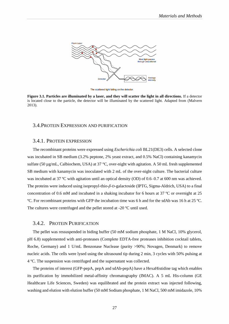
3.3.5. DYNAMIC LIGHT SCATTERING
Dynamic light scattering (DLS) is one of the most popular methods to determine the hydrodynamic
size of particles. Dynamic light scattering measures Brownian motion and relates this to the size of the
particles (Fig. 3.1). The particles are constantly moving due to Brownian motion, i.e. the movement of
particles due to the random collision with the other molecules of the liquid that surrounds the particle.
An important aspect of Brownian motion for DLS is that small particles move quickly and large particles
move more slowly. The particles are illuminated with a laser and the intensity fluctuations of the
scattered light are analyzed. The relationship between the size of a particle and its speed due to Brownian
motion is defined in the Stokes-Einstein equation (Malvern 2013). It is possible to compute the sphere
size distribution and give a description of the particle’s motion in the medium, measuring the diffusion
coefficient of the particle and using the autocorrelation function (Goldburg 1999).
All the samples analyzed by DLS were diluted in filtered (0.2 M) dH2O, and prepared carefully to
decrease external contaminants. The final concentration of iron in each sample was 0.2 g/mL. The zeta
potential of SPIONs was also measured. This parameter allows the study of colloidal stability by
measuring nanoparticle charge when an electrical field is applied. The zeta potential over a large range
of pH (2−11) was measured to determine the best condition. A Horiba SZ-100 was used to analyze the
samples.
Materials and Methods
27
Figure 3.1. Particles are illuminated by a laser, and they will scatter the light in all directions. If a detector
is located close to the particle, the detector will be illuminated by the scattered light. Adapted from (Malvern
2013).
3.4. PROTEIN EXPRESSION AND PURIFICATION
3.4.1. PROTEIN EXPRESSION
The recombinant proteins were expressed using Escherichia coli BL21(DE3) cells. A selected clone
was incubated in SB medium (3.2% peptone, 2% yeast extract, and 0.5% NaCl) containing kanamycin
sulfate (50 µg/mL, Calbiochem, USA) at 37 °C, over-night with agitation. A 50 mL fresh supplemented
SB medium with kanamycin was inoculated with 2 mL of the over-night culture. The bacterial culture
was incubated at 37 ºC with agitation until an optical density (OD) of 0.6–0.7 at 600 nm was achieved.
The proteins were induced using isopropyl-thio-β-D-galactoside (IPTG, Sigma-Aldrich, USA) to a final
concentration of 0.6 mM and incubated in a shaking incubator for 6 hours at 37 ºC or overnight at 25
ºC. For recombinant proteins with GFP the incubation time was 6 h and for the sdAb was 16 h at 25 ºC.
The cultures were centrifuged and the pellet stored at -20 ºC until used.
3.4.2. PROTEIN PURIFICATION
The pellet was ressuspended in biding buffer (50 mM sodium phosphate, 1 M NaCl, 10% glycerol,
pH 6.8) supplemented with anti-proteases (Complete EDTA-free proteases inhibition cocktail tablets,
Roche, Germany) and 1 U/mL Benzonase Nuclease (purity >90%; Novagen, Denmark) to remove
nucleic acids. The cells were lysed using the ultrasound tip during 2 min, 3 cycles with 50% pulsing at
4 ºC. The suspension was centrifuged and the supernatant was collected.
The proteins of interest (GFP-pepA, pepA and sdAb-pepA) have a HexaHistidine tag which enables
its purification by immobilized metal-affinity chromatography (IMAC). A 5 mL His-column (GE
Healthcare Life Sciences, Sweden) was equilibrated and the protein extract was injected following,
washing and elution with elution buffer (50 mM Sodium phosphate, 1 M NaCl, 500 mM imidazole, 10%
Materials and Methods
28
glycerol, pH 6.8). With the purpose to obtain higher purity proteins, an additional step of gel filtration
chromatography (Sephadex G-200, Sigma-Aldrich) was added.
The concentration of the protein in solution was determined through the Bradford assay. The methods
consist in the addition of an acidic dye (Bradford Dye Reagent, BIO-RAD, Germany) and the
measurement of the absorbance at 595 nm in a microplate reader. The absorbance of the sample when
compared to a standard curve provides the protein concentration. The standard curve was obtained using
bovine serum albumin (BSA, Sigma-Aldrich, USA) (Bradford 1976).
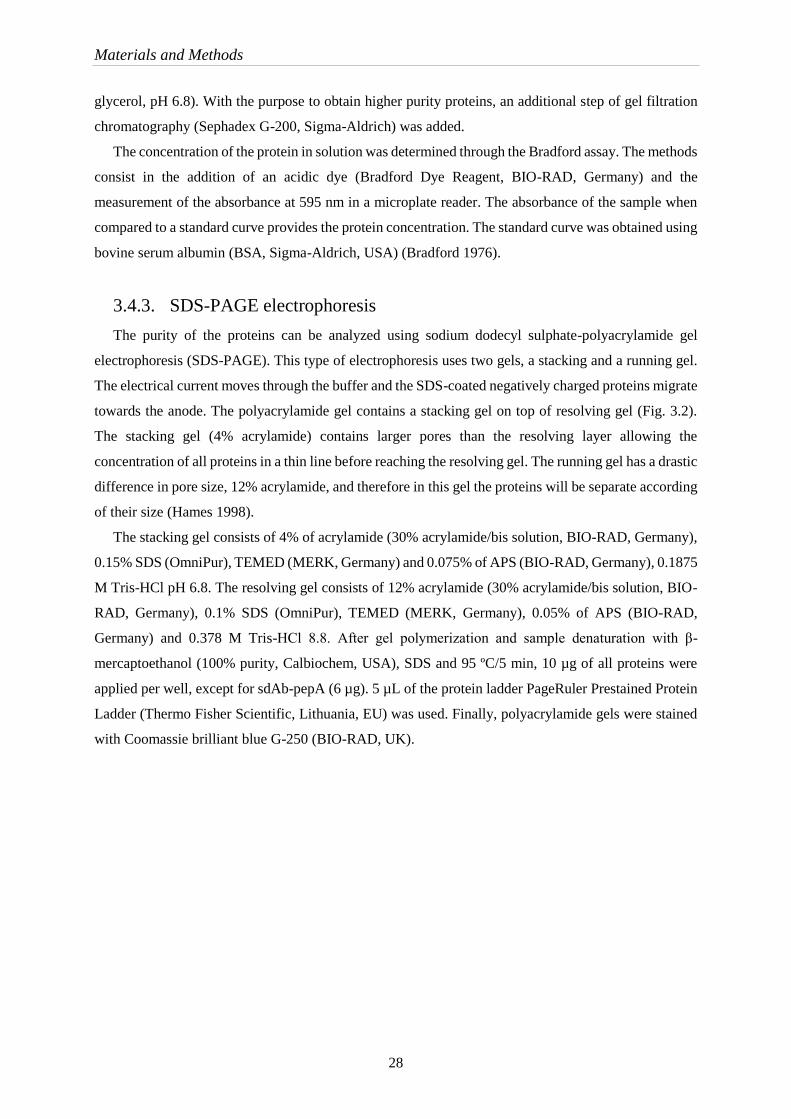
3.4.3. SDS-PAGE electrophoresis
The purity of the proteins can be analyzed using sodium dodecyl sulphate-polyacrylamide gel
electrophoresis (SDS-PAGE). This type of electrophoresis uses two gels, a stacking and a running gel.
The electrical current moves through the buffer and the SDS-coated negatively charged proteins migrate
towards the anode. The polyacrylamide gel contains a stacking gel on top of resolving gel (Fig. 3.2).
The stacking gel (4% acrylamide) contains larger pores than the resolving layer allowing the
concentration of all proteins in a thin line before reaching the resolving gel. The running gel has a drastic
difference in pore size, 12% acrylamide, and therefore in this gel the proteins will be separate according
of their size (Hames 1998).
The stacking gel consists of 4% of acrylamide (30% acrylamide/bis solution, BIO-RAD, Germany),
0.15% SDS (OmniPur), TEMED (MERK, Germany) and 0.075% of APS (BIO-RAD, Germany), 0.1875
M Tris-HCl pH 6.8. The resolving gel consists of 12% acrylamide (30% acrylamide/bis solution, BIO-
RAD, Germany), 0.1% SDS (OmniPur), TEMED (MERK, Germany), 0.05% of APS (BIO-RAD,
Germany) and 0.378 M Tris-HCl 8.8. After gel polymerization and sample denaturation with β-
mercaptoethanol (100% purity, Calbiochem, USA), SDS and 95 ºC/5 min, 10 µg of all proteins were
applied per well, except for sdAb-pepA (6 µg). 5 µL of the protein ladder PageRuler Prestained Protein
Ladder (Thermo Fisher Scientific, Lithuania, EU) was used. Finally, polyacrylamide gels were stained
with Coomassie brilliant blue G-250 (BIO-RAD, UK).
Materials and Methods
29
Figure 3.2. Scheme of SDS-PAGE system. A, Concentration of the proteins in the stacking gel. B, Migration and
separation of the proteins in the resolving gel.
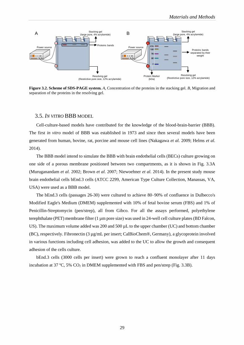
3.5. IN VITRO BBB MODEL
Cell-culture-based models have contributed for the knowledge of the blood-brain-barrier (BBB).
The first in vitro model of BBB was established in 1973 and since then several models have been
generated from human, bovine, rat, porcine and mouse cell lines (Nakagawa et al. 2009; Helms et al.
2014).
The BBB model intend to simulate the BBB with brain endothelial cells (BECs) culture growing on
one side of a porous membrane positioned between two compartments, as it is shown in Fig. 3.3A
(Muruganandam et al. 2002; Brown et al. 2007; Niewoehner et al. 2014). In the present study mouse
brain endothelial cells bEnd.3 cells (ATCC 2299, American Type Culture Collection, Manassas, VA,
USA) were used as a BBB model.
The bEnd.3 cells (passages 26-30) were cultured to achieve 80–90% of confluence in Dulbecco's
Modified Eagle's Medium (DMEM) supplemented with 10% of fetal bovine serum (FBS) and 1% of
Penicillin-Streptomycin (pen/strep), all from Gibco. For all the assays performed, polyethylene
terephthalate (PET) membrane filter (1 µm pore size) was used in 24-well cell culture plates (BD Falcon,
US). The maximum volume added was 200 and 500 µL to the upper chamber (UC) and bottom chamber
(BC), respectively. Fibronectin (3 µg/mL per insert; CalBioChem®, Germany), a glycoprotein involved
in various functions including cell adhesion, was added to the UC to allow the growth and consequent
adhesion of the cells culture.
bEnd.3 cells (3000 cells per insert) were grown to reach a confluent monolayer after 11 days
incubation at 37 ºC, 5% CO2 in DMEM supplemented with FBS and pen/strep (Fig. 3.3B).
Protein Marker
(kDa) Resolving gel
(Restrictive pore size, 12% acrylamide)
Power source
Stacking gel (large pore, 4% acrylamide)
Proteins bands
Resolving gel (Restrictive pore size, 12% acrylamide)
Stacking gel (large pore, 4% acrylamide)
Proteins bands separated by their
weight
A B
Power source

Materials and Methods
30
Figure 3.3. In vitro BBB model. A, The in vitro BBB model consists of a transwell system with an insert in which
bEnd.3 cells are grown separating two chambers A. The insert, or apical side, corresponds to the blood side, while
the base (bottom chamber) corresponds to the brain side. Peptides were added to the top compartment. B, bEnd.3
cells at 2, 4 and 10 days after culturing procedures.
3.5.1. IN VITRO TRANSCYTOSIS ASSAY AND DETECTION OF RECOMBINANT GFP
PROTEINS BY FLUORESCENCE
The first assays were made using GFP recombinant proteins (SPIONs-GFP-pepA) with the main
goal of estimate the amount of translocated protein and its interaction with the cells membrane by simply
using the fluorescence of the GFP (Green Fluorescent Protein).
Different incubation times were tested for both SPIONs-GFP and SPIONs-GFP-pepA. Initially
different time points (30 min 7 h) were tested.
6 µg/mL of SPIONs-GFP-pepA were added to the UC. The samples were diluted in DMEM without
phenol red (Gibco, ThermoFisher, USA) supplemented with 10% of FBS. All the assays were performed
using DMEM without phenol red medium. On the day of the assay, inserts were washed two times with
PBS 1X, at 37 ºC, followed by three washes with DMEM without phenol red, at 37ºC. Then, the samples
were added to the UC and incubated for 6 hours at 37 ºC, 5% CO2. After 6h of incubation, the samples
were collected to determine the apical and basolateral amount of protein. The content of recombinant
GFP proteins in the samples were quantified by fluorescence detection using a black microplate 96
(Eppendorf AG, Hamburg, Germany) in Infinite M200 (TECAN) fluorescence reader, using 395 and
509 nm for the excitation and emission wavelengths, respectively.
After established the best conditions for the GFP fused proteins, the following step was to evaluate
the SPIONs-pepA and SPIONS-sdAb-pepA. The time point of 6 h was chosen and the sample amount
was 50 µg/mL of each protein.
A
B

Materials and Methods
31
3.5.2. DETECTION OF PROTEINS BY WESTERN BLOT
The collected samples from the translocation assay with SPIONs-pepA and SPIONS-sdAb-pepA
were quantified by western blot, using an antibody against the His tag, present in both proteins.
First, samples were resolved by SDS-PAGE electrophoresis. 6 µL of UC and 15 µL of BC samples
previously denatured with β-mercaptoethanol were applied onto the gel. In addition, 100 ng of sdAb-
pepA and pepA control were added to the same gel.
The gel obtained from the SDS-PAGE electrophoresis was compressed with a solid nitrocellulose
membrane (Amersham Protran 0.2 NC, GE Healthcare Life Sciences, Sweden) in a cassette and
immersed in transfer buffer (20% of methanol) (BIO-RAD, Germany) between two parallel electrodes,
forming a sandwich (Fig. 3.4). The transfer occurred at 250 mA, during 1 h, and the membrane was
washed with dH2O several times to remove methanol, and lastly, the protein bands were stained with
Ponceau S solution (Sigma-Aldrich, USA) and washed several times with PBS 1x, 0.2% Tween
(CalBioChem®, Germany).
Figure 3.4. Western Blotting scheme. Transfer of proteins from SDS-PAGE gel to a nitrocellulose membrane.
A transfer “sandwich” is assembled with the following layers in order, from cathode (-) to anode (+): sponge, filter
paper, SDS-PAGE gel, nitrocellulose membrane, filter paper and sponge. Adapted from (BIO-RAD 2016).
Before incubation with the primary antibody, the membrane was incubated 1 h at RT with blocking
buffer (5% nonfat milk in PBS 1x, 0.2% Tween) to prevent the nonspecific binding of the antibodies.
Finally, the nitrocellulose membrane was incubated with 0.02 U/mL of a conjugate antibody (Anti-His6-
HRPeroxidase, Roche, Germany) diluted in 1% nonfat milk in PBS 1x with 0.2% Tween for 1 h at RT
with agitation.
For detection, the membrane was washed in PBS 1x to remove Tween and the ECL Prime solution
(GE Healthcare Life Sciences, Buckinghamshire, UK) was added. The solution contains a
Sponge
Filter paper
Gel
Membrane
Filter paper
Sponge
Cathode (-)
Current (I)
Anode (+)
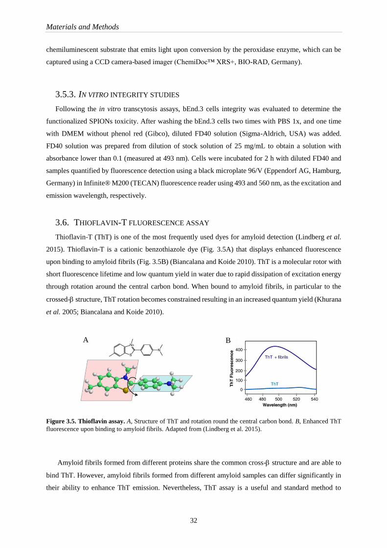
Materials and Methods
32
chemiluminescent substrate that emits light upon conversion by the peroxidase enzyme, which can be
captured using a CCD camera-based imager (ChemiDoc™ XRS+, BIO-RAD, Germany).
3.5.3. IN VITRO INTEGRITY STUDIES
Following the in vitro transcytosis assays, bEnd.3 cells integrity was evaluated to determine the
functionalized SPIONs toxicity. After washing the bEnd.3 cells two times with PBS 1x, and one time
with DMEM without phenol red (Gibco), diluted FD40 solution (Sigma-Aldrich, USA) was added.
FD40 solution was prepared from dilution of stock solution of 25 mg/mL to obtain a solution with
absorbance lower than 0.1 (measured at 493 nm). Cells were incubated for 2 h with diluted FD40 and
samples quantified by fluorescence detection using a black microplate 96/V (Eppendorf AG, Hamburg,
Germany) in Infinite® M200 (TECAN) fluorescence reader using 493 and 560 nm, as the excitation and
emission wavelength, respectively.
3.6. THIOFLAVIN-T FLUORESCENCE ASSAY
Thioflavin-T (ThT) is one of the most frequently used dyes for amyloid detection (Lindberg et al.
2015). Thioflavin-T is a cationic benzothiazole dye (Fig. 3.5A) that displays enhanced fluorescence
upon binding to amyloid fibrils (Fig. 3.5B) (Biancalana and Koide 2010). ThT is a molecular rotor with
short fluorescence lifetime and low quantum yield in water due to rapid dissipation of excitation energy
through rotation around the central carbon bond. When bound to amyloid fibrils, in particular to the
crossed- structure, ThT rotation becomes constrained resulting in an increased quantum yield (Khurana
et al. 2005; Biancalana and Koide 2010).
Figure 3.5. Thioflavin assay. A, Structure of ThT and rotation round the central carbon bond. B, Enhanced ThT
fluorescence upon binding to amyloid fibrils. Adapted from (Lindberg et al. 2015).
Amyloid fibrils formed from different proteins share the common cross- structure and are able to
bind ThT. However, amyloid fibrils formed from different amyloid samples can differ significantly in
their ability to enhance ThT emission. Nevertheless, ThT assay is a useful and standard method to
A B

Materials and Methods
33
rapidly measure amyloid fibril formation and to screen for inhibitors able to inhibit the aggregation
process.
The assay was performed with SensoLyte Thioflavin-T Beta-Amyloid (1-42) aggregation Kit
(AnaSpec, Fremont, CA) according to manufacturer’s protocol. Briefly, thioflavin-T (ThT)
measurements were made by tacking ThT aliquots and diluting them in assay buffer (SensoLyte
component A) to 200 M ThT (final concentration). Test samples (sdAb, sdAb-pepA, SPIONs-sdAb,
SPIONs-sdAb-pepA in PBS) were prepared in assay buffer containing 200 M ThT, 46 M A42and
3.7 M sdAb, 8.7 M sdAb-pepA, 1.6M SPIONs-sdAb and 1.5 M SPIONs-sdAb-pepA,
respectively. An inhibitor control was assayed containing ThT, A42 and 100 M of the supplied
aggregation inhibitor morin in parallel with the positive control containing ThT and A42 without
inhibitor. A vehicle control (ThT and PBS), a test sample control (ThT and test sample) and a blank
(ThT and assay buffer) were also performed.
Immediately after sample preparation, the fluorescence intensity was measured in a TECAN Infinite
M200 (Männedorf, Switzerland) at 37°C using an excitation of 440 nm and emission of 484 nm, with
15 sec shaking between reads to facilitate aggregation. The fluorescence was followed 1.5h with
measurements every 5 min. The fluorescence values were corrected by subtracting the background
fluorescence of ThT in assay buffer.


Part IV Results and Discussion

Results and discussion
37
Alzheimer’s disease is characterized by an accumulation of amyloid-β protein aggregates (A
oligomers and fibrils) in the brain tissue leading to an inflammatory response. From all current or under
investigation therapies, the antibody therapeutic approach is a promising path to tackle this devastating
disease. However, the main challenge for all therapies to the treatment of brain disorders is the
permeability of the BBB. Its principal function is to control the exchange of substances between the
blood and the fluids of the brain, imposing an obstacle to the treatment. This is true for either small
molecules or protein-based therapies, as the one assessed in this work (Banks and Erickson 2010; Neves
et al. 2016; Reiman 2016; Tycko 2016).
In the recent years, several strategies have been developed to delivery therapeutic molecules to the
brain, such as the use of cell penetrating peptides (CPPs) with capacity to translocate various types of
cargo molecules to interior of cells. Recently, CPP designed from an -helical domain of the dengue
virus capsid protein, pepH3, were studied and revealed a great ability to cross the BBB and return to the
blood circulation to be eliminated (Freire et al. 2013; Corte-Real et al. 2016; Kristensen et al. 2016).
The goal of the work described was to further develop a BBB translocation system with a therapeutic
and diagnostic entity. The translocation properties of CPP were combined with the therapeutic properties
of an antibody anti -amyloid into a delivery system amenable to magnetic resonance imaging.
Superparamagnetic iron oxide nanoparticles (SPIONs) were selected to provide magnetic properties to
the system. When iron oxide nanoparticles are below 20 nm, they have a single domain and became
superparamagnetic acting as a “single super spin” with high magnetic susceptibility. When an external
magnetic field is applied, the particles became magnetized until their saturation magnetization. Upon
removal of external magnetic field, SPIONs lose their magnetism, a property that can be useful in
biomedical applications (Wahajuddin and Arora 2012; Li et al. 2013; Santhosh and Ulrih 2013;
Shevtsov et al. 2015).
SPIONs are usually employed as a contrast agent in magnetic resonance imaging with better
properties than common gadolinium-based agents (Frisoni et al. 2010; Hola et al. 2015; Shevtsov et al.
2015). They might contribute to early detection of the -amyloid in the affected brain tissue, when
coupled to antibodies that recognize A
The work is divided in five parts: synthesis of SPIONs; surface modification; functionalization of
SPIONs with all proteins tested (SPIONs-GFP, SPIONs-GFP-pepA, SPIONs-pepA and SPIONs-sdAb-
pepA); evaluation of the system in a cell based BBB model and; assessment of the therapeutic properties
of the antibody in the functionalized nanoparticle.
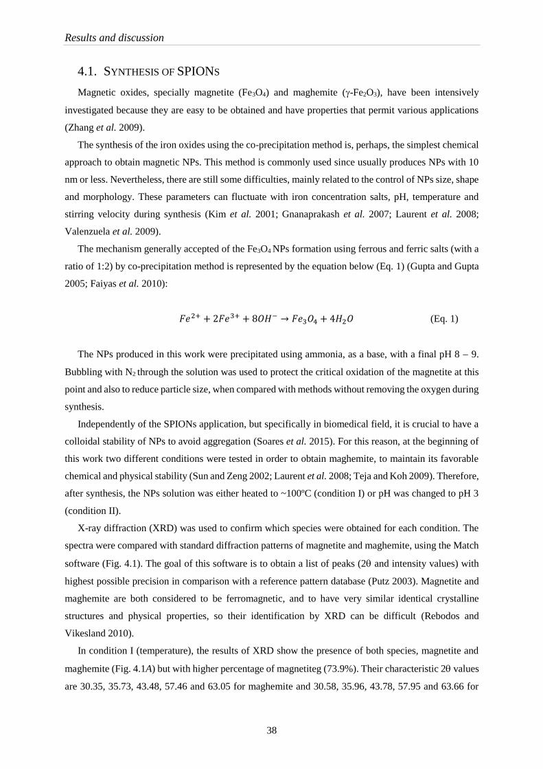
Results and discussion
38
4.1. SYNTHESIS OF SPIONS
Magnetic oxides, specially magnetite (Fe3O4) and maghemite (-Fe2O3), have been intensively
investigated because they are easy to be obtained and have properties that permit various applications
(Zhang et al. 2009).
The synthesis of the iron oxides using the co-precipitation method is, perhaps, the simplest chemical
approach to obtain magnetic NPs. This method is commonly used since usually produces NPs with 10
nm or less. Nevertheless, there are still some difficulties, mainly related to the control of NPs size, shape
and morphology. These parameters can fluctuate with iron concentration salts, pH, temperature and
stirring velocity during synthesis (Kim et al. 2001; Gnanaprakash et al. 2007; Laurent et al. 2008;
Valenzuela et al. 2009).
The mechanism generally accepted of the Fe3O4 NPs formation using ferrous and ferric salts (with a
ratio of 1:2) by co-precipitation method is represented by the equation below (Eq. 1) (Gupta and Gupta
2005; Faiyas et al. 2010):
𝐹𝑒2+ + 2𝐹𝑒3+ + 8𝑂𝐻− → 𝐹𝑒3𝑂4 + 4𝐻2𝑂 (Eq. 1)
The NPs produced in this work were precipitated using ammonia, as a base, with a final pH 8 9.
Bubbling with N2 through the solution was used to protect the critical oxidation of the magnetite at this
point and also to reduce particle size, when compared with methods without removing the oxygen during
synthesis.
Independently of the SPIONs application, but specifically in biomedical field, it is crucial to have a
colloidal stability of NPs to avoid aggregation (Soares et al. 2015). For this reason, at the beginning of
this work two different conditions were tested in order to obtain maghemite, to maintain its favorable
chemical and physical stability (Sun and Zeng 2002; Laurent et al. 2008; Teja and Koh 2009). Therefore,
after synthesis, the NPs solution was either heated to ~100ºC (condition I) or pH was changed to pH 3
(condition II).
X-ray diffraction (XRD) was used to confirm which species were obtained for each condition. The
spectra were compared with standard diffraction patterns of magnetite and maghemite, using the Match
software (Fig. 4.1). The goal of this software is to obtain a list of peaks (2 and intensity values) with
highest possible precision in comparison with a reference pattern database (Putz 2003). Magnetite and
maghemite are both considered to be ferromagnetic, and to have very similar identical crystalline
structures and physical properties, so their identification by XRD can be difficult (Rebodos and
Vikesland 2010).
In condition I (temperature), the results of XRD show the presence of both species, magnetite and
maghemite (Fig. 4.1A) but with higher percentage of magnetiteg (73.9%). Their characteristic 2 values
are 30.35, 35.73, 43.48, 57.46 and 63.05 for maghemite and 30.58, 35.96, 43.78, 57.95 and 63.66 for

Results and discussion
39
magnetite. In condition II (pH) (Fig. 4.1B) the prevalent species is magnetite (75.6%) with a lower
percentage of maghemite (24.4%). Their characteristic 2 values for magnetite are 30.32, 35.59, 43.28,
53.80, 57.14, and for the maghemite species the 2 values are 62.86 and 30.12, 35.59, 43.25, 53.67,
57.28 and 62.28 (Appendix II).
The magnetite (Fe2+) species is oxidized into the more stable maghemite (Fe3+) during both processes
(Rebodos and Vikesland 2010). However, the oxidation of the NPs by pH is less efficient when
compared with oxidation by temperature.
Figure 4.1. XRD of SPIONs oxidized by different conditions. A, SPIONs oxidized by temperature. B, SPIONs
oxidized by acidic pH, 3. C, SPIONs oxidized by pH and surface modification with 0.3 M DMSA. The Miller
indices (hkl) are represented in panel A.
The average grain size was calculated for naked NPs (condition I and II) and DMSA coated SPIONs
using Scherrer’s equation (Eq. 2), where is equivalent to the grain average core diameter, K is the grain
shape factor (K = 0.94), is the incident x-ray wavelength, denotes the full width at half-maximum
(in radians) of the highest intensity and is the corresponding diffraction angle (2) (Soares et al. 2015).
𝜏 =𝐾𝜆
𝛽 cos𝜃 (Eq. 2)
The average grain obtained using equation 2, for naked SPIONs using condition I (2 = 35.58) was
8.9 nm and using condition II (2 = 35.53) was 8 nm, which indicates that the oxidation method does
not change the average particle size of the NPs.
The purpose of using iron oxide NPs in this work is to explore their magnetic properties in magnetic
resonance imaging, as a contrast agent. The magnetite has a higher magnetic saturation than maghemite
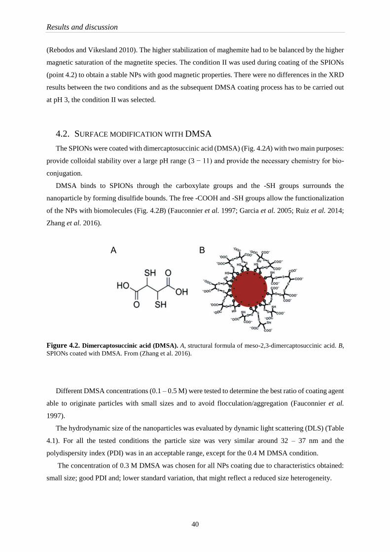
Results and discussion
40
(Rebodos and Vikesland 2010). The higher stabilization of maghemite had to be balanced by the higher
magnetic saturation of the magnetite species. The condition II was used during coating of the SPIONs
(point 4.2) to obtain a stable NPs with good magnetic properties. There were no differences in the XRD
results between the two conditions and as the subsequent DMSA coating process has to be carried out
at pH 3, the condition II was selected.
4.2. SURFACE MODIFICATION WITH DMSA
The SPIONs were coated with dimercaptosuccinic acid (DMSA) (Fig. 4.2A) with two main purposes:
provide colloidal stability over a large pH range (3 − 11) and provide the necessary chemistry for bio-
conjugation.
DMSA binds to SPIONs through the carboxylate groups and the -SH groups surrounds the
nanoparticle by forming disulfide bounds. The free -COOH and -SH groups allow the functionalization
of the NPs with biomolecules (Fig. 4.2B) (Fauconnier et al. 1997; Garcia et al. 2005; Ruiz et al. 2014;
Zhang et al. 2016).
Figure 4.2. Dimercaptosuccinic acid (DMSA). A, structural formula of meso-2,3-dimercaptosuccinic acid. B,
SPIONs coated with DMSA. From (Zhang et al. 2016).
Different DMSA concentrations (0.1 – 0.5 M) were tested to determine the best ratio of coating agent
able to originate particles with small sizes and to avoid flocculation/aggregation (Fauconnier et al.
1997).
The hydrodynamic size of the nanoparticles was evaluated by dynamic light scattering (DLS) (Table
4.1). For all the tested conditions the particle size was very similar around 32 – 37 nm and the
polydispersity index (PDI) was in an acceptable range, except for the 0.4 M DMSA condition.
The concentration of 0.3 M DMSA was chosen for all NPs coating due to characteristics obtained:
small size; good PDI and; lower standard variation, that might reflect a reduced size heterogeneity.
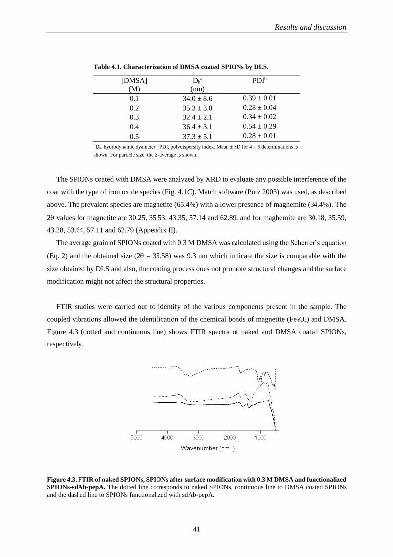
Results and discussion
41
Table 4.1. Characterization of DMSA coated SPIONs by DLS.
aDh, hydrodynamic dyameter. bPDI, polydispersity index. Mean ± SD for 4 – 6 determinations is
shown. For particle size, the Z-average is shown.
The SPIONs coated with DMSA were analyzed by XRD to evaluate any possible interference of the
coat with the type of iron oxide species (Fig. 4.1C). Match software (Putz 2003) was used, as described
above. The prevalent species are magnetite (65.4%) with a lower presence of maghemite (34.4%). The
2 values for magnetite are 30.25, 35.53, 43.35, 57.14 and 62.89; and for maghemite are 30.18, 35.59,
43.28, 53.64, 57.11 and 62.79 (Appendix II).
The average grain of SPIONs coated with 0.3 M DMSA was calculated using the Scherrer’s equation
(Eq. 2) and the obtained size (2 = 35.58) was 9.3 nm which indicate the size is comparable with the
size obtained by DLS and also, the coating process does not promote structural changes and the surface
modification might not affect the structural properties.
FTIR studies were carried out to identify of the various components present in the sample. The
coupled vibrations allowed the identification of the chemical bonds of magnetite (Fe3O4) and DMSA.
Figure 4.3 (dotted and continuous line) shows FTIR spectra of naked and DMSA coated SPIONs,
respectively.
Figure 4.3. FTIR of naked SPIONs, SPIONs after surface modification with 0.3 M DMSA and functionalized
SPIONs-sdAb-pepA. The dotted line corresponds to naked SPIONs, continuous line to DMSA coated SPIONs
and the dashed line to SPIONs functionalized with sdAb-pepA.
[DMSA]
(M)
Dha
(nm)
PDIb
0.1 34.0 ± 8.6 0.39 ± 0.01
0.2 35.3 ± 3.8 0.28 ± 0.04
0.3 32.4 ± 2.1 0.34 ± 0.02
0.4 36.4 ± 3.1 0.54 ± 0.29
0.5 37.3 ± 5.1 0.28 ± 0.01

Results and discussion
42
From the spectra it was possible to observe the characteristic bands of magnetite at 560 cm-1, the
stretching vibration mode of Fe−O. At around 3200 and 1580 cm-1 the stretching vibration mode of −OH
was observed due to the presence of water vapor (Soares et al. 2014). The presence of DMSA at the
surface of the NPs was identified by the characteristic asymmetric and symmetric stretching of carbonyl
groups at 1550 and 1310 cm-1, respectively. These peaks are shifted to lower wavenumbers compared
with the carbonyl absorption in free DMSA at around 1700 cm-1 (Palma et al. 2015). The absorption
bands around 1090 and 930 cm-1 are due the vibration of C−O and −NH2 of the bound proteins (peptide
A and antibody) (Zhang et al. 2009). It is possible to conclude that the coating process and the
functionalization with sdAb-pepA was successful.
The charge or electrostatic potential – zeta potential − at the surface of the nanoparticle has a
significant importance in the colloidal stability of the system and can be considered an indicator of the
colloidal stability due to the electrostatic interaction (Mahmoudi et al. 2011). NPs with values higher
than +25 mV and values lower than -25 mV usually are stable, otherwise they will have a propensity to
aggregate due to Van Der Wall inter-particle attraction (Mahmoudi et al. 2011).
Zeta potential was measured over a large pH range to determine the interval where SPIONs are
stable (Table 4.2). The best zeta potential was -73.39 mV at pH 9. However, the selected pH for the
functionalization procedure was 7.4 (of the PBS buffer), where the zeta-potential value is acceptable to
avoid particle aggregation.
Table 4.2. The pH effect in the zeta potential of the DMSA coated
SPIONs. SPIONs in solution at different pH, coated with 0.3 M DMSA.
Mean ± SD, for 3 – 6 determinations is shown.
Naked SPIONs have a propensity to self-associate leading to the formation of clusters, that were
observed by TEM (Fig. 4.5A,B). The coat with 0.3 M DMSA provides stabilization to the NPs in solution
pH Zeta Potential (mV)
2 -4.7 ± 1.8
3 -30.0 ± 9.2
4 -47.9 ± 2.1
5 -29.5 ± 2.9
6 -33.2 ± 1.9
7 -63.1 ± 8.1
8 -67.6 ± 0.8
9 -73.4 ± 2.3
10 -58.9 ± 1.8
11 0.92 ± 0.2
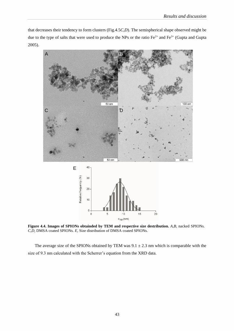
Results and discussion
43
that decreases their tendency to form clusters (Fig.4.5C,D). The semispherical shape observed might be
due to the type of salts that were used to produce the NPs or the ratio Fe2+ and Fe3+ (Gupta and Gupta
2005).
Figure 4.4. Images of SPIONs obtainded by TEM and respective size destribution. A,B, nacked SPIONs.
C,D, DMSA coated SPIONs. E, Size distribution of DMSA coated SPIONs.
The average size of the SPIONs obtained by TEM was 9.1 ± 2.3 nm which is comparable with the
size of 9.3 nm calculated with the Scherrer’s equation from the XRD data.
E
A B
C D
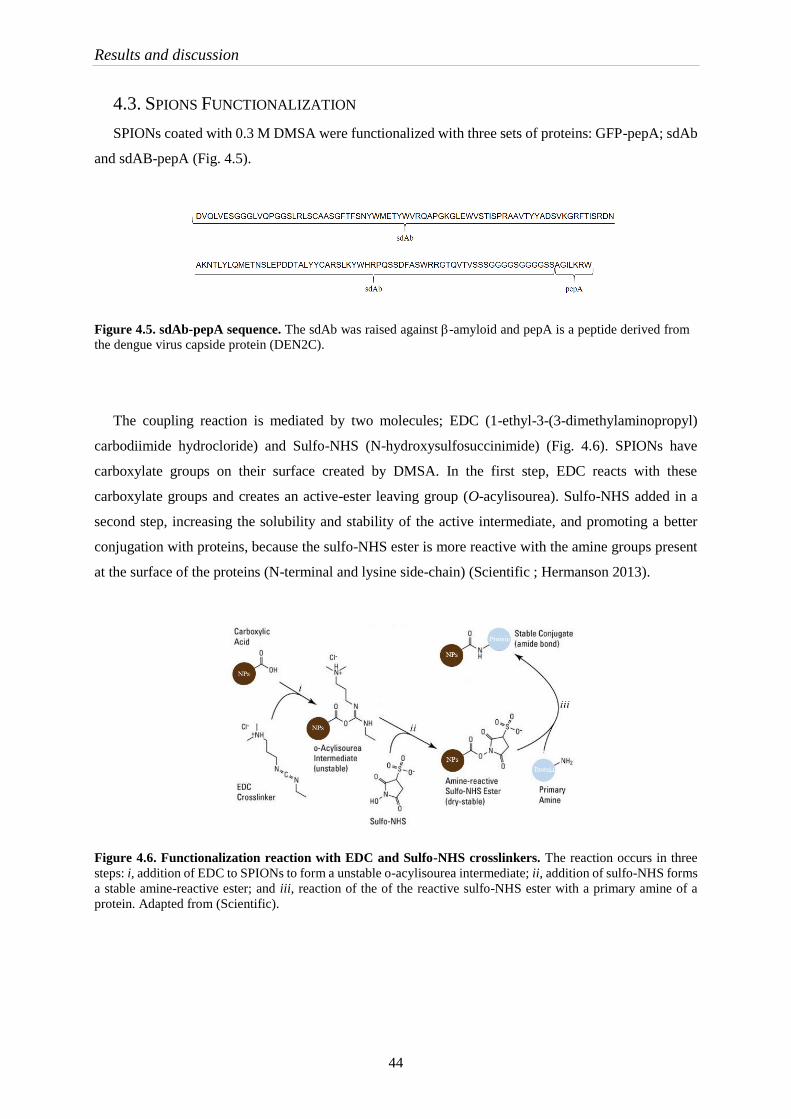
Results and discussion
44
4.3. SPIONS FUNCTIONALIZATION
SPIONs coated with 0.3 M DMSA were functionalized with three sets of proteins: GFP-pepA; sdAb
and sdAB-pepA (Fig. 4.5).
Figure 4.5. sdAb-pepA sequence. The sdAb was raised against -amyloid and pepA is a peptide derived from
the dengue virus capside protein (DEN2C).
The coupling reaction is mediated by two molecules; EDC (1-ethyl-3-(3-dimethylaminopropyl)
carbodiimide hydrocloride) and Sulfo-NHS (N-hydroxysulfosuccinimide) (Fig. 4.6). SPIONs have
carboxylate groups on their surface created by DMSA. In the first step, EDC reacts with these
carboxylate groups and creates an active-ester leaving group (O-acylisourea). Sulfo-NHS added in a
second step, increasing the solubility and stability of the active intermediate, and promoting a better
conjugation with proteins, because the sulfo-NHS ester is more reactive with the amine groups present
at the surface of the proteins (N-terminal and lysine side-chain) (Scientific ; Hermanson 2013).
Figure 4.6. Functionalization reaction with EDC and Sulfo-NHS crosslinkers. The reaction occurs in three
steps: i, addition of EDC to SPIONs to form a unstable o-acylisourea intermediate; ii, addition of sulfo-NHS forms
a stable amine-reactive ester; and iii, reaction of the of the reactive sulfo-NHS ester with a primary amine of a
protein. Adapted from (Scientific).

Results and discussion
45
SPIONs were functionalized with GFP-pepA (~30 kDa) and observed by fluorescence microscopy,
as a first approach to easily assess if the bio-conjugation was effective (Fig. 4.7). It was possible to
observe clusters of SPIONs with GFP-pepA. The large size of the clusters might be due to the stirring
process employed during coating of the NPs. This batch of SPIONs were produced using preliminary
conditions, before the surface modification process with DMSA had been improved (point 4.2). A
critical point during optimization of the surface modification process was the change from a mechanical
stirring to an ultrasound based method during incubation with DMSA. Under mechanical stirring the
dispersion could not be homogeneous, but using an ultra sounds bath during incubation of SPIONs with
0.3 M DMSA avoided this problem.
Figure 4.7. Images of SPIONs-GFP-pepA obtained by fluorescence microscopy.
After the selection of the best condition to achieve small nanoparticle size, the functionalization
reaction was evaluated at two different temperatures: room temperature (~25 ºC) and 4 ºC. It was
important to verify if the temperature could influence protein integrity during the 18 h of the
functionalization procedure. Both conditions were analyzed by SDS-PAGE (Fig. 4.8).
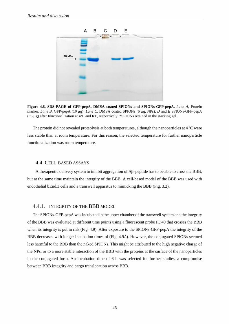
Results and discussion
46
Figure 4.8. SDS-PAGE of GFP-pepA, DMSA coated SPIONs and SPIONs-GFP-pepA. Lane A, Protein
marker; Lane B, GFP-pepA (10 g); Lane C, DMSA coated SPIONs (6 g, NPs); D and E SPIONs-GFP-pepA
(~5 g) after functionalization at 4ºC and RT, respectively. *SPIONs retained in the stacking gel.
The protein did not revealed proteolysis at both temperatures, although the nanoparticles at 4 ºC were
less stable than at room temperature. For this reason, the selected temperature for further nanoparticle
functionalization was room temperature.
4.4. CELL-BASED ASSAYS
A therapeutic delivery system to inhibit aggregation of A-peptide has to be able to cross the BBB,
but at the same time maintain the integrity of the BBB. A cell-based model of the BBB was used with
endothelial bEnd.3 cells and a transwell apparatus to mimicking the BBB (Fig. 3.2).
4.4.1. INTEGRITY OF THE BBB MODEL
The SPIONs-GFP-pepA was incubated in the upper chamber of the transwell system and the integrity
of the BBB was evaluated at different time points using a fluorescent probe FD40 that crosses the BBB
when its integrity is put in risk (Fig. 4.9). After exposure to the SPIONs-GFP-pepA the integrity of the
BBB decreases with longer incubation times of (Fig. 4.9A). However, the conjugated SPIONs seemed
less harmful to the BBB than the naked SPIONs. This might be attributed to the high negative charge of
the NPs, or to a more stable interaction of the BBB with the proteins at the surface of the nanoparticles
in the conjugated form. An incubation time of 6 h was selected for further studies, a compromise
between BBB integrity and cargo translocation across BBB.
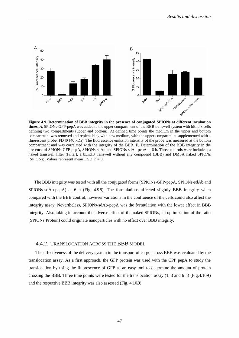
Results and discussion
47
Figure 4.9. Determination of BBB integrity in the presence of conjugated SPIONs at different incubation
times. A, SPIONs-GFP-pepA was added to the upper compartment of the BBB transwell system with bEnd.3 cells
defining two compartments (upper and bottom). At defined time points the medium in the upper and bottom
compartment was removed and replenishing with new medium, with the upper compartment supplemented with a
fluorescent probe, FD40 (40 kDa). The fluorescence emission intensity of the probe was measured at the bottom
compartment and was correlated with the integrity of the BBB. B, Determination of the BBB integrity in the
presence of SPIONs-GFP-pepA, SPIONs-sdAb and SPIONs-sdAb-pepA at 6 h. Three controls were included: a
naked transwell filter (Filter), a bEnd.3 transwell without any compound (BBB) and DMSA naked SPIONs
(SPIONs). Values represent mean SD, n = 3.
The BBB integrity was tested with all the conjugated forms (SPIONs-GFP-pepA, SPIONs-sdAb and
SPIONs-sdAb-pepA) at 6 h (Fig. 4.9B). The formulations affected slightly BBB integrity when
compared with the BBB control, however variations in the confluence of the cells could also affect the
integrity assay. Nevertheless, SPIONs-sdAb-pepA was the formulation with the lower effect in BBB
integrity. Also taking in account the adverse effect of the naked SPIONs, an optimization of the ratio
(SPIONs:Protein) could originate nanoparticles with no effect over BBB integrity.
4.4.2. TRANSLOCATION ACROSS THE BBB MODEL
The effectiveness of the delivery system in the transport of cargo across BBB was evaluated by the
translocation assay. As a first approach, the GFP protein was used with the CPP pepA to study the
translocation by using the fluorescence of GFP as an easy tool to determine the amount of protein
crossing the BBB. Three time points were tested for the translocation assay (1, 3 and 6 h) (Fig.4.10A)
and the respective BBB integrity was also assessed (Fig. 4.10B).
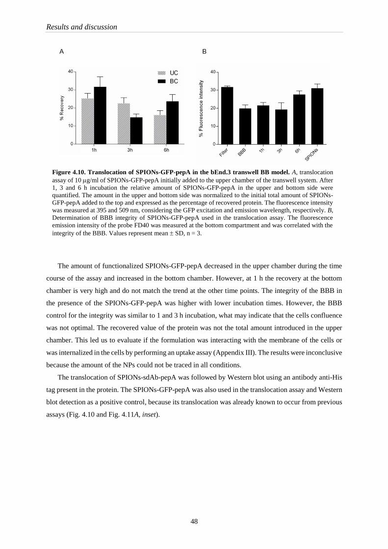
Results and discussion
48
Figure 4.10. Translocation of SPIONs-GFP-pepA in the bEnd.3 transwell BB model. A, translocation
assay of 10 g/ml of SPIONs-GFP-pepA initially added to the upper chamber of the transwell system. After
1, 3 and 6 h incubation the relative amount of SPIONs-GFP-pepA in the upper and bottom side were
quantified. The amount in the upper and bottom side was normalized to the initial total amount of SPIONs-
GFP-pepA added to the top and expressed as the percentage of recovered protein. The fluorescence intensity
was measured at 395 and 509 nm, considering the GFP excitation and emission wavelength, respectively. B,
Determination of BBB integrity of SPIONs-GFP-pepA used in the translocation assay. The fluorescence
emission intensity of the probe FD40 was measured at the bottom compartment and was correlated with the
integrity of the BBB. Values represent mean SD, n = 3.
The amount of functionalized SPIONs-GFP-pepA decreased in the upper chamber during the time
course of the assay and increased in the bottom chamber. However, at 1 h the recovery at the bottom
chamber is very high and do not match the trend at the other time points. The integrity of the BBB in
the presence of the SPIONs-GFP-pepA was higher with lower incubation times. However, the BBB
control for the integrity was similar to 1 and 3 h incubation, what may indicate that the cells confluence
was not optimal. The recovered value of the protein was not the total amount introduced in the upper
chamber. This led us to evaluate if the formulation was interacting with the membrane of the cells or
was internalized in the cells by performing an uptake assay (Appendix III). The results were inconclusive
because the amount of the NPs could not be traced in all conditions.
The translocation of SPIONs-sdAb-pepA was followed by Western blot using an antibody anti-His
tag present in the protein. The SPIONs-GFP-pepA was also used in the translocation assay and Western
blot detection as a positive control, because its translocation was already known to occur from previous
assays (Fig. 4.10 and Fig. 4.11A, inset).

Results and discussion
49
Figure 4.11. Translocation of SPIONs-GFP-pepA and SPIONs-sdAb-pepA in the bEnd.3 transwell BBB
evaluated by western blot. A, Translocation assay of 10 g/ml of SPIONs-GFP-pepA or SPIONs-sdAb-pepA
initially added to the upper chamber of the transwell system. The detection of SPIONs-GFP-pepA of this
formulation batch was also tested by fluorescence detection (inset). B, after 6 h incubation the relative amount of
protein in the upper and bottom sides was quantified by Western blot. For immunodetection, a conjugate anti-His-
HRP mAb was used. As positive control of the anti-His-HRP detection, 100 ng of purified GFP-pepA or sdAb-
pepA was used. The (%) of recovery was determined as the ratio of the protein detected in each chamber after 6 h
to the total amount of protein added to the upper chamber at the beginning of the assay. n.d., not detected.
The translocation of the SPIONs-GFP-pepA was also confirmed by Western blot detection (Fig.
4.11A). The recovered values obtained by fluorescence detection and Western blot were 31.4 3.3 and
13.2 2.9 %, respectively.
In the translocation assay of SPIONs-sdAb-pepA no protein was detected in the bottom chamber
and only a small fraction was detected in the upper chamber. One possibility for this difference is the
fact that the fusion protein of sdAb-pepA could affect the cell penetrating function of pepA. If this turns
out to be the case, a different N-terminal fusion maybe be evaluated (SPIONs-pepA-sdAb). Other
possibility is the internalization of the SPIONs-sdAb-pepA and hence the cellular uptake of this
formulation should be evaluated, as well as the type of interaction of the SPIONs-sdAb-pepA with the
cell membrane (receptor mediator transcytosis – RMT versus adsorptive mediated transcytosis – AMT)
(Jones and Shusta 2007).
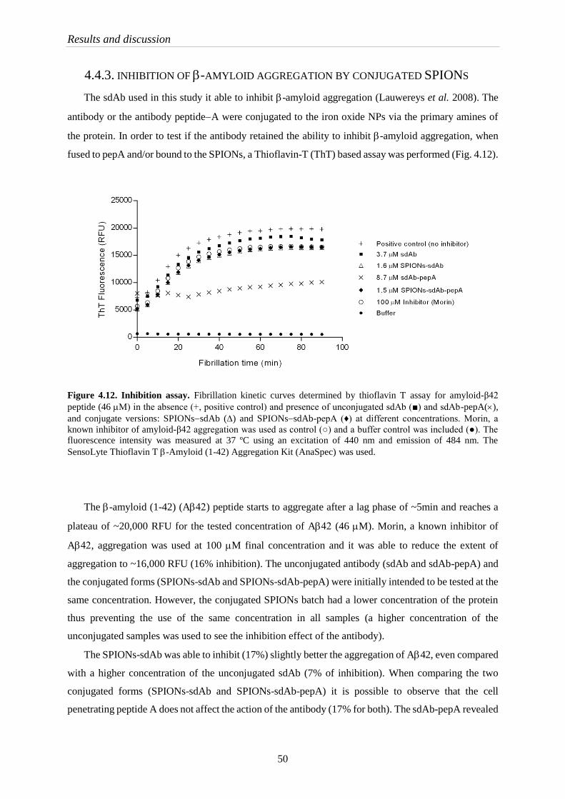
Results and discussion
50
4.4.3. INHIBITION OF -AMYLOID AGGREGATION BY CONJUGATED SPIONS
The sdAb used in this study it able to inhibit -amyloid aggregation (Lauwereys et al. 2008). The
antibody or the antibody peptideA were conjugated to the iron oxide NPs via the primary amines of
the protein. In order to test if the antibody retained the ability to inhibit -amyloid aggregation, when
fused to pepA and/or bound to the SPIONs, a Thioflavin-T (ThT) based assay was performed (Fig. 4.12).
Figure 4.12. Inhibition assay. Fibrillation kinetic curves determined by thioflavin T assay for amyloid-β42
peptide (46 M) in the absence (+, positive control) and presence of unconjugated sdAb (■) and sdAb-pepA(),
and conjugate versions: SPIONssdAb () and SPIONssdAb-pepA (♦) at different concentrations. Morin, a
known inhibitor of amyloid-β42 aggregation was used as control (○) and a buffer control was included (●). The
fluorescence intensity was measured at 37 ºC using an excitation of 440 nm and emission of 484 nm. The
SensoLyte Thioflavin T -Amyloid (1-42) Aggregation Kit (AnaSpec) was used.
The -amyloid (1-42) (A) peptide starts to aggregate after a lag phase of ~5min and reaches a
plateau of ~20,000 RFU for the tested concentration of A (46 M). Morin, a known inhibitor of
A, aggregation was used at 100 M final concentration and it was able to reduce the extent of
aggregation to ~16,000 RFU (16% inhibition). The unconjugated antibody (sdAb and sdAb-pepA) and
the conjugated forms (SPIONs-sdAb and SPIONs-sdAb-pepA) were initially intended to be tested at the
same concentration. However, the conjugated SPIONs batch had a lower concentration of the protein
thus preventing the use of the same concentration in all samples (a higher concentration of the
unconjugated samples was used to see the inhibition effect of the antibody).
The SPIONs-sdAb was able to inhibit (17%) slightly better the aggregation of A42, even compared
with a higher concentration of the unconjugated sdAb (7% of inhibition). When comparing the two
conjugated forms (SPIONs-sdAb and SPIONs-sdAb-pepA) it is possible to observe that the cell
penetrating peptide A does not affect the action of the antibody (17% for both). The sdAb-pepA revealed

Results and discussion
51
the highest inhibition of A42 aggregation (52%), however this form was tested at a higher
concentration.
The two nanoparticles formulations (SPIONs-sdAb and SPIONs-sdAb-pepA) were tested at a low
concentration but exhibited a similar inhibition level (17%) when compared with the inhibitor morin
(16%). Therefor the conjugation of the antibody to the nanoparticles does not appear to affect its
function.


Part V Concluding Remarks and
Future Perspectives


Concluding Remarks and Future Perspectives
55
Central nervous system (CNS) disorders affect up to 1 billion people worldwide. It is estimated that
Alzheimer and other dementias constitute 3% of the total deaths in 2015. The number is expected to
double every 20 years. There is a lack of therapies to treat these disorders due to the properties of blood
brain barrier (BBB). The BBB restricts the transfer of therapeutic molecules between the blood and the
brain. Therefore, there is a need to find molecules that can overcome this obstacle and carry with them
the drugs. These can either be achieved through targeting of brain receptors, via receptor-mediated
transport (RMT); or through adsorptive-mediated transcytosis (AMT). Cell penetrating peptides (CPP)
have been investigated has a drug delivery system due to their ability to cross cell membrane, and are
expected to also be able to transport different cargos across the BBB.
Alzheimer’s disease is characterized by an accumulation of amyloid aggregates in the brain.
Antibodies that recognize can be used not only for targeted imaging but also as promising
immunotherapy for AD, by stopping aggregation.
There is an urgent need to detect AD in early stages, avoiding the decline of the brain functions and
cell death. Usually, the disease is detected using imaging techniques (magnetic resonance of imaging
(MRI) or positron emission tomography), that only allow to detect later stages of the disease.
The possibility to develop a theranostic system that combines the early diagnostic and the therapy
might be a progress for the treatment of AD.
The main goal of this project was to develop a drug delivery system able to cross the blood brain
barrier (BBB) with therapeutic properties to tackle Alzheimer’s disease. The system comprises
superparamagnetic iron oxide nanoparticles (SPIONs), a cell penetrating peptide (pepA) and an antibody
against -amyloid (sdAb).
Initially, iron oxide nanoparticles were synthetized by chemical co-precipitation method with
subsequent oxidation. The main purpose of the oxidation was to obtain maghemite species (-Fe2O3), a
more stable specie. The oxidation of the SPIONs was evaluated in two different conditions: temperature
(condition I) and pH (condition II). For both conditions a mixture of maghemite and magnetite was
present. The oxidation condition through pH was the one selected for three main reasons i) SPIONs
were more stable by pH oxidation; ii) after characterization it was possible to conclude that the size of
the nanoparticles was similar between both conditions (8 and 8.9 nm for condition I and II, respectively)
and iii) the subsequent NPs surface modification reaction had to proceed at pH 3.
The SPIONs surface modification with DMSA was tested at different concentrations. The 0.3 M
DMSA condition was selected due to lower size of the NPs obtained, determined by DLS. The properties
of the iron oxide nanoparticles were analyzed after modification and no alteration of properties was
observed with coating (size, crystalline structure). In addition, the coating consefered further stability to
the nanoparticles.
Concluding Remarks and Future Perspectives
56
Furhter functionalization of DMSA coated SPIONS with GFP-pepA, sdAb and sdAb-pepA was
successful achieved. Reactions at either at 4 ºC or room temperature, resulted in conservation of protein
integrity as analyzed by SDS-PAGE.
The drug delivery system was tested in endothelial cells (bEnd.3) using a transwell system to
mimicking the BBB. The integrity of the cells and the translocation were evaluated. SPIONs and
SPIONs-GFP-pepA had some effect on the BBB integrity, but SPIONs-sdAb-pepA was the formulation
with lower impact on the endothelial cell barrier (SPIONs-sdAb-pepA < SPIONs-GFP-pepA <
SPIONs). After 6 h, 23.7 ± 3.7 % of SPIONs-GFP-pepA successfully crossed the BBB, but the same
was not observed for SPIONs-sdAb-pepA. Further studies need to be carried out to understand this
behavior, but a possible explanation might be related to protein rearrangement during functionalization
and consequent blocking of pepA properties to cross the BBB.
sdAb, sdAb-pepA, SPIONs-sdAb and SPIONs-sdAb-pepA were able to inhibit the aggregation of -
amyloid protein, when compared with a Morin (commercially available inhibitor). The functionalization
of the SPIONs with the antibody does not seem to affect function of the latter. Moreover, SPIONs-sdAb
is able to inhibit -amyloid aggregation at a lower concentration where compared with sdAb alone.
In general, the theranostic drug delivery system was successfully developed, but further optimization
will be necessary. SPIONs-sdAb-pepA formulations have to be modified due to the lack translocation
across the BBB. The order of the proteins in the fusion protein can be reversed with the CPP at the N-
terminal (SPIONs-pepA-sdAb), or different size linkers between the two proteins can be evaluated
(SPIONs-sdAb-(linker)n-pepA), in order to maintain the cell penetration ability of the peptide.
The ratio of SPIONs:protein should also be studied during the functionalization and optimized.
Naked SPIONs affected the integrity of the BBB, but the effect decreased with functionalization. A
lower ratio of SPIONs:protein might decrease even further the adverse effect on the integrity of the
BBB.
The system has also to be tested by magnetic resonance imaging to evaluate the full potential of the
system.
In conclusion, the system is able to play their dual function: the diagnostic (using the SPIONs as a
contrast agent) and the therapy, (using the sdAb to bind the A peptides). Although, this system has
other advantage: the pepA, allows it to cross the BBB and get to the target in the brain, to exert its
function.

Concluding Remarks and Future Perspectives
57
Application of this drug delivery system to other CNS diseases might be discovered in the future, for
instance, through the replacement of sdAb by other antibody that recognize the protein aggregates
related to Parkinson disease.


References

References
61
Albert M., DeCarli C., DeKosky S., Leon M., Foster N. L., Fox N., Frank R., Frackowiak R., Jack C.,
Jagust W. J., Knopman D., Morris J. C., Petersen R. C., Reiman E., Scheltens P., Small G.,
Soininen H., Thal L., Wahlund L., Thies W., Weiner M. W. and Khachaturian Z., The Use of
MRI and PET for Clinical Diagnosis of Dementia and Investigation of Cognitive Impairment:
A Consensus Report, in: Alzheimer’s Association Neuroimaging Work Group Consensus
Report.
Andrieux K. and Couvreur P. (2013) Nanomedicine as a promising approach for the treatment and
diagnosis of brain diseases: the example of Alzheimer's disease. Ann. Pharm. Fr. 71: 225-233.
Banks W. A. and Erickson M. A. (2010) The blood-brain barrier and immune function and dysfunction.
Neurobiol. Dis. 37: 26-32.
Biancalana M. and Koide S. (2010) Molecular mechanism of Thioflavin-T binding to amyloid fibrils.
Biochim. Biophys. Acta 1804: 1405-1412.
Bradford M. M. (1976) A rapid and sensitive method for the quantitation of microgram quantities of
protein utilizing the principle of protein-dye binding. Anal. Biochem. 72: 248-254.
Brown R. C., Morris A. P. and O'Neil R. G. (2007) Tight junction protein expression and barrier
properties of immortalized mouse brain microvessel endothelial cells. Brain Res. 1130: 17-30.
Busquets M. A., Sabate R. and Estelrich J. (2014) Potential applications of magnetic particles to detect
and treat Alzheimer's disease. Nanoscale Res Lett 9: 538.
Calero M., Gutierrez L., Salas G., Luengo Y., Lazaro A., Acedo P., Morales M. P., Miranda R. and
Villanueva A. (2014) Efficient and safe internalization of magnetic iron oxide nanoparticles:
two fundamental requirements for biomedical applications. Nanomedicine 10: 733-743.
Castanho M. A., Neves V. and Aires da Silva F., Design of Peptides for brain delivery, in, vol. IMM
INPI20151000007982, 2015.
Cecchelli R., Berezowski V., Lundquist S., Culot M., Renftel M., Dehouck M. P. and Fenart L. (2007)
Modelling of the blood-brain barrier in drug discovery and development. Nat Rev Drug Discov
6: 650-661.
Corte-Real S., Neves V., Canhão P., Outeiro T., Castanheira N. and Santiago S., Antibody molecules
and peptide delivery systems for use in Alzheimer's disease and related disorders, in, Portugal,
2016.
Costa D. M., Grave E., Bakker P. M. A. and Vandenberghe V. E. (1994) Synthesis and Characterization
of Some Iron Oxides by Sol-Gel Method. J. Solid State Chem. 113: 405-412.
Cummings J. L., Morstorf T. and Zhong K. (2014) Alzheimer's disease drug-development pipeline: few
candidates, frequent failures. Alzheimers Res. Ther. 6: 37.
Daneschvar H. L., Aronson M. D. and Smetana G. W. (2015) Do statins prevent Alzheimer's disease?
A narrative review. Eur. J. Intern. Med.

References
62
de Boer A. G. and Gaillard P. J. (2007) Drug targeting to the brain. Annu. Rev. Pharmacol. Toxicol. 47:
323-355.
Deguchi Y. (2002) Application of in vivo brain microdialysis to the study of blood-brain barrier
transport of drugs. Drug Metab. Pharmacokinet. 17: 395-407.
Eiriksdottir E., Konate K., Langel U., Divita G. and Deshayes S. (2010) Secondary structure of cell-
penetrating peptides controls membrane interaction and insertion. Biochim. Biophys. Acta 1798:
1119-1128.
Faiyas A. P. A., Vinod E. M., Joseph J., Ganesan R. and Pandey R. K. (2010) Dependence of pH and
surfactant effect in the synthesis of magnetite (Fe3O4) nanoparticles and its properties. J. Magn.
Magn. Mater. 322: 400-404.
Fauconnier N., Pons J. N., Roger J. and Bee A. (1997) Thiolation of Maghemite Nanoparticles by
Dimercaptosuccinic Acid. J. Colloid Interface Sci. 194: 427-433.
Freire J. M., Veiga A. S., Conceicao T. M., Kowalczyk W., Mohana-Borges R., Andreu D., Santos N.
C., Da Poian A. T. and Castanho M. A. (2013) Intracellular nucleic acid delivery by the
supercharged dengue virus capsid protein. PLoS One 8: e81450.
Freire J. M., Veiga A. S., Rego de Figueiredo I., de la Torre B. G., Santos N. C., Andreu D., Da Poian
A. T. and Castanho M. A. (2014) Nucleic acid delivery by cell penetrating peptides derived
from dengue virus capsid protein: design and mechanism of action. FEBS J 281: 191-215.
Frisoni G. B., Fox N. C., Jack C. R., Scheltens P. and Thompson P. M. (2010) The clinical use of
structural MRI in Alzheimer disease. Nature Reviews Neurology 6: 67-77.
Garcia M. P., Parca R. M., Chavesa S. M., Silva L. P., Santosa A. D., Lacava Z. G., Morais P. C. and
Azevedo R. B. (2005) Morphological analysis of mouse lungs after treatment with magnetite-
based magnetic fluid stabilized with DMSA. J. Magn. Magn. Mater. 293 277-282.
Ge G., Wu H., Xiong F., Zhang Y., Guo Z., Bian Z., Xu J., Gu C., Gu N., Chen X. and Yang D. (2013)
The cytotoxicity evaluation of magnetic iron oxide nanoparticles on human aortic endothelial
cells. Nanoscale Res Lett 8: 215.
Giacobini E. and Gold G. (2013) Alzheimer disease therapy--moving from amyloid-beta to tau. Nat.
Rev. Neurol. 9: 677-686.
Gnanaprakash G., Mahadevan S., Jayakumar T., Kalyanasundaram P., Philip J. and Raj B. (2007) Effect
of initial pH and temperature of iron salt solutions on formation of magnetite nanoparticles.
Mater. Chem. Phys. 103: 168-175.
Goldburg W. I. (1999) Dynamic light scattering. American Association of Physics Teachers 67: 1152-
1160.
Gupta A. K. and Gupta M. (2005) Synthesis and surface engineering of iron oxide nanoparticles for
biomedical applications. Biomaterials 26: 3995-4021.
Hadj Farhat M. A. and Joubert J. C. (1984) Hydrothermal synthesis and characterisation of the
hexagonal ferrite Fe2-Y. J. Magn. Magn. Mater. 62: 353-358.
References
63
Hames B. D., Gel electrophoresis of proteins : a practical approach, 3rd ed., Oxford University Press,
Oxford ; New York, 1998.
Helms H. C., Hersom M., Kuhlmann L. B., Badolo L., Nielsen C. U. and Brodin B. (2014) An
electrically tight in vitro blood-brain barrier model displays net brain-to-blood efflux of
substrates for the ABC transporters, P-gp, Bcrp and Mrp-1. AAPS J 16: 1046-1055.
Heppner F. L., Ransohoff R. M. and Becher B. (2015) Immune attack: the role of inflammation in
Alzheimer disease. Nat. Rev. Neurosci. 16: 358-372.
Herholz K., Carter S. F. and Jones M. (2007) Positron emission tomography imaging in dementia. Br.
J. Radiol. 80 Spec No 2: S160-167.
Hermanson G. T., Bioconjugate Techniques, Third edition ed., Elsevier, London, 2013.
Hola K., Markova Z., Zoppellaro G., Tucek J. and Zboril R. (2015) Tailored functionalization of iron
oxide nanoparticles for MRI, drug delivery, magnetic separation and immobilization of
biosubstances. Biotechnol Adv 33: 1162-1176.
Holmes C., Wilkinson D., Dean C., Vethanayagam S., Olivieri S., Langley A., Pandita-Gunawardena
N. D., Hogg F., Clare C. and Damms J. (2004) The efficacy of donepezil in the treatment of
neuropsychiatric symptoms in Alzheimer disease. Neurology 63: 214-219.
Jafari S., Maleki Dizaj S. and Adibkia K. (2015) Cell-penetrating peptides and their analogues as novel
nanocarriers for drug delivery. Bioimpacts 5: 103-111.
Johnson K. A., Fox N. C., Sperling R. A. and Klunk W. E. (2012) Brain imaging in Alzheimer disease.
Cold Spring Harb. Perspect. Med. 2: a006213.
Jones A. R. and Shusta E. V. (2007) Blood-brain barrier transport of therapeutics via receptor-mediation.
Pharm. Res. 24: 1759-1771.
Khurana R., Coleman C., Ionescu-Zanetti C., Carter S. A., Krishna V., Grover R. K., Roy R. and Singh
S. (2005) Mechanism of thioflavin T binding to amyloid fibrils. J. Struct. Biol. 151: 229-238.
Kim D. K., Zhang Y., Voit W., Rao K. V. and Muhammed M. (2001) Synthesis and characterization of
surfactant-coated superparamagnetic monodispersed iron oxide nanoparticles. J. Magn. Magn.
Mater. 225: 30-36.
Kristensen M., Birch D. and Morck Nielsen H. (2016) Applications and Challenges for Use of Cell-
Penetrating Peptides as Delivery Vectors for Peptide and Protein Cargos. Int J Mol Sci 17.
Kummer M. P. and Heneka M. T. (2014) Truncated and modified amyloid-beta species. Alzheimers Res.
Ther. 6: 28.
Laurent S., Forge D., Port M., Roch A., Robic C., Vander Elst L. and Muller R. N. (2008) Magnetic
iron oxide nanoparticles: synthesis, stabilization, vectorization, physicochemical
characterizations, and biological applications. Chem. Rev. 108: 2064-2110.
Lauwereys M., Van Leuven F., Van Der Auwera I., Wera S. and Merchiers P., Nanobodies Tm Against
Amyloid-Beta and Polypeptides Comprising the Same for the Treatment of Degenerative Neural
Diseases Such as Alzheimer's Disease, in, Google Patents, 2008.
References
64
Lee N. and Hyeon T. (2012) Designed synthesis of uniformly sized iron oxide nanoparticles for efficient
magnetic resonance imaging contrast agents. Chem. Soc. Rev. 41: 2575-2589.
Li L., Jiang W., Luo K., Song H., Lan F., Wu Y. and Gu Z. (2013) Superparamagnetic iron oxide
nanoparticles as MRI contrast agents for non-invasive stem cell labeling and tracking.
Theranostics 3: 595-615.
Lim E. K., Jang E., Lee K., Haam S. and Huh Y. M. (2013a) Delivery of cancer therapeutics using
nanotechnology. Pharmaceutics 5: 294-317.
Lim J., Yeap S. P., Che H. X. and Low S. C. (2013b) Characterization of magnetic nanoparticle by
dynamic light scattering. Nanoscale Res Lett 8: 381.
Lindberg D. J., Wranne M. S., Gilbert Gatty M., Westerlund F. and Esbjorner E. K. (2015) Steady-state
and time-resolved Thioflavin-T fluorescence can report on morphological differences in
amyloid fibrils formed by Abeta(1-40) and Abeta(1-42). Biochem. Biophys. Res. Commun. 458:
418-423.
Ling D. and Hyeon T. (2013) Chemical design of biocompatible iron oxide nanoparticles for medical
applications. Small 9: 1450-1466.
Liu J., Yang B., Ke J., Li W. and Suen W. C. (2016) Antibody-Based Drugs and Approaches Against
Amyloid-beta Species for Alzheimer's Disease Immunotherapy. Drugs Aging.
López-Pérez J. A., López-Quintela M. A., Mira J., Rivas J. and Charles S. W. (1997) Advances in the
Preparation of Magnetic Nanoparticles by the Microemulsion Method. The journal of physical
chemistry B 101.
Lu J. X., Qiang W., Yau W. M., Schwieters C. D., Meredith S. C. and Tycko R. (2013) Molecular
structure of beta-amyloid fibrils in Alzheimer's disease brain tissue. Cell 154: 1257-1268.
Madani F., Lindberg S., Langel U., Futaki S. and Graslund A. (2011) Mechanisms of cellular uptake of
cell-penetrating peptides. J Biophys 2011: 414729.
Mahmoudi M., Sant S., Wang B., Laurent S. and Sen T. (2011) Superparamagnetic iron oxide
nanoparticles (SPIONs): development, surface modification and applications in chemotherapy.
Adv Drug Deliv Rev 63: 24-46.
Mairuhu A. T., Wagenaar J., Brandjes D. P. and van Gorp E. C. (2004) Dengue: an arthropod-borne
disease of global importance. Eur. J. Clin. Microbiol. Infect. Dis. 23: 425-433.
Malvern, Zetasizer Nano User Manual, Malvern, Worcestershire, 2013.
Martin L., Latypova X., Wilson C. M., Magnaudeix A., Perrin M. L., Yardin C. and Terro F. (2013) Tau
protein kinases: involvement in Alzheimer's disease. Ageing Res Rev 12: 289-309.
Massart R., Magnetic fluids and process for obtaining them, in: Agence Nationale de Valorisation de la
Recherche (ANVAR) (Ed.), France, 1980.
Matharu B., Spencer N., Howe F. and Austen B. (2015) Gadolinium-complexed Abeta-binding contrast
agents for MRI diagnosis of Alzheimer's Disease. Neuropeptides 53: 63-70.
References
65
Maurer K., Volk S. and Gerbaldo H. (1997) Auguste D and Alzheimer's disease. Lancet 349: 1546-
1549.
Mejias R., Perez-Yague S., Roca A. G., Perez N., Villanueva A., Canete M., Manes S., Ruiz-Cabello J.,
Benito M., Labarta A., Batlle X., Veintemillas-Verdaguer S., Morales M. P., Barber D. F. and
Serna C. J. (2010) Liver and brain imaging through dimercaptosuccinic acid-coated iron oxide
nanoparticles. Nanomedicine (Lond) 5: 397-408.
Mejias R., Perez-Yague S., Gutierrez L., Cabrera L. I., Spada R., Acedo P., Serna C. J., Lazaro F. J.,
Villanueva A., Morales Mdel P. and Barber D. F. (2011) Dimercaptosuccinic acid-coated
magnetite nanoparticles for magnetically guided in vivo delivery of interferon gamma for cancer
immunotherapy. Biomaterials 32: 2938-2952.
Mendiola-Precoma J., Berumen L. C., Padilla K. and Garcia-Alcocer G. (2016) Therapies for Prevention
and Treatment of Alzheimer's Disease. Biomed Res Int 2016: 2589276.
Meyer-Luehmann M., Coomaraswamy J., Bolmont T., Kaeser S., Schaefer C., Kilger E.,
Neuenschwander A., Abramowski D., Frey P., Jaton A. L., Vigouret J. M., Paganetti P., Walsh
D. M., Mathews P. M., Ghiso J., Staufenbiel M., Walker L. C. and Jucker M. (2006) Exogenous
induction of cerebral beta-amyloidogenesis is governed by agent and host. Science 313: 1781-
1784.
Murphy M. P. and LeVine H., 3rd (2010) Alzheimer's disease and the amyloid-beta peptide. J.
Alzheimers Dis. 19: 311-323.
Muruganandam A., Tanha J., Narang S. and Stanimirovic D. (2002) Selection of phage-displayed llama
single-domain antibodies that transmigrate across human blood-brain barrier endothelium.
FASEB J. 16: 240-242.
Nakagawa S., Deli M. A., Kawaguchi H., Shimizudani T., Shimono T., Kittel A., Tanaka K. and Niwa
M. (2009) A new blood-brain barrier model using primary rat brain endothelial cells, pericytes
and astrocytes. Neurochem. Int. 54: 253-263.
Neves V., Aires-da-Silva F., Corte-Real S. and Castanho M. A. R. B. (2016) Antibody Approaches To
Treat Brain Diseases. Trends Biotechnol. 34.
Niewoehner J., Bohrmann B., Collin L., Urich E., Sade H., Maier P., Rueger P., Stracke J. O., Lau W.,
Tissot A. C., Loetscher H., Ghosh A. and Freskgard P. O. (2014) Increased brain penetration
and potency of a therapeutic antibody using a monovalent molecular shuttle. Neuron 81: 49-60.
Nordberg A. (2015) Dementia in 2014. Towards early diagnosis in Alzheimer disease. Nat. Rev. Neurol.
11: 69-70.
Obermeier B., Daneman R. and Ransohoff R. M. (2013) Development, maintenance and disruption of
the blood-brain barrier. Nat. Med. 19: 1584-1596.
Palma S. I., Marciello M., Carvalho A., Veintemillas-Verdaguer S., Morales Mdel P. and Roque A. C.
(2015) Effects of phase transfer ligands on monodisperse iron oxide magnetic nanoparticles. J.
Colloid Interface Sci. 437: 147-155.
References
66
Pardridge W. M. (2005) The blood-brain barrier: bottleneck in brain drug development. NeuroRx 2: 3-
14.
Pérez M. J., Jara C., Muñoz‐Urrutia E. and Quintanilla R. A., New Targets for Diagnosis and Treatment
Against Alzheimer’s Disease: The Mitochondrial Approach, in, 2016.
Prades R., Oller-Salvia B., Schwarzmaier S. M., Selva J., Moros M., Balbi M., Grazu V., de La Fuente
J. M., Egea G., Plesnila N., Teixido M. and Giralt E. (2015) Applying the retro-enantio approach
to obtain a peptide capable of overcoming the blood-brain barrier. Angew. Chem. Int. Ed. Engl.
54: 3967-3972.
Putz H., MATCH! Pahse identification from Powder Diffraction, in, Germany, 2003.
Rebodos R. L. and Vikesland P. J. (2010) Effects of oxidation on the magnetization of nanoparticulate
magnetite. Langmuir 26: 16745-16753.
Reiman E. M. (2016) Alzheimer's disease: Attack on amyloid-beta protein. Nature 537: 36-37.
Reitz C. (2012) Alzheimer's disease and the amyloid cascade hypothesis: a critical review. Int. J.
Alzheimers Dis. 2012: 369808.
Risacher S. L. and Saykin A. J. (2013) Neuroimaging and other biomarkers for Alzheimer's disease: the
changing landscape of early detection. Annu. Rev. Clin. Psychol. 9: 621-648.
Ruiz A., Morais P. C., Azevedo R. B., Lacava Z. G. M., Villanueva A. and Morale M. P. (2014)
Magnetic nanoparticles coated with dimercaptosuccinic acid: development, characterization,
and application in biomedicine. J Nanopart Res 16: 2589.
Rümenapp C., Gleich B. and Haase A. (2012) Magnetic Nanoparticles in Magnetic Resonance Imaging
and Diagnostics. Pharm. Res. 29: 1165-1179.
Ryu J. H., Lee S., Son S., Kim S. H., Leary J. F., Choi K. and Kwon I. C. (2014) Theranostic
nanoparticles for future personalized medicine. J. Control. Release 190: 477-484.
Sala Frigerio C. and De Strooper B. (2016) Alzheimer's Disease Mechanisms and Emerging Roads to
Novel Therapeutics. Annu. Rev. Neurosci. 39: 57-79.
Santhosh P. B. and Ulrih N. P. (2013) Multifunctional superparamagnetic iron oxide nanoparticles:
promising tools in cancer theranostics. Cancer Lett. 336: 8-17.
Santos S., Torcato I. and Castanho M. A. (2012) Biomedical applications of dipeptides and tripeptides.
Biopolymers 98: 288-293.
Sarko D., Beijer B., Garcia Boy R., Nothelfer E. M., Leotta K., Eisenhut M., Altmann A., Haberkorn U.
and Mier W. (2010) The pharmacokinetics of cell-penetrating peptides. Mol. Pharm. 7: 2224-
2231.
Scientific T. F. "Carbodiimide Crosslinker Chemistry." Retrieved October, 10, 2016, from
https://www.thermofisher.com/us/en/home/life-science/protein-biology/protein-biology-
learning-center/protein-biology-resource-library/pierce-protein-methods/carbodiimide-
crosslinker-chemistry.html.
References
67
Sevigny J., Chiao P., Bussiere T., Weinreb P. H., Williams L., Maier M., Dunstan R., Salloway S., Chen
T., Ling Y., O'Gorman J., Qian F., Arastu M., Li M., Chollate S., Brennan M. S., Quintero-
Monzon O., Scannevin R. H., Arnold H. M., Engber T., Rhodes K., Ferrero J., Hang Y.,
Mikulskis A., Grimm J., Hock C., Nitsch R. M. and Sandrock A. (2016) The antibody
aducanumab reduces Abeta plaques in Alzheimer's disease. Nature 537: 50-56.
Shevtsov M. A., Nikolaev B. P., Ryzhov V. A., Yakovleva L. Y., Dobrodumov A. V., Marchenko Y.
Y., Margulis B. A., Pitkin E. and Guzhova I. V. (2015) Brain tumor magnetic targeting and
biodistribution of superparamagnetic iron oxide nanoparticles linked with 70-kDa heat shock
protein study by nonlinear longitudinal response. J. Magn. Magn. Mater. 388: 123-134.
Sisodia S. S. and St George-Hyslop P. H. (2002) gamma-Secretase, Notch, Abeta and Alzheimer's
disease: where do the presenilins fit in? Nat. Rev. Neurosci. 3: 281-290.
Smith B. C., Fundamentals of Fourier Transform Infrared Spectroscopy, Second Edition ed., 2011.
Soares P. I., Alves A. M., Pereira L. C., Coutinho J. T., Ferreira I. M., Novo C. M. and Borges J. P.
(2014) Effects of surfactants on the magnetic properties of iron oxide colloids. J. Colloid
Interface Sci. 419: 46-51.
Soares P. I., Lochte F., Echeverria C., Pereira L. C., Coutinho J. T., Ferreira I. M., Novo C. M. and
Borges J. P. (2015) Thermal and magnetic properties of iron oxide colloids: influence of
surfactants. Nanotechnology 26: 425704.
Stuart B., Infrared Spectroscopy: Fundamentals and Applications, 2004.
Sun S. and Zeng H. (2002) Size-controlled synthesis of magnetite nanoparticles. J. Am. Chem. Soc. 124:
8204-8205.
Swanson H. E., Standard x-ray diffraction powder patterns, U.S. Dept. of Commerce For sale by the
Supt. of Docs., U.S. G.P.O., Washington, DC, 1972.
Talelli M., Rijcken C. J., Lammers T., Seevinck P. R., Storm G., van Nostrum C. F. and Hennink W. E.
(2009) Superparamagnetic iron oxide nanoparticles encapsulated in biodegradable
thermosensitive polymeric micelles: toward a targeted nanomedicine suitable for image-guided
drug delivery. Langmuir 25: 2060-2067.
Tartaj P., Morales M. P., Gonzalez-Carreno T., Veintemillas-Verdaguer S. and Serna C. J. (2011) The
iron oxides strike back: from biomedical applications to energy storage devices and
photoelectrochemical water splitting. Adv Mater 23: 5243-5249.
Teja A. S. and Koh P. Y. (2009) Synthesis, properties, and applications of magnetic iron oxide
nanoparticles. Progress in Crystal Growth and Characterization of Materials 55: 22-45.
Trabulo S., Cardoso A. L., Mano M. and De Lima M. C. (2010) Cell-Penetrating Peptides-Mechanisms
of Cellular Uptake and Generation of Delivery Systems. Pharmaceuticals (Basel) 3: 961-993.
Tycko R. (2016) Alzheimer's disease: Structure of aggregates revealed. Nature 537: 492-493.

References
68
Valenzuela R., Fuentes M. C., Parra C., Baeza J., Duran N., Sharma S. K., Knobel M. and Freer J. (2009)
Influence of stirring velocity on the synthesis of magnetite nanoparticles (Fe3O4) by the co-
precipitation method. Journal of Alloys and Compounds 488: 227–231.
Wahajuddin and Arora S. (2012) Superparamagnetic iron oxide nanoparticles: magnetic nanoplatforms
as drug carriers. Int J Nanomedicine 7: 3445-3471.
Weiner M. W., Veitch D. P., Aisen P. S., Beckett L. A., Cairns N. J., Green R. C., Harvey D., Jack C.
R., Jagust W., Liu E., Morris J. C., Petersen R. C., Saykin A. J., Schmidt M. E., Shaw L., Shen
L., Siuciak J. A., Soares H., Toga A. W., Trojanowski J. Q. and Alzheimer's Disease
Neuroimaging I. (2013) The Alzheimer's Disease Neuroimaging Initiative: a review of papers
published since its inception. Alzheimers Dement 9: e111-194.
Wilhelm I. and Krizbai I. A. (2014) In vitro models of the blood-brain barrier for the study of drug
delivery to the brain. Mol. Pharm. 11: 1949-1963.
Wisniewski T. and Frangione B. (1996) Molecular biology of brain aging and neurodegenerative
disorders. Acta Neurobiol. Exp. (Wars.) 56: 267-279.
Wolburg H. and Lippoldt A. (2002) Tight junctions of the blood-brain barrier: development,
composition and regulation. Vascul. Pharmacol. 38: 323-337.
Wu Y., Yang X., Yi X., Liu Y., Chen Y., Liu G. and Li R. W. (2015) Magnetic Nanoparticle for
Biomedicine Applications. Nanotechnology: Nanomedicine & Nanobiotechnology 2.
Wyss-Coray T. and Rogers J. (2012) Inflammation in Alzheimer disease-a brief review of the basic
science and clinical literature. Cold Spring Harb. Perspect. Med. 2: a006346.
Xie H., Zhu Y., Jiang W., Zhou Q., Yang H., Gu N., Zhang Y., Xu H., Xu H. and Yang X. (2011)
Lactoferrin-conjugated superparamagnetic iron oxide nanoparticles as a specific MRI contrast
agent for detection of brain glioma in vivo. Biomaterials 32: 495-502.
Yang L., Rieves D. and Ganley C. (2012) Brain amyloid imaging--FDA approval of florbetapir F18
injection. N. Engl. J. Med. 367: 885-887.
Zhang S., Chen X., Gu C., Zhang Y., Xu J., Bian Z., Yang D. and Gu N. (2009) The Effect of Iron Oxide
Magnetic Nanoparticles on Smooth Muscle Cells. NANO EXPRESS 4: 70-77.
Zhang Y., Dringen R., Petters C., Rastedt W., Köser J., Filserd J. and Stolte S. (2016) Toxicity of
dimercaptosuccinate-coated and unfunctionalized magnetic iron oxide nanoparticles towards
aquatic organisms. The Royal Society of Chemistry 3: 754-767.
Zhang Y. W., Thompson R., Zhang H. and Xu H. (2011) APP processing in Alzheimer's disease. Mol
Brain 4: 3.
Zheng H. and Koo E. H. (2011) Biology and pathophysiology of the amyloid precursor protein. Mol.
Neurodegener. 6: 27.
Zou L. L., Ma J. L., Wang T., Yang T. B. and Liu C. B. (2013) Cell-penetrating Peptide-mediated
therapeutic molecule delivery into the central nervous system. Curr. Neuropharmacol. 11: 197-
208.

Appendix

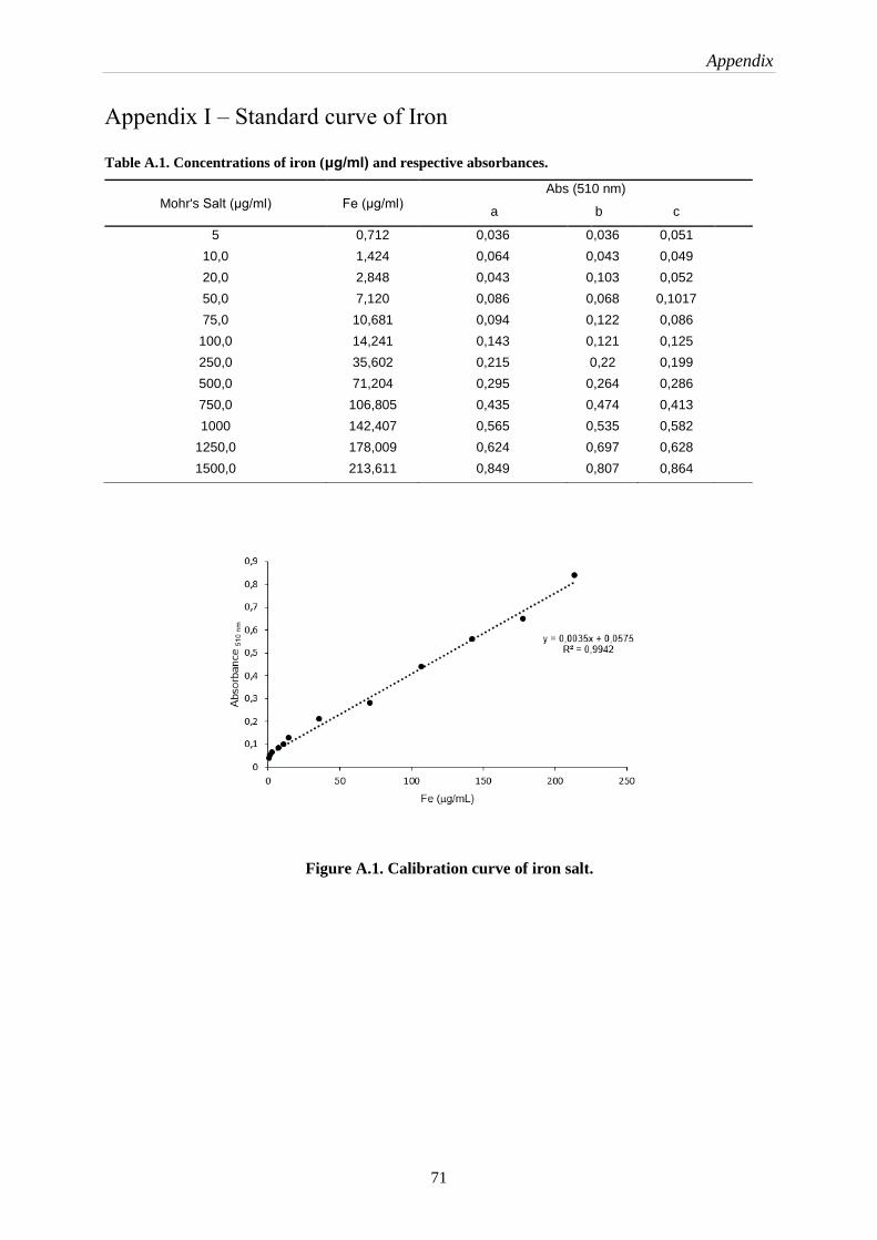
Appendix
71
Appendix I – Standard curve of Iron
Table A.1. Concentrations of iron (μg/ml) and respective absorbances.
Figure A.1. Calibration curve of iron salt.
Mohr's Salt (μg/ml) Fe (μg/ml) Abs (510 nm)
a b c
5 0,712 0,036 0,036 0,051
10,0 1,424 0,064 0,043 0,049
20,0 2,848 0,043 0,103 0,052
50,0 7,120 0,086 0,068 0,1017
75,0 10,681 0,094 0,122 0,086
100,0 14,241 0,143 0,121 0,125
250,0 35,602 0,215 0,22 0,199
500,0 71,204 0,295 0,264 0,286
750,0 106,805 0,435 0,474 0,413
1000 142,407 0,565 0,535 0,582
1250,0 178,009 0,624 0,697 0,628
1500,0 213,611 0,849 0,807 0,864

Appendix
72
Appendix II – XRD Report by Match software
Figure A.2. Iron oxide species report obtained from the condition I by Match software.
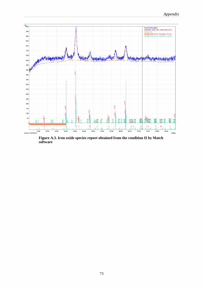
Appendix
73
Figure A.3. Iron oxide species report obtained from the condition II by Match software
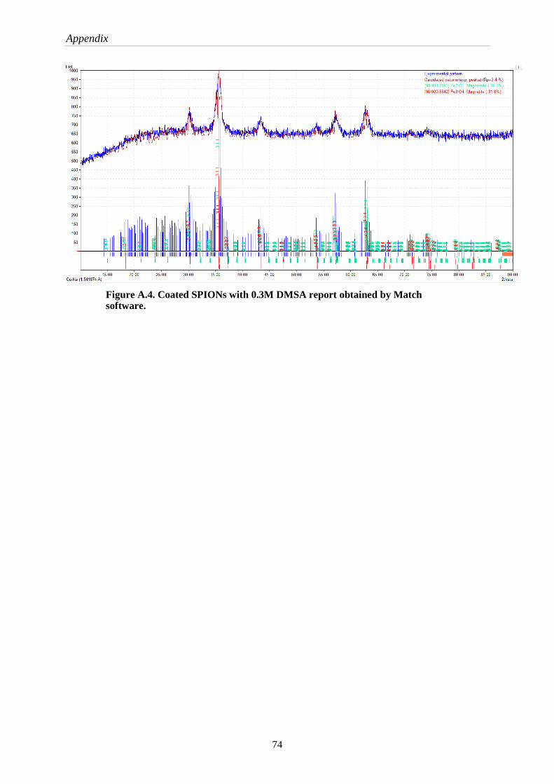
Appendix
74
Figure A.4. Coated SPIONs with 0.3M DMSA report obtained by Match software.
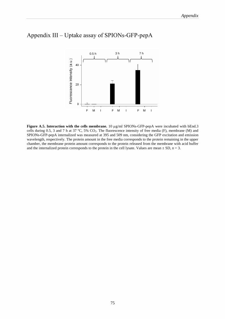
Appendix
75
Appendix III – Uptake assay of SPIONs-GFP-pepA
Figure A.5. Interaction with the cells membrane. 10 g/ml SPIONs-GFP-pepA were incubated with bEnd.3
cells during 0.5, 3 and 7 h at 37 ºC, 5% CO2. The fluorescence intensity of free media (F), membrane (M) and
SPIONs-GFP-pepA internalized was measured at 395 and 509 nm, considering the GFP excitation and emission
wavelength, respectively. The protein amount in the free media corresponds to the protein remaining in the upper
chamber, the membrane protein amount corresponds to the protein released from the membrane with acid buffer
and the internalized protein corresponds to the protein in the cell lysate. Values are mean ± SD, n = 3.
0.5 h 3 h 7 h
F F F M M M I I I