Embed Size (px)
Citation preview
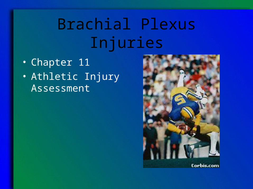
Brachial Plexus Injuries
• Chapter 11• Athletic Injury
Assessment
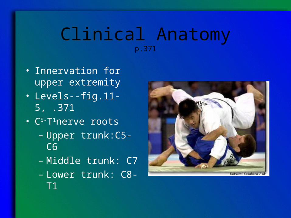
Clinical Anatomyp.371
• Innervation for upper extremity
• Levels--fig.11-5, .371• C5-T1nerve roots
– Upper trunk:C5-C6– Middle trunk: C7– Lower trunk: C8-T1
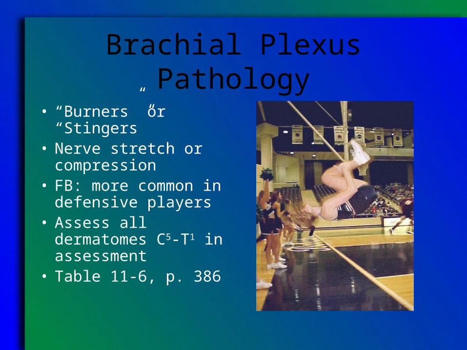
Brachial Plexus Pathology
• “Burners” or “Stingers”
• Nerve stretch or compression
• FB: more common in defensive players
• Assess all dermatomes C5-T1 in assessment
• Table 11-6, p. 386
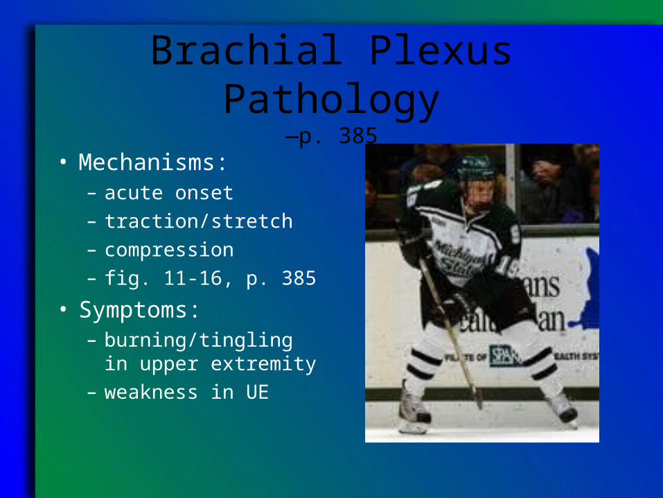
Brachial Plexus Pathology—p. 385
• Mechanisms:– acute onset– traction/stretch– compression– fig. 11-16, p. 385
• Symptoms:– burning/tingling in
upper extremity– weakness in UE
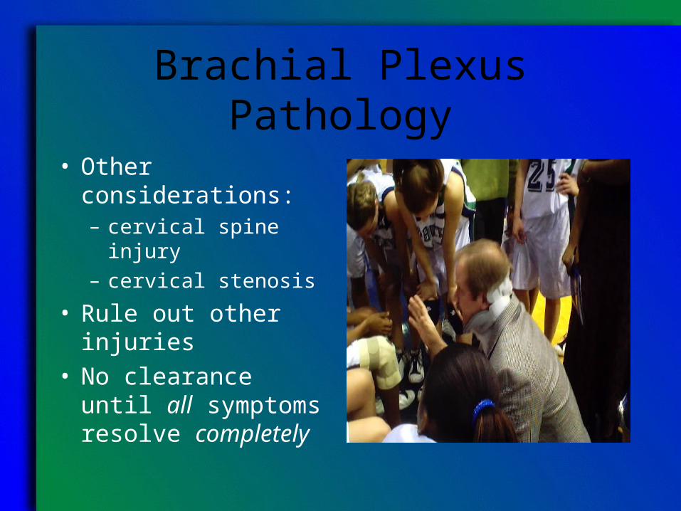
Brachial Plexus Pathology
• Other considerations:– cervical spine injury– cervical stenosis
• Rule out other injuries• No clearance until all
symptoms resolve completely
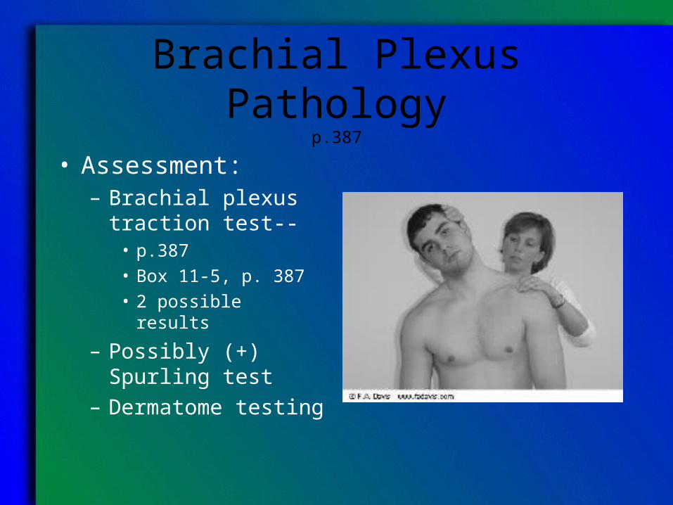
Brachial Plexus Pathologyp.387
• Assessment:– Brachial plexus
traction test--• p.387• Box 11-5, p. 387• 2 possible results
– Possibly (+) Spurling test
– Dermatome testing
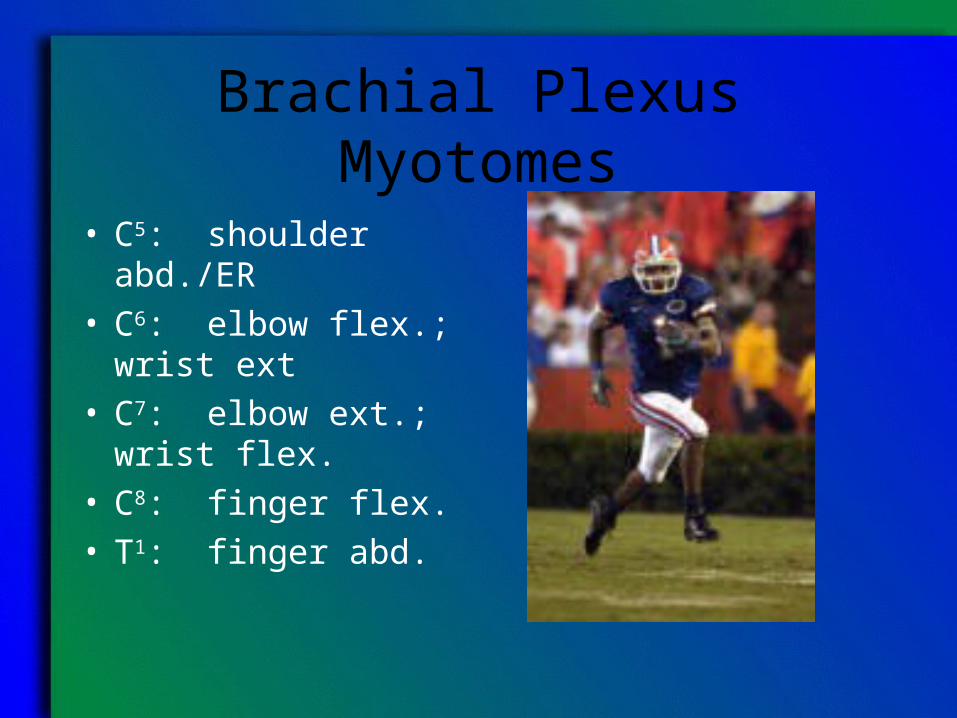
Brachial Plexus Myotomes
• C5: shoulder abd./ER• C6: elbow flex.; wrist
ext• C7: elbow ext.; wrist
flex.• C8: finger flex.• T1: finger abd.
Brachial Plexus Injury: Treatment
• Remove from contact• Test/Retest• Rule out other
pathologies– Bilateral weakness– Weakness outside
brachial plexus (shoulder shrug)
Brachial Plexus Injury: Return to Play Guidelines
• Full painfree AROM in UE and neck
• Normal sensation• Correction of
technique/ equipment to prevent re-injury
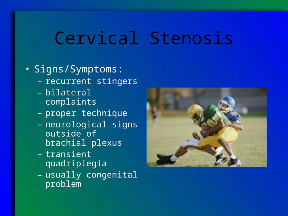
Cervical Stenosis
• Signs/Symptoms:– recurrent stingers– bilateral complaints– proper technique– neurological signs
outside of brachial plexus
– transient quadriplegia– usually congenital
problem
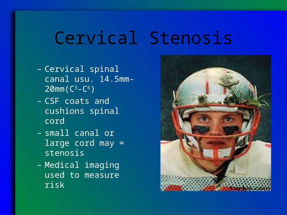
Cervical Stenosis
– Cervical spinal canal usu. 14.5mm-20mm(C3-C6)
– CSF coats and cushions spinal cord
– small canal or large cord may = stenosis
– Medical imaging used to measure risk
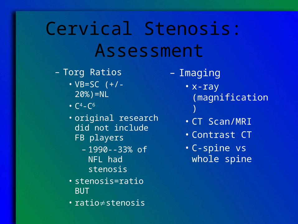
Cervical Stenosis: Assessment
– Torg Ratios• VB=SC (+/-
20%)=NL• C4-C6
• original research did not include FB players
– 1990--33% of NFL had stenosis
• stenosis=ratio BUT • ratiostenosis
– Imaging• x-ray (magnification)• CT Scan/MRI• Contrast CT• C-spine vs whole
spine
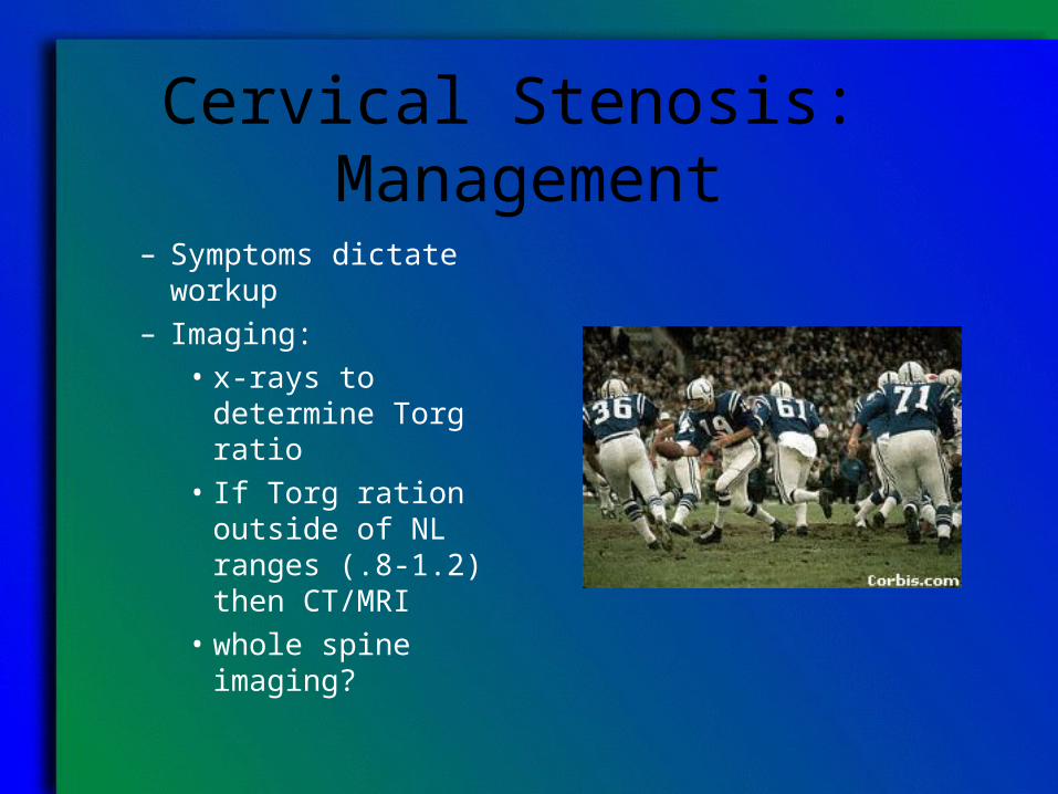
Cervical Stenosis: Management– Symptoms dictate
workup– Imaging:
• x-rays to determine Torg ratio
• If Torg ration outside of NL ranges (.8-1.2) then CT/MRI
• whole spine imaging?
Cervical Stenosis: Management
– Return to Play• Assess risks based on
imaging/ratios• Assess possible
technique changes• if stenotic-avoid all
sports which threaten the C-spine
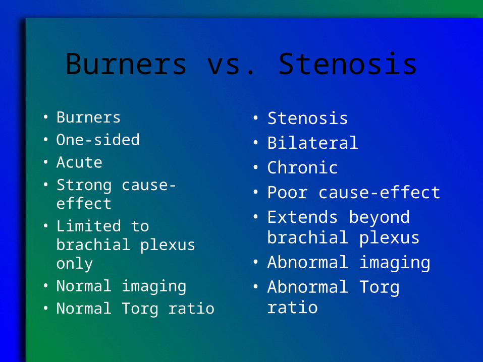
Burners vs. Stenosis
• Burners• One-sided• Acute• Strong cause-effect• Limited to brachial
plexus only• Normal imaging• Normal Torg ratio
• Stenosis• Bilateral• Chronic• Poor cause-effect• Extends beyond
brachial plexus• Abnormal imaging• Abnormal Torg ratio