Embed Size (px)
Citation preview
Case ReportA Case of Apoplexy of Rathke’s Cleft Cyst Followed byCerebral Infarction
Yu-ichiro Ohnishi,1 Yasunori Fujimoto,2 Koichi Iwatsuki,1 and Toshiki Yoshimine1
1Department of Neurosurgery, Osaka University Medical School, Suita, Osaka 565-0871, Japan2Department of Neurosurgery, Osaka Neurological Institute, Toyonaka, Osaka 565-0871, Japan
Correspondence should be addressed to Yu-ichiro Ohnishi; [email protected]
Received 19 December 2014; Accepted 14 February 2015
Academic Editor: Mehmet Turgut
Copyright © 2015 Yu-ichiro Ohnishi et al. This is an open access article distributed under the Creative Commons AttributionLicense, which permits unrestricted use, distribution, and reproduction in any medium, provided the original work is properlycited.
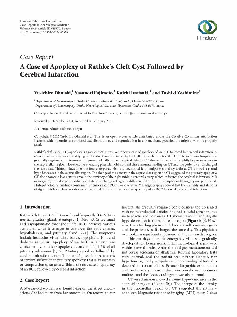
Rathke’s cleft cyst (RCC) apoplexy is a rare clinical entity.We report a case of apoplexy of an RCC followed by cerebral infarction. A67-year-old woman was found lying on the street unconscious. She had fallen from her motorbike. On referral to our hospital shegradually regained consciousness and presented with no neurological deficits. CT showed a round and slightly hyperdense area inthe suprasellar region. However, the attending physician did not find this abnormal finding on CT and the patient was dischargedthe same day. Thirteen days after the first emergency visit she developed left hemiparesis and dysarthria. CT showed a roundhypodense area in the suprasellar region.The change of the density in the suprasellar region onCT suggested the pituitary apoplexy.CT also showed a low density area in the territory of the right middle cerebral artery, which indicated the cerebral infarction. MRangiography revealed poor visibility and stenotic changes of rightmiddle cerebral arteries. Transsphenoidal surgery was performed.Histopathological findings confirmed a hemorrhagic RCC. Postoperative MR angiography showed that the visibility and stenosisof right middle cerebral arteries were recovered. This is the rare case of apoplexy of an RCC followed by cerebral infarction.
1. Introduction
Rathke’s cleft cysts (RCCs) were found frequently (13–22%) innormal pituitary glands at autopsy [1]. Most RCCs are smalland asymptomatic throughout life. RCC presents varioussymptoms when it enlarges to compress the optic chiasm,hypothalamus, and pituitary gland [2–4]. The symptomsinclude headache, visual disturbance, hypopituitarism, anddiabetes insipidus. Apoplexy of an RCC is a very rareclinical entity. Pituitary apoplexy occurs in 0.4–16.6% of allpituitary adenomas [5, 6]. Pituitary apoplexy followed bycerebral infarction is rare. There are 2 possible mechanismsof cerebral infarction in pituitary apoplexy, that is, vasospasmor compression of an artery. This is the rare case of apoplexyof an RCC followed by cerebral infarction.
2. Case Report
A 67-year-old woman was found lying on the street uncon-scious. She had fallen from her motorbike. On referral to our
hospital she gradually regained consciousness and presentedwith no neurological deficits. She had a facial abrasion, butno headache and no nausea. CT showed a round and slightlyhyperdense area in the suprasellar region (Figure 1(a)). How-ever, the attending physician did not consult a neurosurgeonand the patient was discharged the same day. This physicianoverlooked a significant appearance in the suprasellar region.
Thirteen days after the emergency visit, she graduallydeveloped left hemiparesis. Other neurological signs werewithin normal limits. Arterial blood gas measurement didnot reveal acidemia or alkalemia. Routine laboratory testswere normal, and the patient was neither diabetic, norhypertensive, nor hyperlipidemic. Endocrinological tests alsodetected no abnormalities. Echocardiographic examinationand carotid artery ultrasound examination showed no abnor-malities, and the electrocardiogram was also normal.
CT on admission showed a round hypodense area in thesuprasellar region (Figure 1(b)). The change of the densityin the suprasellar region on CT suggested the pituitaryapoplexy. Magnetic resonance imaging (MRI) taken 2 days
Hindawi Publishing CorporationCase Reports in Neurological MedicineVolume 2015, Article ID 645370, 8 pageshttp://dx.doi.org/10.1155/2015/645370
2 Case Reports in Neurological Medicine
(a) (b) (c)
Figure 1: (a) Axial view of the CT showing the round and slightly hyperdense area in the suprasellar region at the first emergency visit. (b, c)Axial view of the CT showing the round hypodense area in the suprasellar region and the low density area in the territory of the right middlecerebral artery at the second emergency visit.
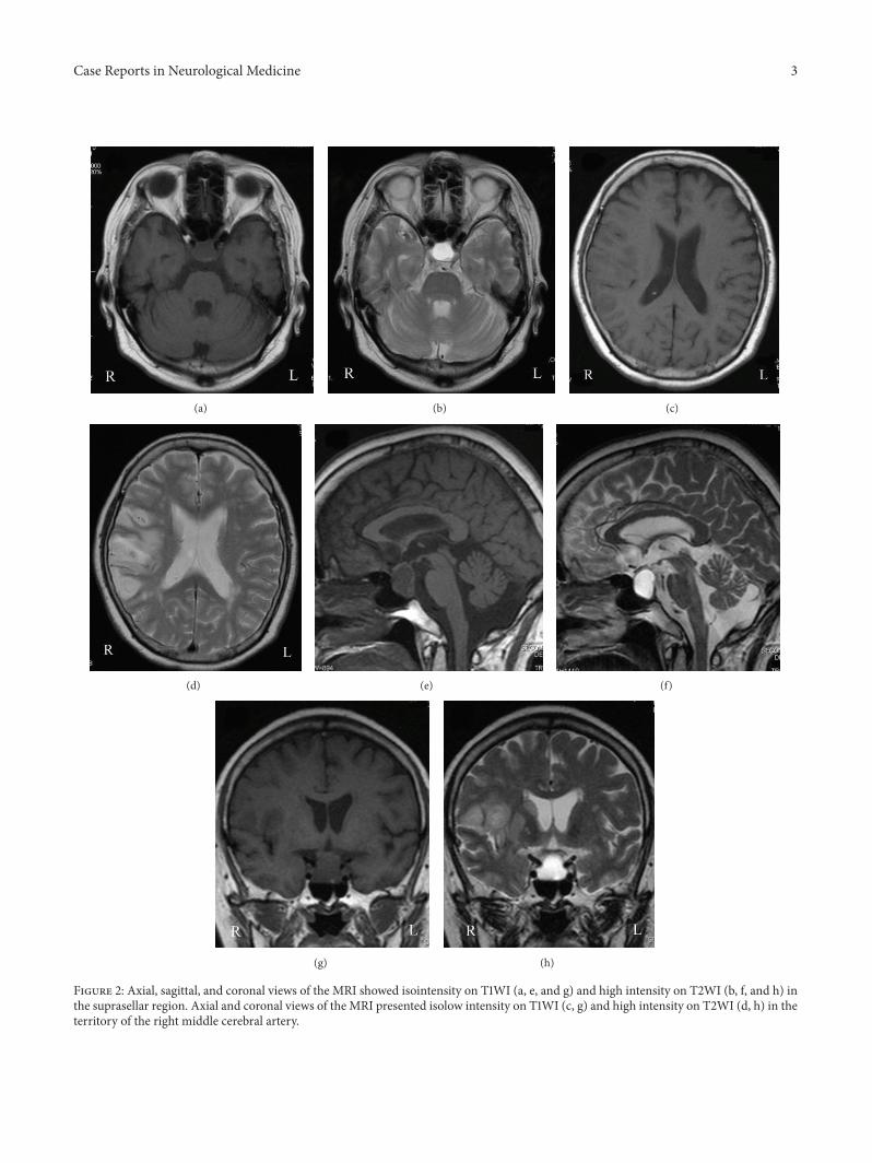
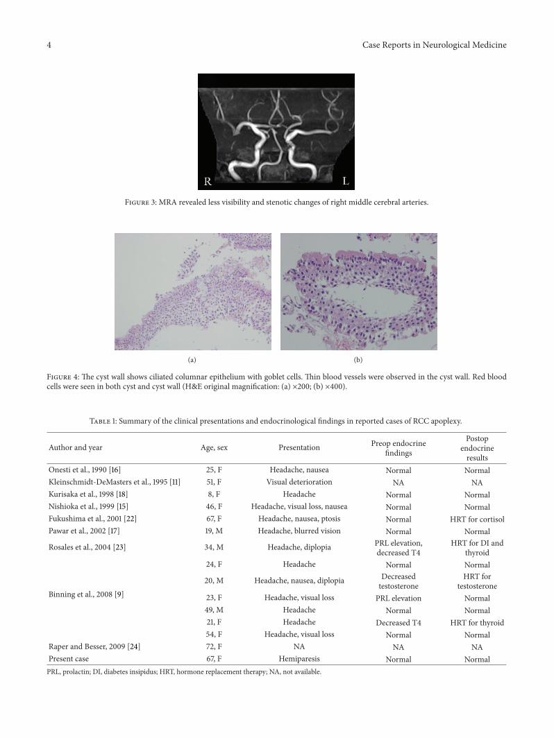
after admission detected isointensity on T1WI and highintensity on T2WI in the suprasellar region (Figure 2). CTalso showed a low density area in the territory of the rightMCA, which indicated the cerebral infarction (Figure 1(c)).MR angiography (MRA) showed signal loss with poor visibil-ity of distal right middle cerebral arteries (MCAs) (Figure 3).The conservative treatment for cerebral infarction improvedher left hemiparesis.
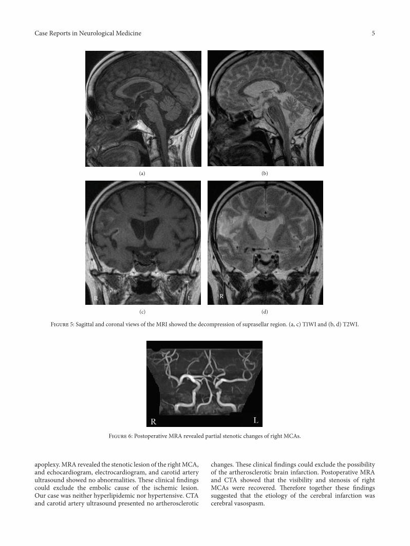
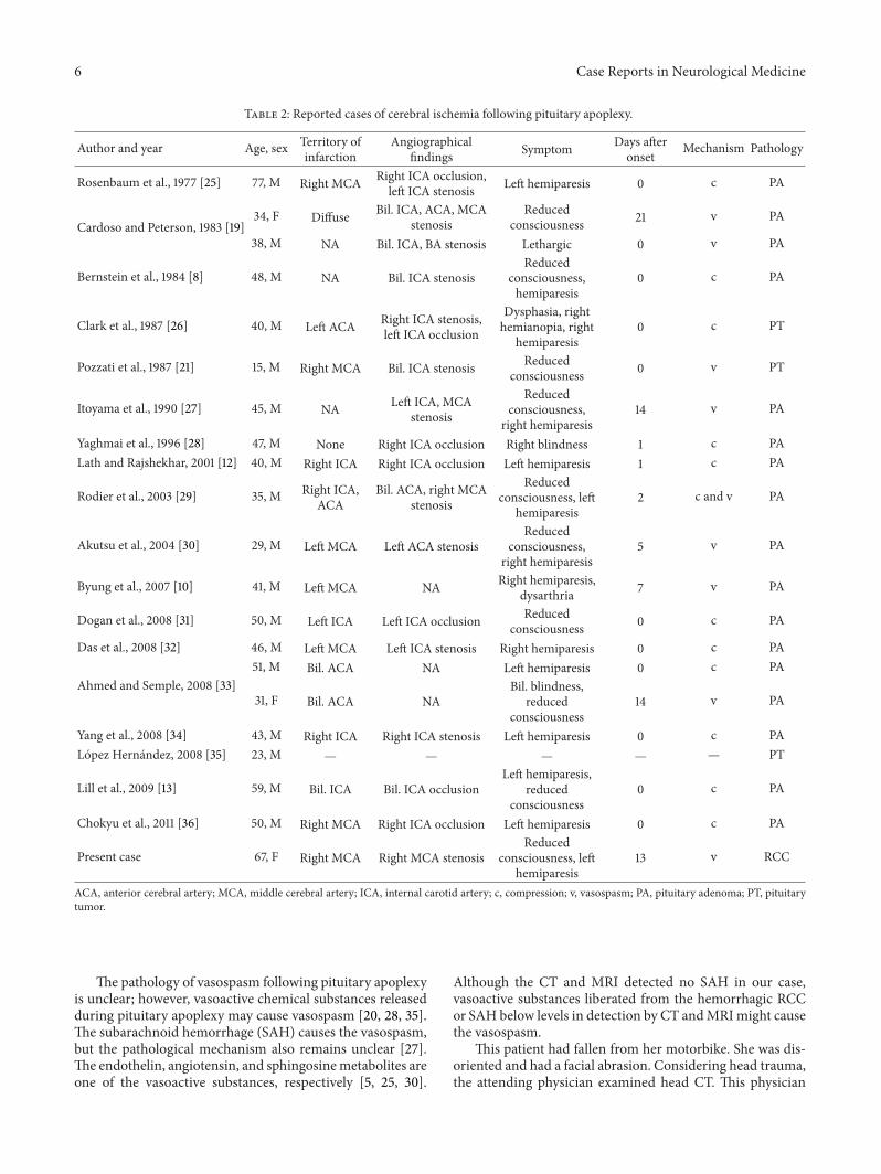
Ten days after admission, endoscopic endonasal trans-sphenoidal surgery was performed. The sella turcica wasfound to be thin. The tumor contained white-tinged viscidfluid.Anormal pituitary glandwas observed on the right side.Histopathological findings confirmed hemorrhagic RCC(Figures 4(a) and 4(b)). The cyst wall was lined by a ciliatedcolumnar cell layer with goblet cells. Thin blood vessels wereobserved in the cyst wall. Red blood cells were seen in bothcyst and cyst wall. Postoperative MRI showed the decom-pression of suprasellar region (Figure 5). Postoperative MRArevealed partial stenotic changes of right MCAs (Figure 6).These findings suggested RCC apoplexy followed by cerebralinfarction.
3. Discussion
RCC apoplexy is a rare clinical entity. Fourteen cases ofapoplexy of RCChave been reported in the literature (Table 1)[6–17]. The mechanism of RCC apoplexy is supposed to bethe repeated minor bleeding from the thin blood vesselsin the cyst wall by the stimulation of cyst contents andthe bleeding from the hypophyseal portal blood vesselsby the compression or shearing stress [7, 15]. The clinicalpresentations of these cases were headache, nausea, visualdisturbance, and cranial nerve palsy. One case in Table 1presented with altered consciousness as our case did. Nawaret al. reported 11 cases with hemorrhage within RCC [14].In their study, although not described in detail of each case,respectively, there were 3 patients with altered consciousness,
there were 10 patients with headache, there were 3 patientswith a visual deficit, and there was 1 patient with cranial nervepalsy.
The pituitary apoplexy is caused by the anticoagulanttherapy, the bromocriptine therapy, the radiation therapy,the hormone loading test, the cerebral angiography, and thecardiovascular surgery [18]. Particularly in macroadenoma,the head trauma can cause the apoplexy between 0 daysand 21 days [19–21]. None of these reviewed cases of RCCapoplexy were caused by the head trauma. In our case, it wasunclear whether the unconsciousness at the first emergencyvisit depended on the head trauma or apoplexy.
Preoperative endocrinological evaluations revealedabnormalities in 4 cases (Table 1). Wakai et al. describedthat preoperative endocrinological examinations revealedabnormalities in 3 cases. Most pituitary apoplexy cases needhormone replacement therapy for partial hypopituitarism[6]. In our case the preoperative endocrinological examina-tions were normal.
A rare complication of pituitary apoplexy is cerebralinfarction, which is caused by either direct compression ofan artery or vasospasm. Twenty cases of pituitary apoplexyfollowed by cerebral infarction have been reported in the lit-erature (Table 2) [1, 20, 22–24, 26, 28, 29, 31–39].The ischemicevents were attributed to mechanical compression by thetumor in 12 cases and to cerebral vasospasm in 8 cases. Thecerebral infarction was located at the anterior cerebral artery(ACA) territory in 4 cases, the MCA territory in 8 cases, andthe ICA territory in 4 cases. Cerebral arterial stenosis wasdetected in the ICA in 15 cases, in the MCA in 3 cases, and inthe ACA in 3 cases.The cerebral infarction occurred between0 and 21 days after the onset of pituitary apoplexy. Mostinfarctions due to cerebral vasospasm occurred between 5and 21 days after onset, and all infarctions by mechanicalcompression occurred within 2 days after onset.
In our case, the cerebral infarction occurred in the rightMCA territory after a 13-day interval from the onset of RCC
Case Reports in Neurological Medicine 3
(a) (b) (c)
(d) (e) (f)
(g) (h)
Figure 2: Axial, sagittal, and coronal views of the MRI showed isointensity on T1WI (a, e, and g) and high intensity on T2WI (b, f, and h) inthe suprasellar region. Axial and coronal views of the MRI presented isolow intensity on T1WI (c, g) and high intensity on T2WI (d, h) in theterritory of the right middle cerebral artery.
4 Case Reports in Neurological Medicine
Figure 3: MRA revealed less visibility and stenotic changes of right middle cerebral arteries.
(a) (b)
Figure 4: The cyst wall shows ciliated columnar epithelium with goblet cells. Thin blood vessels were observed in the cyst wall. Red bloodcells were seen in both cyst and cyst wall (H&E original magnification: (a) ×200; (b) ×400).
Table 1: Summary of the clinical presentations and endocrinological findings in reported cases of RCC apoplexy.
Author and year Age, sex Presentation Preop endocrinefindings
Postopendocrineresults
Onesti et al., 1990 [16] 25, F Headache, nausea Normal NormalKleinschmidt-DeMasters et al., 1995 [11] 51, F Visual deterioration NA NAKurisaka et al., 1998 [18] 8, F Headache Normal NormalNishioka et al., 1999 [15] 46, F Headache, visual loss, nausea Normal NormalFukushima et al., 2001 [22] 67, F Headache, nausea, ptosis Normal HRT for cortisolPawar et al., 2002 [17] 19, M Headache, blurred vision Normal Normal
Rosales et al., 2004 [23] 34, M Headache, diplopia PRL elevation,decreased T4
HRT for DI andthyroid
Binning et al., 2008 [9]
24, F Headache Normal Normal
20, M Headache, nausea, diplopia Decreasedtestosterone
HRT fortestosterone
23, F Headache, visual loss PRL elevation Normal49, M Headache Normal Normal21, F Headache Decreased T4 HRT for thyroid54, F Headache, visual loss Normal Normal
Raper and Besser, 2009 [24] 72, F NA NA NAPresent case 67, F Hemiparesis Normal NormalPRL, prolactin; DI, diabetes insipidus; HRT, hormone replacement therapy; NA, not available.
Case Reports in Neurological Medicine 5
(a) (b)
(c) (d)
Figure 5: Sagittal and coronal views of the MRI showed the decompression of suprasellar region. (a, c) T1WI and (b, d) T2WI.
Figure 6: Postoperative MRA revealed partial stenotic changes of right MCAs.
apoplexy.MRA revealed the stenotic lesion of the rightMCA,and echocardiogram, electrocardiogram, and carotid arteryultrasound showed no abnormalities. These clinical findingscould exclude the embolic cause of the ischemic lesion.Our case was neither hyperlipidemic nor hypertensive. CTAand carotid artery ultrasound presented no artherosclerotic
changes. These clinical findings could exclude the possibilityof the artherosclerotic brain infarction. Postoperative MRAand CTA showed that the visibility and stenosis of rightMCAs were recovered. Therefore together these findingssuggested that the etiology of the cerebral infarction wascerebral vasospasm.
6 Case Reports in Neurological Medicine
Table 2: Reported cases of cerebral ischemia following pituitary apoplexy.
Author and year Age, sex Territory ofinfarction
Angiographicalfindings Symptom Days after
onset Mechanism Pathology
Rosenbaum et al., 1977 [25] 77, M Right MCA Right ICA occlusion,left ICA stenosis Left hemiparesis 0 c PA
Cardoso and Peterson, 1983 [19]34, F Diffuse Bil. ICA, ACA, MCA
stenosisReduced
consciousness 21 v PA
38, M NA Bil. ICA, BA stenosis Lethargic 0 v PA
Bernstein et al., 1984 [8] 48, M NA Bil. ICA stenosisReduced
consciousness,hemiparesis
0 c PA
Clark et al., 1987 [26] 40, M Left ACA Right ICA stenosis,left ICA occlusion
Dysphasia, righthemianopia, right
hemiparesis0 c PT
Pozzati et al., 1987 [21] 15, M Right MCA Bil. ICA stenosis Reducedconsciousness 0 v PT
Itoyama et al., 1990 [27] 45, M NA Left ICA, MCAstenosis
Reducedconsciousness,
right hemiparesis14 v PA
Yaghmai et al., 1996 [28] 47, M None Right ICA occlusion Right blindness 1 c PALath and Rajshekhar, 2001 [12] 40, M Right ICA Right ICA occlusion Left hemiparesis 1 c PA
Rodier et al., 2003 [29] 35, M Right ICA,ACA
Bil. ACA, right MCAstenosis
Reducedconsciousness, left
hemiparesis2 c and v PA
Akutsu et al., 2004 [30] 29, M Left MCA Left ACA stenosisReduced
consciousness,right hemiparesis
5 v PA
Byung et al., 2007 [10] 41, M Left MCA NA Right hemiparesis,dysarthria 7 v PA
Dogan et al., 2008 [31] 50, M Left ICA Left ICA occlusion Reducedconsciousness 0 c PA
Das et al., 2008 [32] 46, M Left MCA Left ICA stenosis Right hemiparesis 0 c PA
Ahmed and Semple, 2008 [33]51, M Bil. ACA NA Left hemiparesis 0 c PA
31, F Bil. ACA NABil. blindness,
reducedconsciousness
14 v PA
Yang et al., 2008 [34] 43, M Right ICA Right ICA stenosis Left hemiparesis 0 c PALopez Hernandez, 2008 [35] 23, M — — — — — PT
Lill et al., 2009 [13] 59, M Bil. ICA Bil. ICA occlusionLeft hemiparesis,
reducedconsciousness
0 c PA
Chokyu et al., 2011 [36] 50, M Right MCA Right ICA occlusion Left hemiparesis 0 c PA
Present case 67, F Right MCA Right MCA stenosisReduced
consciousness, lefthemiparesis
13 v RCC
ACA, anterior cerebral artery; MCA, middle cerebral artery; ICA, internal carotid artery; c, compression; v, vasospasm; PA, pituitary adenoma; PT, pituitarytumor.
The pathology of vasospasm following pituitary apoplexyis unclear; however, vasoactive chemical substances releasedduring pituitary apoplexy may cause vasospasm [20, 28, 35].The subarachnoid hemorrhage (SAH) causes the vasospasm,but the pathological mechanism also remains unclear [27].The endothelin, angiotensin, and sphingosinemetabolites areone of the vasoactive substances, respectively [5, 25, 30].
Although the CT and MRI detected no SAH in our case,vasoactive substances liberated from the hemorrhagic RCCor SAH below levels in detection by CT andMRImight causethe vasospasm.
This patient had fallen from her motorbike. She was dis-oriented and had a facial abrasion. Considering head trauma,the attending physician examined head CT. This physician
Case Reports in Neurological Medicine 7
overlooked a significant appearance in the suprasellar lesion.Therefore the attending physician did not consult a neurosur-geon.
Asymptomatic incidental RCC should be monitoredconservatively. Symptomatic RCC can benefit from surgicaldecompression, and RCC apoplexy should be considered thesurgical decompression to prevent cerebral infarction, even ifsymptoms resolve soon after onset.
Conflict of Interests
The authors declare that there is no conflict of interestsregarding the publication of this paper.
References
[1] P. L. Semple, J. A. Jane Jr., and E. R. Laws Jr., “Clinical relevanceof precipitating factors in pituitary apoplexy,”Neurosurgery, vol.61, no. 5, pp. 956–961, 2007.
[2] K. Eguchi, T. Uozimi, K. Arita et al., “Pituitary functionin patients with Rathke’s cleft cyst: significance of surgicalmanagement,” Endocrine Journal, vol. 41, no. 5, pp. 535–540,1994.
[3] W. El-Mahdy andM. Powell, “Transsphenoidal management of28 symptomatic Rathke’s cleft cysts, with special reference tovisual and hormonal recovery,” Neurosurgery, vol. 42, no. 1, pp.7–16, 1998.
[4] S. Hama, K. Arita, A. Tominaga et al., “Symptomatic Rathke’scleft cyst coexisting with central diabetes insipidus andhypophysitis: case report,” Endocrine Journal, vol. 46, no. 1, pp.187–192, 1999.
[5] N. Sanno, K. Oyama, S. Tahara, A. Teramoto, and Y. Kato, “Asurvey of pituitary incidentaloma in Japan,” European Journalof Endocrinology, vol. 149, no. 2, pp. 123–127, 2003.
[6] S. Wakai, T. Fukushima, A. Teramoto, and K. Sano, “Pituitaryapoplexy: its incidence and clinical significance,” Journal ofNeurosurgery, vol. 55, no. 2, pp. 187–193, 1981.
[7] A. E.Alewijnse, S. L.M. Peters, andM.C.Michel, “Cardiovascu-lar effects of sphingosine-1-phosphate and other sphingomyelinmetabolites,” British Journal of Pharmacology, vol. 143, no. 6, pp.666–684, 2004.
[8] M. Bernstein, R. A. Hegele, F. Gentili et al., “Pituitary apoplexyassociated with a triple bolus test. Case report,” Journal ofNeurosurgery, vol. 61, no. 3, pp. 586–590, 1984.
[9] M. J. Binning, J. K. Liu, J. Gannon, A. G. Osborn, and W.T. Couldwell, “Hemorrhagic and nonhemorrhagic Rathke cleftcysts mimicking pituitary apoplexy,” Journal of Neurosurgery,vol. 108, no. 1, pp. 3–8, 2008.
[10] C. J. Byung, S. P. Yong, S. O. Hyung, S. K. Young, and K. C.Bong, “Pituitary apoplexy complicated by chemical meningitisand cerebral infarction,” Journal of Korean Medical Science, vol.22, no. 6, pp. 1085–1089, 2007.
[11] B. K. Kleinschmidt-DeMasters, K. O. Lillehei, and J. C. Stears,“The pathologic, surgical, and MR spectrum of Rathke cleftcysts,” Surgical Neurology, vol. 44, no. 1, pp. 19–27, 1995.
[12] R. Lath and V. Rajshekhar, “Massive cerebral infarction as afeature of pituitary apoplexy,”Neurology India, vol. 49, no. 2, pp.191–193, 2001.
[13] C. M. Lill, H. Hoch, F.-J. Dieste, H.-P. Vogel, F. Zipp, and F.Paul, “Bilateral stroke following pituitary apoplexy,” Journal ofClinical Neuroscience, vol. 16, no. 12, pp. 1670–1673, 2009.
[14] R. N. Nawar, D. Abdelmannan,W. R. Selman, and B.M. Arafah,“Pituitary tumor apoplexy: a review,” Journal of Intensive CareMedicine, vol. 23, no. 2, pp. 75–90, 2008.
[15] H. Nishioka, H. Ito, T. Miki, T. Hashimoto, H. Nojima, and H.Matsumura, “Rathke’s cleft cyst with pituitary apoplexy: casereport,” Neuroradiology, vol. 41, no. 11, pp. 832–834, 1999.
[16] S. T. Onesti, T.Wisniewski, and K. D. Post, “Clinical versus sub-clinical pituitary apoplexy: presentation, surgical management,and outcome in 21 patients,” Neurosurgery, vol. 26, no. 6, pp.980–986, 1990.
[17] S. J. Pawar, R. R. Sharma, S. D. Lad, E. Dev, and R. V. Devadas,“Rathke’s cleft cyst presenting as pituitary apoplexy,” Journal ofClinical Neuroscience, vol. 9, no. 1, pp. 76–79, 2002.
[18] M. Kurisaka, N. Fukui, T. Sakamoto, K. Mori, T. Okada, andK. Sogabe, “A case of Rathke’s cleft cyst with apoplexy,” Child’sNervous System, vol. 14, no. 7, pp. 343–347, 1998.
[19] E. R. Cardoso and E. W. Peterson, “Pituitary apoplexy andvasospasm,” Surgical Neurology, vol. 20, no. 5, pp. 391–395, 1983.
[20] J. T. Chaiban, D. Abdelmannan, M. Cohen, W. R. Selman, andB.M. Arafah, “Rathke cleft cyst apoplexy: a newly characterizeddistinct clinical entity,” Journal of Neurosurgery, vol. 114, no. 2,pp. 318–324, 2011.
[21] E. Pozzati, G. Frank, M. T. Nasi, and G. Giuliani, “Pituitaryapoplexy, bilateral carotid vasospasm, and cerebral infarction ina 15-year-old boy,” Neurosurgery, vol. 20, no. 1, pp. 56–59, 1987.
[22] Y. Fukushima, H. Oka, S. Utsuki, and K. Fujii, “A symptomaticRathke’s cleft cyst with pituitary apoplexy: a case report,”Neurological Surgery, vol. 29, no. 12, pp. 1183–1187, 2001.
[23] M. Y. Rosales, T. W. Smith, and M. Safran, “HemorrhagicRathke’s cleft cyst presenting as diplopia,” Endocrine Practice,vol. 10, no. 2, pp. 129–134, 2004.
[24] D. M. S. Raper and M. Besser, “Clinical features, managementand recurrence of symptomatic Rathke’s cleft cyst,” Journal ofClinical Neuroscience, vol. 16, no. 3, pp. 385–389, 2009.
[25] T. J. Rosenbaum, O. W. Houser, and E. R. Laws, “Pituitaryapoplexy producing internal carotid artery occlusion. Casereport,” Journal of Neurosurgery, vol. 47, no. 4, pp. 599–604, 1977.
[26] J. D. Clark, C. E. Freer, and T. Wheatley, “Pituitary apoplexy: anunusual cause of stroke,” Clinical Radiology, vol. 38, pp. 75–77,1987.
[27] Y. Itoyama, S. Goto, M. Miura, J.-I. Kuratsu, and Y. T. UshioMatsumoto, “Intracranial arterial vasospasm associated withpituitary apoplexy after head trauma—case report,” NeurologiaMedico-Chirurgica, vol. 30, no. 5, pp. 350–353, 1990.
[28] R. Yaghmai,W. J. Olan, S. O’Malley, andW. O. Bank, “Nonhem-orrhagic pituitarymacroadenoma producing reversible internalcarotid artery occlusion: case report,” Neurosurgery, vol. 38, no.6, pp. 1245–1248, 1996.
[29] G. Rodier, Y. Mootien, F. Battaglia, O. Martinet, and E. Cohen,“Bilateral stroke secondary to pituitary apoplexy,” Journal ofNeurology, vol. 250, no. 4, pp. 494–495, 2003.
[30] H. Akutsu, S. Noguchi, T. Tsunoda, M. Sasaki, and A. Mat-sumura, “Cerebral infarction following pituitary apoplexy: casereport,” Neurologia Medico-Chirurgica, vol. 44, no. 9, pp. 479–483, 2004.
[31] S.Dogan,H.Kocaeli, F. Abas, andE.Korfali, “Pituitary apoplexyas a cause of internal carotid artery occlusion,” Journal ofClinical Neuroscience, vol. 15, no. 4, pp. 480–483, 2008.
[32] N. K. Das, S. Behari, and D. Banerji, “Pituitary apoplexyassociated with acute cerebral infarct,” Journal of Clinical Neu-roscience, vol. 15, no. 12, pp. 1418–1420, 2008.
8 Case Reports in Neurological Medicine
[33] S. K. Ahmed and P. L. Semple, “Cerebral ischaemia in pituitaryapoplexy,” Acta Neurochirurgica, vol. 150, no. 11, pp. 1193–1196,2008.
[34] S.-H. Yang, K.-S. Lee, K.-Y. Lee, S. W. Lee, and Y.-K. Hong,“Pituitary apoplexy producing internal carotid artery compres-sion: a case report,” Journal of Korean Medical Science, vol. 23,no. 6, pp. 1113–1117, 2008.
[35] N. Lopez Hernandez, A. Garcıa Escriva, J. M. Molto Jorda, andN. Garcıa Barragan, “Massive cerebral infarction secondary toapoplexy due to pituitary adenoma,” Neurologia, vol. 23, no. 4,pp. 248–255, 2008.
[36] I. Chokyu, N. Tsuyuguchi, T. Goto, K. Chokyu, M. Chokyu, andK. Ohata, “Pituitary apoplexy causing internal carotid arteryocclusion—case report,” Neurologia Medico-Chirurgica, vol. 51,no. 1, pp. 48–51, 2011.
[37] R. Dev, S. K. Singh, M. C. Sharma, P. Khetan, and A. Chugh,“Post traumatic pituitary apoplexy with contiguous intra cere-bral hematoma operated through endonasal route—a casereport,” Pituitary, vol. 10, no. 3, pp. 291–294, 2007.
[38] R. L. Macdonald, R. M. Pluta, and J. H. Zhang, “Cerebralvasospasm after subarachnoid hemorrhage: the emerging revo-lution,”Nature Clinical Practice Neurology, vol. 3, no. 5, pp. 256–263, 2007.
[39] D. Regoli, S. Dion, N.-E. Rhaleb, G. Drapeau, and P. D’Orleans-Juste, “Vasoactive peptides and their receptors,” Blood Vessels,vol. 27, no. 2–5, pp. 137–145, 1990.

Submit your manuscripts athttp://www.hindawi.com
Stem CellsInternational
Hindawi Publishing Corporationhttp://www.hindawi.com Volume 2014
Hindawi Publishing Corporationhttp://www.hindawi.com Volume 2014
MEDIATORSINFLAMMATION
of
Hindawi Publishing Corporationhttp://www.hindawi.com Volume 2014
Behavioural Neurology
EndocrinologyInternational Journal of
Hindawi Publishing Corporationhttp://www.hindawi.com Volume 2014
Hindawi Publishing Corporationhttp://www.hindawi.com Volume 2014
Disease Markers
Hindawi Publishing Corporationhttp://www.hindawi.com Volume 2014
BioMed Research International
OncologyJournal of
Hindawi Publishing Corporationhttp://www.hindawi.com Volume 2014
Hindawi Publishing Corporationhttp://www.hindawi.com Volume 2014
Oxidative Medicine and Cellular Longevity
Hindawi Publishing Corporationhttp://www.hindawi.com Volume 2014
PPAR Research
The Scientific World JournalHindawi Publishing Corporation http://www.hindawi.com Volume 2014
Immunology ResearchHindawi Publishing Corporationhttp://www.hindawi.com Volume 2014
Journal of
ObesityJournal of
Hindawi Publishing Corporationhttp://www.hindawi.com Volume 2014
Hindawi Publishing Corporationhttp://www.hindawi.com Volume 2014
Computational and Mathematical Methods in Medicine
OphthalmologyJournal of
Hindawi Publishing Corporationhttp://www.hindawi.com Volume 2014
Diabetes ResearchJournal of
Hindawi Publishing Corporationhttp://www.hindawi.com Volume 2014
Hindawi Publishing Corporationhttp://www.hindawi.com Volume 2014
Research and TreatmentAIDS
Hindawi Publishing Corporationhttp://www.hindawi.com Volume 2014
Gastroenterology Research and Practice
Hindawi Publishing Corporationhttp://www.hindawi.com Volume 2014
Parkinson’s Disease
Evidence-Based Complementary and Alternative Medicine
Volume 2014Hindawi Publishing Corporationhttp://www.hindawi.com


![Ang Bingot (cleft lip o cleft palate) [Pananaliksik]](https://img.pdfslide.tips/doc/110x75/552029d24a79595e718b467b/ang-bingot-cleft-lip-o-cleft-palate-pananaliksik.jpg)