Embed Size (px)
Citation preview
การรกษาทางทนตกรรมจดฟนในผปวยอะมโลเจนเนซส อมเพอเฟคตา: ชนดไฮโปพลาสตก 2 ราย
ณฐพล ตงจตร* สวรรณ ลภนะพรลาภ* ชวลต สจรตวรยะกล**
รายงานผปวย
Case Report
บทคดยออะมโลเจนเนซส อมเพอเฟคตา เปนความผดปกตทพบไดยาก โดยมความผดปกตของการสรางผวเคลอบฟน หรอชนภายนอก
ของตวฟน ความผดปกตนเปนกลมหนงของสภาวะทถายทอดไดทางพนธกรรมซงสามารถแบงเปน 4 ชนดใหญๆ ไดดงน 1. แบบ
ไฮโปพลาสตก 2. แบบไฮโปแคลซไฟด 3. แบบไฮโปแมททเลชน 4. แบบไฮโปแมททเลชน-ไฮโปเพลเซยรวมกบทอโรดอนทซม การรกษา
ทางทนตกรรมจดฟนในผปวยอะมโลเจนเนซส อมเพอเฟคตา สามารถสรางปญหาไดเนองจากคณภาพและปรมาณของผวเคลอบฟน
ทผดปกต บทความน รายงานถงผปวยอะมโลเจนเนซส อมเพอเฟคตา 2 ราย: ชนดไฮโปพลาสตกแบบผวเรยบและแบบจดทมารบ
การรกษาทางทนตกรรมจดฟน เนองจากผปวยทงสองรายเปนชนดไฮโปพลาสตก เคลอบฟนจะมการพอกพนของแคลเซยมเปนปกต
แตบางกวา ดงนน การเตรยมผวฟนในผปวยประเภทนกอนการตดเครองมอทางทนตกรรมจดฟนแบบตดแนน รวมทงการถอด
เครองมอสามารถท�าไดตามปกต ไมปรากฏการหลดบอยของเครองมอจดฟนหรอความเสยหายจากผวเคลอบฟนตลอดระยะเวลา
ในการจดฟน ทงสองรายประสบความส�าเรจในการรกษาทางทนตกรรมจดฟนแบบปกตทวไป อยางไรกตามการจดการจ�าเปนตอง
มการประสานงานอยางใกลชดระหวางสหสาขาวชาชพ
ค�าส�าคญ: อะมโลเจนเนซส อมเพอเฟคตา, ไฮโปพลาสตก, สหสาขาวชาชพ, ทนตกรรมจดฟน
* ภาควชาทนตกรรมจดฟน คณะทนตแพทยศาสตร มหาวทยาลยมหดล
** นกศกษาหลงปรญญา หลกสตรฝกอบรมทนตแพทยเฉพาะทาง สาขาทนตกรรมจดฟน
* Department of Orthodontics, Faculty of Dentistry, Mahidol University
** Specialty training student in Orthodontics, Faculty of Dentistry, Mahidol University
ว ออน ไ ล น ท น ต จ ดฟ น ป ท 6 ฉ . 1 2 5 5 9 3 ณ ฐ พ ล ต ง จ ต ร แ ล ะ ค ณ ะ
Orthodontic Treatment of Amelogenesis Imperfecta Patients: Two Cases of Hypoplastic Type
Nathaphon Tangjit* Suwannee Luppanapornlarp* Chavalit Sujaritviriyakul**
AbstractAmelogenesis imperfecta (AI) is a rare inherited disorder characterized by abnormal formation of the
enamel or external layer of the crown of teeth. This anomality is a group of hereditary conditions that can
be divided into four major types: hypoplastic AI, hypocalcified AI, hypomaturation AI and hypomaturation-
hypoplastic AI with taurodontism. Orthodontic treatment in a patient with amelogenesis imperfecta could be
problematic due to quality and quantity of enamel surface. This article presented 2 cases: smooth and pitted
hypoplastic types of AI undergoing orthodontic treatment. Because this type of AI showed normal calcification
with thin enamel, the preparation of tooth surface before bonding fixed appliances and management in
removing appliance were performed conventionally. Neither the multiple bond failure nor the enamel damage
had been presented throughout the treatment. These two cases were successfully treated with conventional
orthodontic appliances. However, the management required an interdisciplinary approach.
Keywords: Amelogenesis imperfecta, Hypoplastic, Interdisciplinary, Orthodontics
IntroductionAmelogenesis imperfecta (AI) is a complicated
group of conditions that demonstrate developmental
alterations in the structure of the enamel. This condition
causes teeth, in many levels, to be unusually small,
discolored, pitted or grooved, and prone to rapid wear
and breakage. Other dental abnormalities are also
possible. Enamel formation or “amelogenesis” is under
genetic control that AI is defected in the genes encoding
enamel resulted in inheritable malformations of enamel
by mutation and altered expression at some genes.(1-3)
In fact, AI is not associated with defects in other parts
of the body or other health problems.(4)
EtiologyDental enamel is a highly mineralized tissue
with over 95% of its volume occupied by large, highly
organized, hydroxyapatite crystals. The formation
of this highly organization occurs in ameloblasts
through the interaction of a number of organic matrix
molecules including several genes such as enamelin
(ENAM), amelogenin (AMELX), ameloblastin (AMBN),
tuftelin (TUFT), amelotin, dentine sialophosphoprotein
(DSPP), enzymes such as kallikrein4 (KLK4), and
matrix metalloproteinase 20 (MMP20). The ENAM,
AMELX, and MMP20 genes provide instructions for
manufacturing proteins that are essential for normal
4 O J T h a i A s s o c O r t h o d V o l 6 N o 1 2 0 1 6 N a t h a p h o n T a n g j i t e t a l .

tooth development.(4-8) Most of these proteins are
involved in the formation of enamel which is the
hard, calcium-rich material that forms the protective
outer layer of each tooth. The ENAM gene represents
approximately 1% to 5% of enamel matrix, and
mutations of the ENAM gene have been correlated
with some autosomal dominant and recessive
patterns of hypoplastic amelogenesis imperfecta,
ranging from minor pitting to diffuse generalized thin
enamel.(6) The MMP-20 gene codes for a proteinase
named enamelysin; mutation of this gene has been
reported to associate with the autosomal recessive
pigmented hypomaturation variant of amelogenesis
imperfecta.(4,8) The mutation of protease KLK4 has
been also reported to be involved with some forms
of hypomaturation amelogenesis imperfecta. Both
enamelysin and kallikrein 4 are thought necessary
for the removal of enamel matrix proteins during the
maturation stage of enamel development.(4,10) Another
strong report is the AMBN gene that codes for the
protein ameloblastin, which constituted about 5%
of enamel matrix. Although, it is not proven to be
associated with amelogenesis imperfecta, this gene
locus is strong candidate for some of the autosomal
dominant patterns.(7,10) The distal-less homeobox 3
(DLX3) gene is in a group of genes that code for a
number of proteins that are critical for craniofacial,
tooth, hair, brain, and neural development; mutation
of this gene has been associated with the hypoplastic-
hypomaturation variants of amelogenesis imperfecta
with taurodontism.(9-10) Recently, FAM83H gene (Family
With Sequence Similarity 83, Member H) is believed
to be involved in the formation of enamel, although
the function of the protein produced from this gene
is unknown.(11-12)
Classifif icationAmelogenesis imperfecta varies in clinical
appearance that depended on the pattern of
inheritance, the mutation involved, expression of matrix
proteins and biochemical changes associated with
the mutation. Many classifications of AI have evolved
since the original classification of “hypoplastic” and
“hypocalcified” types was established in 1945.(1,13-14)
Until now, the multiplicity of classification systems
which based primarily or exclusively on phenotype
can be confusing, and it is not always possible to
cross-reference between the various subtypes used,
or to know which classification system might have
been applied to a particular case.(13) It is urged on
researchers to classify AI conditions by genome and by
subsequent biochemistry for ultimately usefulness.(14)
Because many studies have been reported that pattern
of inheritance, abnormal phenotypes and molecular
disorders, biochemical analysis of the enamel, etc.,
should be taken into account, consequently the
number of AI subtypes mentioned in the majority of
the reports are at least 10 to 15 types.(13-15) The widely
accepted classifications developed are mostly relied on
the phenotype and pedigree (i.e., clinical appearance
and apparent pattern of inheritance; Table 1). However
base on the clinical and radiographic basis alone, the
characteristics of AI can be divided into 4 major types:
1) hypoplasia appearance (enamel is thin and stained
with smooth or pitted surface, but normally calcified),
2) hypomaturation appearance (enamel is of normal
thickness, but of reduced hardness or harder than the
hypocalcified form and its color varies from yellow/
brown to red/brown), 3) hypocalcified appearance
(soft enamel that can be removed without difficulty),
and 4) Hypomaturation-hypoplasia with taurodontism
(Table 1).(15-16) Other clinical features of AI that may
ว ออน ไ ล น ท น ต จ ดฟ น ป ท 6 ฉ . 1 2 5 5 9 5 ณ ฐ พ ล ต ง จ ต ร แ ล ะ ค ณ ะ

be found are: delay in dental eruption, microdontia,
deviant crown and morphology, root resorption, short
roots, enlarge pulp chamber, pulp stones, dens in
dente, and tooth agenesis.(15)
Orthodontic Management in AIAI management is often complex and takes a
significant amount of time especially in childhood to
early adulthood.(16) Because clinical manifestations
of AI vary according to subtype and its severity(15,17),
the orthodontic treatment hence requires an
interdisciplinary approach.(18) For patients with
amelogenesis imperfecta teeth, enamel surface is
usually difficult to bond with brackets, and also it
creates the risk to damage the enamel when removing
appliance.(18,21) Moreover, the weakening in enamel
surface of AI has the effect of bond strengths which
are usually lower than that of normal enamel surface
in conventionally bonded (acid-etched)(19). This leads to
multiple bond failures during treatment, and it needs
to step back to rebond these teeth consequently
increasing orthodontic treatment duration. The
malformation in enamel surface is another factor to
cause an inaccuracy in bracket position especially in
pre-adjusted appliance prescription. Thus the final
Type I Hypoplastic IA - Pitted autosomal dominant
IB - Local autosomal dominant
IC - Local autosomal recessive
ID - Smooth autosomal dominant
IE - Smooth X-linked dominant
IF - Rough autosomal dominant
IG - Enamel agenesis, autosomal recessive
Type II Hypomaturation
IIA - Hypomaturation, pigmented autosomal recessive
IIB – Hypomaturation, pigmented X-linked recessive
IIC - Snow-capped teeth, X-linked
IID – Snow-capped teeth, autosomal dominant?
Type III Hypocalcified
IIIA – Autosomal dominant
IIIB – Autosomal recessive
Type IV Hypomaturation – hypoplastic with taurodontism
IVA – Autosomal dominant
IVB - Autosomal recessive
Table 1 Classification of Amelogenesis Imperfecta (AI) proposed by Witkop (1988)
6 O J T h a i A s s o c O r t h o d V o l 6 N o 1 2 0 1 6 N a t h a p h o n T a n g j i t e t a l .

positions of the teeth are essentially adjusted by
second and third order bends of orthodontic mechanic.
Many AI cases have been treated successfully with
conventional acid etch bonding methods, such as the
hypoplastic type of AI. Other types that have problems
of bond failures or difficulty of bracket placement are
suggested to use traditional banded appliance instead
of bonded bracket. Moreover, glass ionomer cement –
based adhesives are recommended to improve
appliance retention because it is less dependent on
microtag formation, and also help in the reduction of
further enamel demineralization. Preformed stainless
steel crowns with welded tubes or brackets are another
choice to prevent further decrease in vertical height and
also sustain the bite to aid in placing final restorations
after orthodontic treatment.
Debonding of the brackets can also cause
fractures to the fragile enamel, and must therefore
be performed with caution. The use of debonding
pliers to apply a shear or tensile force according to the
manufacturer’s instructions is still accepted. However,
pliers with narrow blades created sufficient debonding
strength and led to reduced force levels on the enamel
surface are recommended.(20) The management of more
complex cases with severe malocclusion requires
team interdisciplinary approach. This report describes
two cases of patients with hypoplastic type of AI and
orthodontic management.
Report of Two CasesCase 1. A 23-year-old male was referred to
the orthodontic clinic, Faculty of Dentistry, Mahidol
University for orthodontic treatment. No remarkable
findings were identified in his medical record including
no evidence of systemic disease, nutritional deficiency,
or drug treatments that may have affected dentition
structure during development. His chief compliant
was spacing of maxillary and mandibular teeth. From
intraoral examination, this patient presented with
generalized tooth discoloration, generalize spacing in
upper and lower dentition, loss of mandibular right
first molar, dental caries exposed pulp of mandibular
left first molar, mesial tipping of mandibular left and
right molars. Posterior teeth were yellow in color,
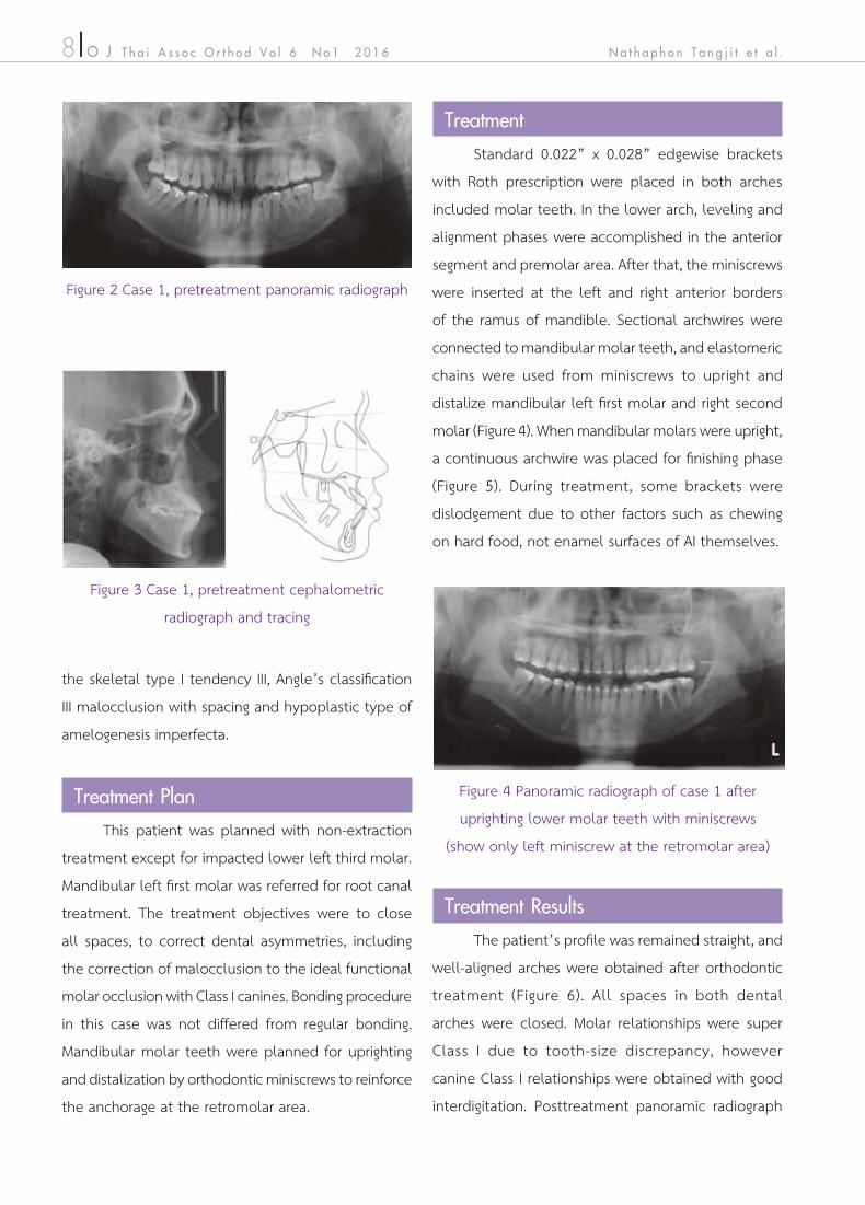
but the enamel was hard (Figure 1). The panoramic
Figure 1 Case 1, pretreatment facial
and intraoral photographs
radiograph showed the absence of maxillary third
molars except impacted mandibular left third molar.
Mandibular left first molar showed caries exposed pulp
with rarified area at the apex (Figure 2). From lateral
cephalometric measurements, this patient presented
with skeletal type I tendency III (ANB = 0.5°), prognathic
mandible (SNB = 86.5°), and with open configuration
(MP-PP = 27°). Upper incisors were proclined (U1-NA
= 38.5°) and lower incisors were in normal inclination
(L1-NB = 30°, Figure 3). This patient was diagnosed as
ว ออน ไ ล น ท น ต จ ดฟ น ป ท 6 ฉ . 1 2 5 5 9 7 ณ ฐ พ ล ต ง จ ต ร แ ล ะ ค ณ ะ
the skeletal type I tendency III, Angle’s classification
III malocclusion with spacing and hypoplastic type of
amelogenesis imperfecta.
Treatment PlanThis patient was planned with non-extraction
treatment except for impacted lower left third molar.
Mandibular left first molar was referred for root canal
treatment. The treatment objectives were to close
all spaces, to correct dental asymmetries, including
the correction of malocclusion to the ideal functional
molar occlusion with Class I canines. Bonding procedure
in this case was not differed from regular bonding.
Mandibular molar teeth were planned for uprighting
and distalization by orthodontic miniscrews to reinforce
the anchorage at the retromolar area.
Treatment Standard 0.022” x 0.028” edgewise brackets
with Roth prescription were placed in both arches
included molar teeth. In the lower arch, leveling and
alignment phases were accomplished in the anterior
segment and premolar area. After that, the miniscrews
were inserted at the left and right anterior borders
of the ramus of mandible. Sectional archwires were
connected to mandibular molar teeth, and elastomeric
chains were used from miniscrews to upright and
distalize mandibular left first molar and right second
molar (Figure 4). When mandibular molars were upright,
a continuous archwire was placed for finishing phase
(Figure 5). During treatment, some brackets were
dislodgement due to other factors such as chewing
on hard food, not enamel surfaces of AI themselves.
Figure 4 Panoramic radiograph of case 1 after
uprighting lower molar teeth with miniscrews
(show only left miniscrew at the retromolar area)
Figure 2 Case 1, pretreatment panoramic radiograph
Figure 3 Case 1, pretreatment cephalometric
radiograph and tracing
Treatment ResultsThe patient’s profile was remained straight, and
well-aligned arches were obtained after orthodontic
treatment (Figure 6). All spaces in both dental
arches were closed. Molar relationships were super
Class I due to tooth-size discrepancy, however
canine Class I relationships were obtained with good
interdigitation. Posttreatment panoramic radiograph
8 O J T h a i A s s o c O r t h o d V o l 6 N o 1 2 0 1 6 N a t h a p h o n T a n g j i t e t a l .

Figure 5 Treatment progress of case 1
treated with fixed appliance and conventional bonding systems (acid-etch)
Figure 6 Case 1, posttreatment facial and
intraoral photographs
ว ออน ไ ล น ท น ต จ ดฟ น ป ท 6 ฉ . 1 2 5 5 9 9 ณ ฐ พ ล ต ง จ ต ร แ ล ะ ค ณ ะ

Figure 8 Case 1, posttreatment cephalometric
radiograph, tracing, and superimposition;
(a) Cranial base, (b) Maxilla, (c) Mandible
(black = pretreatment; red = posttreatment)
Figure 7 Case1, posttreatment panoramic radiograph
after treatment showed the patient’s skeletal
relationship maintained as a skeletal type I (ANB =
1°, AO-BO = -0.5 mm; Figure 8, Table 2). Maxillary and
mandibular incisors were in more normal inclination
(U1-NA = 31°, L1-NB = 24°). Tooth surface of affected AI
teeth were still normal hardness and smooth without
tooth sensitivity.
Case 2: A boy, aged 12.6 years, and his
parents presented to the orthodontic clinic, Faculty of
Dentistry, Mahidol University with the chief complaints
of dental crowding and discolored teeth. This patient
was in a good physical health. However, the family
history revealed that there was a similar problem of
the discolored teeth in the family line. The clinical
examination of this patient showed a symmetrical,
brachyfacial face with straight profile (Figure 9). The
intra-oral examination showed that this patient was
in the very late mixed dentition (second permanent
molars were not erupted yet). His oral hygiene was
fair with upper midline shift 2 mm to the left, a Class
I molar relationship on the right, and cusp-to-cusp
on the left. The maxillary canines were still in the
high positions, and lack of spaces for the alignment.
Maxillary lateral incisors were lingual displacement with
anterior cross-bite, and the maxillary dental arch was
slightly constricted. Teeth were stain with yellow to
brown in color, and most affected were premolars and
anteriors. Enamel was hard and the surface was pitted.
The panoramic radiograph showed all permanent teeth
were present without any pathology (Figure 10). From
the lateral cephalometric measurements (Figure 11),
this patient presented with skeletal type I tendency
III (ANB = 0.9°, AO-BO = -3.6 mm), open configuration
(MP-PP = 27°, mandibular angle = 130.7° ). Upper
incisors were proclined (U1-NA = 34.8°) and lower
incisors were retroclination (L1-NB = 22.7°). This patient
showed mandibular molars were upright and no root
resorption or periodontal bone loss (Figure 7). The
lateral cephalometric analysis and superimpositions
10 O J T h a i A s s o c O r t h o d V o l 6 N o 1 2 0 1 6 N a t h a p h o n T a n g j i t e t a l .

Table 2 Cephalometric values of pre- and posttreatment of case 1
Measurement Thai norm Pretreatment Posttreatment Differences
NS-FH (º) 7 ± 2.58 9 10 1
SNA (º) 84 ± 3.58 87 88 1
SNB (º) 81 ± 3.59 86.5 87 0.5
ANB (º) 3 ± 2.50 0.5 1 0.5
AO-BO (mm) -2 ± 3.49 -0.5 -0.5 0
NS-MP (º) 30 ± 5.61 33 32 -1
NS-PP (º) 9 ± 3.03 6.5 6 -0.5
MP-PP (º) 21 ± 5.25 27 26 -1
Mand angle 118 ± 6.13 126 125 -1
U1-NA (º) 22 ± 5.94 38.5 31 -7.5
U1-NA (mm) 5 ± 2.13 10 6 -4
L1-NB (º) 30 ± 5.61 30 24 -6
L1-NB (mm) 7 ± 2.22 8 6 -2
L1-MP (º) 97 ± 5.97 90 87 -3
U1-L1 (º) 125 ± 8.03 111 122.5 11.5
Nasolabial angle 91 ± 7.98 90 93 3
H-angle 14 ± 3.83 15.1 12 -3.1
L1-E plane (mm) 2 ± 2.03 4 2 -2
ว ออน ไ ล น ท น ต จ ดฟ น ป ท 6 ฉ . 1 2 5 5 9 11 ณ ฐ พ ล ต ง จ ต ร แ ล ะ ค ณ ะ

Figure 9 Case 2, pretreatment photographs
Figure 10 Case 2, pretreatment panoramic radiograph
Figure 11 Case 2, pretreatment cephalometric radiograph and tracing
12 O J T h a i A s s o c O r t h o d V o l 6 N o 1 2 0 1 6 N a t h a p h o n T a n g j i t e t a l .

was diagnosed as the skeletal type I tendency III with
Angle’s Classification III subdivision, upper anterior
crowding, and a hypoplastic type of amelogenesis
imperfecta.
Treatment PlanThe main treatment objective of this patient was
to expand the maxilla with rapid maxillary expansion
(RME) to relieve maxillary anterior crowding and
correct anterior cross-bite with full fixed orthodontic
appliance, and to obtain Class I molar and canine
relationships with normal overjet and overbite.
Because of the discoloration of the anterior teeth
and the young age of the patient, an interdisciplinary
consultation with a pediatric dentist, a restorative
dentist, and a prosthodontist were planned. After
ideal dental and skeletal relationships were achieved,
restorative treatment was required. Adhesive composite
restoration, glass ionomer cement, veneer front teeth
or crowns were explained to the patient for choices
of restoration. The importance of oral hygiene, caries
control, and cooperation was emphasized. The patient
was told to see the pediatric dentist periodically. The
patient and his parents gave their informed consent.
Treatment A RME was inserted to correct the mild transverse
discrepancy of the maxillary arch, to gain spaces
for the maxillary canines, and to correct anterior
crossbite of the maxillary lateral incisors (Figure 12 a).
Standard 0.022" x 0.028" edgewise brackets with
Roth prescription were placed on all maxillary and
mandibular teeth for leveling and aligning. To maintain
incisor torque and coordinate the dental arches,
rectangular stainless steel wires were used in both
arches. Because this patient presented with skeletal
type III tendency, mandibular growth had started and
accelerated with growth spurt; therefore Class III elastics
were used to control horizontal growth. Finally, this
patient was told to wear the up and down elastics in
order to seat the cusps (Figure 12 b).
Treatment ResultsThe posttreatment photographs show that the
patient’s profile was still straight profile, and the anterior
crossbite was corrected to normal overjet and overbite.
The posterior teeth illustrate good interdigitation, and
the correction of crowding. Molar and canine Class I
relationships were achieved on both sides (Figure 13).
The treatment time of this patient was 3.6 years. The
lateral cephalometric analysis after treatment (Figure
14 and Table 3) showed that the patient’s skeletal
was still within type I (ANB = -2.23°, AO-BO = -2.18
mm). The superimpositions (Figure 15) show the spurt
of horizontal mandibular growth slightly greater than
vertical growth (SNA = 82.18°, SNB = 84.42°). Maxillary
incisors were slightly proclined and mandibular incisors
were slightly retroclined (U1-NA = 39.8°, L1-NB = 19.67°).
Patient’s cooperation was good with care of his oral
hygiene. After debonding and retention, patient was
sent for restorative treatment of AI teeth: posterior
crowns of molars and veneers of maxillary front teeth.
Figure 12 Treatment progress of case 2
(a) using RME and (b) full fixed appliance.
a
b
ว ออน ไ ล น ท น ต จ ดฟ น ป ท 6 ฉ . 1 2 5 5 9 13 ณ ฐ พ ล ต ง จ ต ร แ ล ะ ค ณ ะ

Figure 13 Case 2, posttreatment facial and
intraoral photographs
Figure Post-treatment panoramic radiograph
Figure Post-treatment cephalometric radiograph
Figure 14 Case 2, posttreatment cephalometric radiograph and tracing.
14 O J T h a i A s s o c O r t h o d V o l 6 N o 1 2 0 1 6 N a t h a p h o n T a n g j i t e t a l .

Table 3 Cephalometric values of pre- and posttreatment of case 2
Figure 15 Case 2, cephalometric superimposition; (a) Cranial base, (b) Maxilla,
(c) Mandible (black = pretreatment; red = posttreatment)
Measurement Thai norm Pre-treatment Post-treatment Differences
NS-FH (º) 7 ± 2.58 7.94 5.76 -2.15
SNA (º) 84 ± 3.58 81.70 82.18 0.48
SNB (º) 81 ± 3.59 80.81 84.42 3.61
ANB (º) 3 ± 2.50 0.89 -2.23 -3.12
AO-BO (mm) -2 ± 3.49 -3.57 -2.18 1.39
NS-MP (º) 30 ± 5.61 34.37 26.02 -8.35
NS-PP (º) 9 ± 3.03 6.58 5.74 -0.84
MP-PP (º) 21 ± 5.25 27.79 20.28 -7.51
Mand angle 118 ± 6.13 130.66 123.82 -6.84
U1-NA (º) 22 ± 5.94 34.85 39.80 4.95
U1-NA (mm) 5 ± 2.13 9.16 9.99 0.83
L1-NB (º) 30 ± 5.61 22.68 19.67 -3.01
L1-NB (mm) 7 ± 2.22 5.60 5.51 -0.09
L1-MP (º) 97 ± 5.97 87.50 89.24 1.74
U1-L1 (º) 125 ± 8.03 121.58 122.76 1.18
Nasolabial angle 91 ± 7.98 89.19 81.39 -7.80
H-angle 14 ± 3.83 14.42 7.29 -7.13
L1-E plane (mm) 2 ± 2.03 0.78 -3.25 -4.03
ว ออน ไ ล น ท น ต จ ดฟ น ป ท 6 ฉ . 1 2 5 5 9 15 ณ ฐ พ ล ต ง จ ต ร แ ล ะ ค ณ ะ

Because of the high cost for complete restorations, he
wanted to do only maxillary incisor veneers. At 3 years
retention he was called back, and it was found that his
posterior teeth showed chips of enamel surfaces with
extended caries (Figure 16). However, his occlusion
was still in good interdigitation. This patient was told
to have immediately crown procedure of the remaining
affected AI teeth.
DiscussionClinical problems of AI patients are usually
associated with esthetics, oral health with tooth
sensitivity, and dental caries. The interdisciplinary
treatments that usually involved are adhesive
restorative techniques with tooth-to reconstruction
bond properties and prosthodontic rehabilitation of AI
teeth. Consideration of AI patient’s treatment modality
includes a patient’s age, severity of AI, periodontal
condition, and financial implications for the patient’s
family, and long-term prognosis.(21-22) Because of normal
enamel calcification, orthodontic management of
these 2 cases was considered to use conventional
resin composite bonding. Moreover, the information
of these defected teeth and how to manage them
were told to motivate patients for good cooperation
and oral hygiene care.
Although these two cases were diagnosed as
hypoplastic AI, the case-1 patient came for orthodontic
treatment at the adult stage and the affected AI teeth
showed thin, hard, smooth, and glossy enamel, which
varied in color from white to cream-brown (Figure 1).
This patient’s clinical feature was subdivided into a
smooth autosomal dominant hypoplastic type (Table
1).(15) While the case 2’s AI teeth showed small, discrete,
pinpoint-to-pinhead sized pits, which were arranged in
horizontal rows (Figure 9). The case 2’s clinical feature
was subdivided into a pitted autosomal dominant
hypoplastic type. In comparison, it seems that the
case 1 teeth with smooth glossy enamels were shown
lower severity than that of the case 2 with pitting surfaces,
and with aged. During treatment, some brackets of the
case 1 were dislodgement but no incidence of multiple
bond failures, while in case 2 multiple bond failures
were found and led to the increase of orthodontic
treatment duration. Soew et al.(23) studied the effect of
acid-etching on enamel from different clinical variants
of AI and found that the pitted hypoplastic type
showed a predominant etch pattern of type 1 in which
the prism cores of enamel were preferential removed.
In contrast, no typical etch patterns were found from
the enamel affected by smooth hypoplastic variant.
Moreover, high failure rates of adhesive restoration
on AI affected teeth could be due to fractures within
weak enamel or dentin supporting the restorations.
In hypocalcified AI, pretreating the tooth surface with
5% sodium hypochlorite (NaOCl) for 1 minute would
remove excess protein to make the enamel crystals
more accessible to the etching solution resulting in
enhancing the bonding of orthodontic brackets to the
affected tooth.(24,25)
Figure 16 Case 2, recall intraoral photographs at
3 years retention with restorative treatment of
maxillary front teeth. Posterior teeth show reduced
enamel surfaces with some fractures and caries.
16 O J T h a i A s s o c O r t h o d V o l 6 N o 1 2 0 1 6 N a t h a p h o n T a n g j i t e t a l .

However, orthodontic treatments of these 2
cases were achieved; the case-1 patient treated with
miniscrews to upright molars, and the case-2 patient
treated with RME to correct crowding. Orthodontic
retentions were provided with wraparound retainers
in both cases, and the patients had been informed
of new retainers in case that the old ones were not
suitable with the restored AI affected teeth.
After orthodontic management, the cost of
restorative or prosthodontic treatments in the AI
patients was in consideration,(22) because the basic
health insurance could not cover the overall cost of
restorative and prosthodontic treatments. In case 2,
therefore the patient wanted the anterior teeth to be
restored first due to esthetics and financial limitation.
The recall 3 years after treatment photographs of case
2 showed that the posterior teeth had reduced enamel
surfaces and were prone to caries, while the case 1
was still observed without any problem.
ConclusionThe 2 cases report has been presented
to illustrate the fact that patients who have the
hypoplastic type of amelogenesis imperfecta can be
successfully treated with conventional orthodontic
appliances. The orthodontic management of AI patient
has special conditions to be considerate as describe
above. Moreover, the AI patients need to be continually
motivated during and after orthodontic treatment using
various techniques and appliances that aim to preserve
the fragile tooth structure.
References1. Weinman JP, Svoboda JF, Woods RW. Hereditary
disturbances of enamel formation and calcification. J Am
Dent Assoc 1945;32:397-418.
2. Aldred MJ, Crawford PJM, Roberts E, Gillespie CM, Thomas
NT, Fenton I, Sandkuijl LA, Harper PS. Genetic hetero-
geneity in X-linked amelogenesis imperfecta. Genomics
1992a;14:567-73.
3. Aldred MJ, Crawford PJM, Roberts E, Thomas NST.
Identification of a nonsense mutation in the amelogenin
gene (AMELX) in a family with X-linked amelogenesis
imperfecta (AIH1). Hum Genet 1992b;90:413-6.
4. Wright JT. The molecular etiologies and associated
phenotypes of amelogenesis imperfecta. Am J Med Genet
A 2006;140:2547-55.
5. Wright JT, Hart PS, Aldred MJ, Seow K, Crawford PJ, Hong
SP, et al. Relationship of phenotype and genotype in
X-linked amelogenesis imperfecta. Connect Tissue Res
2003;44(Suppl 1):72-8.
6. Kim JW, Seymen F, Lin BP, Kiziltan B, Gencay K, Simmer
JP, et al. ENAM mutations in autosomal-dominant
amelogenesis imperfecta. J Dent Res 2005;84:278-82.
7. Delsuc F, Gasse B, Sire JY. Evolutionary analysis of selective
constraints identifies ameloblastin (AMBN) as a potential
candidate for amelogenesis imperfecta. BMC Evol Biol.
2015;15:148.
8. Caterina JJ, Skobe Z, Shi J, Ding Y, Simmer JP, Birkedal-
Hansen H, et al. Enamelysin (matrix metalloproteinase
20)-deficient mice display an amelogenesis imperfecta
phenotype. J Biol Chem. 2002;277:49598-604.
9. Dong J, Amor D, Aldred MJ, Gu T, Escamilla M, MacDougall
M. DLX3 mutation associated with autosomal dominant
amelogenesis imperfecta with taurodontism. Am J Med
Genet A. 2005;133A:138-41.
10. Stephanopoulos G, Garefalaki ME, Lyroudia K. Genes and
related proteins involved in amelogenesis imperfecta.
J Dent Res 2005;84:1117-26.
11. Chan HC, Estrella NM, Milkovich RN, Kim JW, Simmer JP, Hu
JC. Target gene analyses of 39 amelogenesis imperfecta
kindreds. Eur J Oral Sci 2011;119(Suppl 1):311-23.
12. Wang SK, Hu Y, Yang J, Smith CE, Richardson AS, Yamakoshi
Y, el al. Fam83h null mice support a neomorphic
mechanism for human ADHCAI. Mol Genet Genomic Med
2015;4:46-67.
ว ออน ไ ล น ท น ต จ ดฟ น ป ท 6 ฉ . 1 2 5 5 9 17 ณ ฐ พ ล ต ง จ ต ร แ ล ะ ค ณ ะ

13. Witkop CJ. Amelogenesis imperfecta, dentinogenesis
imperfecta and dentin dysplasia revisited, problems in
classification. J Oral Pathol 1989;17:547–53.
14. Aldred MJ, Savarirayan R, Crawford PJ. Amelogenesis
imperfecta: a classification and catalogue for the 21st
century. Oral Dis 2003;9:19–23.
15. Seow WK. Clinical diagnosis and management strategies
of amelogenesis imperfecta variants. Pediatr Dent
1993;15:384-93.
16. Aren G, Ozdemir D, Firatli S, Uygur C, Sepet E, Firatli
E. Evaluation of oral, systemic manifestations in an
amelogenesis imperfecta population. J Dent 2003;31:585-
91.
17. Pavlic A, Battelino T, Podkrajsek KT, Ovsenik M. Craniofacial
characteristics and genotypes of amelogenesis imperfecta
patients. Eur J Orthod 2011;33:325‐31.
18. N. Arkutu, K. Gadhia, S. McDonald, K. Malik, L. Currie.
Amelogenesis imperfecta : the orthodontic prespective.
Br Dent J 2012;212:485-9.
19. Gisler V, Enkling N, Zix J, Kim K, Kellerhoff NM, Mericske-
Stern R. A multidisciplinary approach to the functional
and esthetic rehabilitation of amelogenesis imperfecta
and open bite deformity: a case report. J Esthet Restor
Dent 2010;22:282-93.
20. Bishara SE, Ostby AW. Bonding and debonding from metal
to ceramic: Research and its clinical application. Semin
Orthod. 2010;16: 24- 36.
21. Millet C, Duprez JP, Khoury C, Morgon L, Richard B.
Interdisciplinary Care for a Patient with Amelogenesis
Imperfecta: A Clinical Report. J Prosthodont 2015;24:424-
31.
22. Patel M, McDonnell ST, Iram S, Chan MF. Amelogenesis
imperfecta – l i felong management. Restorative
management of the adult patient. Br Dent J 2013;215:449-
57.
23. Seow WK, Amaratunge A. The effects of acid-etching on
enamel from different clinical variants of amelogenesis
imperfecta: an SEM study. Pediatr Dent 1998;20:37-42.
24. Venezie RD, Vadiakas G, Christensen JR, Wright JT. Enamel
pretreatment with sodium hypochlorite to enhance
bonding in hypocalcified amelogenesis imperfecta: case
report and SEM analysis. Pediatr Dent 1994;16:433-36.
25. Saroğlu I, Aras S, Oztaş D. Effect of deproteinization on
composite bond strength in hypocalcified amelogenesis
imperfecta. Oral Dis 2006;12:305-08.
18 O J T h a i A s s o c O r t h o d V o l 6 N o 1 2 0 1 6 N a t h a p h o n T a n g j i t e t a l .