Embed Size (px)
Citation preview
İletişim Bilgileri: Asıf Yıldırım e-mail: [email protected]
Marmara Medical Journal 2006;19(2);77-79
SB İstanbul Göztepe Eğitim ve Araştırma Hastanesi, Üroloji, İstanbul, Türkiye
CASE REPORTS
NEONATAL MIXED SEX-CORD STROMAL TUMOR OF THE TESTIS: A CASE REPORT
Asıf Yıldırım1, Erem Başok1, Adnan Başaran1, Ebru Zemheri2, Reşit Tokuç1 1SB İstanbul Göztepe Eğitim ve Araştırma Hastanesi, Üroloji, İstanbul, Türkiye 2SB İstanbul Göztepe Eğitim
ve Araştırma Hastanesi, Patoloji, İstanbul, Türkiye
ABSTRACT Sex cord-stromal tumors of the testis are rare. We report a mixed type sex-cord stromal tumor in neonate. According to published reports, sex-cord stromal testis tumors exhibit benign behavior in prepubertal patients. However, undifferentiated stromal tumor with a large number of mitotic figures may exhibit metastatic behavior. Keywords: testis, sex cord-stromal tumor, neonate
NEONATAL TESTİS MİKST SEKS-KORD STROMAL TÜMÖR: OLGU SUNUMU
ÖZET Testisin seks-kord stromal tümörü nadir olarak görülmektedir. Bu yazıda, neonatal testisin mikst tipte seks-kord stromal tümör olgusu sunuldu. Literatüre bakıldığında, prepübertal dönemde testisin seks-kord stromal tümörleri benign seyir göstermektedir. Buna karşın, çok sayıda mitotik aktivite ile diferansiye-olmayan stromal tümör malign davranış gösterebilir. Anahtar Kelimeler: testis, seks kord-stromal tümör, neonatal INTRODUCTION
Testis tumors in the neonate are extremely rare and with the exception of the previous reports of the Tumor Registry data, few cases have been documented in the literature.1 Sex-cord stromal testis tumors represent only 5% of all testicular neoplasms, and include Leydig cell, Sertoli cell, juvenile granulosa cell, thecal cell tumors, mixed cell types and undifferentiated tumors.2 We report a case of neonatal mixed sex-cord stromal tumor and discuss its diagnosis and treatment. To our knowledge no such cases have been reported in the literature. CASE PRESENTATION
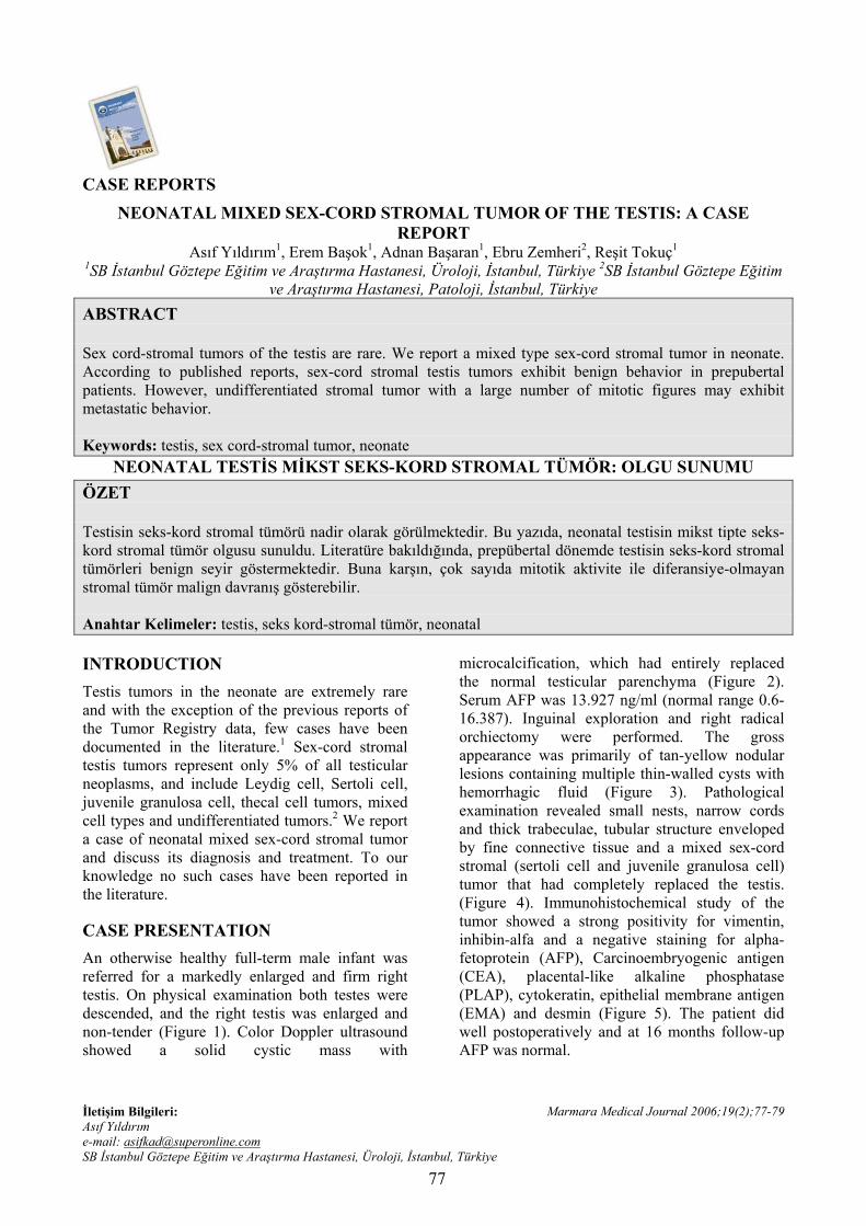
An otherwise healthy full-term male infant was referred for a markedly enlarged and firm right testis. On physical examination both testes were descended, and the right testis was enlarged and non-tender (Figure 1). Color Doppler ultrasound showed a solid cystic mass with
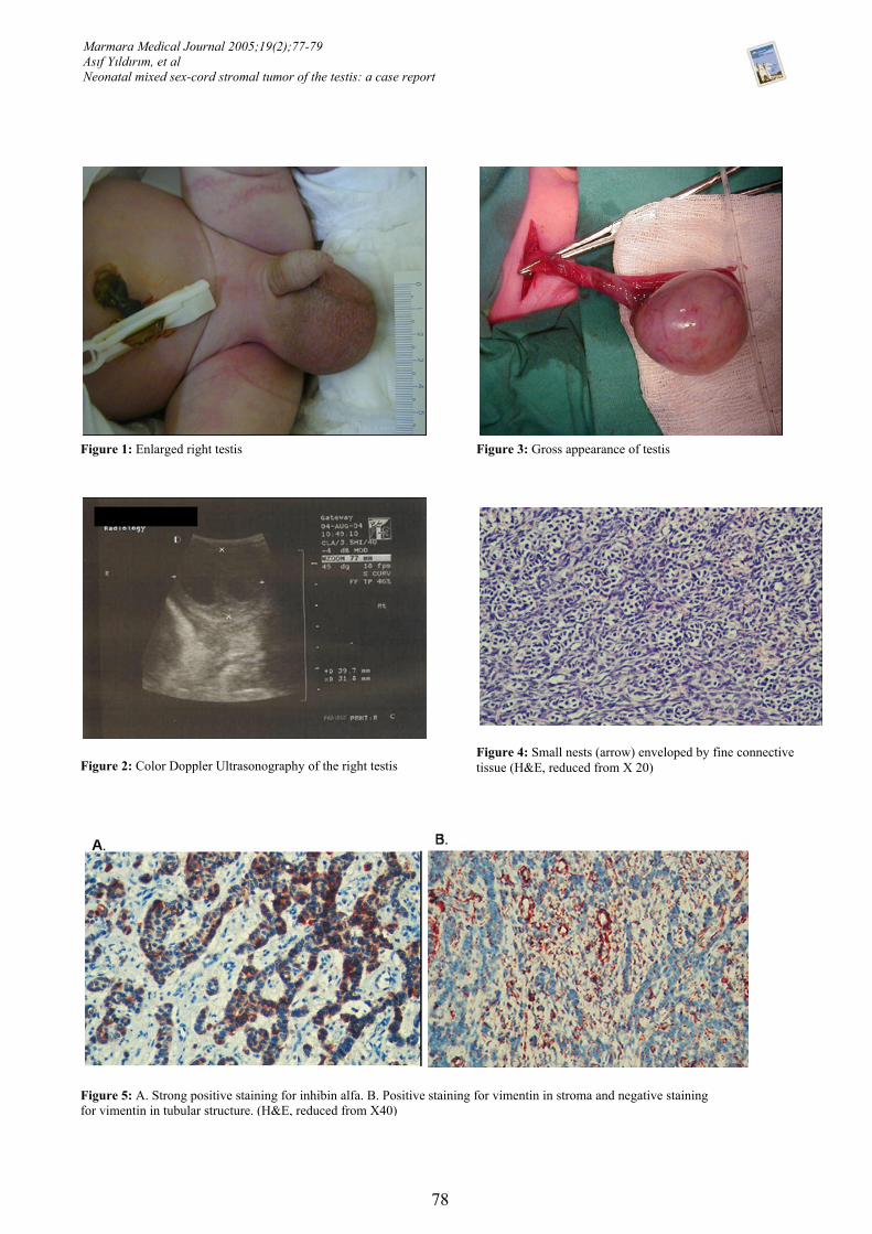
microcalcification, which had entirely replaced the normal testicular parenchyma (Figure 2). Serum AFP was 13.927 ng/ml (normal range 0.6-16.387). Inguinal exploration and right radical orchiectomy were performed. The gross appearance was primarily of tan-yellow nodular lesions containing multiple thin-walled cysts with hemorrhagic fluid (Figure 3). Pathological examination revealed small nests, narrow cords and thick trabeculae, tubular structure enveloped by fine connective tissue and a mixed sex-cord stromal (sertoli cell and juvenile granulosa cell) tumor that had completely replaced the testis. (Figure 4). Immunohistochemical study of the tumor showed a strong positivity for vimentin, inhibin-alfa and a negative staining for alpha-fetoprotein (AFP), Carcinoembryogenic antigen (CEA), placental-like alkaline phosphatase (PLAP), cytokeratin, epithelial membrane antigen (EMA) and desmin (Figure 5). The patient did well postoperatively and at 16 months follow-up AFP was normal.
77
Marmara Medical Journal 2005;19(2);77-79 Asıf Yıldırım, et al Neonatal mixed sex-cord stromal tumor of the testis: a case report
Figure 1: Enlarged right testis Figure 3: Gross appearance of testis
Figure 2: Color Doppler Ultrasonography of the right testisFigure 4: Small nests (arrow) enveloped by fine connective tissue (H&E, reduced from X 20)
Figure 5: A. Strong positive staining for inhibin alfa. B. Positive staining for vimentin in stroma and negative staining for vimentin in tubular structure. (H&E, reduced from X40)
78
Marmara Medical Journal 2005;19(2);77-79 Asıf Yıldırım, et al Neonatal mixed sex-cord stromal tumor of the testis: a case report
DISCUSSION
On microscopic examination granulosa cell tumors contain granulose-like cells that line a cystic space, which is intermixed with solid nodular areas. These tumors can be reliably differentiated from yolk sac tumors because they demonstrate positive immunostaining with inhibin and negative immunostaining with AFP.1,3,4 Microscopically, sertoli cell tumors consist of an interlacing network of loose fibrous stroma with a cellular parenchyma composed of moderately small cells with irregular hyperchromatic nuclei.2,3
According to a recent report from the Prepubertal Testis Tumor Registry (1980-2001), of the 43 registered cases of stromal tumors, 8 were mixed or undifferentiated stromal tumor.3 Interpretation of tumor markers in neonates and infants is difficult. In a recent report average AFP for a full-term newborn was 48.406 ng/ml. This value decreases to less than 8.5 ng/ml by age 8 months4
.
The data in the registry confirm that the vast majority of stromal tumors are benign. No patient with leydig cell, juvenile granulosa cell or sertoli cell tumor had metastatic disease during an average 24.6 months of followup.3 However, in the literature at least 4 malignant sertoli cell tumors have been reported in prepubertal patients.3 Juvenile granulosa cell tumor (JGCT) is a distinct variety of gonadal sex-cord stromal tumor that has most often reported in the ovaries of premenarchal girls, rarely in adults, and only recently in the infantile testis.5 The first case of JGCT of the infantile testis was reported by Crump in 1983.6 Subsequently, Lawrence et al described 14 cases and established this tumor as a distinct clinicopathologic entity.4 The immunohistochemical profile of ovarian sex cord-stromal tumors has been well characterized, but few studies have detailed the immunophenotype of the corresponding testicular neoplasms.7 Inhibin is a peptide hormone that is produced by ovarian granulosa cells and testicular sertoli cells8
. McCluggage et al found inhibin to be a good
marker of testicular sex cord-stromal tumors.7 Similar to previous studies, the tumor exhibited strong positive staining for inhibin and vimentin in the present case.8
Management of sex cord-stromal testis tumors is still debated, since the rarity of such tumors makes it difficult to develop a consensus on a standardized approach.
CONCLUSION
Stromal testis tumors are rare. Data from the Prepubertal Testis Tumor Registry confirm the benign behavior of most of these tumors. Periodic follow-up of the remaining testicle through puberty and self-examination thereafter seem reasonable.
REFERENCES
1. Uehling DT, Smith JE, Logan R and Hafez GR. Newborn granulosa cell tumor of the testis. J Urol. 1987; 138: 385-386.
2. Emanuele C, Sara SM, Nicola F, Cristian F, Paola B, Dario F. Undifferentiated sex cord-stromal testis tumor. J Urol. 2004; 171 (6): 2375.
3. Thomas JC, Ross JH and Kay R. Stromal testis tumors in children: e report from the Prepubertal Testis Tumor Registry. J Urol. 2001; 166: 2338-2340.
4. Lawrence WD, Young RH and Scully RE. Juvenile granulosa cell tumor of the infant testis. A report of 14 cases. Am J Surg Pathol. 1985; 9: 87-94.
5. Antonio PA, Nancy J, Howard M. Juvenile Granulosa Cell Tumor of the Infantile Testis: Evidence of a dual epithelial-smooth muscle differentiation. Am J Surg Pathol. 1996; 20 (1): 72-79.
6. Crump WD. Juvenile granulosa cell (sex cord-stromal) tumor of fetal testis. J Urol. 1983; 129: 1057-1058.
7. McCluggage WG, Shanks J, Whiteside C, Maxwell P, Benerjee S, Biggart JD. Immunohistochemical study of testicular sex cord-stromal tumors, including staining with anti-inhibin antibody. Am J of Surg Pathol. 1998; 22 (5): 615-619.
8. Merchenthaler I, Culler MD, Petrusz P, Negro-Viler A. Immunohistochemical localization of inhibin in rat and human reproductive tissues. Mol Cell Endocrinol. 1988; 54: 239-243.
79
![GİS Tümörleri .ppt [Uyumluluk Modu]±şındaLasertedavileri,elektrokoagülasyon,hipertermi tedavileriözofagusobstrüksüyonlarındadenenmektedir. Özefagus kanserinin lazer tedavisi](https://img.pdfslide.tips/doc/110x75/5b1b79f07f8b9a28258e9845/gis-tuemoerleri-ppt-uyumluluk-modu-sindalasertedavilerielektrokoaguelasyonhipertermi.jpg)