Embed Size (px)
Citation preview
1
Chapter 12
DNA & RNA
The Genetic Material: Protein or DNA?
- scientists knew that chromosomes consisted of DNA and protein
- but did not know which was responsible for carrying the genetic material
- both were possible
- proteins seemed the more likely choice because they were thought to be more chemically complex than
DNA
- little was known about DNA in the 1940’s
- DNA seemed too simple to carry all the genetic information
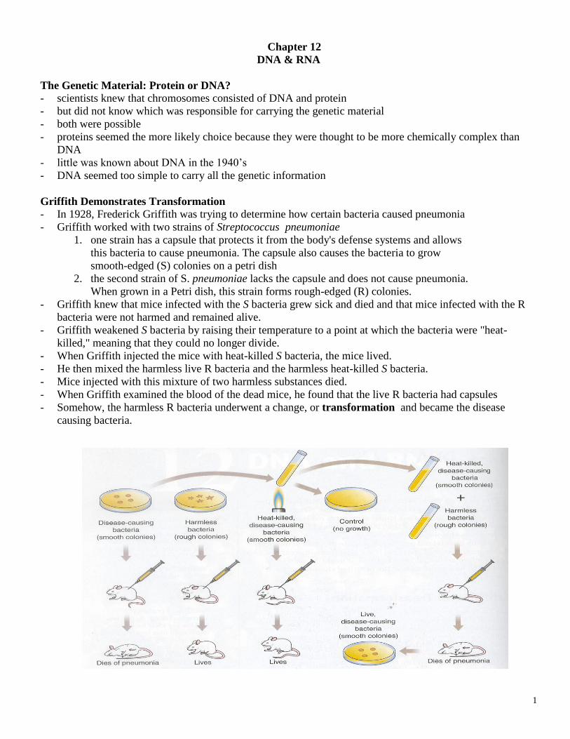
Griffith Demonstrates Transformation
- In 1928, Frederick Griffith was trying to determine how certain bacteria caused pneumonia
- Griffith worked with two strains of Streptococcus pneumoniae
1. one strain has a capsule that protects it from the body's defense systems and allows
this bacteria to cause pneumonia. The capsule also causes the bacteria to grow
smooth-edged (S) colonies on a petri dish
2. the second strain of S. pneumoniae lacks the capsule and does not cause pneumonia.
When grown in a Petri dish, this strain forms rough-edged (R) colonies.
- Griffith knew that mice infected with the S bacteria grew sick and died and that mice infected with the R
bacteria were not harmed and remained alive.
- Griffith weakened S bacteria by raising their temperature to a point at which the bacteria were "heat-
killed," meaning that they could no longer divide.
- When Griffith injected the mice with heat-killed S bacteria, the mice lived.
- He then mixed the harmless live R bacteria and the harmless heat-killed S bacteria.
- Mice injected with this mixture of two harmless substances died.
- When Griffith examined the blood of the dead mice, he found that the live R bacteria had capsules
- Somehow, the harmless R bacteria underwent a change, or transformation and became the disease
causing bacteria.
2
Avery
- In 1944, Oswald Avery and co-workers demonstrated that DNA was responsible for transformation
- They showed that the activity of the material responsible for transformation was not affected by protein-
destroying enzymes but was destroyed when a DNA-destroying enzymes were present.
- Avery and his colleagues made the announcement that the genetic material was DNA. .
Hershey and Chase Show That Genes Are Made of DNA
- Avery's experiments concluded that the genetic material was composed of DNA but many scientist
remained skeptical.
- They thought that DNA was relevant only to certain kinds of bacteria and preferred to think that protein
was the genetic material.
- In 1952, Alfred Hershey and Martha Chase, performed an experiment that settled the controversy.
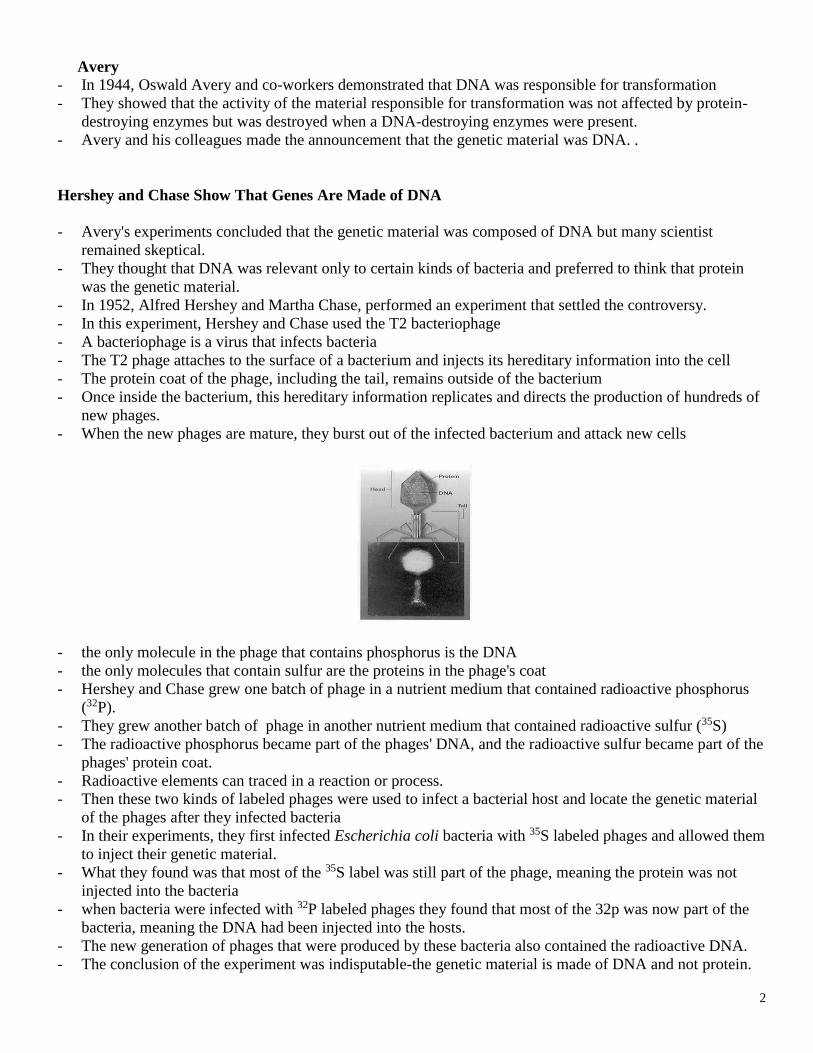
- In this experiment, Hershey and Chase used the T2 bacteriophage
- A bacteriophage is a virus that infects bacteria
- The T2 phage attaches to the surface of a bacterium and injects its hereditary information into the cell
- The protein coat of the phage, including the tail, remains outside of the bacterium
- Once inside the bacterium, this hereditary information replicates and directs the production of hundreds of
new phages.
- When the new phages are mature, they burst out of the infected bacterium and attack new cells
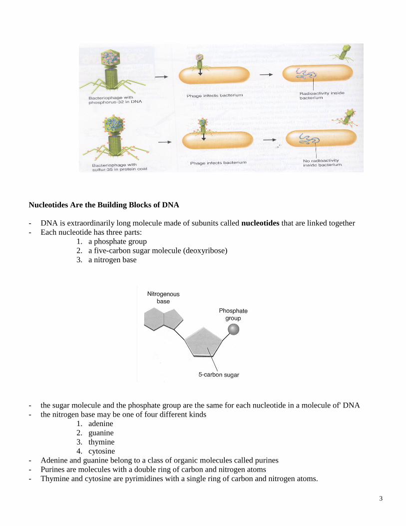
- the only molecule in the phage that contains phosphorus is the DNA
- the only molecules that contain sulfur are the proteins in the phage's coat
- Hershey and Chase grew one batch of phage in a nutrient medium that contained radioactive phosphorus
(32P).
- They grew another batch of phage in another nutrient medium that contained radioactive sulfur (35S)
- The radioactive phosphorus became part of the phages' DNA, and the radioactive sulfur became part of the
phages' protein coat.
- Radioactive elements can traced in a reaction or process.
- Then these two kinds of labeled phages were used to infect a bacterial host and locate the genetic material
of the phages after they infected bacteria
- In their experiments, they first infected Escherichia coli bacteria with 35S labeled phages and allowed them
to inject their genetic material.
- What they found was that most of the 35S label was still part of the phage, meaning the protein was not
injected into the bacteria
- when bacteria were infected with 32P labeled phages they found that most of the 32p was now part of the
bacteria, meaning the DNA had been injected into the hosts.
- The new generation of phages that were produced by these bacteria also contained the radioactive DNA.
- The conclusion of the experiment was indisputable-the genetic material is made of DNA and not protein.
3
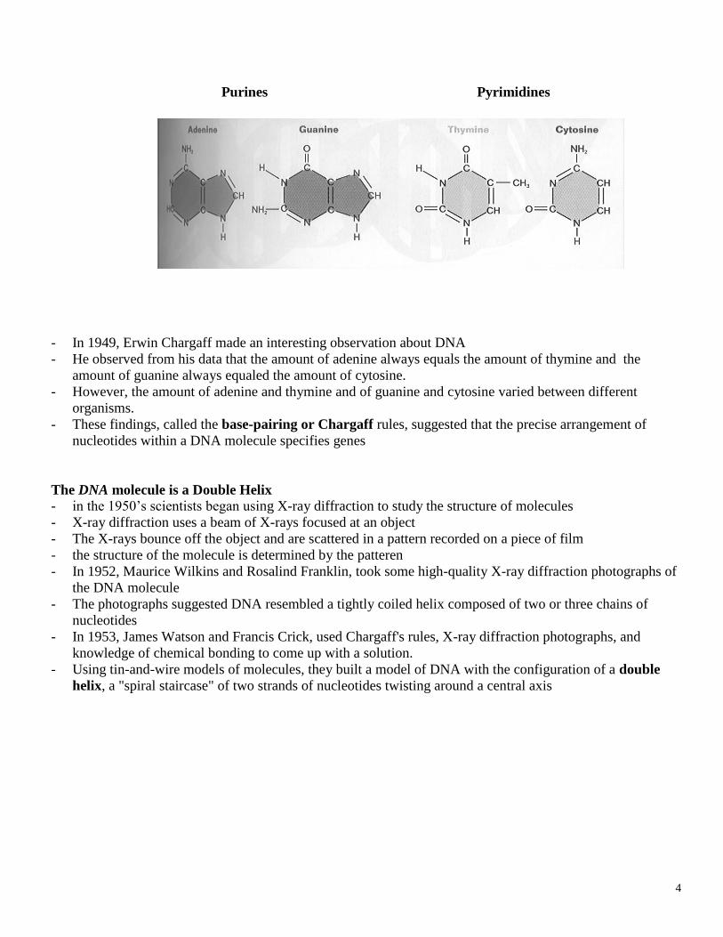
Nucleotides Are the Building Blocks of DNA
- DNA is extraordinarily long molecule made of subunits called nucleotides that are linked together
- Each nucleotide has three parts:
1. a phosphate group
2. a five-carbon sugar molecule (deoxyribose)
3. a nitrogen base
- the sugar molecule and the phosphate group are the same for each nucleotide in a molecule of' DNA
- the nitrogen base may be one of four different kinds
1. adenine
2. guanine
3. thymine
4. cytosine
- Adenine and guanine belong to a class of organic molecules called purines
- Purines are molecules with a double ring of carbon and nitrogen atoms
- Thymine and cytosine are pyrimidines with a single ring of carbon and nitrogen atoms.
4
Purines Pyrimidines
- In 1949, Erwin Chargaff made an interesting observation about DNA
- He observed from his data that the amount of adenine always equals the amount of thymine and the
amount of guanine always equaled the amount of cytosine.
- However, the amount of adenine and thymine and of guanine and cytosine varied between different
organisms.
- These findings, called the base-pairing or Chargaff rules, suggested that the precise arrangement of
nucleotides within a DNA molecule specifies genes
The DNA molecule is a Double Helix
- in the 1950’s scientists began using X-ray diffraction to study the structure of molecules
- X-ray diffraction uses a beam of X-rays focused at an object
- The X-rays bounce off the object and are scattered in a pattern recorded on a piece of film
- the structure of the molecule is determined by the patteren
- In 1952, Maurice Wilkins and Rosalind Franklin, took some high-quality X-ray diffraction photographs of
the DNA molecule
- The photographs suggested DNA resembled a tightly coiled helix composed of two or three chains of
nucleotides
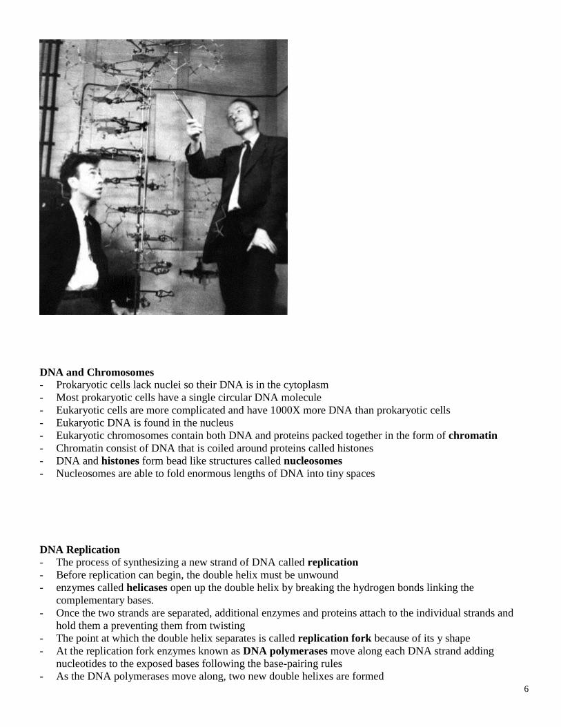
- In 1953, James Watson and Francis Crick, used Chargaff's rules, X-ray diffraction photographs, and
knowledge of chemical bonding to come up with a solution.
- Using tin-and-wire models of molecules, they built a model of DNA with the configuration of a double
helix, a "spiral staircase" of two strands of nucleotides twisting around a central axis
5
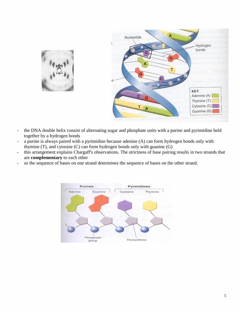
- the DNA double helix consist of alternating sugar and phosphate units with a purine and pyrimidine held
together by a hydrogen bonds
- a purine is always paired with a pyrimidine because adenine (A) can form hydrogen bonds only with
thymine (T), and cytosine (C) can form hydrogen bonds only with guanine (G)
- this arrangement explains Chargaff's observations. The strictness of base pairing results in two strands that
are complementary to each other
- so the sequence of bases on one strand determines the sequence of bases on the other strand.
6
DNA and Chromosomes
- Prokaryotic cells lack nuclei so their DNA is in the cytoplasm
- Most prokaryotic cells have a single circular DNA molecule
- Eukaryotic cells are more complicated and have 1000X more DNA than prokaryotic cells
- Eukaryotic DNA is found in the nucleus
- Eukaryotic chromosomes contain both DNA and proteins packed together in the form of chromatin
- Chromatin consist of DNA that is coiled around proteins called histones
- DNA and histones form bead like structures called nucleosomes
- Nucleosomes are able to fold enormous lengths of DNA into tiny spaces
DNA Replication
- The process of synthesizing a new strand of DNA called replication
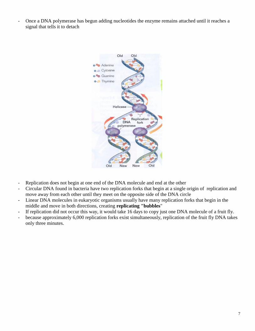
- Before replication can begin, the double helix must be unwound
- enzymes called helicases open up the double helix by breaking the hydrogen bonds linking the
complementary bases.
- Once the two strands are separated, additional enzymes and proteins attach to the individual strands and
hold them a preventing them from twisting
- The point at which the double helix separates is called replication fork because of its y shape
- At the replication fork enzymes known as DNA polymerases move along each DNA strand adding
nucleotides to the exposed bases following the base-pairing rules
- As the DNA polymerases move along, two new double helixes are formed
7
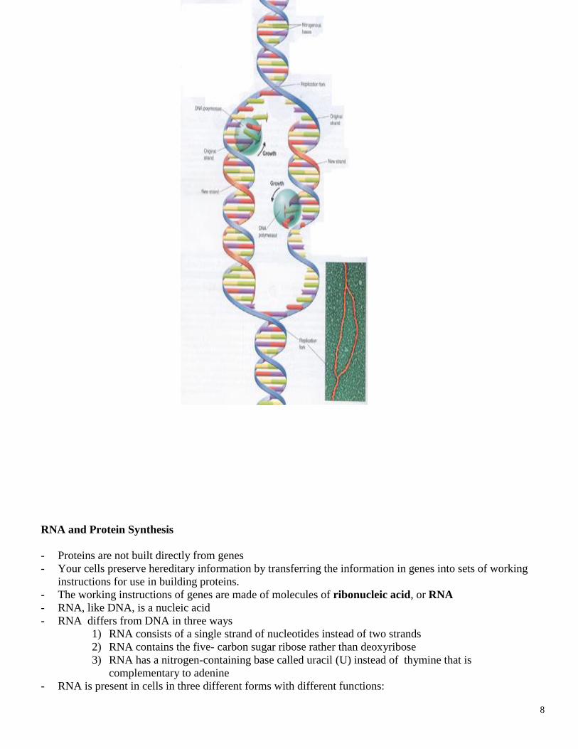
- Once a DNA polymerase has begun adding nucleotides the enzyme remains attached until it reaches a
signal that tells it to detach
- Replication does not begin at one end of the DNA molecule and end at the other
- Circular DNA found in bacteria have two replication forks that begin at a single origin of replication and
move away from each other until they meet on the opposite side of the DNA circle
- Linear DNA molecules in eukaryotic organisms usually have many replication forks that begin in the
middle and move in both directions, creating replicating "bubbles"
- If replication did not occur this way, it would take 16 days to copy just one DNA molecule of a fruit fly.
- because approximately 6,000 replication forks exist simultaneously, replication of the fruit fly DNA takes
only three minutes.
8
RNA and Protein Synthesis
- Proteins are not built directly from genes
- Your cells preserve hereditary information by transferring the information in genes into sets of working
instructions for use in building proteins.
- The working instructions of genes are made of molecules of ribonucleic acid, or RNA
- RNA, like DNA, is a nucleic acid
- RNA differs from DNA in three ways
1) RNA consists of a single strand of nucleotides instead of two strands
2) RNA contains the five- carbon sugar ribose rather than deoxyribose
3) RNA has a nitrogen-containing base called uracil (U) instead of thymine that is
complementary to adenine
- RNA is present in cells in three different forms with different functions:
9
1) messenger RNA (mRNA)
2) ribosomal RNA (rRNA)
3) transfer RNA (tRNA)
- Messenger RNA is an RNA copy of a gene used as a blueprint for a protein
- When a cell needs a particular protein, a specific mRNA is made
- mRNA is appropriately named because it carries hereditary information from DNA and delivers it to the
site of translation
- During translation, mRNA serves as a template for the assembly of amino acids
- tRNA acts as an interpreter molecule, translating mRNA sequences into amino acid sequences
- rRNA plays a structural role in ribosomes, the organelles that function as the sites of translation
-
- All three types of RNA process the information from DNA into proteins, a process called gene expression
- Gene expression occurs in two stages:
1) Transcription - the information in DNA is transferred to mRNA
2) Translation - the information in mRNA is used to make a protein
Transcription Making RNA
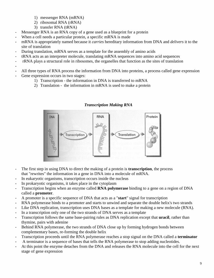
- The first step in using DNA to direct the making of a protein is transcription, the process
that "rewrites" the information in a gene in DNA into a molecule of mRNA.
- In eukaryotic organisms, transcription occurs inside the nucleus
- In prokaryotic organisms, it takes place in the cytoplasm
- Transcription begins when an enzyme called RNA polymerase binding to a gene on a region of DNA
called a promoter.
- A promoter is a specific sequence of DNA that acts as a "start" signal for transcription
- RNA polymerase binds to a promoter and starts to unwind and separate the double helix's two strands
- Like DNA replication, transcription uses DNA bases as a template for making a new molecule (RNA).
- In a transcription only one of the two strands of DNA serves as a template
- Transcription follows the same base-pairing rules as DNA replication except that uracil, rather than
thymine, pairs with adenine
- Behind RNA polymerase, the two strands of DNA close up by forming hydrogen bonds between
complementary bases, re-forming the double helix
- Transcription proceeds until the RNA polymerase reaches a stop signal on the DNA called a terminator
- A terminator is a sequence of bases that tells the RNA polymerase to stop adding nucleotides.
- At this point the enzyme detaches from the DNA and releases the RNA molecule into the cell for the next
stage of gene expression
10
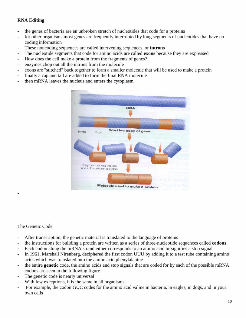
RNA Editing
- the genes of bacteria are an unbroken stretch of nucleotides that code for a proteins
- for other organisms most genes are frequently interrupted by long segments of nucleotides that have no
coding information
- These noncoding sequences are called intervening sequences, or introns
- The nucleotide segments that code for amino acids are called exons because they are expressed
- How does the cell make a protein from the fragments of genes?
- enzymes chop out all the introns from the molecule
- exons are "stitched" back together to form a smaller molecule that will be used to make a protein
- finally a cap and tail are added to form the final RNA molecule
- then mRNA leaves the nucleus and enters the cytoplasm
-
-
The Genetic Code
- After transcription, the genetic material is translated to the language of proteins
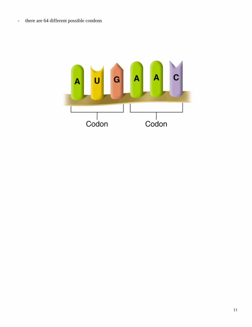
- the instructions for building a protein are written as a series of three-nucleotide sequences called codons
- Each codon along the mRNA strand either corresponds to an amino acid or signifies a stop signal
- In 1961, Marshall Nirenberg, deciphered the first codon UUU by adding it to a test tube containing amino
acids which was translated into the amino acid phenylalanine
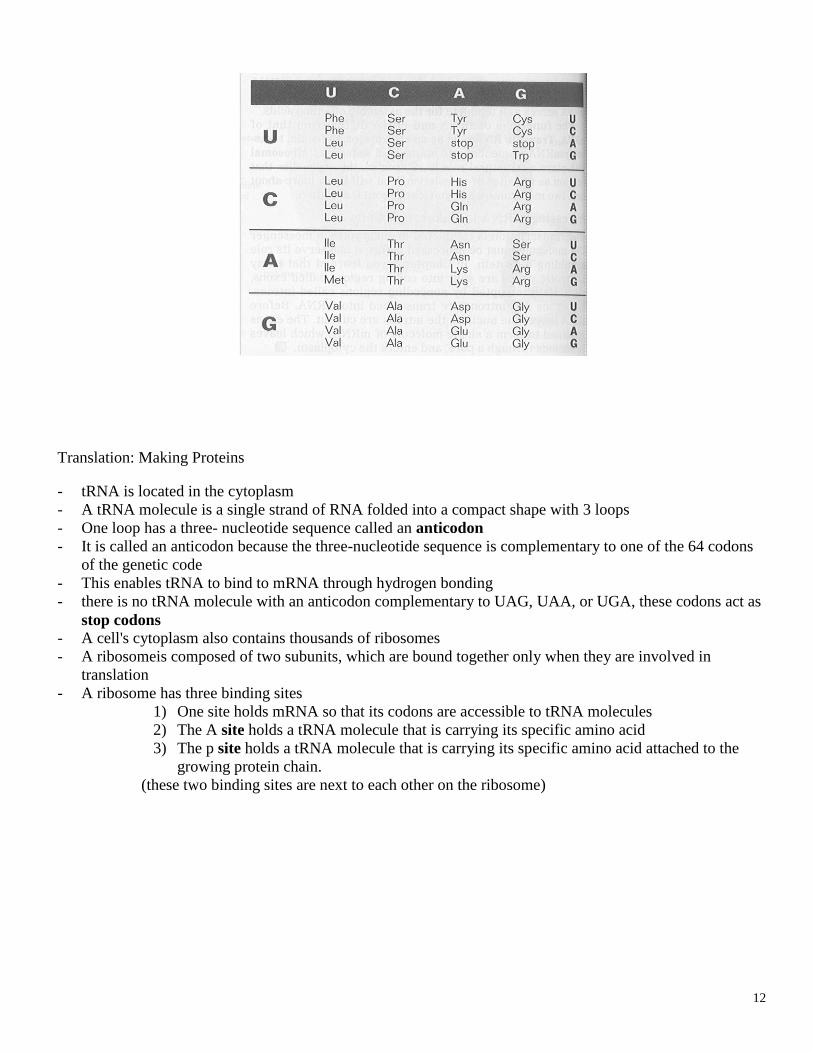
- the entire genetic code, the amino acids and stop signals that are coded for by each of the possible mRNA
codons are seen in the following figure
- The genetic code is nearly universal
- With few exceptions, it is the same in all organisms
- For example, the codon GUC codes for the amino acid valine in bacteria, in eagles, in dogs, and in your
own cells
11
- there are 64 different possible condons
12
Translation: Making Proteins
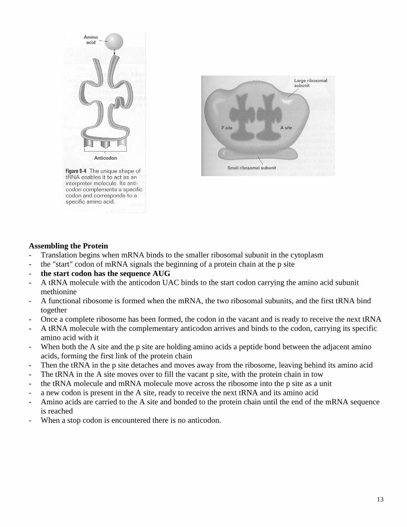
- tRNA is located in the cytoplasm
- A tRNA molecule is a single strand of RNA folded into a compact shape with 3 loops
- One loop has a three- nucleotide sequence called an anticodon
- It is called an anticodon because the three-nucleotide sequence is complementary to one of the 64 codons
of the genetic code
- This enables tRNA to bind to mRNA through hydrogen bonding
- there is no tRNA molecule with an anticodon complementary to UAG, UAA, or UGA, these codons act as
stop codons - A cell's cytoplasm also contains thousands of ribosomes
- A ribosomeis composed of two subunits, which are bound together only when they are involved in
translation
- A ribosome has three binding sites
1) One site holds mRNA so that its codons are accessible to tRNA molecules
2) The A site holds a tRNA molecule that is carrying its specific amino acid
3) The p site holds a tRNA molecule that is carrying its specific amino acid attached to the
growing protein chain.
(these two binding sites are next to each other on the ribosome)
13
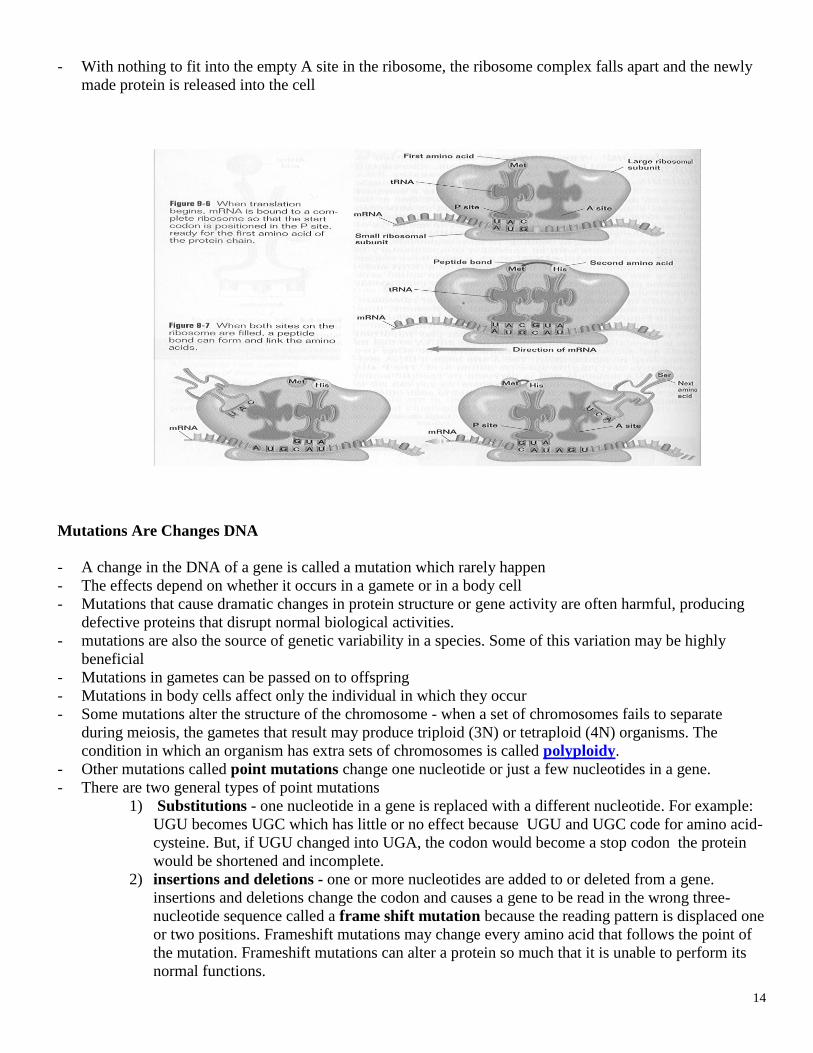
Assembling the Protein
- Translation begins when mRNA binds to the smaller ribosomal subunit in the cytoplasm
- the "start" codon of mRNA signals the beginning of a protein chain at the p site
- the start codon has the sequence AUG - A tRNA molecule with the anticodon UAC binds to the start codon carrying the amino acid subunit
methionine
- A functional ribosome is formed when the mRNA, the two ribosomal subunits, and the first tRNA bind
together
- Once a complete ribosome has been formed, the codon in the vacant and is ready to receive the next tRNA
- A tRNA molecule with the complementary anticodon arrives and binds to the codon, carrying its specific
amino acid with it
- When both the A site and the p site are holding amino acids a peptide bond between the adjacent amino
acids, forming the first link of the protein chain
- Then the tRNA in the p site detaches and moves away from the ribosome, leaving behind its amino acid
- The tRNA in the A site moves over to fill the vacant p site, with the protein chain in tow
- the tRNA molecule and mRNA molecule move across the ribosome into the p site as a unit
- a new codon is present in the A site, ready to receive the next tRNA and its amino acid
- Amino acids are carried to the A site and bonded to the protein chain until the end of the mRNA sequence
is reached
- When a stop codon is encountered there is no anticodon.
14
- With nothing to fit into the empty A site in the ribosome, the ribosome complex falls apart and the newly
made protein is released into the cell
Mutations Are Changes DNA
- A change in the DNA of a gene is called a mutation which rarely happen
- The effects depend on whether it occurs in a gamete or in a body cell
- Mutations that cause dramatic changes in protein structure or gene activity are often harmful, producing
defective proteins that disrupt normal biological activities.
- mutations are also the source of genetic variability in a species. Some of this variation may be highly
beneficial
- Mutations in gametes can be passed on to offspring
- Mutations in body cells affect only the individual in which they occur
- Some mutations alter the structure of the chromosome - when a set of chromosomes fails to separate
during meiosis, the gametes that result may produce triploid (3N) or tetraploid (4N) organisms. The
condition in which an organism has extra sets of chromosomes is called polyploidy.
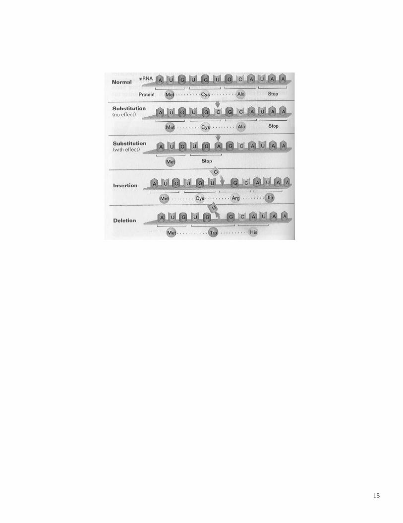
- Other mutations called point mutations change one nucleotide or just a few nucleotides in a gene.
- There are two general types of point mutations
1) Substitutions - one nucleotide in a gene is replaced with a different nucleotide. For example:
UGU becomes UGC which has little or no effect because UGU and UGC code for amino acid-
cysteine. But, if UGU changed into UGA, the codon would become a stop codon the protein
would be shortened and incomplete.
2) insertions and deletions - one or more nucleotides are added to or deleted from a gene.
insertions and deletions change the codon and causes a gene to be read in the wrong three-
nucleotide sequence called a frame shift mutation because the reading pattern is displaced one
or two positions. Frameshift mutations may change every amino acid that follows the point of
the mutation. Frameshift mutations can alter a protein so much that it is unable to perform its
normal functions.
15
16
What Causes Mutations?
- Some mutations are chemical mishaps that arise spontaneously
- Other mutations are induced by exposure to environmental agents called mutagens
- Mutagens include X rays and gamma rays, ultraviolet light, and certain chemicals or irritants referred to as
carcinogens
- Carcinogens are cancer-causing agents
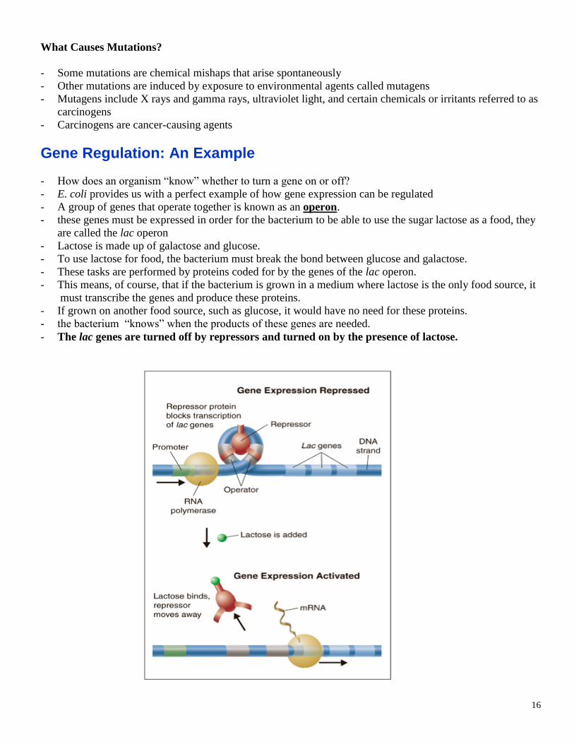
Gene Regulation: An Example
- How does an organism “know” whether to turn a gene on or off?
- E. coli provides us with a perfect example of how gene expression can be regulated
- A group of genes that operate together is known as an operon.
- these genes must be expressed in order for the bacterium to be able to use the sugar lactose as a food, they
are called the lac operon
- Lactose is made up of galactose and glucose.
- To use lactose for food, the bacterium must break the bond between glucose and galactose.
- These tasks are performed by proteins coded for by the genes of the lac operon.
- This means, of course, that if the bacterium is grown in a medium where lactose is the only food source, it
must transcribe the genes and produce these proteins.
- If grown on another food source, such as glucose, it would have no need for these proteins.
- the bacterium “knows” when the products of these genes are needed.
- The lac genes are turned off by repressors and turned on by the presence of lactose.
17
- The general principles of gene regulation in prokaryotes also apply to eukaryotic cells, although there are
some important differences.
- Operons are generally not found in eukaryotes.
- Most eukaryotic genes are controlled individually and have regulatory sequences that are much
more complex than those of the lac operon.
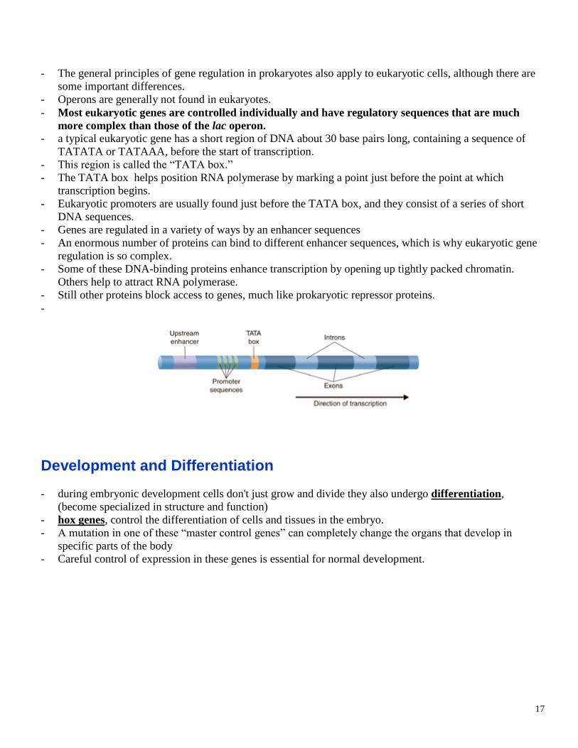
- a typical eukaryotic gene has a short region of DNA about 30 base pairs long, containing a sequence of
TATATA or TATAAA, before the start of transcription.
- This region is called the “TATA box.”
- The TATA box helps position RNA polymerase by marking a point just before the point at which
transcription begins.
- Eukaryotic promoters are usually found just before the TATA box, and they consist of a series of short
DNA sequences.
- Genes are regulated in a variety of ways by an enhancer sequences
- An enormous number of proteins can bind to different enhancer sequences, which is why eukaryotic gene
regulation is so complex.
- Some of these DNA-binding proteins enhance transcription by opening up tightly packed chromatin.
Others help to attract RNA polymerase.
- Still other proteins block access to genes, much like prokaryotic repressor proteins.
-
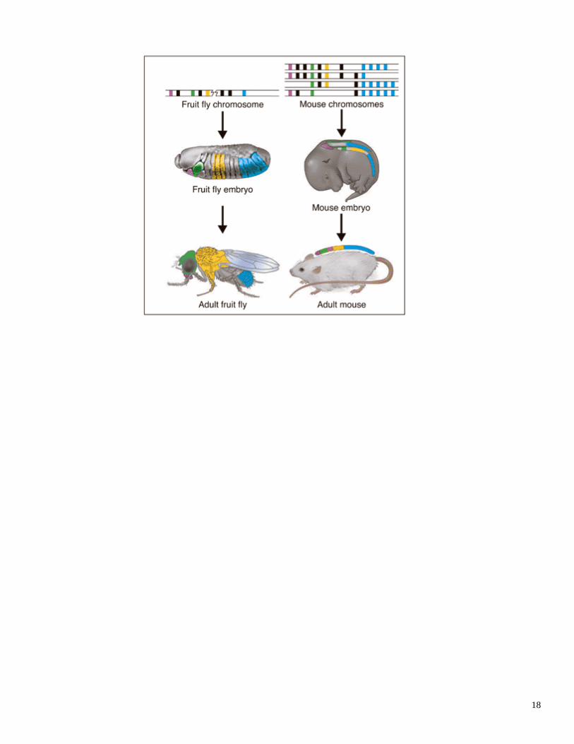
Development and Differentiation
- during embryonic development cells don't just grow and divide they also undergo differentiation,
(become specialized in structure and function)
- hox genes, control the differentiation of cells and tissues in the embryo.
- A mutation in one of these “master control genes” can completely change the organs that develop in
specific parts of the body
- Careful control of expression in these genes is essential for normal development.
18


![DNA RNA [in Turkish]](https://img.pdfslide.tips/doc/110x75/5599a5741a28abfd688b46e7/dna-rna-in-turkish.jpg)