Embed Size (px)
Citation preview
Hindawi Publishing CorporationJournal of Biomedicine and BiotechnologyVolume 2009, Article ID 165637, 10 pagesdoi:10.1155/2009/165637
Research Article
Characterization of Multidrug Resistant ESBL-ProducingEscherichia coli Isolates from Hospitals in Malaysia
King-Ting Lim,1 Rohani Yasin,2 Chew-Chieng Yeo,3 Savithri Puthucheary,4
and Kwai-Lin Thong1
1 Microbiology Division, Institute of Biological Science, Faculty of Science, University of Malaya,50603 Kuala Lumpur, Malaysia
2 Specialized Diagnostic Centre, Institute of Medical Research, Jalan Pahang, 50588 Kuala Lumpur, Malaysia3 Faculty of Agriculture and Biotechnology, Darul Iman University, Malaysia, 20400 Kuala Terengganu, Malaysia4 Department of Medical Microbiology, Faculty of Medicine, University of Malaya, 50603 Kuala Lumpur, Malaysia
Correspondence should be addressed to Kwai-Lin Thong, [email protected]
Received 11 February 2009; Accepted 11 June 2009
Recommended by Vanessa Sperandio
The emergence of Escherichia coli that produce extended spectrum β-lactamases (ESBLs) and are multidrug resistant (MDR)poses antibiotic management problems. Forty-seven E. coli isolates from various public hospitals in Malaysia were studied. Allisolates were sensitive to imipenem whereas 36 were MDR (resistant to 2 or more classes of antibiotics). PCR detection usinggene-specific primers showed that 87.5% of the ESBL-producing E. coli harbored the blaTEM gene. Other ESBL-encoding genesdetected were blaOXA, blaSHV, and blaCTX-M. Integron-encoded integrases were detected in 55.3% of isolates, with class 1 integron-encoded intI1 integrase being the majority. Amplification and sequence analysis of the 5′CS region of the integrons showed knownantibiotic resistance-encoding gene cassettes of various sizes that were inserted within the respective integrons. Conjugation andtransformation experiments indicated that some of the antibiotic resistance genes were likely plasmid-encoded and transmissible.All 47 isolates were subtyped by PFGE and PCR-based fingerprinting using random amplified polymorphic DNA (RAPD),repetitive extragenic palindromes (REPs), and enterobacterial repetitive intergenic consensus (ERIC). These isolates were verydiverse and heterogeneous. PFGE, ERIC, and REP-PCR methods were more discriminative than RAPD in subtyping the E. coliisolates.
Copyright © 2009 King-Ting Lim et al. This is an open access article distributed under the Creative Commons Attribution License,which permits unrestricted use, distribution, and reproduction in any medium, provided the original work is properly cited.
1. Introduction
Escherichia coli are one of the main bacterial pathogensresponsible for nosocomial infections especially in immuno-compromised patients [1]. Extended-spectrum β-lactamases(ESBLs) are enzymes produced by Gram-negative bacillithat mediate resistance to penicillin, cephalosporins, andmonobactams and are commonly recognized in Enterobac-teriaceae and Pseudomonas aeruginosa [2]. Although mostESBLs are mutants of TEM and SHV enzymes, the CTX-Mtype β-lactamases which have become important, originatedfrom β-lactamases found in environmental species of thegenus Kluyvera, and this enzyme hydrolyzes cefotaxime andcefriaxone but is weakly active against ceftazidime [3, 4].These enzymes are present worldwide with more than 50variants [4]. The emergence of ESBL-producers along with
multiple resistant isolates poses a serious problem in thehospital setting. The widespread uses of antibiotics coupledwith the transmissibility of resistance determinants mediatedby plasmids, transposons, and gene cassettes in integrons arefactors that contribute to the increase in antibiotic resistancein bacterial pathogens [1].
Rapid and discriminative subtyping methods are essen-tial for determining the epidemiology of isolates in orderto design rational control methods. Available subtypingmethods for E. coli include PFGE, plasmid profiling, ribo-typing and PCR-based typing methods such as arbitrary-primed PCR, repetitive extragenic palindromes (REPs), andenterobacterial repetitive intergenic consensus (ERIC) [5].
A study from Malaysia reported the presence of the ampCgene and SHV-5 ESBL in clinical isolates of E. coli [6].
2 Journal of Biomedicine and Biotechnology
However, the genotypic characterization of other resistantisolates has not been reported so far. The objectives of thisstudy were to determine the antimicrobial resistance andESBL profiles of E. coli isolated from 5 public Malaysianhospitals and to determine their genetic diversity using PCR-based fingerprinting techniques and PFGE. The presence ofresistance genes and integrons was also determined via PCR,and their transferability was determined by conjugation andtransformation.
2. Materials and Methods
2.1. Bacterial Strains. In this retrospective study, 47 nonre-peat E. coli isolates were collected in 2004 from 47 differentpatients selected randomly from intensive care units of 5public hospitals located in different parts of Malaysia. Theparticipating hospitals were Kota Bharu Hospital (n = 10),Sultanah Aminah Hospital (n = 23), Kuala Lumpur Hospital(n = 10), Ipoh Hospital (n = 2), and Queen ElizabethHospital (n = 2). The isolates were from tracheal aspirates(n = 18), urine (n = 6), body fluids (n = 1), blood(n = 7), pus (n = 6), bile (n = 1), catheter tips (n = 6),and unknown (n = 2). They were identified by standardlaboratory methods in the respective hospitals. They werestored in cryovials containing Luria-Bertani broth with 50%glycerol (Invitrogen, USA) at −20◦C and −85◦C.
2.2. Genotyping by RAPD, REP, and ERIC. Crude DNA fromthe E. coli obtained by direct cell lysate was used for randomamplified polymorphic DNA (RAPD) analysis using primersOPAB04 [7] and OPB17 [8] with cycling conditions aspreviously described (Table 1).
Enterobacterial repetitive intergenic consensus (ERIC)analysis was performed using primer ERIC-1R (OperonBiotechnologies GmBH, Germany) while repetitive extra-genic palindrome (REP) analysis was carried out usingREP oligonucleotides (Operon Biotechnologies GmBH, Ger-many) as primers previously reported [9] (Table 1).
Each PCR reaction was carried out in a 25 μL volumeusing 1.5 U of Taq DNA polymerase (Promega, Madi-son, Wis, USA) in the reaction buffer provided by themanufacturer containing 2.5 mM MgCl2, 50 μM of eachdeoxynucleoside triphosphate, 0.3 μM of the selected primerand 5 μL of DNA template. Aliquots (10 μL) of each PCRproduct were subjected to electrophoresis on 1.5% agarosegel.
2.3. Genotyping by PFGE. PFGE was performed according topreviously described protocols [20] with minor variations.Equal volumes of 1% Seakem Gold agarose (Cambrex BioScience, Rockland, USA) and standardized cell suspension(OD610 = 1.4; approximately 1 × 10⁸ cfu/mL) were mixed toform agarose plugs, and the bacteria lysed within the plugswith cell lysis buffer (50 mM Tris; 50 mM EDTA (pH 8.0),1% Sacrosine, 1 mg/mL proteinase K) and incubated at 54◦Cfor 3 hours. Plugs were then washed with sterile deionisedwater (twice) and TE buffer (4 times), then digested with 12U of XbaI (Promega, Madison, Wis, USA), and incubated
overnight at 37◦C. The XbaI-digested DNA was separatedon a CHEF-DRIII (BioRad, Hercules, CA, USA) with pulsetimes of 2.2–54.2 seconds at 200 V for 24 hours, and gels werephotographed under UV light after staining with 0.5 μg/mLethidium bromide.
2.4. Fingerprint Pattern Analysis. The banding patterns gen-erated by RAPD, ERIC-PCR, REP-PCR, and PFGE wereanalyzed using GelCompar II, version 2.5 (Applied Maths,Kortrijk, Belgium). PCR fingerprints and PFGE profiles wereassigned arbitrary designation and analyzed by defining asimilarity (Dice) coefficient F [21]. Cluster analysis based onthe unweighted pair group method with arithmetic averages(UPGMA) with a position tolerance of 0.15 was done usingthe GelCompar II software.
2.5. Antimicrobial Susceptibility Testing and Screening forESBL. Eighteen antimicrobial agents were tested by thedisk diffusion method in accordance with CLSI guide-lines [22]: ampicillin 10 μg, piperacillin 100 μg, amoxicillin-clavulanic acid 20 μg/10 μg, cefriaxone 30 μg, ceftazidime30 μg, cefepime 30 μg, cefoperazone 75 μg, aztreonam30 μg, imipenem 10 μg, amikacin 30 μg, streptomycin 10 μg,gentamicin 30 μg, kanamycin 30 μg, tetracycline 30 μg,chloramphenicol 30 μg, ciprofloxacin 5 μg, trimethoprim-sulfamethoxazole 75 μg, and nalidixic acid 30 μg (Oxoid Ltd.,Basingtoke, Hampshire, England). Isolates were screened forESBL production by the CLSI disk diffusion method usingcefriaxone 30 μg, ceftazidime 30 μg, and aztreonam 30 μg[22]. The double-disk synergy test was performed accordingto established protocols and results interpreted as describedpreviously [23].
Phenotypic confirmatory test was performed with 30 μgceftazidime, 30/10 μg ceftazidime-clavulanic acid (Becton,Diskson & Company, Maryland, USA), 30 μg cefotaximeand 30/10 μg cefotaxime-clavulanic acid (Becton, Diskson &Company, Maryland, USA) disks on Mueller-Hinton agar.The results were interpreted as described [22]. E. coli isolatesATCC 25922 and ATCC 35218, Klebsiella pneumoniae ATCC700603, and P. aeruginosa ATCC 27853 were used as controls.
2.6. Detection of ESBL Genes. β-lactamase genes (blaTEM,blaSHV, blaCTX-M, blaOXA, blaVEB, blaDHA) were detected byPCR using reverse and forward primer pairs listed in Table 1.Boiled suspension of bacterial cells was used as DNA tem-plate, and cycling parameters were as previously described[10–13] with minor modifications (Table 1). Primers 1R,1, 8F, 8R, 2F, 2R, 9F, and 9R [15] were used for furthersubgrouping of the blaCTX-M gene into CTX-M groups 1, 2,8/25, and 9. All amplified products obtained were sequencedto validate their identities.
Two ESBL-producing isolates (EC19 and EC31) werefurther tested by using primers specific for blaACT, blaGES,blaVIM, blaPER, and blaFOX genes using conditions as listed inTable 1.
Clinical isolates of E. coli were used as the positivecontrols for blaTEM, blaSHV, blaCTX-M, and blaOXA genes.No positive controls were available for detection of blaVEB,
Journal of Biomedicine and Biotechnology 3
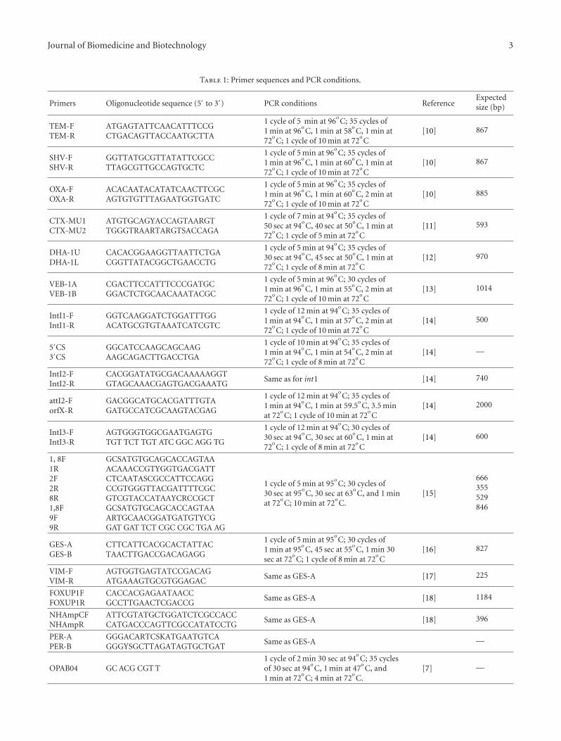
Table 1: Primer sequences and PCR conditions.
Primers Oligonucleotide sequence (5′ to 3′) PCR conditions ReferenceExpectedsize (bp)
TEM-FTEM-R
ATGAGTATTCAACATTTCCGCTGACAGTTACCAATGCTTA
1 cycle of 5 min at 96oC; 35 cycles of
1 min at 96oC, 1 min at 58
oC, 1 min at
72oC; 1 cycle of 10 min at 72
oC
[10] 867
SHV-FSHV-R
GGTTATGCGTTATATTCGCCTTAGCGTTGCCAGTGCTC
1 cycle of 5 min at 96oC; 35 cycles of
1 min at 96oC, 1 min at 60
oC, 1 min at
72oC; 1 cycle of 10 min at 72
oC
[10] 867
OXA-FOXA-R
ACACAATACATATCAACTTCGCAGTGTGTTTAGAATGGTGATC
1 cycle of 5 min at 96oC; 35 cycles of
1 min at 96oC, 1 min at 60
oC, 2 min at
72oC; 1 cycle of 10 min at 72
oC
[10] 885
CTX-MU1CTX-MU2
ATGTGCAGYACCAGTAARGTTGGGTRAARTARGTSACCAGA
1 cycle of 7 min at 94oC; 35 cycles of
50 sec at 94oC, 40 sec at 50
oC, 1 min at
72oC; 1 cycle of 5 min at 72
oC
[11] 593
DHA-1UDHA-1L
CACACGGAAGGTTAATTCTGACGGTTATACGGCTGAACCTG
1 cycle of 5 min at 94oC; 35 cycles of
30 sec at 94oC, 45 sec at 50
oC, 1 min at
72oC; 1 cycle of 8 min at 72
oC
[12] 970
VEB-1AVEB-1B
CGACTTCCATTTCCCGATGCGGACTCTGCAACAAATACGC
1 cycle of 5 min at 96oC; 30 cycles of
1 min at 96oC, 1 min at 55
oC, 2 min at
72oC; 1 cycle of 10 min at 72
oC
[13] 1014
IntI1-FIntI1-R
GGTCAAGGATCTGGATTTGGACATGCGTGTAAATCATCGTC
1 cycle of 12 min at 94oC; 35 cycles of
1 min at 94oC, 1 min at 57
oC, 2 min at
72oC; 1 cycle of 10 min at 72
oC
[14] 500
5′CS3′CS
GGCATCCAAGCAGCAAGAAGCAGACTTGACCTGA
1 cycle of 10 min at 94oC; 35 cycles of
1 min at 94oC, 1 min at 54
oC, 2 min at
72oC; 1 cycle of 8 min at 72
oC
[14] —
IntI2-FIntI2-R
CACGGATATGCGACAAAAAGGTGTAGCAAACGAGTGACGAAATG
Same as for int1 [14] 740
attI2-ForfX-R
GACGGCATGCACGATTTGTAGATGCCATCGCAAGTACGAG
1 cycle of 12 min at 94oC; 35 cycles of
1 min at 94oC, 1 min at 59.5
oC, 3.5 min
at 72oC; 1 cycle of 10 min at 72
oC
[14] 2000
IntI3-FIntI3-R
AGTGGGTGGCGAATGAGTGTGT TCT TGT ATC GGC AGG TG
1 cycle of 12 min at 94oC; 30 cycles of
30 sec at 94oC, 30 sec at 60
oC, 1 min at
72oC; 1 cycle of 8 min at 72
oC
[14] 600
1, 8F1R2F2R8R1,8F9F9R
GCSATGTGCAGCACCAGTAAACAAACCGTYGGTGACGATTCTCAATASCGCCATTCCAGGCCGTGGGTTACGATTTTCGCGTCGTACCATAAYCRCCGCTGCSATGTGCAGCACCAGTAAARTGCAACGGATGATGTYCGGAT GAT TCT CGC CGC TGA AG
1 cycle of 5 min at 95oC; 30 cycles of
30 sec at 95oC, 30 sec at 63
oC, and 1 min
at 72oC; 10 min at 72
oC.
[15]
666355529846
GES-AGES-B
CTTCATTCACGCACTATTACTAACTTGACCGACAGAGG
1 cycle of 5 min at 95oC; 30 cycles of
1 min at 95oC, 45 sec at 55
oC, 1 min 30
sec at 72oC; 1 cycle of 8 min at 72
oC
[16] 827
VIM-FVIM-R
AGTGGTGAGTATCCGACAGATGAAAGTGCGTGGAGAC
Same as GES-A [17] 225
FOXUP1FFOXUP1R
CACCACGAGAATAACCGCCTTGAACTCGACCG
Same as GES-A [18] 1184
NHAmpCFNHAmpR
ATTCGTATGCTGGATCTCGCCACCCATGACCCAGTTCGCCATATCCTG
Same as GES-A [18] 396
PER-APER-B
GGGACARTCSKATGAATGTCAGGGYSGCTTAGATAGTGCTGAT
Same as GES-A —
OPAB04 GC ACG CGT T1 cycle of 2 min 30 sec at 94
oC; 35 cycles
of 30 sec at 94oC, 1 min at 47
oC, and
1 min at 72oC; 4 min at 72
oC.
[7] —
4 Journal of Biomedicine and Biotechnology
Table 1: Continued.
Primers Oligonucleotide sequence (5′ to 3′) PCR conditions ReferenceExpectedsize (bp)
OPB17 AGGGAACGAG Same as OPAB04 [8] —
REP GCG CCG ICA TGC GGC ATT1 cycle of 7 min at 94
oC; 30 cycles of
30 sec at 94oC, 1 min at 44
oC, 8 min at
72oC for 30 cycles; 16 min at 65
oC.
[9] —
ERIC-1R ATGTAACGTCCTGGGGATTCAC1 cycle of 2 min and 30 sec at 94
oC; 35
cycles of 30 sec at 94oC, 1 min at 47
oC,
1 min at 72oC; 1 cycle of 4 min at 72
oC
[19] —
blaACT, blaGES, blaVIM, blaPER, blaFOX, and blaDHA genes andsubgrouping of CTX-M group amplified 2, 8/25, and 9.
2.7. Detection of Class 1, 2, and 3 Integrons. Class 1, 2, and3 integrons were detected by PCR using established primersand conditions as listed in Table 1. Selected amplifiedproducts were sequenced to corroborate their identities.
2.8. Transfer of Antibiotic Resistance Determinants. Transferof resistance genes was attempted in broth using nalidixicresistant recipient E. coli JM109 (endA1, recA1, gyrA96,thi, hsdR17 (rk−, mk
+), relA1, supE44, λ-, Δ (lac-proAB),(F′, traD36, proAB, lacIqZΔZM15). Transconjugants wereselected on Luria-Bertani agar supplemented with ampicillin(100 μg/mL) plus nalidixic acid (100 μg/mL) (Sigma Aldrich,USA).
Transformation experiments were carried out for isolatesin which conjugation failed to produce positive results.Plasmid DNA was extracted from ESBL-producing E. coliusing the QIAprep Spin Miniprep Kit (QIAGEN, Hilden,Germany) and was transformed by electroporation intoelectrocompetent E. coli DH10B (F−mcrA Δ(mrr-hsdRMS-mcrBC) Φ80lacZ ΔlacX74 recA1 endA1 araD139 Δ (ara,leu)7697 galU galK λ−rpsL nupG tonA). Transformants wereselected on Luria-Bertani agar plates containing 100 μg/mLampicillin (Sigma Aldrich, USA).
Size determination of the plasmids from transconjugantsand transformants was carried out by digestion with EcoRIor SphI (Promega, Madison, Wis, USA), and the productsseparated in 0.8% agarose gels at 70 V for 4 hours.
3. Results
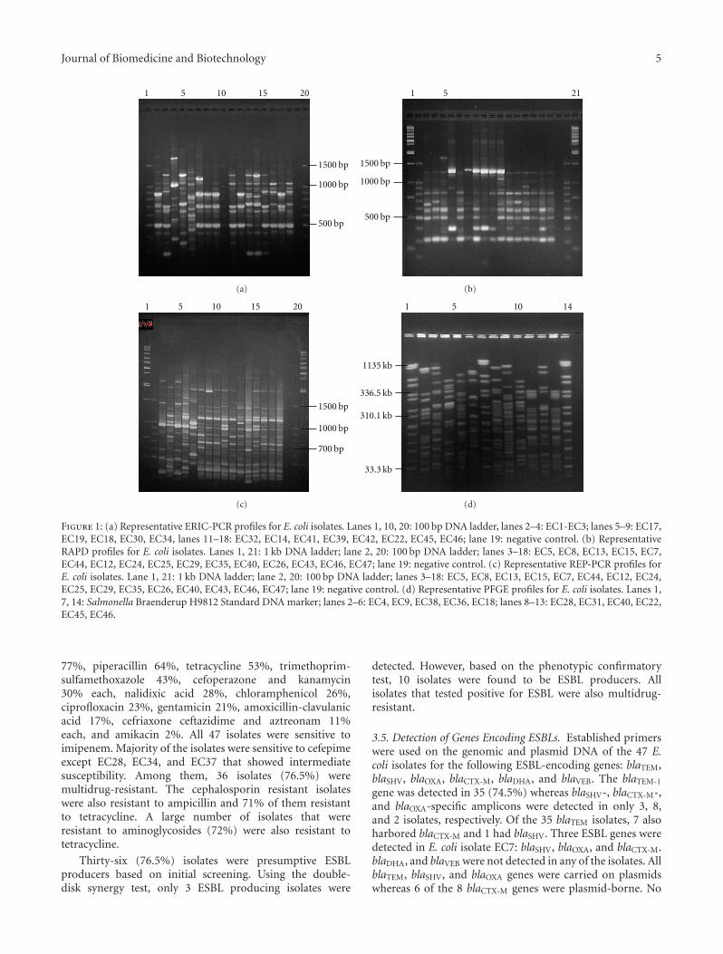
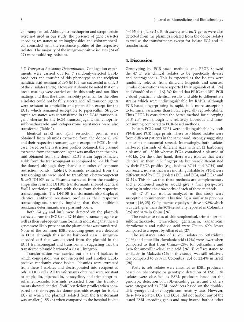
3.1. PCR-Based Fingerprinting. Three PCR-based DNA fin-gerprinting methods were used to subtype the 47 E. coliisolates. ERIC-PCR analysis differentiated the 47 isolatesinto 45 unique profiles (F = 0.54–1.0) whereas RAPDusing the OPAB04 and OPB17 primers generated 44 and 43profiles, respectively, (F = 0.41–1.0 for the OPAB04 primerand F = 0.36–1.0 for the OPB17 primer, see Figures 1(a)and 1(b)). REP-PCR differentiated the 47 isolates into 45distinct profiles (F = 0.53–1.0, see Figure 1(c)). All threePCR-based methods were reproducible as identical profileswere obtained in separate experiments using the same set ofisolates.
Two isolates, EC14 and EC34, from the same hospitalbut from different wards, yielded identical profiles by allthe 3 methods. Two other blood isolates, EC12 and EC24,from 2 different patients in the same ward, were alsoindistinguishable by their ERIC, REP, and RAPD profiles.ESBL-producing isolates EC4, EC9, and EC20 were clonallyrelated by both RAPD and REP-PCR. Isolates EC4 and EC9were indistinguishable by RAPD using the OPAB04 primerwhereas isolates EC4 and EC20 were indistinguishable byRAPD using the OPB17 primer. However, isolates EC4 andEC9 were in the same cluster (92% similarity) based on theirERIC-PCR profiles.
3.2. PFGE with XbaI-Digested Genomic DNA. XbaI-digestedgenomic DNA of the 47 E. coli isolates resulted in 44 distinctpulsed-field profiles (PFPs) comprising 12–26 restrictionfragments. The 2 E. coli strains that had identical ERIC-PCR,REP-PCR and RAPD profiles (i.e., EC12 and EC24) weresimilarly indistinguishable by their PFPs with both sharingall 14 restriction fragments.
Two other isolates, EC1 and EC4 which were indistin-guishable by PFGE but were distinguishable in their ERIC,RAPD and REP-PCR profiles, had 39%–68% similarities.Similarly, ERIC, RAPD, and REP-PCR differentiated isolatesEC37 and EC39 that displayed identical PFPs. Both EC37and EC39 were isolated from the same hospital. On theother hand, isolates EC14 and EC34 that displayed identicalERIC, RAPD, and REP-PCR profiles could be differentiatedby PFGE (F = 0.81).
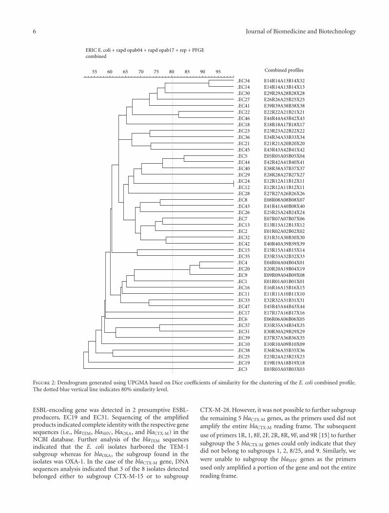
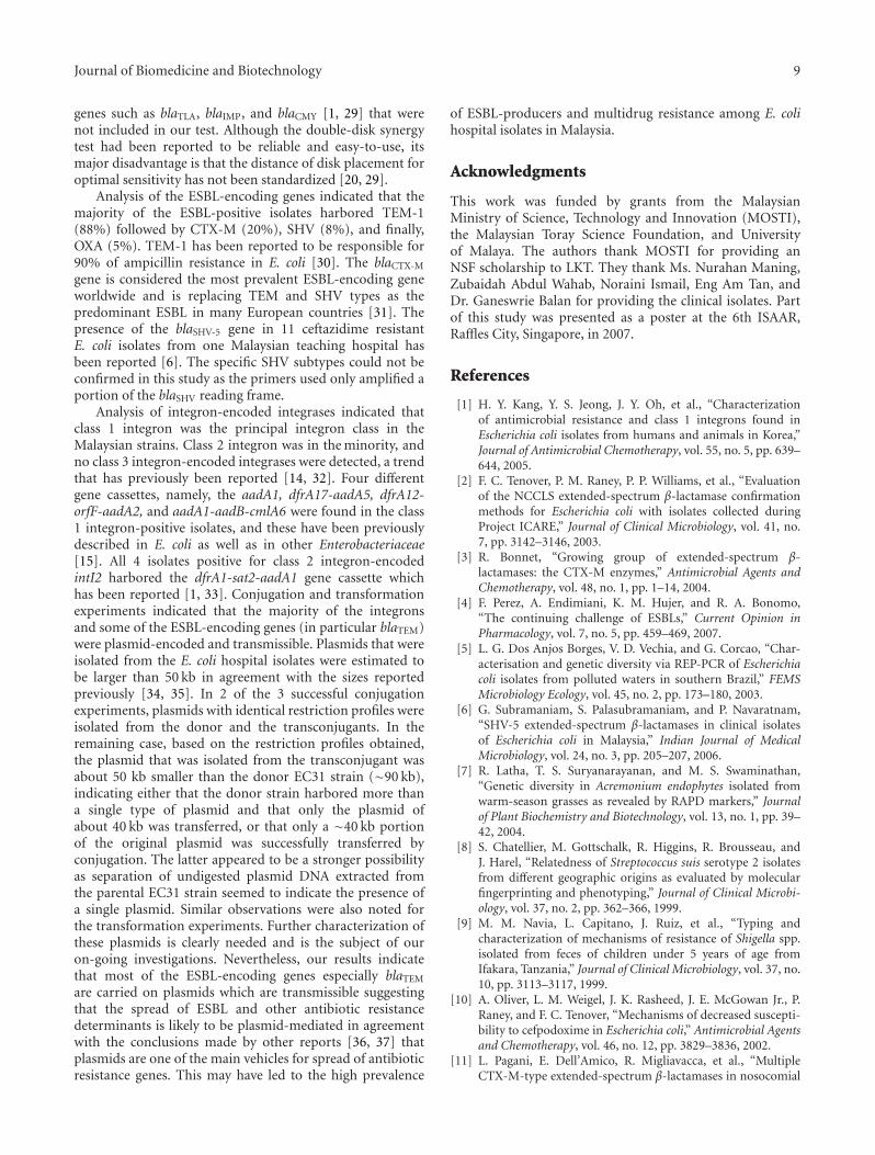
3.3. Combined Analysis. A dendrogram based on the com-bined fingerprints generated by ERIC-PCR, RAPD, REP-PCR, and PFGE was constructed (Figure 2). All the 47isolates were differentiated into 46 combined subtypes(Table 2). Two isolates, EC12 and EC24, with the combinedprofile E12R12A11B12X11 were identical in their ERIC,RAPD, REP, and PFGE profiles. Three other isolates (EC4,EC9, and EC20) were grouped within the same cluster andwere clonally related (more than 85% similarity). IsolatesEC14 and EC34 were also grouped together within the samecluster and were clonally related (more than 94% similarity).
3.4. Antimicrobial Susceptibility. The antibiotic resistantrates for the E. coli isolates were as follows: ampicillin
Journal of Biomedicine and Biotechnology 5
1500 bp
1000 bp
500 bp
1 5 10 15 20
(a)
1500 bp
1000 bp
500 bp
1 5 21
(b)
1500 bp
1000 bp
700 bp
1 5 10 15 20
(c)
1135 kb
336.5 kb
310.1 kb
33.3 kb
1 5 10 14
(d)
Figure 1: (a) Representative ERIC-PCR profiles for E. coli isolates. Lanes 1, 10, 20: 100 bp DNA ladder, lanes 2–4: EC1-EC3; lanes 5–9: EC17,EC19, EC18, EC30, EC34, lanes 11–18: EC32, EC14, EC41, EC39, EC42, EC22, EC45, EC46; lane 19: negative control. (b) RepresentativeRAPD profiles for E. coli isolates. Lanes 1, 21: 1 kb DNA ladder; lane 2, 20: 100 bp DNA ladder; lanes 3–18: EC5, EC8, EC13, EC15, EC7,EC44, EC12, EC24, EC25, EC29, EC35, EC40, EC26, EC43, EC46, EC47; lane 19: negative control. (c) Representative REP-PCR profiles forE. coli isolates. Lane 1, 21: 1 kb DNA ladder; lane 2, 20: 100 bp DNA ladder; lanes 3–18: EC5, EC8, EC13, EC15, EC7, EC44, EC12, EC24,EC25, EC29, EC35, EC26, EC40, EC43, EC46, EC47; lane 19: negative control. (d) Representative PFGE profiles for E. coli isolates. Lanes 1,7, 14: Salmonella Braenderup H9812 Standard DNA marker; lanes 2–6: EC4, EC9, EC38, EC36, EC18; lanes 8–13: EC28, EC31, EC40, EC22,EC45, EC46.
77%, piperacillin 64%, tetracycline 53%, trimethoprim-sulfamethoxazole 43%, cefoperazone and kanamycin30% each, nalidixic acid 28%, chloramphenicol 26%,ciprofloxacin 23%, gentamicin 21%, amoxicillin-clavulanicacid 17%, cefriaxone ceftazidime and aztreonam 11%each, and amikacin 2%. All 47 isolates were sensitive toimipenem. Majority of the isolates were sensitive to cefepimeexcept EC28, EC34, and EC37 that showed intermediatesusceptibility. Among them, 36 isolates (76.5%) weremultidrug-resistant. The cephalosporin resistant isolateswere also resistant to ampicillin and 71% of them resistantto tetracycline. A large number of isolates that wereresistant to aminoglycosides (72%) were also resistant totetracycline.
Thirty-six (76.5%) isolates were presumptive ESBLproducers based on initial screening. Using the double-disk synergy test, only 3 ESBL producing isolates were
detected. However, based on the phenotypic confirmatorytest, 10 isolates were found to be ESBL producers. Allisolates that tested positive for ESBL were also multidrug-resistant.
3.5. Detection of Genes Encoding ESBLs. Established primerswere used on the genomic and plasmid DNA of the 47 E.coli isolates for the following ESBL-encoding genes: blaTEM,blaSHV, blaOXA, blaCTX-M, blaDHA, and blaVEB. The blaTEM-1
gene was detected in 35 (74.5%) whereas blaSHV-, blaCTX-M-,and blaOXA-specific amplicons were detected in only 3, 8,and 2 isolates, respectively. Of the 35 blaTEM isolates, 7 alsoharbored blaCTX-M and 1 had blaSHV. Three ESBL genes weredetected in E. coli isolate EC7: blaSHV, blaOXA, and blaCTX-M.blaDHA, and blaVEB were not detected in any of the isolates. AllblaTEM, blaSHV, and blaOXA genes were carried on plasmidswhereas 6 of the 8 blaCTX-M genes were plasmid-borne. No
6 Journal of Biomedicine and Biotechnology
ERIC E. coli + rapd opab04 + rapd opab17 + rep + PFGE combined
.EC34
.EC14
.EC30
.EC27
.EC41
.EC22
.EC46
.EC18
.EC23
.EC36
.EC21
.EC45
.EC5
.EC44
.EC40
.EC29
.EC24
.EC12
.EC28
.EC8
.EC43
.EC26
.EC7
.EC13
.EC2
.EC32
.EC42
.EC15
.EC35
.EC4
.EC20
.EC9
.EC1
.EC16
.EC11
.EC33
.EC47
.EC17
.EC6
.EC37
.EC31
.EC39
.EC10
.EC38
.EC25
.EC19
.EC3
E14R14A13B14X32E14R14A13B14X13E29R29A28B28X28E26R26A25B25X25E39R39A38B38X38E22R22A21B21X21E44R44A43B42X43E18R18A17B18X17E23R23A22B22X22E34R34A33B33X34E21R21A20B20X20E43R43A42B41X42E05R05A05B05X04E42R42A41B40X41E38R38A37B37X37E28R28A27B27X27E12R12A11B12X11E12R12A11B12X11E27R27A26B26X26E08R08A08B08X07E41R41A40B08X40E25R25A24B24X24E07R07A07B07X06E13R13A12B13X12E01R02A02B02X02E31R31A30B30X30E40R40A39B39X39E15R15A14B15X14E33R33A32B32X33E04R04A04B04X01E20R20A19B04X19E09R09A04B09X08E01R01A01B01X01E16R16A15B16X15E11R11A10B11X10E32R32A31B31X31E45R45A44B43X44E17R17A16B17X16E06R06A06B06X05E35R35A34B34X35E30R30A29B29X29E37R37A36B36X35E10R10A09B10X09E36R36A35B35X36E23R24A23B23X23E19R19A18B19X18E03R03A03B03X03
Combined profiles 55 60 65 70 75 80 85 90 95
Figure 2: Dendrogram generated using UPGMA based on Dice coefficients of similarity for the clustering of the E. coli combined profile.The dotted blue vertical line indicates 80% similarity level.
ESBL-encoding gene was detected in 2 presumptive ESBL-producers, EC19 and EC31. Sequencing of the amplifiedproducts indicated complete identity with the respective genesequences (i.e., blaTEM, blaSHV, blaOXA, and blaCTX-M) in theNCBI database. Further analysis of the blaTEM sequencesindicated that the E. coli isolates harbored the TEM-1subgroup whereas for blaOXA, the subgroup found in theisolates was OXA-1. In the case of the blaCTX-M gene, DNAsequences analysis indicated that 3 of the 8 isolates detectedbelonged either to subgroup CTX-M-15 or to subgroup
CTX-M-28. However, it was not possible to further subgroupthe remaining 5 blaCTX-M genes, as the primers used did notamplify the entire blaCTX-M reading frame. The subsequent
use of primers 1R, 1, 8F, 2F, 2R, 8R, 9F, and 9R [15] to furthersubgroup the 5 blaCTX-M genes could only indicate that theydid not belong to subgroups 1, 2, 8/25, and 9. Similarly, wewere unable to subgroup the blaSHV genes as the primersused only amplified a portion of the gene and not the entirereading frame.
Journal of Biomedicine and Biotechnology 7
Table 2: Antimicrobial resistance, size of plasmids and ESBL genes detected in the selected donor E. coli isolates, and their respectivetransconjugants and transformants.
No. Resistanceprofile
ESBL-encodinggenedetected byPCR
Plasmiddonor(kb)
Plasmid transfor-mant/transconjugant(kb)
Resistance profile oftransformant/transconjugant
ESBL-encoding genetransferred
Mode of transfer
EC7
AMP, PIP,TET, CRO,FEP, NAL,SXT, CHL,CFP, CAZ,KAN,ATM
blaOXA,blaSHV,blaCTX-M
135 55AMP, PIP, TET, CRO,FEP, NAL, SXT, CFP,KAN, ATM
blaOXA, blaSHV, Transformation
EC10
AMP, PIP,TET, NAL,SXT, CHL,CAZ,KAN, CIP,STR, GEN
blaTEM 310 310AMP, PIP, TET, NAL,SXT, CHL, CIP, STR,GEN
blaTEM Transformation
EC12
AMP, PIP,TET, NAL,SXT, CHL,KAN, STR
blaTEM 50 50AMP, PIP, TET, SXT,KAN, STR
blaTEM Transformation
EC18AMP, PIP,TET, SXT,STR, CRO
blaTEM 190 190 AMP blaTEM Conjugation
EC24
AMP, PIP,TET, NAL,SXT, CHL,KAN, CIP,GEN
blaTEM,blaCTX-M
60 60AMP, PIP, TET, NAL,SXT, CHL, CIP, GEN
blaTEM,blaCTX-M
Transformation
EC31
AMP, PIP,TET, SXT,CHL, CFP,KAN,CRO, STR
ND 90 40 AMP, PIP, SXT, CFP ND Conjugation
EC36
AMP, PIP,TET, SXT,CFP, CIP,STR, KAN,GEN,NAL,CHL, SXT
blaTEM 300 300AMP, PIP, TET, SXT,CIP, STR, GEN, CHL,NAL
blaTEM Transformation
EC46 AMP, PIP,STR
blaTEM 50 50 AMP, PIP, STR blaTEM Conjugation
3.6. Class 1, 2, and 3 Integrons. Forty-seven E. coli isolateswere screened for the presence of integrases encoded onclass 1, 2, and 3 integrons. The class 1 integron-encodedintI1 integrase gene was detected in 25 isolates while 4isolates tested positive for class 2-encoded intI2 integrase.One isolate, EC24, was found to harbor both intI1 and intI2.No class 3 integron was detected. Majority of the integronswere found to be plasmid-encoded (16 of the 25 intI1 and 3of the 4 intI2 detected).
Isolates that were positive for class 1 and 2 integronswere further analyzed for the presence of inserted genecassettes within the variable region by using the primer pair
5′ CS/3′ CS for class 1 integrons and primer pair orfx/attI2for class 2 integrons. Amplified products of different sizeswere obtained from 17 of the 25 intI1-positive isolates andsequencing indicated the presence of 5 different types ofknown gene cassettes: aadA5-dfrA17, dfrA7, aadA1-aadB-cmlA6, dfrA12-aadA2-orfF and aadA1. Using the attI2/orfXprimer pair for intI2-positive isolates resulted in a 2 kbamplified product which, when sequenced, contained thedfrA1-aadA1-sat2 gene cassette. The aadA2, aadA5, andaadB genes encode resistance to aminoglycosides whereassat2 encode resistance to strepthoricin. Both dfrA12 anddfrA17 encode resistance to trimethoprim and cmlA6 to
8 Journal of Biomedicine and Biotechnology
chloramphenicol. Although trimethoprim and strepthoricinwere not used in our study, the presence of gene cassettesencoding resistance to aminoglycosides and chlorampheni-col coincided with the resistance profiles of the respectiveisolates. The majority of the integron-positive isolates (24 of27) were multidrug-resistant.
3.7. Transfer of Resistance Determinants. Conjugation exper-iments were carried out for 7 randomly-selected ESBL-producers and transfer of this phenotype to the recipientnalidixic acid-resistant E. coli JM109 was successful in only 3of the 7 isolates (38%). However, it should be noted that onlybroth matings were carried out in this study and not filtermatings and thus the transmissibility potential for the other4 isolates could not be fully ascertained. All transconjugantswere resistant to ampicillin and piperacillin except for theEC18 which remained susceptible to piperacillin. Strepto-mycin resistance was cotransferred in the EC46 transconju-gant whereas for the EC31 transconjugant, trimethoprim-sulfomethaxazole and cefoperazone resistances were alsotransferred (Table 2).
Identical EcoRI and SphI restriction profiles wereobtained from plasmids extracted from the donor E. coliand their respective transconjugants except for EC31. In thiscase, based on the restriction profiles obtained, the plasmidextracted from the transconjugant was smaller than the plas-mid obtained from the donor EC31 strain (approximately40 kb from the transconjugant as compared to ∼90 kb fromthe donor) although they shared a number of commonrestriction bands (Table 2). Plasmids extracted from thetransconjugants were used to transform electrocompetentE. coli DH10B cells. Plasmids extracted from the resultingampicillin resistant DH10B transformants showed identicalEcoRI restriction profiles with those from their respectivetransconjugants. The DH10B transformants also displayedidentical antibiotic resistance profiles as their respectivetransconjugants, strongly implying that these antibioticresistance determinants were plasmid-borne.
Both blaTEM and intI1 were detected on the plasmidsextracted from the EC18 and EC46 donor, transconjugants aswell as their subsequent transformants indicating that these 2genes were likely present on the plasmid that was transferred.None of the common ESBL-encoding genes were detectedin EC31 although this isolate harbored class 1 integron-encoded intI that was detected from the plasmid in theEC31 transconjugant and transformant suggesting that thetransferred plasmid harbored a class 1 integron.
Transformation was carried out for the 4 isolates inwhich conjugation was not successful and another ESBL-positive randomly chose isolate. Plasmids were extractedfrom these 5 isolates and electroporated into recipient E.coli DH10B cells. All transformants obtained were resistantto ampicillin, piperacillin, tetracycline, and trimethoprim-sulfamethoxazole. Plasmids extracted from the transfor-mants showed identical EcoRI restriction profiles when com-pared to their respective donor plasmids except for isolateEC7 in which the plasmid isolated from the transformantwas smaller (∼55 kb) when compared to the hospital isolate
(∼135 kb) (Table 2). Both blaTEM and intI1 genes were alsodetected from the plasmids isolated from the donor isolatesas well as the transformants except for isolate EC7 and itstransformant.
4. Discussion
Genotyping by PCR-based methods and PFGE showedthe 47 E. coli clinical isolates to be genetically diverseand heterogeneous. This is expected as the isolates wererandomly selected from different hospitals and sources.Similar observations were reported by Mugnaioli et al. [24]and Woodford et al. [16]. We found that ERIC and REP-PCRyielded practically identical results and able to differentiatestrains which were indistinguishable by RAPD. AlthoughPCR-based fingerprinting is rapid, it is more susceptibleto technical variations than PFGE especially reproducibility.Thus PFGE is considered the better method for subtypingof E. coli, even though it is relatively laborious and time-consuming compared to PCR-based methods.
Isolates EC12 and EC24 were indistinguishable by bothPFGE and PCR fingerprints. These two blood isolates werefrom different patients in the same ward, strongly suggestinga possible nosocomial spread. Interestingly, both isolatesharbored plasmids of different sizes with EC12 harboringa plasmid of ∼50 kb whereas EC24 contained a plasmid of∼60 kb. On the other hand, there were isolates that wereidentical in their PCR fingerprints but were differentiatedby their PFGE profiles (e.g., isolates EC14 and EC34) andconversely, isolates that were indistinguishable by PFGE weredifferentiated by PCR (isolates EC1 and EC4, and EC37 andEC39). This shows that these methods are complementaryand a combined analysis would give a finer perspectivebearing in mind the drawbacks of each of these methods.
All 47 E. coli isolates analyzed in this study weresusceptible to imipenem. This finding is similar to previousreports [16, 25]. Cefepime was equally sensitive at 98% whichis a rate higher than the 80% sensitivity reported in Colombia[25] and 70% in China [26].
The resistance rates of chloramphenicol, trimethoprim-sulfomethaxazole, tetracycline, gentamicin, kanamycin,ciprofloxacin and nalidixic acid were 7% to 69% lowercompared to a report by Alhaj et al. [27].
The resistance rates of E. coli isolates to ceftazidime(11%) and amoxillin-clavulanic acid (17%) were lower whencompared to that from China—28% for ceftazidime and84% for amoxillin-clavulanic acid [26]. E. coli resistance toamikacin in Malaysia (2% in this study) was still relativelylow compared to 27% in Colombia [25] or 22.4% in Israel[28].
Forty E. coli isolates were classified as ESBL producersbased on phenotypic or genotypic detection of ESBL: 38isolates were classified as ESBL producers based on thegenotypic detection of ESBL-encoding genes, and 2 otherswere categorized as ESBL producers based on the double-disk synergy and phenotypic confirmatory tests. However,these two isolates, EC7 and EC31, did not harbor any of thetested ESBL-encoding genes and may instead harbor other
Journal of Biomedicine and Biotechnology 9
genes such as blaTLA, blaIMP, and blaCMY [1, 29] that werenot included in our test. Although the double-disk synergytest had been reported to be reliable and easy-to-use, itsmajor disadvantage is that the distance of disk placement foroptimal sensitivity has not been standardized [20, 29].
Analysis of the ESBL-encoding genes indicated that themajority of the ESBL-positive isolates harbored TEM-1(88%) followed by CTX-M (20%), SHV (8%), and finally,OXA (5%). TEM-1 has been reported to be responsible for90% of ampicillin resistance in E. coli [30]. The blaCTX-M
gene is considered the most prevalent ESBL-encoding geneworldwide and is replacing TEM and SHV types as thepredominant ESBL in many European countries [31]. Thepresence of the blaSHV-5 gene in 11 ceftazidime resistantE. coli isolates from one Malaysian teaching hospital hasbeen reported [6]. The specific SHV subtypes could not beconfirmed in this study as the primers used only amplified aportion of the blaSHV reading frame.
Analysis of integron-encoded integrases indicated thatclass 1 integron was the principal integron class in theMalaysian strains. Class 2 integron was in the minority, andno class 3 integron-encoded integrases were detected, a trendthat has previously been reported [14, 32]. Four differentgene cassettes, namely, the aadA1, dfrA17-aadA5, dfrA12-orfF-aadA2, and aadA1-aadB-cmlA6 were found in the class1 integron-positive isolates, and these have been previouslydescribed in E. coli as well as in other Enterobacteriaceae[15]. All 4 isolates positive for class 2 integron-encodedintI2 harbored the dfrA1-sat2-aadA1 gene cassette whichhas been reported [1, 33]. Conjugation and transformationexperiments indicated that the majority of the integronsand some of the ESBL-encoding genes (in particular blaTEM)were plasmid-encoded and transmissible. Plasmids that wereisolated from the E. coli hospital isolates were estimated tobe larger than 50 kb in agreement with the sizes reportedpreviously [34, 35]. In 2 of the 3 successful conjugationexperiments, plasmids with identical restriction profiles wereisolated from the donor and the transconjugants. In theremaining case, based on the restriction profiles obtained,the plasmid that was isolated from the transconjugant wasabout 50 kb smaller than the donor EC31 strain (∼90 kb),indicating either that the donor strain harbored more thana single type of plasmid and that only the plasmid ofabout 40 kb was transferred, or that only a ∼40 kb portionof the original plasmid was successfully transferred byconjugation. The latter appeared to be a stronger possibilityas separation of undigested plasmid DNA extracted fromthe parental EC31 strain seemed to indicate the presence ofa single plasmid. Similar observations were also noted forthe transformation experiments. Further characterization ofthese plasmids is clearly needed and is the subject of ouron-going investigations. Nevertheless, our results indicatethat most of the ESBL-encoding genes especially blaTEM
are carried on plasmids which are transmissible suggestingthat the spread of ESBL and other antibiotic resistancedeterminants is likely to be plasmid-mediated in agreementwith the conclusions made by other reports [36, 37] thatplasmids are one of the main vehicles for spread of antibioticresistance genes. This may have led to the high prevalence
of ESBL-producers and multidrug resistance among E. colihospital isolates in Malaysia.
Acknowledgments
This work was funded by grants from the MalaysianMinistry of Science, Technology and Innovation (MOSTI),the Malaysian Toray Science Foundation, and Universityof Malaya. The authors thank MOSTI for providing anNSF scholarship to LKT. They thank Ms. Nurahan Maning,Zubaidah Abdul Wahab, Noraini Ismail, Eng Am Tan, andDr. Ganeswrie Balan for providing the clinical isolates. Partof this study was presented as a poster at the 6th ISAAR,Raffles City, Singapore, in 2007.
References
[1] H. Y. Kang, Y. S. Jeong, J. Y. Oh, et al., “Characterizationof antimicrobial resistance and class 1 integrons found inEscherichia coli isolates from humans and animals in Korea,”Journal of Antimicrobial Chemotherapy, vol. 55, no. 5, pp. 639–644, 2005.
[2] F. C. Tenover, P. M. Raney, P. P. Williams, et al., “Evaluationof the NCCLS extended-spectrum β-lactamase confirmationmethods for Escherichia coli with isolates collected duringProject ICARE,” Journal of Clinical Microbiology, vol. 41, no.7, pp. 3142–3146, 2003.
[3] R. Bonnet, “Growing group of extended-spectrum β-lactamases: the CTX-M enzymes,” Antimicrobial Agents andChemotherapy, vol. 48, no. 1, pp. 1–14, 2004.
[4] F. Perez, A. Endimiani, K. M. Hujer, and R. A. Bonomo,“The continuing challenge of ESBLs,” Current Opinion inPharmacology, vol. 7, no. 5, pp. 459–469, 2007.
[5] L. G. Dos Anjos Borges, V. D. Vechia, and G. Corcao, “Char-acterisation and genetic diversity via REP-PCR of Escherichiacoli isolates from polluted waters in southern Brazil,” FEMSMicrobiology Ecology, vol. 45, no. 2, pp. 173–180, 2003.
[6] G. Subramaniam, S. Palasubramaniam, and P. Navaratnam,“SHV-5 extended-spectrum β-lactamases in clinical isolatesof Escherichia coli in Malaysia,” Indian Journal of MedicalMicrobiology, vol. 24, no. 3, pp. 205–207, 2006.
[7] R. Latha, T. S. Suryanarayanan, and M. S. Swaminathan,“Genetic diversity in Acremonium endophytes isolated fromwarm-season grasses as revealed by RAPD markers,” Journalof Plant Biochemistry and Biotechnology, vol. 13, no. 1, pp. 39–42, 2004.
[8] S. Chatellier, M. Gottschalk, R. Higgins, R. Brousseau, andJ. Harel, “Relatedness of Streptococcus suis serotype 2 isolatesfrom different geographic origins as evaluated by molecularfingerprinting and phenotyping,” Journal of Clinical Microbi-ology, vol. 37, no. 2, pp. 362–366, 1999.
[9] M. M. Navia, L. Capitano, J. Ruiz, et al., “Typing andcharacterization of mechanisms of resistance of Shigella spp.isolated from feces of children under 5 years of age fromIfakara, Tanzania,” Journal of Clinical Microbiology, vol. 37, no.10, pp. 3113–3117, 1999.
[10] A. Oliver, L. M. Weigel, J. K. Rasheed, J. E. McGowan Jr., P.Raney, and F. C. Tenover, “Mechanisms of decreased suscepti-bility to cefpodoxime in Escherichia coli,” Antimicrobial Agentsand Chemotherapy, vol. 46, no. 12, pp. 3829–3836, 2002.
[11] L. Pagani, E. Dell’Amico, R. Migliavacca, et al., “MultipleCTX-M-type extended-spectrum β-lactamases in nosocomial
10 Journal of Biomedicine and Biotechnology
isolates of Enterobacteriaceae from a hospital in NorthernItaly,” Journal of Clinical Microbiology, vol. 41, no. 9, pp. 4264–4269, 2003.
[12] H. Pai, C.-I. Kang, J.-H. Byeon, et al., “Epidemiology andclinical features of bloodstream infections caused by AmpC-type-β-lactamase-producing Klebsiella pneumoniae,” Antimi-crobial Agents and Chemotherapy, vol. 48, no. 10, pp. 3720–3728, 2004.
[13] X. Jiang, Z. Zhang, M. Li, D. Zhou, F. Ruan, and Y. Lu,“Detection of extended-spectrum β-lactamases in clinicalisolates of Pseudomonas aeruginosa,” Antimicrobial Agents andChemotherapy, vol. 50, no. 9, pp. 2990–2995, 2006.
[14] E. Machado, R. Canton, F. Baquero, et al., “Integron contentof extended-spectrum-β-lactamase-producing Escherichia colistrains over 12 years in a single hospital in Madrid, Spain,”Antimicrobial Agents and Chemotherapy, vol. 49, no. 5, pp.1823–1829, 2005.
[15] V. M. Ensor, D. M. Livermore, and P. M. Hawkey, “A novelreverse-line hybridization assay for identifying genotypes ofCTX-M-type extended-spectrum β-lactamases,” Journal ofAntimicrobial Chemotherapy, vol. 59, no. 3, pp. 387–395, 2007.
[16] N. Woodford, M. E. Ward, M. E. Kaufmann, et al., “Commu-nity and hospital spread of Escherichia coli producing CTX-M extended-spectrum β-lactamases in the UK,” Journal ofAntimicrobial Chemotherapy, vol. 54, no. 4, pp. 735–743, 2004.
[17] P. Giakkoupi, A. Xanthaki, M. Kanelopoulo, et al., “VIM-1metallo-β-lactamase-producing Klebsiella pneumoniae strainsin Greek hospitals,” Journal of Clinical Microbiology, vol. 41,no. 8, pp. 3893–3896, 2003.
[18] P. E. Coudron, N. D. Hanson, and M. W. Climo, “Occur-rence of extended-spectrum and ampC beta-lactamases inbloodstream isolates of Klebsiella pneumoniae isolates harborplasmid-mediated FOX-5 and ACT-1 AmpC beta-lactamases,”Journal of Clinical Microbiology, vol. 41, no. 2, pp. 772–777,2003.
[19] F. J. De Bruijn, “Use of repetitive (repetitive extragenic palin-dromic and enterobacterial repetitive intergeneric consensus)sequences and the polymerase chain reaction to fingerprint thegenomes of Rhizobium meliloti isolates and other soil bacteria,”Applied and Environmental Microbiology, vol. 58, no. 7, pp.2180–2187, 1992.
[20] S. Tofteland, B. Haldorsen, K. H. Dahl, et al., “Effectsof phenotype and genotype on methods for detection ofextended-spectrum-β-lactamase-producing clinical isolates ofEscherichia coli and Klebsiella pneumoniae in Norway,” Journalof Clinical Microbiology, vol. 45, no. 1, pp. 199–205, 2007.
[21] K. L. Thong, K. S. Lai, S. D. Puthucheary, Y. T. Koh, N.Ahmad, and R. M. Yassin, “Subtyping of Salmonella entericaserovar Muenchen by pulsed-field gel electrophoresis, plasmidprofiling and antimicrobial susceptibility testing,” MalaysianJournal of Science, vol. 26, no. 2, pp. 1–13, 2007.
[22] Clinical and Laboratory Standards Institute, “Performancestandards for antimicrobial susceptibility testing,” Fifteenthinformational supplement. Approved standard MS100-S16.Wayne, PA: CLSI; 2006.
[23] V. Jarlier, M. H. Nicolas, G. Fournier, and A. Philippon,“Extended broad-spectrum beta-lactamases conferring trans-ferable resistance to newer beta-lactam agents in Enterobac-teriaceae: hospital prevalence and susceptibility patterns,”Reviews of Infectious Diseases, vol. 10, no. 4, pp. 867–878, 1988.
[24] C. Mugnaioli, F. Luzzaro, F. De Luca, et al., “CTX-M-typeextended-spectrum β-lactamases in Italy: molecular epidemi-ology of an emerging countrywide problem,” AntimicrobialAgents and Chemotherapy, vol. 50, no. 8, pp. 2700–2706, 2006.
[25] M. V. Villegas, A. Correa, F. Perez, M. C. Miranda, T.Zuluaga, and J. P. Quinn, “Prevalence and characterization ofextended-spectrum β-lactamases in Klebsiella pneumoniae andEscherichia coli isolates from Colombian hospitals,” DiagnosticMicrobiology and Infectious Disease, vol. 49, no. 3, pp. 217–222,2004.
[26] Y. Yu, S. Ji, Y. Chen, et al., “Resistance of strains producingextended-spectrum β-lactamases and genotype distribution inChina,” Journal of Infection, vol. 54, no. 1, pp. 53–57, 2007.
[27] N. Alhaj, N. S. Mariana, A. R. Raha, and Z. Ishak, “Prevalenceof antibiotic resistance among Escherichia coli from differentsources in Malaysia,” Research Journal of Pharmacology, vol. 1,pp. 44–49, 2007.
[28] R. Colodner, Z. Samra, N. Keller, et al., “First national surveil-lance of susceptibility of extended-spectrum β-lactamase-producing Escherichia coli and Klebsiella spp. to antimicrobialsin Israel,” Diagnostic Microbiology and Infectious Disease, vol.57, no. 2, pp. 201–205, 2007.
[29] P. A. Bradford, “Extended-spectrum β-lactamases in the 21stcentury: characterization, epidemiology, and detection of thisimportant resistance threat,” Clinical Microbiology Reviews,vol. 14, no. 4, pp. 933–951, 2001.
[30] D. M. Livermore, “β-lactamases in laboratory and clinicalresistance,” Clinical Microbiology Reviews, vol. 8, no. 4, pp.557–584, 1995.
[31] D. M. Livermore, R. Canton, M. Gniadkowski, et al., “CTX-M:changing the face of ESBLs in Europe,” Journal of AntimicrobialChemotherapy, vol. 59, no. 2, pp. 165–174, 2007.
[32] A. van Essen-Zandbergen, H. Smith, K. Veldman, and D.Mevius, “Occurrence and characteristics of class 1, 2 and 3integrons in Escherichia coli, Salmonella and Campylobacterspp. in the Netherlands,” Journal of Antimicrobial Chemother-apy, vol. 59, no. 4, pp. 746–750, 2007.
[33] I. S. Henriques, F. Fonseca, A. Alves, M. J. Saavedra, andA. Correia, “Occurrence and diversity of integrons and β-lactamase genes among ampicillin-resistant isolates fromestuarine waters,” Research in Microbiology, vol. 157, no. 10,pp. 938–947, 2006.
[34] C. Eckert, V. Gautier, M. Saladin-Allard, et al., “Dissemina-tion of CTX-M-type β-lactamases among clinical isolates ofEnterobacteriaceae in Paris, France,” Antimicrobial Agents andChemotherapy, vol. 48, no. 4, pp. 1249–1255, 2004.
[35] S. Lavilla, J. J. Gonzalez-Lopez, M. Sabate, et al., “Prevalence ofqnr genes among extended-spectrum β-lactamase-producingenterobacterial isolates in Barcelona, Spain,” Journal of Antimi-crobial Chemotherapy, vol. 61, no. 2, pp. 291–295, 2008.
[36] C.-R. Li, Y. Li, and P.-A. Zhang, “Dissemination and spread ofCTX-M extended-spectrum β-lactamases among clinical iso-lates of Klebsiella pneumoniae in central China,” InternationalJournal of Antimicrobial Agents, vol. 22, no. 5, pp. 521–525,2003.
[37] D. Sompolinsky, Y. Nitzan, S. Tetry, et al., “Integron-mediatedESBL resistance in rare serotypes of Escherichia coli causinginfections in an elderly population of Israel,” Journal ofAntimicrobial Chemotherapy, vol. 55, no. 1, pp. 119–122, 2005.