Embed Size (px)
Citation preview

TOSHIBA FDX4343Rdr detector• Cesium Iodide (CsI) Scintillator with Amorphous Silicon (a-Si) Photodiode• 17” x 17” Detector Size (512 x 495 x 43mm)• 16.9 x 17.3 inch active area• 143μm Pixel Size• 9 Megapixel (3,008 x 3,072 pixels)• 3.5 lp/mm• 6 second cycle time (Shot to shot)• MTF (2.0 lp/mm, 70 kVp, 1x1) 30% typ.• DQE (0), Quantum-Limited >70%• Less than one (1) second to image preview• Operating temperature +50° F to +95° F• 14 Bit A/D Conversion• 16 bit Digital Output (1000 Base-T)• External X-Ray Synchronization Control Using included external Synchronization Device
SPECIFICATIONS
Toll Free: (800) 800-6006Fax: (847) 288-7011Email: [email protected] N. Gary Ave., Suite B, Roselle, IL 60172
UMGMedicalImaging
Toll Free: (800) 261-9808Fax: (914) 835-6111
Email: [email protected] www.umgxray.com
28 Calvert Street, Harrison, NY 10528
Del Medical is an ISO 13485 & ISO9001 Certified Facility. Specifications are subject to change without notice. 07/10 #
9000 DW
vet
DELWORKS VETERINARY DELWORKSDELWORKS
A complete DR upgrade package for upgrading existing film-based or CCD imaging systems
Complete package includes:• Toshiba FDX4343R Detector• DelWorks Veterinary Acquisition & Processing Software• Dell Optiplex 780; Intel Core 2 Duo 3.0 Ghz, 4GB RAM, 250GB HD• 19” touchscreen flat panel, color display, keyboard and mouse
19.5 in. (495 mm)
19.7 in. (501 mm)
Power Supply ConnectorLAN Connector
LED Display for Mode
D Sub Connector
Wired21 in. (532 mm)
1.7 in.(43 mm)
13.4
in. (3
4 mm)
12.4
in. (
31.4
mm
)
3.65 in. (9.26 mm)
processing software• Extensive measuring functions• CD/DVD Burning for patient images• DICOM Store, Print, and MWL• Generator Integration (on select models)• Advanced Image Processing features• Unique Animal Positioning Guide• Grid Suppression software• Extensive tool library with measuring, layout adjustments, export options and more• Patient CD creation
acquisition station hardware• Dell Optiplex 780 Small Form Factor Computer with Intel Core 2 Duo 3.0GHz, 4GB RAM, weight 15 lbs. (6.80 kg)• 19” t ouch screen, flat-panel display, weight 18.74 lbs. (8.5 kg). Display rotates 90 degrees landscape/vertical.Approved for medical use, complies with international medical safety and emission standards (such as CE, UL, EN etc.), - Active screen diagonal 19 in. (483 mm); width 14.80 in. (376 mm); height 11.85 in. (301 mm) - Pixel pitch 0.294 mm, 16.8 million colors• Keyboard and mouse 16.46 in. (418 mm)
13.6
2 in
. (34
6 m
m)
Display Depth2.76 in.(70 mm)
Depth with Pedestal 8.75 in.
(222.25 mm)
verti
cal t
rave
l5.
5 in
. (14
0 m
m)

PREVIEWyour image
DELWORKSVETERINARY DRIf you are considering transitioning your practice to digital
radiography, consider the DelWorks Veterinary DR Retrofit Package, the easy and affordable way to
upgrade your existing imaging system to a flat panel (DR) digital system. This upgrade kit
includes a Cesium (CsI) flat panel detector that is easy to install and will consistently provide high quality, professional images.
Standard Processing with Advanced Image Processing
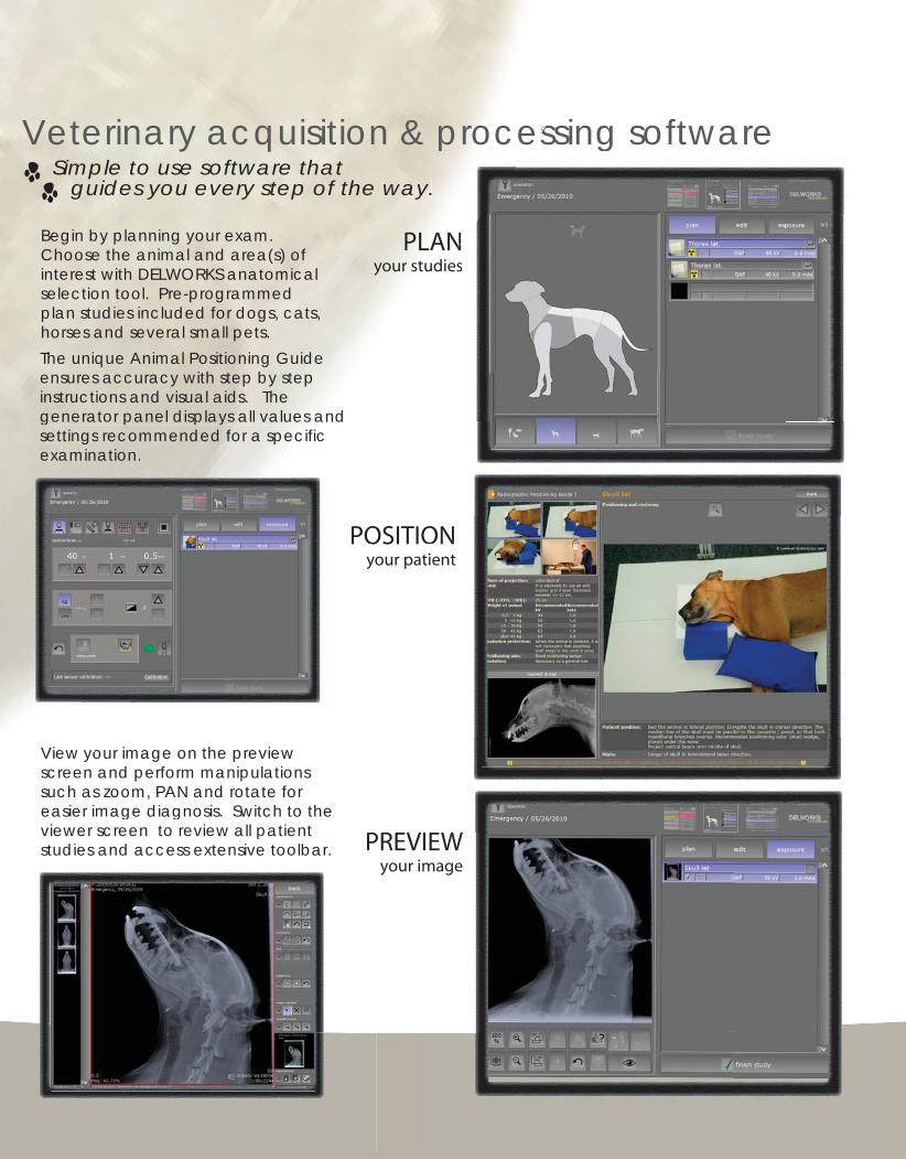
PLANyour studies
Begin by planning your exam. Choose the animal and area(s) of interest with DELWORKS anatomical selection tool. Pre-programmed plan studies included for dogs, cats, horses and several small pets.
POSITIONyour patient
The unique Animal Positioning Guide ensures accuracy with step by step instructions and visual aids. The generator panel displays all values and settings recommended for a specific examination.
View your image on the preview screen and perform manipulations such as zoom, PAN and rotate for easier image diagnosis. Switch to the viewer screen to review all patient studies and access extensive toolbar.
Veterinary acquisition & processing software Simple to use software that guides you every step of the way.
U p g r a d e P a c k a g e NLANdiesudi
& essing softwareprocesy.
ONtient
IEWmage
POy
g p p yic settings recommended for a specific
examination.
your image on the previewView yo
Pviewer screen to review all patient studies and access extensive toolbar.
The user-friendly DelWorks Veterinary specific acquisition software is easy to operate and features a wide range of tools for a variety of image manipulations. After exposure, images display in less than 1 second on the TFT color LCD clinical review display with touch screen interface. This DICOM compliant, high definition display provides more accurate grayscales and better contrast than standard monitors.
Automatic image optimization is achieved with the integrated Advanced Image Processing software, so you can get the image you need fast to make your diagnosis quickly and efficiently.
PREVIEWyour image
DELWORKSVETERINARY DRIf you are considering transitioning your practice to digital
radiography, consider the DelWorks Veterinary DR Retrofit Package, the easy and affordable way to
upgrade your existing imaging system to a flat panel (DR) digital system. This upgrade kit
includes a Cesium (CsI) flat panel detector that is easy to install and will consistently provide high quality, professional images.
Standard Processing with Advanced Image Processing
PLANyour studies
Begin by planning your exam. Choose the animal and area(s) of interest with DELWORKS anatomical selection tool. Pre-programmed plan studies included for dogs, cats, horses and several small pets.
POSITIONyour patient
The unique Animal Positioning Guide ensures accuracy with step by step instructions and visual aids. The generator panel displays all values and settings recommended for a specific examination.
View your image on the preview screen and perform manipulations such as zoom, PAN and rotate for easier image diagnosis. Switch to the viewer screen to review all patient studies and access extensive toolbar.
Veterinary acquisition & processing software Simple to use software that guides you every step of the way.
U p g r a d e P a c k a g e NLANdiesudi
& essing softwareprocesy.
ONtient
IEWmage
POy
g p p yic settings recommended for a specific
examination.
your image on the previewView yo
Pviewer screen to review all patient studies and access extensive toolbar.
The user-friendly DelWorks Veterinary specific acquisition software is easy to operate and features a wide range of tools for a variety of image manipulations. After exposure, images display in less than 1 second on the TFT color LCD clinical review display with touch screen interface. This DICOM compliant, high definition display provides more accurate grayscales and better contrast than standard monitors.
Automatic image optimization is achieved with the integrated Advanced Image Processing software, so you can get the image you need fast to make your diagnosis quickly and efficiently.

TOSHIBA FDX4343Rdr detector• Cesium Iodide (CsI) Scintillator with Amorphous Silicon (a-Si) Photodiode• 17” x 17” Detector Size (512 x 495 x 43mm)• 16.9 x 17.3 inch active area• 143μm Pixel Size• 9 Megapixel (3,008 x 3,072 pixels)• 3.5 lp/mm• 6 second cycle time (Shot to shot)• MTF (2.0 lp/mm, 70 kVp, 1x1) 30% typ.• DQE (0), Quantum-Limited >70%• Less than one (1) second to image preview• Operating temperature +50° F to +95° F• 14 Bit A/D Conversion• 16 bit Digital Output (1000 Base-T)• External X-Ray Synchronization Control Using included external Synchronization Device
SPECIFICATIONS
Toll Free: (800) 800-6006Fax: (847) 288-7011Email: [email protected] N. Gary Ave., Suite B, Roselle, IL 60172
UMGMedicalImaging
Toll Free: (800) 261-9808Fax: (914) 835-6111
Email: [email protected] www.umgxray.com
28 Calvert Street, Harrison, NY 10528
Del Medical is an ISO 13485 & ISO9001 Certified Facility. Specifications are subject to change without notice.
07/10 #9000 D
Wvet
DELWORKS VETERINARY DELWORKSDELWORKS
A complete DR upgrade package for upgrading existing film-based or CCD imaging systems
Complete package includes:• Toshiba FDX4343R Detector• DelWorks Veterinary Acquisition & Processing Software• Dell Optiplex 780; Intel Core 2 Duo 3.0 Ghz, 4GB RAM, 250GB HD• 19” touchscreen flat panel, color display, keyboard and mouse
19.5 in. (495 mm)
19.7 in. (501 mm)
Power Supply ConnectorLAN Connector
LED Display for Mode
D Sub Connector
Wired21 in. (532 mm)
1.7 in.(43 mm)
13.4
in. (3
4 mm)
12.4
in. (
31.4
mm
)
3.65 in. (9.26 mm)
processing software• Extensive measuring functions• CD/DVD Burning for patient images• DICOM Store, Print, and MWL• Generator Integration (on select models)• Advanced Image Processing features• Unique Animal Positioning Guide• Grid Suppression software• Extensive tool library with measuring, layout adjustments, export options and more• Patient CD creation
acquisition station hardware• Dell Optiplex 780 Small Form Factor Computer with Intel Core 2 Duo 3.0GHz, 4GB RAM, weight 15 lbs. (6.80 kg)• 19” t ouch screen, flat-panel display, weight 18.74 lbs. (8.5 kg). Display rotates 90 degrees landscape/vertical.Approved for medical use, complies with international medical safety and emission standards (such as CE, UL, EN etc.), - Active screen diagonal 19 in. (483 mm); width 14.80 in. (376 mm); height 11.85 in. (301 mm) - Pixel pitch 0.294 mm, 16.8 million colors• Keyboard and mouse 16.46 in. (418 mm)
13.6
2 in
. (34
6 m
m)
Display Depth2.76 in.(70 mm)
Depth with Pedestal 8.75 in.
(222.25 mm)
verti
cal t
rave
l5.
5 in
. (14
0 m
m)