Embed Size (px)
Citation preview
저 시-비 리- 경 지 2.0 한민
는 아래 조건 르는 경 에 한하여 게
l 저 물 복제, 포, 전송, 전시, 공연 송할 수 습니다.
다 과 같 조건 라야 합니다:
l 하는, 저 물 나 포 경 , 저 물에 적 된 허락조건 명확하게 나타내어야 합니다.
l 저 터 허가를 면 러한 조건들 적 되지 않습니다.
저 에 른 리는 내 에 하여 향 지 않습니다.
것 허락규약(Legal Code) 해하 쉽게 약한 것 니다.
Disclaimer
저 시. 하는 원저 를 시하여야 합니다.
비 리. 하는 저 물 리 목적 할 수 없습니다.
경 지. 하는 저 물 개 , 형 또는 가공할 수 없습니다.
치의과학박사 학위논문
심미적이고 효과적인 상아질 지각과민증 처치용
타닌-금속 복합체
Aesthetically effective dental desensitizer using tannin-metal complexes
2017년 8월
서울대학교 대학원
치의과학과 치과생체재료과학 전공
주 성 원
심미적이고 효과적인
상아질 지각과민증 처치용타닌-금속 복합체
Aesthetically effective dental desensitizer using tannin-metal complexes
지도교수 안 진 수
이 논문을 치의과학 박사학위논문으로 제출함2017년 4월
서울대학교 대학원치의과학과 치과생체재료과학 전공
주 성 원
주성원의 박사학위논문을 인준함2017년 6월
위 원 장 (인)
부 위 원 장 (인)
위 원 (인)
위 원 (인)
위 원 (인)
<Abstract>
Aesthetically effective dental desensitizer using tannin-metal complexes
Sung-Won Ju
Department of Dental Biomaterials Science
School of Dentistry
Seoul National University
(Directed by Professor, Jin-Soo Ahn, D.D.S., Ph.D.)
Introduction: The aim of this study was to evaluate desensitizing solutions which were made tannin acid ion and various metal ion oxides. Aesthetically improved vis-à-vis significant bioremineralization of the hydroxyapatite (HAp) crystals for the treatment of dentinal hypersensitivity has been explored following the tanin acid-metal (ions and oxides) complex mediated mineralization.
Methods: Demineralized human molar disks were treated with TA combined with 6 different daily intake metal ions and metal oxide namely Sr(NO3)2, SiO2, Fe2O3, TiO2, CaCl2, and VCl3 in separate experiments. The samples were coated in the complex solution for 4 min followed by immersion in artificial saliva for 7 days. Biomineralized HAp crystals were well characterized by Field Emission Scanning Electron microscope (FE-SEM), Transmission Electron Microscope (TEM), Scanning Electron Microscope (SEM) and Selected Area Electron Diffraction (SAED).
Results: TA-Sr(NO3)2 TA-TiO2 exhibited the most significant results
by occluding the Ca. 79% and 68% dentinal tubules, respectively with brighter color changes after treatments.
Conclusions: TA combined desensitizing solutions had improved aesthetics of the exposed dentin by its whitening effect using chemically inert and biocompatible, Sr and Ti oxides which may be potential to develop facile ‘at-home’ aesthetically effective dentin hypersensitivity solution.
Keywords: Aesthetics, Dentin hypersensitivity, Dentinal tubule, Remineralization, Tannin-metal complexes Student Number: 2014-30696
Contents
1. Introduction
2. Materials and Methods2.1 Materials2.2 Preparation of acid-etched tooth slices2.3 Coating and remineralization methods2.4 Characterizations of the precipitates of remineralization2.5 Dentinal fluid permeability assay2.6 Colorimetric measurement2.7 Human periodontal ligament (hPDL) cell culture
3. Results and Discussions3.1 Pyrogallol-mediated HAp remineralization3.2 Characterizations3.3 Dentinal fluid test and topological occlusion of dentinal tubules3.4 Color changes and whiteness index measurements3.5 Cell proliferation of human periodontal ligament (hPDL) cells
4. Conclusions References Figures Korean Abstract
- 1 -
1. Introduction
Dentinal hypersensitivity, a common elusive oral health troublesome in the world with a prevalence of about 15–57% of the general population, occurs when the dentin which usually overlaid by enamel or cementum of a tooth is exposed to the direct oral environment by conditions such as bacterial demineralization, abrasion or gingival recession.1 Dentin, composed of 20% of type I collagen fibrils reinforced with 70% Hydroxyapatite (HAp) and 10% plasma like fluid, is a biomineralized hard tissue containing tiny capillaries called tubules.2
Inside each tubule lies a nerve branch that comes from the pulp cavity. When the underlying dentin is exposed, thermal, tactile, osmotic or chemical stimuli in the saliva flowing around provoke movement in the intradentinal fluid which transmits to the afferent intradental nerves and results in the transient severe pain that term as dentinal hypersensitivity.1 Additionally, the color of exposed dentin is yellower than enamel and it defines overall teeth color. Therefore, occluding exposed dentinal tubules is proposed to be a fundamental strategy for reducing stimuli-evoked fluid moves to desensitize dentin as well as to reduce the yellowish staining of the teeth. Procedures to cure or reduce the dentinal hypersensitivity ranges from ‘at-home’ -self-administered desensitizing therapies to ‘in-office’-more complex clinical process, includes dental sealants or laser therapy, by dental professionals in the dental office. However, until now, there is no universally satisfied regimen which significantly cures and relieves the dentinal hypersensitivity. Over the decades, several attempts have been made to occlude the tubules by various commercially available remineralization systems. However, most of them contain glutaraldehyde, oxalate or acrylate as an active ingredient in the
- 2 -
system in high concentrations.3 Furthermore, these chemicals reported to show a high toxic effect on the human gingival fibroblasts, the most abundant structural cells in the periodontal tissues, therefore, it requires the proper administration of the correct dosage, leading to the need of a clinician to administer it.4 In our continuous attempts to develop efficient dentinal hypersensitivity treatment system, bio-inspired catechol-metal complexion strategies have been studied so far mimicking the tunicate’s wound healing mechanism. Significant results have been obtained using gallic acid-Fe3+ as an active ingredient with 87% occlusion of the dentinal tubules after 7 days5 and in the case of Tannin Acid (TA) combined with Fe3+, resulted in about 47% occlusions to the dentinal tubules.6
Although previous reports offer potential applied uses, the high cost of gallic acid, as well as iron ions were reported to forms harmful insoluble hydroxides and phosphates precipitation at physiological and higher pH.7 Tooth discoloration may be impediments for the practical application, since some metal complexes showed tooth discoloration after the treatment.6, 8
Tannin is a common, low-cost and edible plant polyphenols present in red wine, fruits, tea and virtually all families of plants. It could be a promising candidate because of their building blocks and trihydroxyphenyl-containing molecules can use as precursors for the formation of multifunctional coatings.9 In the presence of a mild aqueous solution of metal ions and metal oxides, many pyrogallol groups in TA are able to form complexes and adhere to various substrates within 30 sec.10 In addition, pyrogallol groups of TA can strongly capture calcium ions, a key component of HAp, and supports the HAp nucleation leading the dentinal tubules occlusion.6 However, the comprehensive study of tannic acid with different metal ions metaloxides (e.g. Sr2+, Ca2+
, Fe3+, V3+TiO2, SiO2, Fe2O3), as well as aesthetic evolution for tannin-metal ion complex, have not been studied,
- 3 -
concerning their effects on the dentinal occlusion and practical application. In the course of the investigation with the aim to disclose major sources, potential exploration of this low-cost plant polyphenols concerning its complex formation with various metal ions and metal oxides for the biomineralization of HAp which cures the dentinal hypersensitivity treatment as well as aesthetic evolution by measuring the color changes after the treatment is desired. The comprehensive data set of tannin-metal complexes for dentinal hypersensitivity treatment along with aesthetic improvement is furnished for their potential applications.
2. Materials and Methods
2.1 Materials Tannic acid, Sr(NO3)2, TiO2 (particle size : 21 nm), SiO2 (particle size : 100 nm), Fe2O3, CaCl2, VCl3, antibiotic mixtures, and trypsin were purchased from Sigma-Aldrich (St. Louis, MO, USA). KH2PO4
was purchased from Samchun (Seoul, South. Korea) and tris (hydroxymethyl) aminomethane was purchased from EM Science (Gibbstown, NJ, USA). DMEM and FBS were purchased from Hyclone (Logan, UT, USA). All the chemicals used were of analytical grade and were used as received unless otherwise noted.
2.2 Preparation of acid-etched tooth slices The teeth slice from human molar were treated with 3% sodium hypochlorite solution to sterilize from bacteria and rinsed with phosphate buffered saline (PBS). The dentin samples were taken from the tooth crown and were perpendicularly sliced to the longitudinal axis into 1 mm thick discs with a diameter of approximately 8−10 mm
- 4 -
using a low-speed diamond saw blade. The tooth slices were then acid-etched with 3% HNO3 solution for 1 min followed by rinsing with sufficient deionized water. The guideline of the institutional review board (IRB AN14259-001) of the Korea University Medical Centre (Seoul, Korea) was followed to obtain human molars.
2.3 Coating and remineralization methods Six different aqueous solutions of Sr(NO3)2, SiO2, Fe2O3, TiO2, CaC12, and VCl3 (1.2 mM) concentration were freshly prepared for separate experiments. TA (0.47 mM) and metal ion solution were vigorously mixed and the pH raised to 8.0 by slowly adding NaOH (0.1 M) solution. The tooth discs were immersed in the TA-metal complex solution for 4 min, changing solution after 1-minute intervals. Then, these tooth slices were immersed in deionized water and used wherever applicable. The calcification solution referred to here as artificial saliva was prepared following the previously reported procedure. Briefly, the solution contained calcium (CaCl2·2H2O) (8 mM), phosphate (KH2PO4) (1.55 M), fluoride (NaF) (1 mg/L), NaCl (180 mM) was buffered by tris(hydroxymethyl)aminomethane-HCl (50 mM). The pH of the calcification solution was adjusted to 7.6 using HCl (0.1 N) and NaOH (0.1 M) solutions and stored at 4°C prior to use. Dental slices with coating and without coating were immersed at the bottom of polyethylene tubes filled with calcification solution (10 mL). All samples were placed in a shaking incubator at 100 rpm and 37°C, and the calcification solution was refreshed everyday. The tooth slices were removed after 7 days, cleaned using ultrasonication in deionized water, and rinsed 3 or 4 times with deionized water followed by drying at the room temperature prior to characterizations.
- 5 -
2.4 Characterizations of the precipitates of remineralization The surface morphologies of the control and the treated dentin samples following 4 min treatment and subsequent 7 days remineralization treatment in the artificial saliva solution were analyzed using a Field Emission Scanning Electron microscope (FE-SEM, JEOL, Tokyo, Japan) operated at 10kV. The average diameters and lengths of the HAp nanorods in the composite were measured from Transmission Electron Microscope (TEM, JEOL) and SEM images using ImageJ software. The diffraction pattern was observed by selected area electron diffraction (SAED).
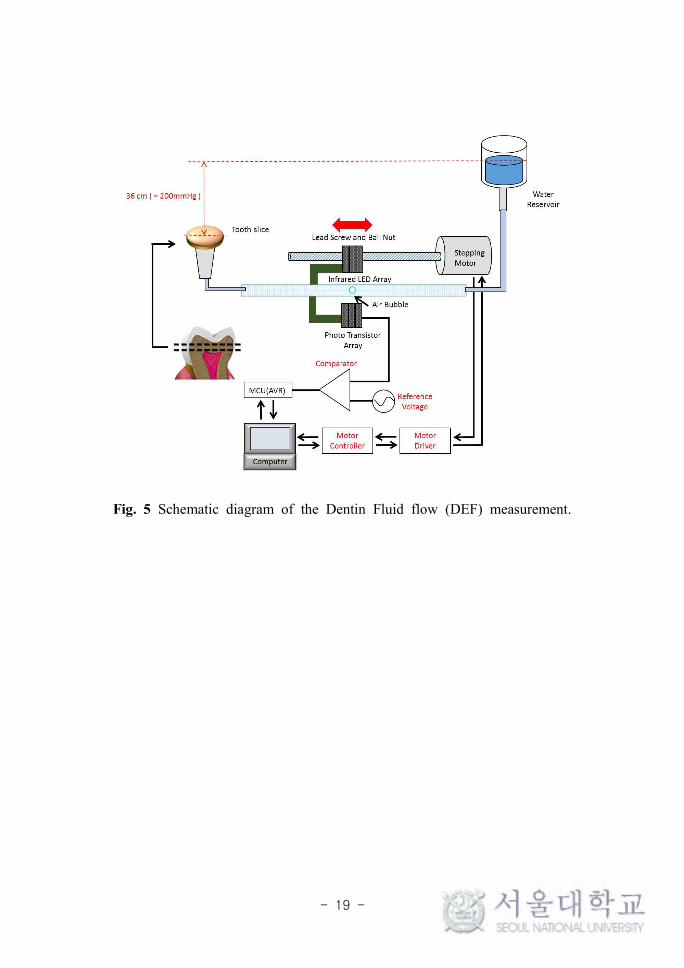
2.5 Dentin fluid permeability assay Dental hypersensitivity originates from neural stimulation by fluid flow within the dentinal tubules that is driven by physiological pulpal pressure of approximately 200 mmH2O. In vitro dentin fluid permeability assay was performed using a fluid filtration system following our previous study to evaluate the TA-metal complex induced biomineralization of HAp, Briefly, the amount of water infiltration in an untreated tooth slice for 10 min at 200 mmH2O pressure was first measured as the standard. The water infiltration amounts after the TA-metal treatment for 4 min followed by incubation in artificial saliva for 7 days were again measured. The permeability of each sample was expressed as a percentage (%) of the fluid flow.
2.6 Colorimetric measurement To evaluate the color change by the TA-metal treatment, colorimetric measurements of samples were performed using a reflectance colorimetric device (PR-670, SpectraScan, Photo Research, Chatsworth, CA, USA). This device was set to produce color parameters based on average daylight (D65: 6404 K). The optical geometry of this system consists of 45-degree illumination angle and
- 6 -
0-degree viewing angle to measure an area of 3 mm in diameter. The color parameters were recorded in the L*a*b* color space, as established by ′the Commission Internationale de l'Eclairage (CIELAB)′ in 1976. The mean of the results was determined by three separate measurements in succession. Color differences (∆E), equation (1) and whiteness index (W), equation (2) were calculated according to the following formulas, as reported in the literature:11, 12
∆ (1)
(2)
Here, L* is a group of lightness, a* a group of a red varying set, and b* a group of a yellow varying set. The samples were tested before treatment (denoted as 0), after 4 min (denoted as 1) and after 7 days remineralization (denoted as 2).
2.7 Human periodontal ligament (hPDL) cell culture To evaluate the direct cytotoxicity and physiological adaptation of hPDL cells, the 4th cell passage cells was examined. The proliferation was measured in three types of culture dishes: uncoated, TA/Sr2+-coated, and remineralized sample using essential medium-alpha (MEM-α; Hyclone, USA) media supplemented with 10% (v/v) fetal bovine serum (FBS; Hyclone) and 1% penicillin/streptomycin (Hyclone) at 37°C in a humidified atmosphere of 5% CO2 and 95% air. Subconfluent cells were detached using 0.25% trypsin-EDTA (Hyclone) then viable cells were identified using a trypan blue assay and counted using a hemocytometer. After that, the 5 × 10⁴ of the cells (> 95% viable) in MEM-α with 10% FBS were placed on each sample-coated glass disk, which was put in a 24-well plate. To count the number of viable cells, a Cell Counting Kit-8 (CCK-8; Dojindo Laboratories, Japan) solution was used; this solution produces a yellow formazan dye in the presence
- 7 -
of viable cells. One to three days after cell seeding, 50 μl of CCK-8 was added to the wells for 3 h at 37 °C to allow the formation of formazan crystals, then absorbance was measured at 450 nm using a microplate reader (Bio-Rad, Richmond, CA, USA).
3. Results and Discussions
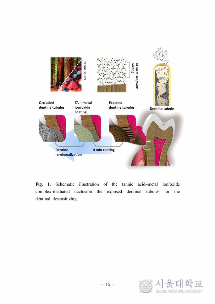
3.1 Pyrogallol-mediated HAp remineralization Hydroxyl groups of TA were initially participated to conjugate with metal ions and metal oxides, and incorporate the coating layer on the dentin tubule.13 The presumable hypothesis may be, some of the unconjugated hydroxyl groups of TA may interact with calcium ions of artificial saliva and forms complex and thereby induce the HAp remineralization.14 On the other hand, TA complex may also interact with the organic interface of the dentin, thereby stabilizing the exposed collagen matrix. This interaction is a predominantly essential process in the cell adhesion and growth.15 Generally, TA/metal complex potentially induce cross-links in the dentin collagen and grow a densely packed and well-aligned rod-like shape of remineralized HAp crystals. This remineralized densely-packed HAp crystals hinder an osmotic fluid exchange with internal dentinal nerves and prevent the stimulation of dentinal nerves which leads relieve from the dentinal hypersensitivity. The schematic presentation of the pyrogallol-mediated HAp remineralization and dentin tubule occlusion is shown in Fig. 1.
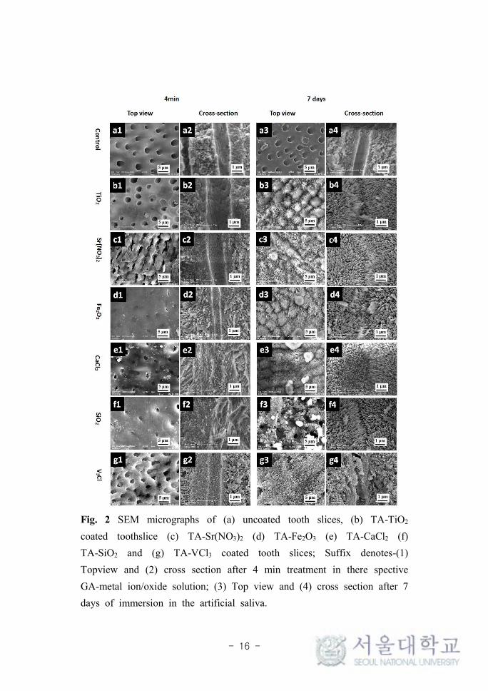
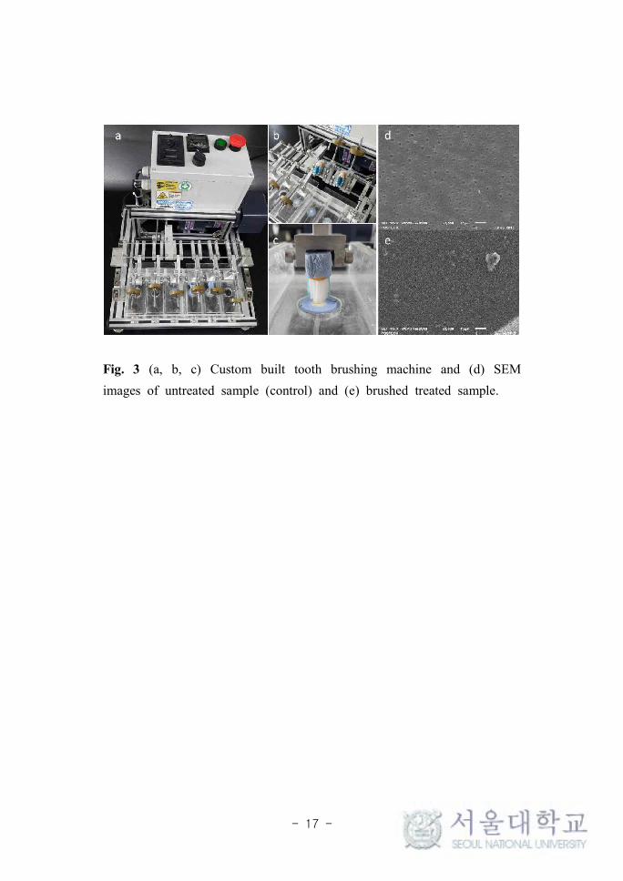
3.2 Characterizations In order to evaluate the surface deposits and topology of the tubule occlusion after treatment of TA-metal, treated and untreated dentin disks were examined as top and cross-view by SEM (Fig. 2). The untreated sample showed well defined and regular tubules (2-4 μm diameters)
- 8 -
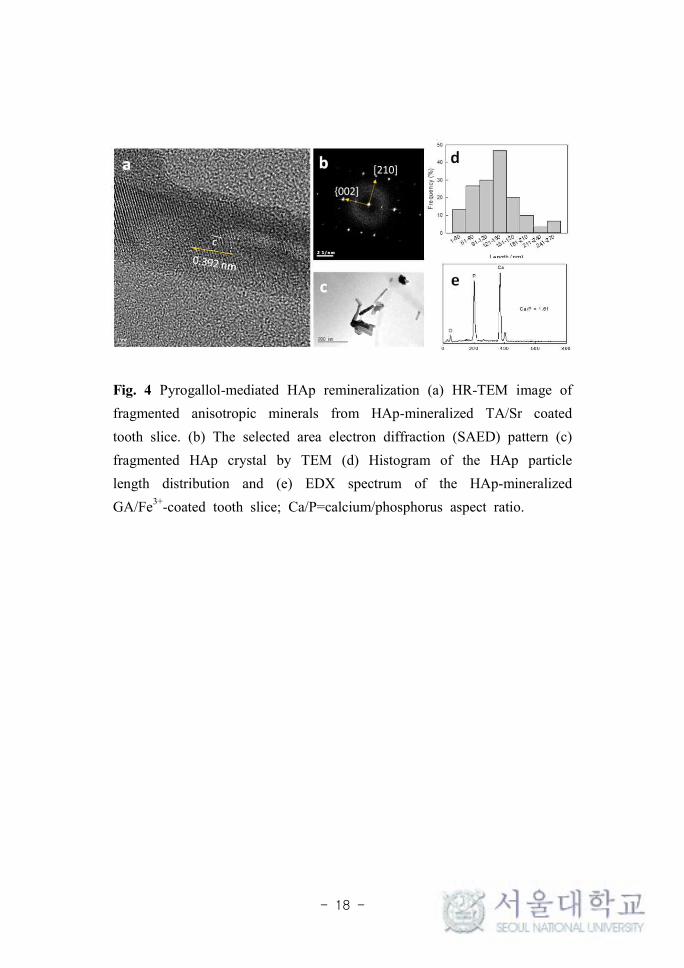
that penetrate dentin perpendicularly to the surface (Fig. 2a). After 4 min of the TA-metal treatment, tubules entrances were observed narrow down, although the middle of the tubules remained unchanged. The result indicates that the TA/metal ion complex form films and deposited to surface within 4 min. After 7 days remineralization in the artificial saliva, all tubules were significantly filled with needle-like HAp (Fig. 2b-g). Although the human body has natural self-remineralization ability, the process is very slow which is good in agreement with the SEM image of the untreated sample. Thus, considering the low concentration (< 0.016%) of the TA/metal used for this coating, TA/metal coating shown efficient piled up on the dentin compared to any commercial desensitizer. Furthermore, the mechanical stability of the biomineralized HAp was checked on samples after 7days treatment, which is stable after vigorous mechanical brushing over 1000 strokes (Fig. 3). This good mechanical stability can be well explained by the fact that the pyrogallol groups have strong adhesion strength on the substrate and high density of cross-links with metal ions/oxides, similar to the natural behavior in tunicate, where they glue their torn tissues under the sea by the pyrogallol-mediated cross-links. The results obtained here are favourable for the oral environment which is under various mechanical stresses such as chewing, brushing, and temperature-driven air pressure. The needle-like mineral deposited on the tubule surfaces were characterized by TEM and EDX (Fig. 4). The mineral-like material was fragmented using ultra-sonicator in ethanol for 30 min. EDX analysis revealed that the Ca/P ratio of the need-like minerals was 1.61 having 100-300 nm length and 10-20 aspect ratio. The selected area electron diffraction (SAED) pattern exhibited characteristic peaks: (002), (004), (112), (211), and (300) which are similar to those of HAp peaks and confirmed the mineral as HAp.16 The high-resolution TEM image evidently showed the (002) crystal plane is perpendicular to c-axis, a
- 9 -
longitudinal axis of HAp, that suggests anisotropic growth of needle-like HA occurred along the (002) plane which is similar to that of reported in the literature.16, 17 The absence of HAp minerals in the untreated sample suggested the involvement of pyrogallol groups in the HAp nucleation. The result reconfirmed that the pyrogallol groups initiate the TA-mediated anisotropic HAp remineralization. Overall, pyrogallol groups of TA contributed to adhesion to substrate, to mechanical reinforcement by forming TA/metal complexes, and to highly anisotropic HAp formation by Ca2+ ion adsorption from the saliva. The highly anisotropic HAp has been considered as to have good bio-reactivity and bio-absorbability. In addition, mechanical properties of naturally occurring HAp-embedded materials are modulated by aspect ratio and arrangements of HAp mineral.
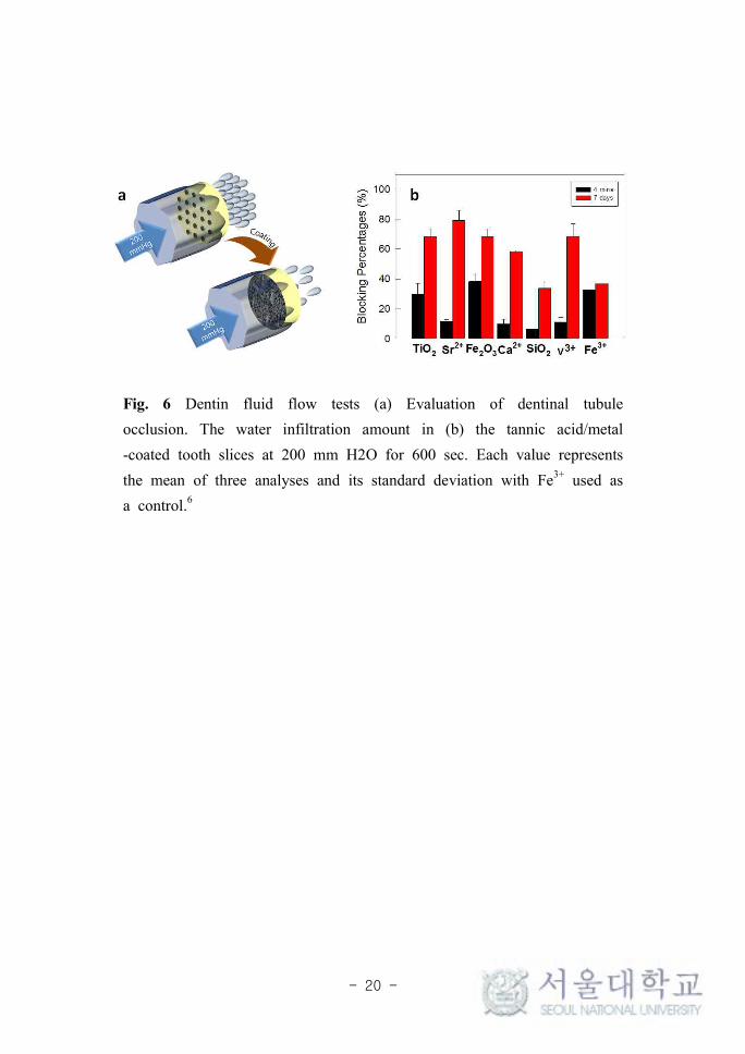
3.3 Dentin fluid test and topological occlusion of dentinal tubules The TA/metal coating was expected to function as a dentinal sealant and to block the dentin’s open tubules. To evaluate the occlusion effect of the TA/metal coating, dentin disks prepared from human molar disks were used to measure the fluid permeability through dentin. Permeability is derived from the hydraulic conductance or ease of fluid flow through the dentin (Fig 5). The TA/metal treatment blocked the water infiltration and results were depicted in fig. 6b. TA with TiO2, Sr2+, Fe2O3, Ca2+, SiO2, V3+ and Fe3+ blocked 29.44, 11.68, 38, 34, 9.86, 6.69, 11.25 and 32.65% of the tubules after 4 min, respectively and 68.25, 79.31, 68.12, 58.50, 33.91, 68.16 and 36.73% after 7 days ,respectively. The result suggests that the topologically observed TA/metal complex actually restrained the dentinal fluid flow and the regenerated HAp significantly gave the additional dentinal fluid blockage. The greater nucleation of HAp in the case of Sr ions may be
- 10 -
explained by the fact that the human body absorbs strontium as if it were calcium due to the chemical resemblance of the elements. Interestingly, these serendipitous results are very encouraging, because Ti and Sr ions are chemically inert and stable in the organism having good biocompatibility and tissue compatibility. Strontium ions promote early bone formation and its inherent ability to osseointegration has aroused prominent clinical interest,18,19 on the other hand, TiO2 is considered as a promising bone scaffolding material because it promotes osteoblast adhesion which induces bone formation and its ability to osseointegrate, enabling use in dental implants.20
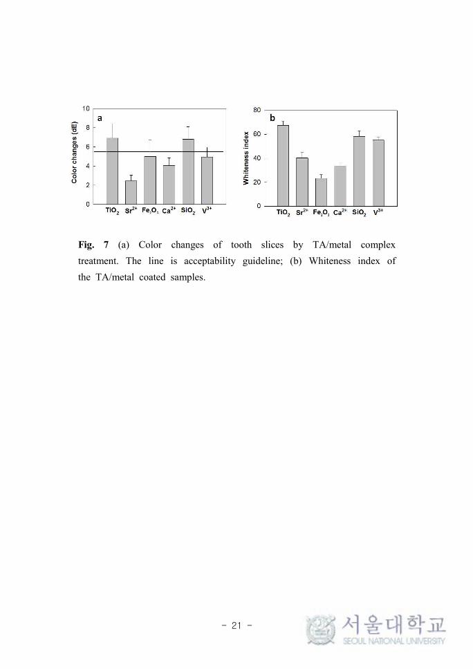
3.4 Color changes and whiteness index measurements The tooth staining after therapy is an important aesthetic concern in clinical dentistry. The staining of the dentin was quantified by measuring the color change (∆E) and whiteness index (W) of the dentin after the TA-metal treatment (Fig. 7). The enamel layer, the exterior and visible part of the tooth, was not stained after the TA-metal treatment, whereas the dentin that is generally invisible because it is covered by the enamel layer and gum became slightly discolored after the TA-metal coating. Interestingly, the color changes after the TA-metal treatment did not exceed the acceptability (∆E = 5.5). Some of the metal ion Ti2+and Si4+ changed to brighter as previously reported and recently use as the component of tooth whitening agents.21 Dentin is covered by enamel or gingiva which by abrasion of enamel, dental caries or recession of gingiva causes exposure of dentin. Usually, dentin has more yellowish and higher chroma than enamel therefore exposed dentin is responsible to both hypersensitivity and undesirable color change of teeth.22 Results are quite promising in case of TA with Ti and Sr ions considering dentin occlusion vis-à-vis aesthetic improvement. This study provides potential application of TA-metal complex due to the fact that the color changes
- 11 -
to brighter are desirable considering the aesthetic improvement, an additional advantage of the hypersensitivity treatment in dentistry.
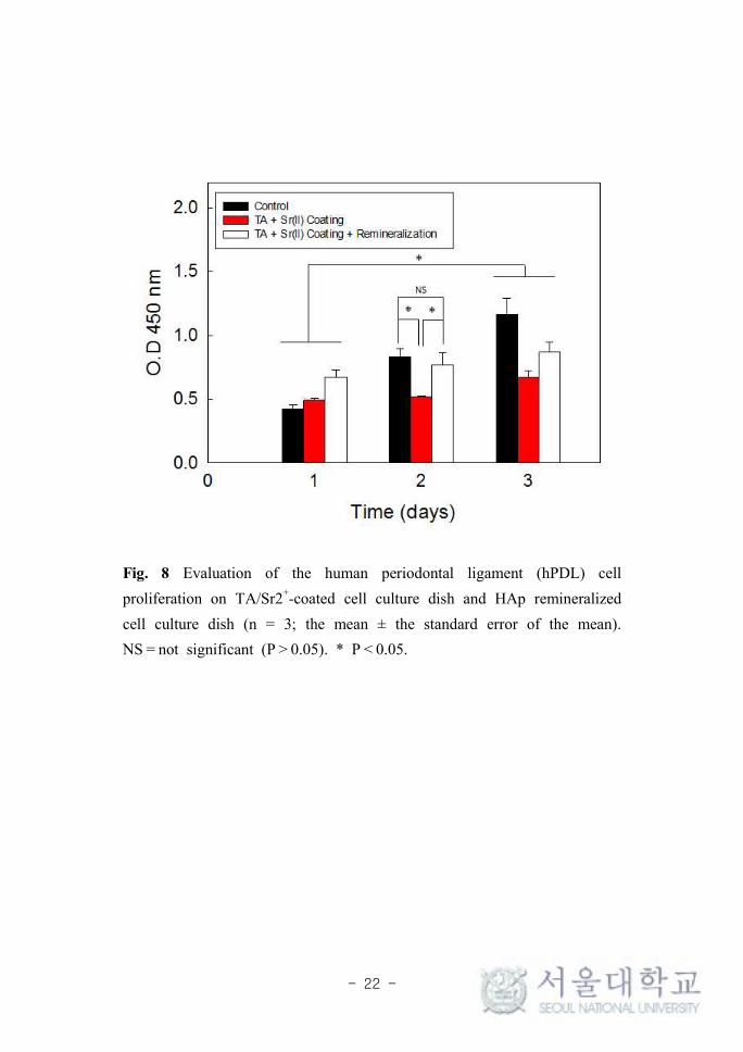
3.5 Cell proliferation of human periodontal ligament (hPDL) cells Tannin has been introduced to human and animal as an ingredient of food and tea from the origin of the life, its safety has already been proven.23 To quantitatively evaluate the cytotoxicity of the TA/Metal ion coating to a human periodontal ligament cells (hPDL), cell proliferation on TA-Sr(II) 4 min coating and after 7 days remineralization were monitored for 3 days using a colorimetric assay, while empty wells were used as control (Fig. 8). The cell proliferation test showed viable cell numbers on TA only coated and TA/Metal ion coated wells gradually moderately increased. Hence, TA-Sr2+, the optimized candidate of this study, proved low cytotoxicity to the hPDL cells.
4. Conclusions
In this study, we proved that the TA/metal coating occluded the exposed dentinal tubules by directly narrowing down to the tubule by inducing HAp remineralization in the artificial saliva. The low concentration (< 0.016%) of TA-Sr(NO3)2 and TA-TiO2 coatings were efficient and facile to occlude the tubules with improved aesthetic effect to the treatment of dentinal hypersensitivity. It formed a mechanically stable complex film on the micro-sized tubules only within 4 min and nucleate the densely packed HAp crystals after 7 days remineralization which resulted in to ~79 % occlusion in the case of TA/Sr2+ complex. This study may be potentially useful to develop sustainable, cost-competitive and biocompatible with aesthetically effective dental desensitizer solution such as toothpaste and mouthwash.
- 12 -
References
1. Splieth CH, Tachou A. Epidemiology of dentin hypersensitivity. Clinical Oral Investigations, 2013;17;3-8.
2. Goldberg M, Kulkarni AB, Young M, et al. Dentin: Structure, Composition and Mineralization. Frontiers in Bioscience (Elite Edition), 2011;3;711-735.
3. Schupbach P, Lutz F, Finger WJ. Closing of dentinal tubules by Gluma desensitizer. European Journal of Oral Sciences, 1997;105;414-421.
4. Sengun A, Buyukbas S, Hakki SS. Cytotoxic effects of dental desensitizers on human gingival fibroblasts. Journal of Biomedical Materials Research Part B: Applied Biomaterials, 2006;78B;131-137.
5. Prajatelistia E, Ju SW, Sanandiya ND, et al. Tunicate-Inspired Gallic Acid/Metal Ion Complex for Instant and Efficient Treatment of Dentin Hypersensitivity. Advanced Healthcare Materials, 2016;5;919-927.
6. Oh DX, Prajatelistia E, Ju SW, et al. A rapid, efficient, and facile solution for dental hypersensitivity: The tannin–iron complex. Scientific Reports, 2015;5;10884.
7. Iffat AT, Maqsoodahida ZT, Fatima N. Study of Complex Formation of Fe(Ⅲ) with Tannic Acid. Journal- Chemical Society of Pakistan, 2005;27;174-177.
8. Adcock KG, Hogan SM. Extrinsic Iron Staining in Infant Teeth from Iron-Fortified Formula and Rice Cereal. The Journal of Pediatric Pharmacology and Therapeutics, 2008;13;162-165.
9. Sileika TS, Barrett DG, Zhang R, et al. Colorless Multifunctional Coatings Inspired by Polyphenols Found in Tea, Chocolate, and
- 13 -
Wine. Angewandte Chemie International Edition, 2013;52;10766-10770.
10. Ejima H, Richardson JJ, Liang K, et al. One-Step Assembly of Coordination Complexes for Versatile Film and Particle Engineering. Science, 2013;341;154-157.
11. Douglas RD, Brewer JD. Acceptability of shade differences in metal ceramic crowns. Journal of Prosthetic Dentistry, 1998;79;254-260.
12. Luo W, Westland S, Ellwood R. Development of a whiteness index for dentistry. Journal of Dentistry, 2009;37;21-26.
13. Nakajima A, Sakaguchi T. Uptake and recovery of gold by immobilized persimmon tannin. Journal of Chemical Technology & Biotechnology, 1993;57;321-326.
14. Kim S, Don S, Mainwaring DE. Effect of ion-binding on the formation of temporary viscoelastic networks of proanthocyanidin biopolymers. Journal of Applied Polymer Science, 1997;65; 1795-1805.
15. Velmurugan P, Singam ERA, Jonnalagadda RR, et al. Investigation on interaction of tannic acid with type I collagen and its effect on thermal, enzymatic, and conformational stability for tissue engineering applications. Biopolymers, 2014;101; 471-483.
16. Ryu J, Ku SH, Lee H, et al. Mussel-Inspired Polydopamine Coating as a Universal Route to Hydroxyapatite Crystallization. Advanced Functional Materials, 2010;20;2132-2139.
17. Zhou YZ, Cao Y, Liu W, et al. Polydopamine-Induced Tooth Remineralization. ACS Applied Materials & Interfaces, 2012;4;6901-6910.
18. Andersen OZ, Offermanns V, Sillassen M, et al. Accelerated bone ingrowth by local delivery of strontium from surface functionalized titanium implants. Biomaterials, 2013;34;5883-5890.
19. Schumacher M, Gelinsky M. Strontium modified calcium phosphate
- 14 -
cements – approaches towards targeted stimulation of bone turnover. Journal of Materials Chemistry B, 2015;3; 4626-4640.
20. Wang N, Li H, Lü W, et al. Effects of TiO2 nanotubes with different diameters on gene expression and osseointegration of implants in minipigs. Biomaterials, 2011;32; 6900-6911.
21. El-Meliegy E, Noort RV. Glasses and Glass Ceramics for Medical Applications. Springer New York, 2012.
22. ten Bosch JJ, Coops JC. Tooth Color and Reflectance as Related to Light Scattering and Enamel Hardness. Journal of Dental Research, 1995;74;374-380.
23. Park JH, Kim K, Lee J, et al. A Cytoprotective and Degradable Metal–Polyphenol Nanoshell for Single-Cell Encapsulation. Angewandte Chemie International Edition, 2014;53;12420-12425.
- 15 -
Fig. 1. Schematic illustration of the tannic acid–metal ion/oxide complex-mediated occlusion the exposed dentinal tubules for the dentinal desensitizing.
- 16 -
Fig. 2 SEM micrographs of (a) uncoated tooth slices, (b) TA-TiO2
coated toothslice (c) TA-Sr(NO3)2 (d) TA-Fe2O3 (e) TA-CaCl2 (f) TA-SiO2 and (g) TA-VCl3 coated tooth slices; Suffix denotes-(1) Topview and (2) cross section after 4 min treatment in there spective GA-metal ion/oxide solution; (3) Top view and (4) cross section after 7 days of immersion in the artificial saliva.
- 17 -
Fig. 3 (a, b, c) Custom built tooth brushing machine and (d) SEM images of untreated sample (control) and (e) brushed treated sample.
- 18 -
Fig. 4 Pyrogallol-mediated HAp remineralization (a) HR-TEM image of fragmented anisotropic minerals from HAp-mineralized TA/Sr coated tooth slice. (b) The selected area electron diffraction (SAED) pattern (c) fragmented HAp crystal by TEM (d) Histogram of the HAp particle length distribution and (e) EDX spectrum of the HAp-mineralized GA/Fe3+-coated tooth slice; Ca/P=calcium/phosphorus aspect ratio.
- 19 -
Fig. 5 Schematic diagram of the Dentin Fluid flow (DEF) measurement.
- 20 -
Fig. 6 Dentin fluid flow tests (a) Evaluation of dentinal tubule occlusion. The water infiltration amount in (b) the tannic acid/metal -coated tooth slices at 200 mm H2O for 600 sec. Each value represents the mean of three analyses and its standard deviation with Fe3+ used as a control.6
- 21 -
Fig. 7 (a) Color changes of tooth slices by TA/metal complex treatment. The line is acceptability guideline; (b) Whiteness index of the TA/metal coated samples.
- 22 -
Fig. 8 Evaluation of the human periodontal ligament (hPDL) cell proliferation on TA/Sr2+-coated cell culture dish and HAp remineralized cell culture dish (n = 3; the mean ± the standard error of the mean). NS = not significant (P > 0.05). * P < 0.05.
- 23 -
<국문초록>
심미적이고 효과적인 상아질 지각과민증 처치용
타닌-금속복합체
서울대학교 대학원 치의과학과 치과생체재료과학 전공
(지도교수 안 진 수)
주 성 원
연구목적: 본 연구에서는 상아질 지각과민증 처치를 위한 구강도
포용 조성물로 다양한 금속이온의 타닌-금속이온 복합체를 형성하
여 상아질에 적용 후, 상아세관 내 수산화인회석 결정 유도를 통한
상아세관의 봉쇄 정도와 그 색상의 변화 정도를 측정 평가하여 효
과적이며 심미적인 지각과민 처지제로서의 활용 가능성을 알아보고
자 하였다.
연구대상 및 방법: 발거한 사람 대구치를 치축과 수직된 방향으
로 잘라 디스크형태의 시편으로 만들고 상아질 노출면을 산을 이용
하여 탈회시켰다. 6종의 금속 이온 및 금속 산화물[ Sr(NO3)2, SiO2, Fe2O3, TiO2, CaC12, VCl3 ]을 각각 일일 섭취 허용량을 기준하여 타
닌-금속이온 복합 솔루션을 만들고 이를 시편의 상아질 면에 4분간
코팅하였다. 그런 다음 인공타액에서 담가놓은 7일 후, 수산화 인회
석의 결정화 유도를 통한 상아세관을 봉쇄 정도를 다양한 방법으로
- 24 -
측정하였다. 또한 솔루션의 코팅전후 및 인공타액에서 7일 후의 각
각의 색상변화정도 또한 측정하였다.
결과: 6종의 금속 이온 및 산화물 중에서 TA-Sr(NO3)2 TA-TiO2
이 각각 79% 와 68%의 상아세관 봉쇄율을 보임으로써 가장 현저한
결과를 나타내었고, 색상의 변화도 또한 솔루션의 처치 후에 명도가
높아지는 결과를 얻었다.
결론: Sr 및 Ti 의 금속 산화물과 타닌 이온의 복합체로 만든
지각과민증 처치 용액은 화학적으로 불활성이며 생체적합성이 우수
함은 물론, 상아세관을 효과적으로 봉쇄하면서 노출된 상아질의 색
상으로 인하여 치아가 누렇게 보이는 것을 없애주는 미백효과도 지
님을 알 수 있었다. 이 용액은 '가정에서' 손쉽게 적용할 수 있는
효과적이고 심미적인 상아질 지각과민 처치제 솔루션으로의 활용
및 개발 가능성을 지닌다.
주요어: 심미, 상아질 지각과민증, 상아세관, 재광화, 타닌-금속 복합
체
학 번: 2014-30696