Embed Size (px)
Citation preview
Distinct features of neurotransmitter systems in thehuman brain with focus on the galanin systemin locus coeruleus and dorsal rapheErwan Le Maîtrea,1,2, Swapnali Shantaram Bardea,1,2, Miklos Palkovitsb,c, Rochellys Diaz-Heijtza,and Tomas G. M. Hökfelta,2
aDepartment of Neuroscience, Karolinska Institutet, 17177 Stockholm, Sweden; and bNeuromorphological and Neuroendocrine Research Laboratory,Semmelweis University, and cHungarian Academy of Sciences, 1094, Budapest, Hungary
Contributed by Tomas G. M. Hökfelt, December 8, 2012 (sent for review September 8, 2012)
Using riboprobe in situ hybridization, we studied the localizationof the transcripts for the neuropeptide galanin and its receptors(GalR1–R3), tryptophan hydroxylase 2, tyrosine hydroxylase, andnitric oxide synthase as well as the three vesicular glutamatetransporters (VGLUT 1–3) in the locus coeruleus (LC) and the dorsalraphe nucleus (DRN) regions of postmortem human brains. Quan-titative real-time PCR (qPCR) was used also. Galanin and GalR3mRNA were found in many noradrenergic LC neurons, and GalR3overlapped with serotonin neurons in the DRN. The qPCR analysisat the LC level ranked the transcripts in the following order in theLC: galanin >> GalR3 >> GalR1 > GalR2; in the DRN the rankingwas galanin >> GalR3 >> GalR1 = GalR2. In forebrain regions theranking was GalR1 > galanin > GalR2. VGLUT1 and -2 werestrongly expressed in the pontine nuclei but could not be detectedin LC or serotonin neurons. VGLUT2 transcripts were found in verysmall, nonpigmented cells in the LC and in the lateral and dorsalaspects of the periaqueductal central gray. Nitric oxide synthasewas not detected in serotonin neurons. These findings show dis-tinct differences between the human brain and rodents, especiallyrat, in the distribution of the galanin system and some other trans-mitter systems. For example, GalR3 seems to be the importantgalanin receptor in both the human LC and DRN versus GalR1 and-2 in the rodent brain. Such knowledge may be important whenconsidering therapeutic principles and drug development.
noradrenaline | species difference | transmitter coexistence |monoamines | depression
The locus coeruleus (LC) and the dorsal raphe nucleus(DRN)/raphe median nucleus (MRN) have been the focus of
clinical and preclinical monoamine research for almost halfa century. Using the formaldehyde fluorescence (Falck–Hillarp)method (1), Dahlström and Fuxe (2) originally described thesenuclei in the rat as containing noradrenaline (NA) (the A6group) and 5-hydroxytryptamine (5-HT; serotonin) (the B7/8groups), respectively. The LC harbors 2,800–3,600 neurons withan additional 260 neurons in the subcoeruleus area, the greatmajority of which are noradrenergic (3–6).The DRN forms a rostro-caudal, ventral midline column and is
part of the periaqueductal central gray (PAG) (7) with a largenumber of 5-HT neurons that can be subdivided into several sub-groups (2, 8–11). Both the NA-LC (5, 6, 12) and the 5HT-DRN (8–11, 13) neurons have wide projections to most forebrain areas.In humans the LC is a compact, blue-pigmented nucleus con-
sisting of a total of ∼50,000 neurons (both sides), almost all ofwhich are noradrenergic (14–16). The DRN comprises about165,000 5-HT neurons, which constitute around 70% of all DRNneurons (17). Thus, there are numerous nonserotonergic neuronsin the human DRN, as is also the case in other species, includingthe mouse (18, 19).It now is well established that most neurons synthesize and
release several types of messenger molecules in the process knownas “cotransmission” (20–26). For example, in the rat some sero-
tonergic DRN neurons synthesize nitric oxide (NO), visualizedas NO synthase (NOS) and/or NADPH-diaphorase (27–32),and glutamate, visualized as vesicular glutamate transporter 3(VGLUT3) in both the rat (33–38) and mouse (19, 39). Manyneuron systems have one or more coexisting neuropeptides;for example, galanin is present both in the NA-LC and the5HT-DRN neurons in the rat (40).Galanin, a 29-aa neuropeptide (41) [30 aa in humans (42–44)]
is widely distributed in the rodent brain (45–50) and exerts itseffects via G protein-coupled galanin receptors (51–55) witha wide distribution in rodents (39, 56–60). So far, three of thesereceptors have been identified, GalR1–3.In the rat galanin coexists with NA in most LC neurons (40,
61, 62) and with 5-HT in many DRN neurons (32, 40). It is highlylikely that galanin also is coexpressed in the NA-LC neurons ofthe mouse (39, 45, 49) and human (63–65). However, mouse5HT-DRN neurons do not synthesize galanin (19, 39, 45, 49, 66),nor has galanin expression been detected in human DRN neu-rons (64).Early autoradiographic studies showed binding of iodinated
galanin to LC and DRN in the rat (67–69) and primates, includinghumans (70–72). In situ hybridization (ISH) has demonstrated theGalR1 and -2 transcripts in the rat LC and GalR1 in the ventro-lateral PAG; the relationship to the 5HT-DRN neurons is notclear, although a weak GalR2 mRNA signal was observed in themidline (56, 58, 73). These results generally were in agreementwith the quantitative real-time PCR (qPCR) results (60). How-
Significance
For decades rodents have been used to explore normal brainfunctions and mechanisms underlying brain diseases. Such dataoften have been the basis in the search for new drugs. In thisstudy we selected chemical markers associated with centralnoradrenaline and serotonin neurons, key systems in researchon and current treatment of depression, and studied theirexpression with in situ hybridization in postmortem humanbrains. The results show some distinct species differences be-tween human and rodent noradrenergic and serotonergicneurons which may better inform the development of novelanxiolytic/antidepressant drugs.
Author contributions: E.L.M., S.S.B., R.D.-H., and T.G.M.H. designed research; E.L.M., S.S.B.,and R.D.-H. performed research; M.P. contributed new reagents/analytic tools; E.L.M.,S.S.B., R.D.-H., and T.G.M.H. analyzed data; and E.L.M., S.S.B., R.D.-H., and T.G.M.H. wrotethe paper.
The authors declare no conflict of interest.
Freely available online through the PNAS open access option.1E.L.M., and S.S.B. contributed equally to this work.2To whom correspondence may be addressed. E-mail: [email protected] or [email protected].
This article contains supporting information online at www.pnas.org/lookup/suppl/doi:10.1073/pnas.1221378110/-/DCSupplemental.
E536–E545 | PNAS | Published online January 22, 2013 www.pnas.org/cgi/doi/10.1073/pnas.1221378110
ever, none of the galanin receptor subtypes have been identified sofar in the human LC or DRN.In this study we analyzed sections/slices including the LC and
DRN in human postmortem brains using ISH and riboprobesgenerated to identify galanin and GalR1-3. To identify NA and5-HT neurons, probes were designed for the human catechol-amine-synthesizing enzyme tyrosine hydroxylase (TH) (74, 75)and the 5-HT–synthesizing enzyme tryptophan hydroxylase 2(TPH2), respectively (76). A limited qPCR analysis of thesemarkers was carried out at the pontine level and, for the galaninsystem, in some forebrain areas also.Finally, we attempted to identify a relationship between NA
and 5-HT neurons, on the one hand, and between NOergic andglutamatergic neurons on the other hand, using probes againstnNOS (77, 78) and the three VGLUTs (79–81), respectively.Preliminary results from these studies have been reported atmeetings (82, 83).
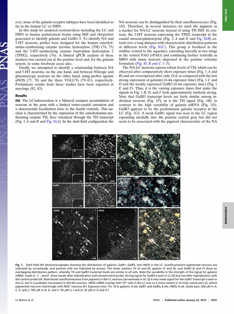
ResultsISH. The LC/subcoeruleus is a bilateral compact accumulation ofneurons in the pons with a limited rostro-caudal extension anda characteristic localization close to the fourth ventricle. This nu-cleus is characterized by the expression of the catecholamine-syn-thesizing enzyme TH, here visualized through the TH transcript(Fig. 1 A and B and Fig. S1A). In the dark-field configuration the
NA neurons can be distinguished by their autofluorescence (Fig.1H). Therefore, in several instances we used the pigment asa marker for NA-LC neurons instead of using TH ISH. In con-trast, the 5-HT neurons expressing the TPH2 transcript in thecaudal mesencephalon/pons (Fig. 2 A and D and Fig. S1B) ex-tend over a long distance with characteristic distribution patternsat different levels (Fig. S1C). This group is localized in themidline ventral to the aqueduct, extending laterally in two wingsin the ventral PAG (vPAG) and continuing further ventrally asMRN with many neurons dispersed in the pontine reticularformation (Fig. S1 B and C 1–5).The NA-LC neurons express robust levels of TH, which can be
observed after comparatively short exposure times (Fig. 1 A andB) and are overexposed after only 10 d, as compared with the lessstrong expression of galanin) (4-wk exposure time) (Fig. 1 C andD and the weakly expressed GalR3 (8-wk exposure time) (Fig. 1E and F). Thus, it is the varying exposure times that make thesignals in Fig. 1 B, D, and F look approximately similarly strong.Note that GalR3 transcript levels are fairly similar among in-dividual neurons (Fig. 1F), as is the TH signal (Fig. 1B), incontrast to the high variability of galanin mRNA (Fig. 1D).GalR3 appears to be the predominant galanin receptor in theLC (Fig. 1G). A weak GalR1 signal was seen in the LC regionexpanding medially into the pontine central gray but did notseem to be associated with the pigment characteristic of the NA
ATH
A’
TH B
Galanin C
C’
Galanin D
GalR3 FGalR3 E
E’
GalR1 nNOS
G
I K
GalR3
nNOS J
LC
GalR3/Sense H
Fig. 1. Dark-field ISH photomicrographs showing the distribution of galanin, GalR1, GalR3, and nNOS in the LC. Autofluorescent pigmented neurons areindicated by arrowheads, and positive cells are indicated by arrows. The three markers TH (A and B), galanin (C and D), and GalR3 (E and F) show anoverlapping distribution pattern, whereby TH and GalR3 transcript levels are similar in all cells. Note the variability in the strength of the signal for galaninmRNA. Insets A9, C 9, and E 9 show results after hybridization with sense/control probe. Strong signal for GalR3 is seen in LC (G) but not after hybridization withthe control probe (H). Note bluish autofluorescence from pigment in NA-LC neurons (arrowheads in H). (I) A very weak signal for the GalR1 transcript is seen inthe LC, but it is probably not present in the NA neurons. nNOS mRNA overlap with TH+ cells in the LC only to a minor extent in its most rostral part (J), wherepigmented neurons intermingle with NOS+ neurons (K). Exposure time: TH, 10 d; galanin, 4 wk; GalR1 and GalR3, 8 wk; nNOS, 6 wk. (Scale bars: 200 μM in A,C, E, and J; 100 μM in B, D, and F; 50 μM in I and H; 25 μM in G and K.)
Le Maître et al. PNAS | Published online January 22, 2013 | E537
NEU
ROSC
IENCE
PNASPL
US

neurons (Fig. 1I) as confirmed in cresyl violet-counterstainedsections analyzed with bright- and dark-field microscopy (see Fig.S3 A and B).A comparison of the distribution of transcripts for TPH2 (Fig.
2 A and D), galanin (Fig. 2 B and E), and GalR3 (Fig. 2 C and F)in the DRN/vPAG shows that TPH2 and GalR3 exhibit a distinctoverlap, but no galanin mRNA can be seen in this particularsubregion (Fig. 2 B and E). (Exposure times for TH, galanin, andGalR3 were approximately as stated above.) However, galaninmRNA is observed at this level just dorsal to the serotonin cellsand extending dorsally/laterally in the vPAG (Fig. 2B). GalaninmRNA was also seen in a number of additional nuclei, includingthe inferior colliculus (Fig. S2 A and D), the tegmental pedun-cular pontis (Fig. S2 B and E), the medial parabrachial nucleus(Fig. S2 C and F), and the subcuneiform nucleus (Fig. S2 G andH). GalR1 and -3 were also observed in other regions in thesections analyzed; e.g., a robust GalR1 signal was observed inthe lateral PAG (Fig. S3C), and GalR3 signal was seen lateral tothe aqueduct. A signal for GalR2 mRNA could not be detectedin any of the sections.nNOS was expressed in many regions of the brainstem, but these
cells overlapped with TH-positive (+) mRNA in the LC only toa minor extent (Fig. 1J) and only in its most rostral part, wherepigmented NA neurons intermingled with NOS+ neurons (Fig. 1K).There was no overlap with the TPH2+ 5-HT neurons in the DRNor MRN (compare A and B in Fig. 3). However, strongly labeledlarge neurons were seen in the ventro-lateral PAG extending intothe reticular formation, and many small, weakly labeled cells wereencountered in the dorso-lateral region of the PAG.VGLUT1 mRNA could not be detected in the vPAG, in-
cluding the DRN (Fig. 4A). However, a very strong VGLUT1mRNA signal was observed in many neurons of the pontinenuclei (Fig. 4 B and C). VGLUT2 also was strongly and fre-quently expressed in these nuclei, overlapping with VGLUT1mRNA. At the level of LC, VGLUT2 mRNA was seen in thepontine central gray substance, in the LC area, and extendingmedially into the region of the dorsal tegmental nucleus (Fig.4D). High-power magnification showed that in most instances
grains did not appear to overlie the autofluorescent noradren-ergic cell bodies but instead represented a separate populationof very small cells, possibly glial cells (Fig. 4E). In contrast toVGLUT1, there was a strong VGLUT2 signal in the PAG, pri-marily in the lateral/dorsolateral region (Fig. 4 G and H). Themidline area lacked detectable transcript, in strong contrast tothe distribution of TPH2 mRNA (compare F and G in Fig. 4).
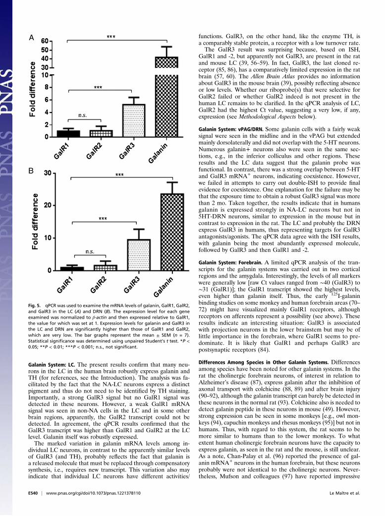
qPCR. The relative expression of galanin and its receptors was an-alyzed by qPCR in tissue slices of containing LC and DRN and inmacrodissected tissue punches from frontal cortex, cingulate cor-tex, and the amygdaloid complex (for details, see SI Materials andMethods). In addition, TH [raw cycle threshold (Ct) value 28] andTPH2 (raw Ct value 25) mRNA levels also were analyzed in suchslices (Table S1). Galanin mRNA expression was ∼42-fold higherthan GalR1 in the LC slices and was ∼25-fold higher than GalR1
Galanin B
Galanin E FGalR3TPH2 D
GalR3 CvPAG
TPH2 A
DRN
vPAG
DRN
DRN
Fig. 2. Dark-field ISH photomicrographs showing the distribution of TPH2 (A and D), galanin (B and E), and GalR3 (C and F) in semiadjacent sections. D, E,and F show higher magnification of boxes in A, B, and C, respectively. Arrows indicate cells positive for the respective marker. Note overlapping distribution ofTPH2 and GalR3 in the posterior DRN (compare D and F). However, no galanin signal is seen in that particular area (B and E). Instead galanin+ cells are seenmore dorsally (B). Exposure time: TPH2, 10 d; galanin, 4 wk; GalR3, 8 wk. (Scale bars: 100 μM in A, B, and C; 50 μM in D, E, and F.)
nNOSA
DRN
Aq
MLF
TPH2B
Aq
DRN
MLF
Fig. 3. Dark-field ISH photomicrographs showing the distribution of nNOS(A) and TPH2 (B) transcripts in semiadjacent sections. There is no overlap withthe TPH2+ 5-HT neurons in the DRN. Aq, Aqueduct; MLF, medial longitudinalfascicle. Exposure time: TPH2, 10 d; nNOS, 6 wk. (Scale bars: 100 μM.)
E538 | www.pnas.org/cgi/doi/10.1073/pnas.1221378110 Le Maître et al.

in the DRN slices (Table S1). GalR3 was the most abundantlyexpressed galanin receptor, being approximately sixfold higher thanGalR1 in LC slices and approximately sevenfold higher in DRNslices. GalR2 was expressed at very low levels (Fig. 5). The raw Ctvalues for galanin and GalR1-3 transcripts are given in Table S1.In the forebrain regions, however, GalR1 was the most abun-
dantly expressed receptor, even higher than galanin. GalaninmRNA was ∼0.6-fold, ∼0.4-fold, and ∼0.8-fold lower than GalR1in the frontal cortex (Fig. S4A), cingulate cortex (Fig. S4B), andamygdaloid complex (Fig. S4C), respectively. However, it is pos-sible that GalR2 and GalR3 were not expressed, because the Ctvalues were >35 for both. Taken together, these results indicatethat GalR3 seems to be the abundantly expressed receptor in thebrainstem nuclei studied here, whereas GalR1 appears to pre-dominate in the forebrain regions.
Controls. Hybridization with control probes resulted in a com-plete absence of signals for TH (Fig. 1A0), galanin (Fig. 1C0), andGalR3 (Fig. 1 E0 and H) in the LC.
DiscussionAlmost 30 y ago Tatemoto, et al. (41) discovered galanin, andsubsequently three receptors were identified, GalR1-3 (51, 53,
55), which are the focus of the present study. Here we studiedthese molecules in selected areas of human postmortem brainswith ISH and qPCR to establish expression patterns and to re-veal possible differences among species. Some other transmitter-related markers, i.e., TH, TPH2, nNOS, and VGLUTs, wereincluded also.The main results of the present study are that, in humans, (i)
galanin is present in NA-LC neurons but not in 5-HT-DRNneurons; (ii) GalR3 is the major galanin receptor in NA-LC neu-rons and probably in 5-HT-DRN neurons; (iii) 5-HT-DRN neu-rons do not express nNOS; (iv) GalR2 is expressed at low levels,if at all, in NA-LC and 5-HT-DRN neurons; (v) GalR3 probablyis not expressed in the forebrain regions studied; instead, GalR1is the predominant receptor; (vi) VGLUT2 is expressed only inthe dorsal and lateral PAG; and, of special importance, (vii)distinct differences exist among species (Table 1).It should be noted that we report only transcripts without
evidence for the expression of the receptor protein. Thus, GalR2and -3 receptors may be present on afferents (presynaptic) to theforebrain regions studied. For example, there is evidence that inthe rat the NA-LC neurons synthesize GalR2, which probably istransported centrifugally to act as a presynaptic receptor inforebrain regions (84).
VGLUT1 A
B
GF HTPH2 VGLUT2 VGLUT2
VGLUT2VGLUT1 C D
E
DRN
MRN
PN
vPAG
DRN
MRN
PAG
LCMLF
MLF
4V
Aq Aq
Aq
LC
Fig. 4. Dark-field ISH photomicrographs showing the distribution of VGLUT1 (A–C), VGLUT2 (D, E, G, and H), and TPH2 (F) mRNA at the pontine level. Thebox in F shows approximately the region displayed in the semiadjacent section in G. There is no detectable signal for VGLUT1 in the vPAG, including the DRN(A), whereas there is a very strong signal for both VGLUT1 (B) and -2 (C) in the pontine nuclei. A distinct VGLUT2 signal is seen in the pontine central gray,including the LC, extending medially to the dorsal tegmental nucleus (D). As seen in the high-power magnification, the cells are very small (arrows) comparedwith the pigmented NA neurons (arrowheads in E). Numerous vGLUT2 mRNA+ cells are observed in the lateral PAG (G and H), although there are very fewcells in the DRN and vPAG (compare G and F). MLF, medial longitudinal fascicle; PN, pontine nuclei. Exposure time: TPH2, 10 d; VGLUT1 and VGLUT2, 8 wk.(Scale bars: 200 μM in A, D, and F; 100 μM in B and G; 50 μM in H; 25 μM in C and E).
Le Maître et al. PNAS | Published online January 22, 2013 | E539
NEU
ROSC
IENCE
PNASPL
US
Galanin System: LC. The present results confirm that many neu-rons in the LC in the human brain robustly express galanin andTH (for references, see the Introduction). The analysis was fa-cilitated by the fact that the NA-LC neurons express a distinctpigment and thus do not need to be identified by TH staining.Importantly, a strong GalR3 signal but no GalR1 signal wasdetected in these neurons. However, a weak GalR1 mRNAsignal was seen in non-NA cells in the LC and in some otherbrain regions, apparently, the GalR2 transcript could not bedetected. In agreement, the qPCR results confirmed that theGalR3 transcript was higher than GalR1 and GalR2 at the LClevel. Galanin itself was robustly expressed.The marked variation in galanin mRNA levels among in-
dividual LC neurons, in contrast to the apparently similar levelsof GalR3 (and TH), probably reflects the fact that galanin isa released molecule that must be replaced through compensatorysynthesis, i.e., requires new transcript. This variation also mayindicate that individual LC neurons have different activities/
functions. GalR3, on the other hand, like the enzyme TH, isa comparably stable protein, a receptor with a low turnover rate.The GalR3 result was surprising because, based on ISH,
GalR1 and -2, but apparently not GalR3, are present in the ratand mouse LC (39, 56–59). In fact, GalR3, the last cloned re-ceptor (85, 86), has a comparatively limited expression in the ratbrain (57, 60). The Allen Brain Atlas provides no informationabout GalR3 in the mouse brain (39), possibly reflecting absenceor low levels. Whether our riboprobe(s) that were selective forGalR2 failed or whether GalR2 indeed is not present in thehuman LC remains to be clarified. In the qPCR analysis of LC,GalR2 had the highest Ct value, suggesting a very low, if any,expression (see Methodological Aspects below).
Galanin System: vPAG/DRN. Some galanin cells with a fairly weaksignal were seen in the midline and in the vPAG but extendedmainly dorsolaterally and did not overlap with the 5-HT neurons.Numerous galanin+ neurons also were seen in the same sec-tions, e.g., in the inferior colliculus and other regions. Theseresults and the LC data suggest that the galanin probe wasfunctional. In contrast, there was a strong overlap between 5-HTand GalR3 mRNA+ neurons, indicating coexistence. However,we failed in attempts to carry out double-ISH to provide finalevidence for coexistence. One explanation for the failure may bethat the exposure time to obtain a robust GalR3 signal was morethan 2 mo. Taken together, the results indicate that in humansgalanin is expressed strongly in NA-LC neurons but not in5HT-DRN neurons, similar to expression in the mouse but incontrast to expression in the rat. The LC and probably the DRNexpress GalR3 in humans, thus representing targets for GalR3antagonists/agonists. The qPCR data agree with the ISH results,with galanin being the most abundantly expressed molecule,followed by GalR3 and then GalR1 and -2.
Galanin System: Forebrain. A limited qPCR analysis of the tran-scripts for the galanin systems was carried out in two corticalregions and the amygdala. Interestingly, the levels of all markerswere generally low [raw Ct values ranged from ∼40 (GalR3) to∼31 (GalR1)]; the GalR1 transcript showed the highest levels,even higher than galanin itself. Thus, the early 125I-galaninbinding studies on some monkey and human forebrain areas (70–72) might have visualized mainly GalR1 receptors, althoughreceptors on afferents represent a possibility (see above). Theseresults indicate an interesting situation: GalR3 is associatedwith projection neurons in the lower brainstem but may be oflittle importance in the forebrain, where GalR1 seems to pre-dominate. It is likely that GalR1 and perhaps GalR3 arepostsynaptic receptors (84).
Differences Among Species in Other Galanin Systems. Differencesamong species have been noted for other galanin systems. In therat the cholinergic forebrain neurons, of interest in relation toAlzheimer’s disease (87), express galanin after the inhibition ofaxonal transport with colchicine (88, 89) and after brain injury(90–92), although the galanin transcript can barely be detected inthese neurons in the normal rat (93). Colchicine also is needed todetect galanin peptide in these neurons in mouse (49). However,strong expression can be seen in some monkeys [e.g., owl mon-keys (94), capuchin monkeys and rhesus monkeys (95)] but not inhumans. Thus, with regard to this system, the rat seems to bemore similar to humans than to the lower monkeys. To whatextent human cholinergic forebrain neurons have the capacity toexpress galanin, as seen in the rat and the mouse, is still unclear.As a note, Chan-Palay et al. (96) reported the presence of gal-anin mRNA+ neurons in the human forebrain, but these neuronsprobably were not identical to the cholinergic neurons. Never-theless, Mufson and colleagues (97) have reported impressive
Fig. 5. qPCR was used to examine the mRNA levels of galanin, GalR1, GalR2,and GalR3 in the LC (A) and DRN (B). The expression level for each geneexamined was normalized to β-actin and then expressed relative to GalR1,the value for which was set at 1. Expression levels for galanin and GalR3 inthe LC and DRN are significantly higher than those of GalR1 and GalR2,which are very low. The bar graphs represent the mean ± SEM (n = 7).Statistical significance was determined using unpaired Student’s t test. *P <0.05; **P < 0.01; ***P < 0.001; n.s., not significant.
E540 | www.pnas.org/cgi/doi/10.1073/pnas.1221378110 Le Maître et al.
data providing evidence that galanin has a protective role inAlzheimer’s disease.A similar situation may exist in the histaminergic/GABAergic
tubero-mammilary neurons that express galanin after colchicineadministration in the rat (48, 98–100) but apparently not in hu-man, although this analysis was performed only with immuno-histochemistry and not with ISH (101).
nNOS and Glutamate. Neither nNOS nor VGLUT3 could bedetected in human 5-HT neurons. The lack of nNOS in human issimilar to mouse (19, 39) and different from rat (27–30, 32) but isin agreement with a study by Carrive and Morgan (102) based onNADPH-d histochemistry showing strong staining in the dorsalPAG but apparently none in the DRN. [NAPDH-d in the rat hasbeen shown to be identical to nNOS (103, 104)]. Here we pro-vide confirmatory evidence by showing the apparent absence ofnNOS transcript in human 5-HT neurons based on ISH buta strong signal in the nearby regions, indicating a working probe.Also the NA-LC neurons had no detectable nNOS signal, even ifnNOS+ cells intermingled with NA neurons to a limited extent.The discovery of VGLUTs (79–81) made it possible, via im-
munohistochemistry and ISH, to identify unequivocally the neu-rons using the excitatory amino acid glutamate as transmitter.However, even though powerful antibodies to the VGLUTs havebeen generated, VGLUT expression in neuronal cell soma can bevisualized only with ISH, because antibodies apparently show thetransporters only in nerve endings. [DRG neuron cell bodies arean exception (105, 106).] The distribution patterns in mouse andrat DRN/PAG are similar (for references, see Introduction): noVGLUT1 mRNA is found in the LC, DRN, or vPAG. There aremany VGLUT2 mRNA+ cell bodies in the rat and mouse vPAGbut not in the LC or in the midline of the vPAG, i.e., the DRN.In contrast, a distinct subpopulation of 5-HT neurons in theDRN express VGLUT3 transcript in rats (33–35), mice (39), andsyrian hamsters (107). However, not all rat VGLUT3+ neuronsin the DRN are serotonergic (34).In humans, as in rats and mice, no VGLUT1 transcript was
found in the LC region or in the vPAG. VGLUT2 mRNA wasfound in small cells (possibly glia) in the LC, partly interminglingwith NA neurons, and was seen only in the dorsal and lateralPAG, in contrast to the distribution throughout the entire PAGin rodents. The lack of a VGLUT3 signal may represent eithera false negative caused by an unsensitive or failed probe or a trueabsence. Thus, it still is uncertain whether VGLUT3 expressionin the 5-HT neurons in the vPAG differs in rodents and humans.
Comparison with Results from the Allen Institute for Brain Research.Scientists at the Allen Institute for Brain Research also haveexplored the human brain (108, 109), as described in SI Dis-cussion. The data analysis revealed some individual variability,but, in general, galanin expression was higher in the supraopticnucleus, hypothalamus, LC, and frontal lobe than in the centralnucleus of the amygdala and MRN. TH and TPH2 transcriptsalso were highly expressed in LC and MRN, respectively. Ex-pression levels for the galanin receptors were generally low andare difficult to compare with the present qPCR and ISH results.Taken together, the results from the Allen Institute are, to a certain
extent, in agreement with the qPCR and ISH results in our study.However, this comparison needs further, in-depth analysis. Non-isotopic ISH for galanin and GalR2 also has been performed incortical and subcortical regions, where a weak signal is observed forboth transcripts (109).
Methodological Aspects. Histochemical approaches always areassociated with methodological problems related to sensitivityand specificity (110, 111). These difficulties are accentuatedwhen studying human postmortem tissue because of variabilityamong human beings, deterioration of the tissue with post-mortem time, varying conditions of death, and subsequent han-dling. Even more problematic are low-abundance transcripts,e.g., for certain receptors. Such proteins have slow turnover andthus low mRNA levels because they, unlike neuropeptides, donot need to be replaced swiftly after release. Presumably suchreceptors can be revealed only by advanced molecular biologycombined with electrophysiology (112, 113).In the present study, the transcripts for robustly expressed
molecules, such as TH and TPH2, could be demonstrated in allbrains but, importantly, after considerable differences in expo-sure time to an autoradiographic film/emulsion. Thus, althoughsections from some brains hybridized for TH or TPH2 neededless than a week of exposure, sections from other brains had tobe exposed for several weeks. Not unexpectedly, such differ-ences were critical for the successful processing of the low-abundance galanin receptor transcripts. Thus, in principle, suchtranscripts could be detected only in brains with short exposuretime for TH and TPH2. Therefore, negative results should beinterpreted with caution.The probe also is essential. We obtained good results for GalR3
with only one of the designed probes. The probe may not be asgood for GalR1 for GalR3, and the probe may have failed com-pletely for GalR2 and VGLUT3. Again, qPCR results indicatevery low GalR1 levels. As a note, GalR2 and -3 have a high degreeof homology; therefore in a previous study we demonstrated thespecificity of the primers for the two receptors (114).Difficulties in detecting galanin receptor transcripts are sup-
ported by the recently published Allen Brain Atlas describing thedistribution of some 20,000 transcripts throughout the mouse brain(39): No results are reported for GalR3, GalR2 is distinctlyexpressed only in some of the total of 52 sections/levels, and GalR1appears less abundant than in rat brain. However, the presence ofthe GalR3 transcript has been reported in many rat brain regionsby Mennicken et al. (57) using ISH and by Waters and Krause (60)using qPCR. In contrast, the Allen Brain Atlas shows robust ex-pression of many other neuropeptide receptors and thus supportsour view that galanin receptor transcripts may be particularly dif-ficult to visualize with ISH and, as shown here, probably are evenmore difficult to visualize in human postmortem tissue.The qPCR analysis was carried out on full coronal sections of
the pons, from a level including either the DRN or LC; in fact,these sections were alternate sections to those taken for ISH (SIMaterials and Methods). However other regions within the sliceexpressing the studied markers to a varying extent have beenincluded also. For the LC level, TH probably is confined to the
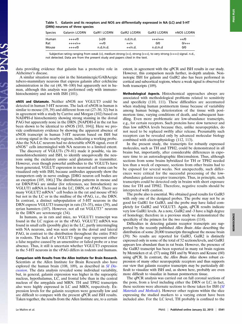
Table 1. Galanin and its receptors and NOS are differentially expressed in NA (LC) and 5-HT(DRN) neurons of three species
Species Galanin LC/DRN GalR1 LC/DRN GalR2 LC/DRN GalR3 LC/DRN nNOS LC/DRN
Human ++++/0 (+)/0 n.d./n.d. +++/+++ ++/0Rat ++++/++ ++/0 ++/+ 0/0 0/+++Mouse ++++/0 n.d./n.d. ++/n.d. n.d./n.d 0/0
Subjective rating ranging from weak (+), medium strong (++), strong (+++), to very strong (++++) signal. n.d.,not detected. Data are from the present study and papers cited in the text.
Le Maître et al. PNAS | Published online January 22, 2013 | E541
NEU
ROSC
IENCE
PNASPL
US
LC and subcoeruleus; GalR1 and -3 were observed only in re-lation to the LC, whereas galanin was expressed in several othernuclei (Fig. S2). TPH2 is fairly widely expressed both in the DRNand MRN and in the reticular formation. Galanin and GalR1also are expressed outside the raphe region, but we observedGalR3 only in the DRN and lateral to the aqueduct. Thus THmRNA is diluted, and results for galanin and GalR1 are notselective for the LC region. In fact, a preliminary qPCR exper-iment with laser capture microdissection (LCM) of the NA-LCnucleus (defined by pigmented neurons) shows a higher level ofTH transcript expression (i.e., a raw Ct value of ∼22 for LCMversus ∼28 for the whole section). Similarly for GalR3 the rawLCM Ct value was ∼29, versus ∼33 for the whole section, thusshowing the expected dilution effect for these markers.
Autoinhibition, Dendro-Somatic Release, and Electrophysiology. Auto-inhibition of LC neurons mediated by NA (115–117) and of DRNneurons mediated by 5-HT (118) may involve not only releasefrom collaterals but also somato-dendritic release (119, 120). Thistype of release also has been shown for neuropeptides (121) andpossibly for galanin in the LC (122). It is assumed that this auto-inhibitionis responsible, at least in part, for the delayed onset ofthe clinical effect of monoamine-reuptake inhibitors (123, 124).Electrophysiological studies in the rat have revealed that gal-
anin hyperpolarizes NA-LC neurons (68, 125, 126), presumablymediated via GalR1 (84), a transcript known to be present inthese neurons (56, 58). In addition, galanin at low concentrations(10−9M) enhances the autoinhibitory effect exerted by NA on LCneurons via α2A adrenoreceptors (127). Moreover, GalR1, butnot -2 or -3, is regulated by galanin signaling in the rat LC (128,129). Galanin also has an inhibitory effect on some 5-HT neu-rons in the DRN. However, it still is unclear if these effects aredirect, via GalR1 (73) or GalR3 (130, 131), indirect via GABAneurons (132), or both. ISH results indicate that GalR1 indeed ispresent in the vPAG but not in 5-HT neurons (56, 58, 73).Moreover, low galanin concentrations enhance the auto-
inhibitory effect of 5-HT via the 5-HT1A receptor in the DRN(73). This effect may be related to the formation of GalR1-5-HT1A receptor heteromers as recently reported by Borroto-Escuela et al. (133). Such receptor complexes increase traffickingof 5-HT1A receptors to the plasma membrane (133) and havebeen suggested to contribute to development of depression (134).
Neuropeptides and Stress-Related Disorders. Several antidepressantsexert their effect via monoamine neurons (124, 135–137). It hasbeen proposed, based on animal experiments over the last fewdecades, that neuropeptide also receptors are putative targets fordevelopment of antidepressants, including receptors for sub-stance P, neuropeptide tyrosine, corticotropin-releasing factor/corticotropin-releasing hormone, melanocyte-concentrating hor-mone, vasopressin, and dynorphin (138–143), as well as the gal-anin system. Thus, galanin interacts with 5-HT1A receptors, andgalanin antagonists and agonists have anxiolytic and antidepres-sive effects in animal experiments (144–151). Interestingly, asso-ciation of genes encoding galanin and/or GalR3 has been reportedfor psychiatric phenotypes (152–154), including panic disorder(155), depression-related parameters (156–158), and nicotine de-pendence (159, 160).The LC plays an important role in the development of stress-
related disorders (161–169), and stress up-regulates galanin ex-pression in the rat LC (170, 171). We therefore hypothesize thatsuch disorders are associated with an increase in firing and withincreased galanin synthesis and release from LC neurons, espe-cially from soma and dendrites (68, 122), resulting in activationof GalR1 autoreceptors, inhibition of firing, and decreased NArelease in the forebrain. Together, these events presumably re-sult in a prodepressive effect. In theory, a similar scenario may betrue for the 5-HT/galanin neurons in the DRN (73). Conse-
quently, attenuating the inhibition and autoinhibition of NA and5-HT neurons by galanin antagonists may have anxiolytic/anti-depressive effects. In fact, these effects may be enhanced by theGalR1-5-HT1A heterodimerization described above. Finally andinterestingly, Murck, et al. (172) have shown that the effects ofi.v.-administered galanin on sleep EEGs in healthy subjects aresimilar to those seen with sleep deprivation (172) and that gal-anin, when given to patients with depression, has an acuteantidepressive effect (173). The site of action of peripherallyadministered galanin remains to be analyzed.Based on the rat experiments, a GalR1 antagonist would be
suitable to obtain such an effect in the LC (56, 58, 61, 84),whereas a GalR3 antagonist (130, 131) may be more appropriatein the DRN. Our study shows that in the human brain the NA-LC, and presumably the 5-HT-DRN neurons, express GalR3,not GalR1 as in the rat (56, 58). The transduction mechanism forGalR3 has not been well characterized. Smith et al. (93) usedXenopus oocytes and coexpressed GalR3 with the potassiumchannel subunits GIRK1 and -4. They found that galanin openspotassium channels and thus hyperpolarize the cell membrane. Ifthe result from this artificial system should turn out to hold truefor human NA LC and 5-HT DRN neurons also, then a GalR3antagonist could have the same effect in humans as in the rat.The investigation of galanin has been hampered by the lack of
selective and powerful pharmacological tools to analyze galaninfunctionality, particularly drugs that can penetrate the blood–brainbarrier (174). However, a small number of such compounds activeat the GalR3 receptor, with anxiolytic and antidepressant activityin various rat models, have been developed (130, 131, 175).
Concluding Remarks. A main message of the present study is thatthere are distinct differences among species with regard to certaintransmitter systems and that results in rodent models cannot al-ways be translated directly to humans. Such species differencesmay be particularly common when studying the coexistence ofvarious transmitters, particularly those related to neuropeptidergicsystems (176), although here we show that species differences alsomay affect NOergic and perhaps glutamatergic systems. However,in general, peptidergic systems are highly conserved. For example,in a comprehensive ISH study, Krolewski, et al. (177) reportedthat several important neuropeptides are distributed similarly inthe human and rodent hypothalamus. Nevertheless, species var-iations should be taken into account when developing drugs forhuman disorders. The present evidence for GalR3 signaling inNA-LC and, presumably, in 5-HT-DRN neurons indicates thata similar mechanism may operate in both types of neurons inhumans and that GalR3 is a relevant target for drugs aiming totreat humans suffering from anxiety and/or depression. However,to our knowledge, no clinical trials have been carried out with theGalR3 antagonists mentioned above.
Materials and MethodsSubjects. Postmortem frozenbrain tissues including LC andDRN from12patientswere obtained by autopsy at different departments of Semmelweis University(Table S2). Written informed consent was obtained, and studies were approvedby the Ethics Committee of Semmelweis University. The postmortem time forthe frozen brains, including LC and DRN, was up to 10 h (Table S2).
RNA Probe Synthesis. RNA probes specific to TH, galanin, GalR1, and GalR3were prepared from human dorsal root ganglion (DRG) mRNA (Clontech).TPH2, GalR2, VGlut 1, VGlut2, VGlut3, and nNOSwere generated from humantotal-brain RNA (Ambion). The human DRG mRNA and total-brain RNA werereverse transcribed to generate cDNA using the Retroscript Kit (Ambion). ThiscDNA then was amplified using specific primers (Table S3), subcloned intoa PCR1II-TOPO vector (Invitrogen), and confirmed by nucleotide sequencing(KIGene). The plasmids were linearized and then transcribed using T7 andSP6 RNA polymerases to generate sense and antisense RNA probes. In vitrotranscription was carried out using the MAXIscript SP6/T7 kit (Ambion) and[α35]-UTP (Perkin-Elmer) according to the manufacturer’s instructions. The
E542 | www.pnas.org/cgi/doi/10.1073/pnas.1221378110 Le Maître et al.
transcripts then were purified using NucAway Spin Columns (Ambion). Senseprobes were used as negative controls.
ISH. Sections (14 μm) for the postmortem human brains were prepared ina cryostat. Fixation, prehybridization, and hybridization were performed asdescribed in SI Materials and Methods. Sections were placed against a filmand/or dipped in an autoradiographic emulsion, developed, and mountedwith glycerol/PBS medium (SI Materials and Methods). Sections also werecounterstained for cresyl violet (Merck) (SI Materials and Methods).
qPCR. Tissue for qPCR analysis was collected as stated in SI Materials andMethods. Total RNA was isolated with the Qiagen RNeasy Mini Kit (Qia-gen). First-strand cDNA was synthesized and subjected to PCR (SI Materialsand Methods).
Microscopic Analysis. Sections were analyzed using a Nikon Eclipse E600 mi-croscope equippedwith a bright- and dark-field condenser and epi-polarizationwith side entrance illumination (Fiberoptic-HeimAG) and epi-fluorescencewithappropriate filters combinations connected to a digital camera (Nikon DXM
1200). In some cases, Kodak T-MAX 400 black-and-white film was used forphotography. Sections were scanned using a Nikon LS-2000 film scanner(Nikon). Scanned and digital images were imported into Adobe PhotoShop 6.0(Adobe Systems, Inc.) and optimized for brightness, contrast, and sharpness.The atlases of Paxinos and Xu-Feng (178) and Olszewski and Baxter (179) wereconsulted throughout this work.
ACKNOWLEDGMENTS. We thank Professor Sandra Ceccatelli, Dr. ChristinaBark and Dr. Roshan Tofighi for advice with regard to qPCR analysis andProfessor Nenad Bogdanovic for valuable advice concerning the neuroanat-omy of the human brain stem and Dr. Csaba Adori, Blanca-Silva Lopez, YuQian, and Mingdong Zhang for their valuable assistance. This study wassupported by the Swedish Research Council (04X-2887); the Marianne andMarcus Wallenberg Foundation; the Knut and Alice Wallenberg Foundation;Grant NEWMOOD; LHSM-CT-2003-503474 from the European Union; theNational Alliance for Research on Schizophrenia and Depression; a grantfrom AFA (the Swedish Insurance company); funds from Karolinska Institu-tet; and the Swedish Brain Foundation. The studies would not have beenpossible without the earlier support of an Unrestricted Bristol-Myers-SquibbNeuroscience grant.
1. Falck B, Hillarp N-Å, Thieme G, Torp A (1962) Fluoresence of catecholamines andrelated compounds with formaldehyde. J Histochem Cytochem 10:348–334.
2. Dahlström A, Fuxe K (1964) Evidence for the existence of monoamine neurons in thecentral nervous system. I. Demonstration of monoamines in the cell bodies ofbrainstem neurons. Acta Physiol Scand 62(Suppl. 232):1–55.
3. Descarries L, Saucier G (1972) Disappearance of the locus coeruleus in the rat afterintraventricular 6-hydroxdopamine. Brain Res 37(2):310–316.
4. Goldman G, Coleman PD (1981) Neuron numbers in locus coeruleus do not changewith age in Fisher 344 rat. Neurobiol Aging 2(1):33–36.
5. Aston-Jones G (2004) Locus coeruleus A5 and A7 noradrenergic cell groups. The RatNervous System, ed Paxinos G (Elsevier, Amsterdam), 3rd Ed, pp 259–294.
6. Moore RY, Bloom FE (1979) Central catecholamine neuron systems: Anatomy andphysiology of the norepinephrine and epinephrine systems. Annu Rev Neurosci2:113–168.
7. Keay KA, Bandler R (2004) Periaqueductal gray. The Rat Nervous System, ed
Paxinos G (Elsevier, Amsterdam), pp 243–257.8. Steinbusch HW (1981) Distribution of serotonin-immunoreactivity in the central
nervous system of the rat-cell bodies and terminals. Neuroscience 6(4):557–618.9. Fuxe K, Hökfelt T, Ungerstedt U (1970) Central monoaminergic tracts. Principles of
Psychopharmacology, eds Clark WG, del Gindice J, (Academic Press, London) pp 87–96.
10. Azmitia EC, Gannon PJ (1986) The primate serotonergic system: A review of humanand animal studies and a report on Macaca fascicularis. Adv Neurol 43:407–468.
11. Törk I, Hornung JP (1990) Raphe nuclei and the serotonergic system. The HumanNervous System, ed Paxinos G (Academic, San Diego), pp 1001–1022.
12. Ungerstedt U (1971) Stereotaxic mapping of the monoamine pathways in the ratbrain. Acta Physiol Scand Suppl 367:1–48.
13. Steinbusch HWM (1984) Serotonin-immunoreactive neurons and their projections inthe CNS. Handbook Of Chemical Neuroanatomy. Classical Transmitters And TransmitterReceptors in the CNS, eds Björklund A, Hökfelt T, Kuhar MJ (Elsevier, Amsterdam),Vol 3, pp 68–140.
14. Baker KG, Törk I, Hornung JP, Halasz P (1989) The human locus coeruleus complex:
An immunohistochemical and three dimensional reconstruction study. Exp Brain Res77(2):257–270.
15. Chan-Palay V, Asan E (1989) Quantitation of catecholamine neurons in the locuscoeruleus in human brains of normal young and older adults and in depression.J Comp Neurol 287(3):357–372.
16. German DC, Walker BS, Manaye K, Smith WK, Woodward DJ, North AJ (1988) Thehuman locus coeruleus: Computer reconstruction of cellular distribution. J Neurosci8:1776–1788.
17. Baker KG, et al. (1991) Distribution, morphology and number of monoamine-synthesizing and substance P-containing neurons in the human dorsal raphe nucleus.Neuroscience 42(3):757–775.
18. Daszuta A, Portalier P (1985) Distribution and quantification of 5-HT nerve cell
bodies in the nucleus raphe dorsalis area of C57BL and BALBc mice. Relationshipbetween anatomy and biochemistry. Brain Res 360(1-2):58–64.
19. Fu W, et al. (2010) Chemical neuroanatomy of the dorsal raphe nucleus and adjacentstructures of the mouse brain. J Comp Neurol 518(17):3464–3494.
20. Nusbaum MP, Blitz DM, Swensen AM, Wood D, Marder E (2001) The roles of co-transmission in neural network modulation. Trends Neurosci 24(3):146–154.
21. Kupfermann I (1991) Functional studies of cotransmission. Physiol Rev 71(3):683–732.22. Burnstock G (1986) Purines and cotransmitters in adrenergic and cholinergic
neurones. Prog Brain Res 68:193–203.23. Merighi A (2002) Costorage and coexistence of neuropeptides in the mammalian
CNS. Prog Neurobiol 66(3):161–190.24. Gutiérrez R (2009) Co-existence and Co-Release of Classical Neurotransmitters
(Springer, New York).25. Hökfelt T, Johansson O, Ljungdahl A, Lundberg JM, Schultzberg M (1980) Peptidergic
neurones. Nature 284(5756):515–521.
26. Lundberg JM (1996) Pharmacology of cotransmission in the autonomic nervoussystem: Integrative aspects on amines, neuropeptides, adenosine triphosphate,amino acids and nitric oxide. Pharmacol Rev 48(1):113–178.
27. Dun NJ, Dun SL, Förstermann U (1994) Nitric oxide synthase immunoreactivity in ratpontine medullary neurons. Neuroscience 59(2):429–445.
28. Wotherspoon G, Albert M, Rattray M, Priestley JV (1994) Serotonin and NADPH-diaphorase in the dorsal raphe nucleus of the adult rat. Neurosci Lett 173(1-2):31–36.
29. Johnson MD, Ma PM (1993) Localization of NADPH diaphorase activity inmonoaminergic neurons of the rat brain. J Comp Neurol 332(4):391–406.
30. Wang QP, Guan JL, Nakai Y (1995) Distribution and synaptic relations of NOSneurons in the dorsal raphe nucleus: A comparison to 5-HT neurons. Brain Res Bull 37(2):177–187.
31. Vincent SR, Kimura H (1992) Histochemical mapping of nitric oxide synthase in therat brain. Neuroscience 46(4):755–784.
32. Xu ZQ, Hökfelt T (1997) Expression of galanin and nitric oxide synthase insubpopulations of serotonin neurons of the rat dorsal raphe nucleus. J ChemNeuroanat 13(3):169–187.
33. Gras C, et al. (2002) A third vesicular glutamate transporter expressed by cholinergicand serotoninergic neurons. J Neurosci 22(13):5442–5451.
34. Hioki H, et al. (2010) Vesicular glutamate transporter 3-expressing nonserotonergicprojection neurons constitute a subregion in the rat midbrain raphe nuclei. J CompNeurol 518(5):668–686.
35. Schäfer MK, Varoqui H, Defamie N, Weihe E, Erickson JD (2002) Molecular cloningand functional identification of mouse vesicular glutamate transporter 3 and itsexpression in subsets of novel excitatory neurons. J Biol Chem 277(52):50734–50748.
36. Amilhon B, et al. (2010) VGLUT3 (vesicular glutamate transporter type 3) contributionto the regulation of serotonergic transmission and anxiety. J Neurosci 30(6):2198–2210.
37. Calizo LH, et al. (2011) Raphe serotonin neurons are not homogenous: Electrophysiological,morphological and neurochemical evidence. Neuropharmacology 61(3):524–543.
38. Jackson J, Bland BH, Antle MC (2009) Nonserotonergic projection neurons in themidbrain raphe nuclei contain the vesicular glutamate transporter VGLUT3. Synapse63(1):31–41.
39. Lein ES, et al. (2007) Genome-wide atlas of gene expression in the adult mouse brain.Nature 445(7124):168–176.
40. Melander T, et al. (1986) Coexistence of galanin-like immunoreactivity withcatecholamines, 5-hydroxytryptamine, GABA and neuropeptides in the rat CNS.J Neurosci 6(12):3640–3654.
41. Tatemoto K, Rökaeus A, Jörnvall H, McDonald TJ, Mutt V (1983) Galanin - a novelbiologically active peptide from porcine intestine. FEBS Lett 164(1):124–128.
42. Bersani M, et al. (1991) Human galanin: Primary structure and identification of twomolecular forms. FEBS Lett 283(2):189–194.
43. Evans HF, Shine J (1991) Human galanin: Molecular cloning reveals a uniquestructure. Endocrinology 129(3):1682–1684.
44. Schmidt WE, et al. (1991) Isolation and primary structure of pituitary humangalanin, a 30-residue nonamidated neuropeptide. Proc Natl Acad Sci USA 88(24):11435–11439.
45. Cheung CC, Hohmann JG, Clifton DK, Steiner RA (2001) Distribution of galaninmessenger RNA-expressing cells in murine brain and their regulation by leptin inregions of the hypothalamus. Neuroscience 103(2):423–432.
46. Rökaeus A, et al. (1984) A galanin-like peptide in the central nervous system andintestine of the rat. Neurosci Lett 47(2):161–166.
47. Skofitsch G, Jacobowitz DM (1986) Quantitative distribution of galanin-likeimmunoreactivity in the rat central nervous system. Peptides 7(4):609–613.
48. Melander T, Hökfelt T, Rökaeus A (1986) Distribution of galaninlikeimmunoreactivity in the rat central nervous system. J Comp Neurol 248(4):475–517.
49. Pérez SE, Wynick D, Steiner RA, Mufson EJ (2001) Distribution of galaninergicimmunoreactivity in the brain of the mouse. J Comp Neurol 434(2):158–185.
50. Skofitsch G, Jacobowitz DM (1985) Immunohistochemical mapping of galanin-likeneurons in the rat central nervous system. Peptides 6(3):509–546.
51. Iismaa TP, Shine J (1999) Galanin and galanin receptors. Results Probl Cell Differ 26:257–291.
Le Maître et al. PNAS | Published online January 22, 2013 | E543
NEU
ROSC
IENCE
PNASPL
US
52. Lang R, Gundlach AL, Kofler B (2007) The galanin peptide family: Receptorpharmacology, pleiotropic biological actions, and implications in health and disease.Pharmacol Ther 115(2):177–207.
53. Branchek TA, Smith KE, Gerald C, Walker MW (2000) Galanin receptor subtypes.Trends Pharmacol Sci 21(3):109–117.
54. Wang S, Gustafson EL (1998) Galanin receptor subtypes. Drug News Perspect 11(8):458–468.
55. Habert-Ortoli E, Amiranoff B, Loquet I, Laburthe M, Mayaux JF (1994) Molecularcloning of a functional human galanin receptor. Proc Natl Acad Sci USA 91(21):9780–9783.
56. Burazin TC, Larm JA, Ryan MC, Gundlach AL (2000) Galanin-R1 and -R2 receptormRNA expression during the development of rat brain suggests differential subtypeinvolvement in synaptic transmission and plasticity. Eur J Neurosci 12(8):2901–2917.
57. Mennicken F, Hoffert C, Pelletier M, Ahmad S, O’Donnell D (2002) Restricteddistribution of galanin receptor 3 (GalR3) mRNA in the adult rat central nervoussystem. J Chem Neuroanat 24(4):257–268.
58. O’Donnell D, Ahmad S, Wahlestedt C, Walker P (1999) Expression of the novelgalanin receptor subtype GALR2 in the adult rat CNS: Distinct distribution fromGALR1. J Comp Neurol 409(3):469–481.
59. O’Donnell D, et al. (2003) Localization of galanin receptor subtypes in the rat CNS.Handbook of Chemical Neuroanatomy. Peptide Receptors, Part II, eds Quirion R,Björklund A, Hökfelt T (Elsevier, Amsterdam), Vol 20, pp 195–244.
60. Waters SM, Krause JE (2000) Distribution of galanin-1, -2 and -3 receptor messengerRNAs in central and peripheral rat tissues. Neuroscience 95(1):265–271.
61. Xu ZQ, Shi TJ, Hökfelt T (1998) Galanin/GMAP- and NPY-like immunoreactivities inlocus coeruleus and noradrenergic nerve terminals in the hippocampal formationand cortex with notes on the galanin-R1 and -R2 receptors. J Comp Neurol 392(2):227–251.
62. Holets VR, Hökfelt T, Rökaeus A, Terenius L, Goldstein M (1988) Locus coeruleusneurons in the rat containing neuropeptide Y, tyrosine hydroxylase or galanin andtheir efferent projections to the spinal cord, cerebral cortex and hypothalamus.Neuroscience 24(3):893–906.
63. Chan-Palay V, Jentsch B, Lang W, Hochli M, Asan E (1990) Distribution ofneuropeptide Y, C-terminal flanking peptide of NPY and galanin coexistence withcatecholamine in the locus coeruleus of normal human, Alzheimer’s dementia andParkinson’s disease brains. Dementia 1:18–31.
64. Kordower JH, Le HK, Mufson EJ (1992) Galanin immunoreactivity in the primatecentral nervous system. J Comp Neurol 319(4):479–500.
65. Miller MA, Kolb PE, Leverenz JB, Peskind ER, Raskind MA (1999) Preservation ofnoradrenergic neurons in the locus ceruleus that coexpress galanin mRNA inAlzheimer’s disease. J Neurochem 73(5):2028–2036.
66. Larm JA, Shen PJ, Gundlach AL (2003) Differential galanin receptor-1 and galaninexpression by 5-HT neurons in dorsal raphé nucleus of rat and mouse: Evidence forspecies-dependent modulation of serotonin transmission. Eur J Neurosci 17(3):481–493.
67. Skofitsch G, Sills MA, Jacobowitz DM (1986) Autoradiographic distribution of 125I-galanin binding sites in the rat central nervous system. Peptides 7(6):1029–1042.
68. Pieribone VA, et al. (1995) Galanin induces a hyperpolarization of norepinephrine-containing locus coeruleus neurons in the brainstem slice. Neuroscience 64(4):861–874.
69. Melander T, et al. (1988) Autoradiographic quantitation and anatomical mapping of125I-galanin binding sites in the rat central nervous system. J Chem Neuroanat 1(4):213–233.
70. Köhler C, Hallman H, Melander T, Hökfelt T, Norheim E (1989) Autoradiographicmapping of galanin receptors in the monkey brain. J Chem Neuroanat 2(5):269–284.
71. Köhler C, et al. (1989) Distribution of galanin-binding sites in the monkey andhuman telencephalon: Preliminary observations. Exp Brain Res 75(2):375–380.
72. Köhler C, Chan-Palay V (1990) Galanin receptors in the post-mortem human brain.Regional distribution of 125I-galanin binding sites using the method of in vitroreceptor autoradiography. Neurosci Lett 120(2):179–182.
73. Xu ZQ, Zhang X, Pieribone VA, Grillner S, Hökfelt T (1998) Galanin-5-hydroxytryptamine interactions: Electrophysiological, immunohistochemical and insitu hybridization studies on rat dorsal raphe neurons with a note on galanin R1 andR2 receptors. Neuroscience 87(1):79–94.
74. Nagatsu T, Levitt M, Udenfriend S (1964) Tyrosine Hydroxylase. The Initial Step inNorepinephrine Biosynthesis. J Biol Chem 239:2910–2917.
75. Grima B, et al. (1987) A single human gene encoding multiple tyrosine hydroxylaseswith different predicted functional characteristics. Nature 326(6114):707–711.
76. Walther DJ, et al. (2003) Synthesis of serotonin by a second tryptophan hydroxylaseisoform. Science 299(5603):76.
77. Bredt DS, et al. (1991) Cloned and expressed nitric oxide synthase structurallyresembles cytochrome P-450 reductase. Nature 351(6329):714–718.
78. Geller DA, et al. (1993) Molecular cloning and expression of inducible nitric oxidesynthase from human hepatocytes. Proc Natl Acad Sci USA 90(8):3491–3495.
79. Fremeau RT, Jr., Voglmaier S, Seal RP, Edwards RH (2004) VGLUTs define subsets ofexcitatory neurons and suggest novel roles for glutamate. Trends Neurosci 27(2):98–103.
80. Kaneko T, Fujiyama F (2002) Complementary distribution of vesicular glutamatetransporters in the central nervous system. Neurosci Res 42(4):243–250.
81. Masson J, Sagné C, Hamon M, El Mestikawy S (1999) Neurotransmitter transportersin the central nervous system. Pharmacol Rev 51(3):439–464.
82. Le Maitre E, Diaz-Heijtz R, Palkovits M, Hökfelt T (2008) The galanin system in thehuman locus coeruleus and dorsal raphe nucleus. J Neural Transm 11:1724–1172.
83. Le Maitre E, Diaz-Heijtz R, Palkovits M, Hökfelt T (2009) The galanin system in thehuman brain and relations to serotonin and noradrenaline neurons. EurNeuropsychopharmacol 19:S18.
84. Ma X, et al. (2001) Effects of galanin receptor agonists on locus coeruleus neurons.Brain Res 919(1):169–174.
85. Smith KE, et al. (1998) Cloned human and rat galanin GALR3 receptors.Pharmacology and activation of G-protein inwardly rectifying K+ channels. J BiolChem 273(36):23321–23326.
86. Wang S, He C, Hashemi T, Bayne M (1997) Cloning and expressional characterizationof a novel galanin receptor. Identification of different pharmacophores withingalanin for the three galanin receptor subtypes. J Biol Chem 272(51):31949–31952.
87. Whitehouse PJ, et al. (1982) Alzheimer’s disease and senile dementia: Loss ofneurons in the basal forebrain. Science 215(4537):1237–1239.
88. Melander T, et al. (1985) Galanin-like immunoreactivity in cholinergic neurons of theseptum-basal forebrain complex projecting to the hippocampus of the rat. Brain Res360(1-2):130–138.
89. Senut MC, Menetrey D, Lamour Y (1989) Cholinergic and peptidergic projections fromthe medial septum and the nucleus of the diagonal band of Broca to dorsalhippocampus, cingulate cortex and olfactory bulb: A combined wheatgerm agglutinin-apohorseradish peroxidase-gold immunohistochemical study. Neuroscience 30(2):385–403.
90. Cortés R, Ceccatelli S, Schalling M, Hökfelt T (1990) Differential effects ofintracerebroventricular colchicine administration on the expression of mRNAs forneuropeptides and neurotransmitter enzymes, with special emphasis on galanin: Anin situ hybridization study. Synapse 6(4):369–391.
91. Agoston DV, Komoly S, Palkovits M (1994) Selective up-regulation of neuropeptidesynthesis by blocking the neuronal activity: Galanin expression in septohippocampalneurons. Exp Neurol 126(2):247–255.
92. Palkovits M (1995) Neuropeptide messenger plasticity in the CNS neurons followingaxotomy. Mol Neurobiol 10(2-3):91–103.
93. Miller MA, Kolb PE, Planas B, Raskind MA (1998) Few cholinergic neurons in the ratbasal forebrain coexpress galanin messenger RNA. J Comp Neurol 391(2):248–258.
94. Melander T, Staines WA (1986) A galanin-like peptide coexists in putative cholinergicsomata of the septum-basal forebrain complex and in acetylcholinesterase-containing fibers and varicosities within the hippocampus in the owl monkey (Aotustrivirgatus). Neurosci Lett 68(1):17–22.
95. Kordower JH, Mufson EJ (1990) Galanin-like immunoreactivity within the primatebasal forebrain: Differential staining patterns between humans and monkeys.J Comp Neurol 294(2):281–292.
96. Chan-Palay V, Köhler C, Ernfors P, Persson H (1991) Senile dementia of the Alzheimertype is accompanied by hypertrophy of galanin axons, reservation of receptors andtissue specific expression of the galanin gene in the nucleus basalis Meynert.Galanin. A New Multifunctional Peptide in the Neuro-Endocrine System, edsHökfelt T, Bartfai T, Jacobowitz D, Ottoson D (MacMillan, London), pp 419–427.
97. Counts SE, Perez SE, Ginsberg SD, Mufson EJ (2010) Neuroprotective role for galaninin Alzheimer’s disease. EXS 102:143–162.
98. Chotard C, et al. (2002) Effects of histamine H3 receptor agonist and antagonist onhistamine co-transmitter expression in rat brain. J Neural Transm 109(3):293–306.
99. Köhler C, et al. (1986) Galanin immunoreactivity in hypothalamic neurons: Furtherevidence for multiple chemical messengers in the tuberomammillary nucleus. JComp Neurol 250(1):58–64.
100. Lantos TA, Görcs TJ, Palkovits M (1995) Immunohistochemical mapping ofneuropeptides in the premamillary region of the hypothalamus in rats. Brain ResBrain Res Rev 20(2):209–249.
101. Trottier S, et al. (2002) Co-localization of histamine with GABA but not with galaninin the human tuberomamillary nucleus. Brain Res 939(1-2):52–64.
102. Carrive P, Morgan MM (2004) Periaqueductal gray. The Human Nervous System, edsPaxinos G, Mai JK (Elsevier, San Diego), 2nd Ed.
103. Hope BT, Michael GJ, Knigge KM, Vincent SR (1991) Neuronal NADPH diaphorase isa nitric oxide synthase. Proc Natl Acad Sci USA 88(7):2811–2814.
104. Bredt DS, et al. (1991) Nitric oxide synthase protein and mRNA are discretelylocalized in neuronal populations of the mammalian CNS together with NADPHdiaphorase. Neuron 7(4):615–624.
105. Oliveira AL, et al. (2003) Cellular localization of three vesicular glutamatetransporter mRNAs and proteins in rat spinal cord and dorsal root ganglia. Synapse50(2):117–129.
106. Brumovsky PR, et al. (2011) Expression of vesicular glutamate transporters type1 and 2 in sensory and autonomic neurons innervating the mouse colorectum. JComp Neurol 519(16):3346–3366.
107. Mintz EM, Scott TJ (2006) Colocalization of serotonin and vesicular glutamatetransporter 3-like immunoreactivity in the midbrain raphe of Syrian hamsters(Mesocricetus auratus). Neurosci Lett 394(2):97–100.
108. Anonymous. Allen Brain Atlas Resources (Allen Institute for Brain Research, Seattle).Available at: www.brain-map.org.
109. Anonymous. Allen Human Cortex Study (Allen Institute for Brain Research, Seattle).Available at http://humancortex.alleninstitute.org.
110. Rhodes KJ, Trimmer JS (2006) Antibodies as valuable neuroscience research toolsversus reagents of mass distraction. J Neurosci 26(31):8017–8020.
111. Saper CB (2005) An open letter to our readers on the use of antibodies. J CompNeurol 493(4):477–478.
112. Bartfai T, Buckley PT, Eberwine J (2012) Drug targets: Single-cell transcriptomicshastens unbiased discovery. Trends Pharmacol Sci 33(1):9–16.
113. Eberwine J, Bartfai T (2011) Single cell transcriptomics of hypothalamic warmsensitive neurons that control core body temperature and fever response Signaling
E544 | www.pnas.org/cgi/doi/10.1073/pnas.1221378110 Le Maître et al.
asymmetry and an extension of chemical neuroanatomy. Pharmacol Ther 129(3):241–259.
114. Tofighi R, et al. (2012) Galanin and its three receptors in human pituitary adenoma.Neuropeptides 46(5):195–201.
115. Svensson TH, Bunney BS, Aghajanian GK (1975) Inhibition of both noradrenergic andserotonergic neurons in brain by the alpha-adrenergic agonist clonidine. Brain Res92(2):291–306.
116. Aghajanian GK, Cedarbaum JM, Wang RY (1977) Evidence for norepinephrine-mediated collateral inhibition of locus coeruleus neurons. Brain Res 136(3):570–577.
117. Cedarbaum JM, Aghajanian GK (1977) Catecholamine receptors on locus coeruleusneurons: Pharmacological characterization. Eur J Pharmacol 44(4):375–385.
118. Aghajanian GK, Wang RY (1978) Physiology and pharmacology of central serotonergicneurons. Psychopharmacology: A Generation of Progress, eds Lipton MA, DiMasco A,Killam KF (Elsevier, Amsterdam).
119. Ludwig M (2005) Dendritic Neurotransmitter Release (Springer, New York).120. Huang HP, et al. (2007) Long latency of evoked quantal transmitter release from
somata of locus coeruleus neurons in rat pontine slices. Proc Natl Acad Sci USA 104(4):1401–1406.
121. Ludwig M, Leng G (2006) Dendritic peptide release and peptide-dependentbehaviours. Nat Rev Neurosci 7(2):126–136.
122. Vila-Porcile E, et al. (2009) Dendritic synthesis and release of the neuropeptidegalanin: Morphological evidence from studies on rat locus coeruleus neurons. JComp Neurol 516(3):199–212.
123. Artigas F, Romero L, de Montigny C, Blier P (1996) Acceleration of the effect ofselected antidepressant drugs in major depression by 5-HT1A antagonists. TrendsNeurosci 19(9):378–383.
124. Millan MJ (2006) Multi-target strategies for the improved treatment of depressivestates: Conceptual foundations and neuronal substrates, drug discovery andtherapeutic application. Pharmacol Ther 110(2):135–370.
125. Seutin V, Verbanck P, Massotte L, Dresse A (1989) Galanin decreases the activity oflocus coeruleus neurons in vitro. Eur J Pharmacol 164(2):373–376.
126. Sevcik J, Finta EP, Illes P (1993) Galanin receptors inhibit the spontaneous firing oflocus coeruleus neurones and interact with mu-opioid receptors. Eur J Pharmacol230(2):223–230.
127. Xu ZQ, Tong YG, Hökfelt T (2001) Galanin enhances noradrenaline-induced outwardcurrent on locus coeruleus noradrenergic neurons. Neuroreport 12(8):1779–1782.
128. Hawes JJ, Brunzell DH, Wynick D, Zachariou V, Picciotto MR (2005) GalR1, but notGalR2 or GalR3, levels are regulated by galanin signaling in the locus coeruleusthrough a cyclic AMP-dependent mechanism. J Neurochem 93(5):1168–1176.
129. Mitsukawa K, Lu X, Bartfai T (2009) Bidirectional regulation of stress responses bygalanin in mice: Involvement of galanin receptor subtype 1. Neuroscience 160(4):837–846.
130. Swanson CJ, et al. (2005) Anxiolytic- and antidepressant-like profiles of the galanin-3receptor (Gal3) antagonists SNAP 37889 and SNAP 398299. Proc Natl Acad Sci USA102(48):17489–17494.
131. Barr AM, et al. (2006) A novel, systemically active, selective galanin receptor type-3ligand exhibits antidepressant-like activity in preclinical tests. Neurosci Lett 405(1-2):111–115.
132. Sharkey LM, Madamba SG, Siggins GR, Bartfai T (2008) Galanin alters GABAergicneurotransmission in the dorsal raphe nucleus. Neurochem Res 33(2):285–291.
133. Borroto-Escuela DO, et al. (2010) Galanin receptor-1 modulates 5-hydroxtryptamine-1A signaling via heterodimerization. Biochem Biophys Res Commun 393(4):767–772.
134. Fuxe K, et al. (2008) Receptor-receptor interactions within receptor mosaics. Impacton neuropsychopharmacology. Brain Res Brain Res Rev 58(2):415–452.
135. Maes M, Meltzer HY (1995) The serotonin hypothesis of major depression.Psychopharmacology: The Fourth Generation of Progress, eds Bloom FE, Kupfer DJ(Raven, New York), pp 933–944.
136. Montgomery SA (2006) Why do we need new and better antidepressants? ClinPsychopharmacol 21(Suppl 1):S1–S10.
137. Schatzberg AF, Schildkrant JJ (1995) Recent studies on norepinephrine systems inmood disorders. Pharmacology: The Fourth Generation of Progress, eds Bloom FE,Kupfer DJ (Raven, New York), pp 933–944.
138. Maubach KA, Rupniak NM, Kramer MS, Hill RG (1999) Novel strategies forpharmacotherapy of depression. Curr Opin Chem Biol 3(4):481–488.
139. Nemeroff CB, Vale WW (2005) The neurobiology of depression: Inroads to treatmentand new drug discovery. J Clin Psychiatry 66(Suppl 7):5–13.
140. Reul JM, Holsboer F (2002) Corticotropin-releasing factor receptors 1 and 2 inanxiety and depression. Curr Opin Pharmacol 2(1):23–33.
141. Sajdyk TJ, Shekhar A, Gehlert DR (2004) Interactions between NPY and CRF in theamygdala to regulate emotionality. Neuropeptides 38(4):225–234.
142. Holmes A, Heilig M, Rupniak NM, Steckler T, Griebel G (2003) Neuropeptide systemsas novel therapeutic targets for depression and anxiety disorders. Trends PharmacolSci 24(11):580–588.
143. Nestler EJ, Carlezon WA, Jr. (2006) The mesolimbic dopamine reward circuit indepression. Biol Psychiatry 59(12):1151–1159.
144. Weiss JM, Bonsall RW, Demetrikopoulos MK, Emery MS, West CHK (1998) Galanin: Asignificant role in depression? Ann N Y Acad Sci 863:364–382.
145. Fuxe K, et al. (1991) Galanin/5-HT interactions in the rat central nervous system.Relevance for depression. Galanin: A New Multifunctional Peptide in the Neuro-Endocrine System. Wenner-Gren Center International Symposium Series, Vol. 58, edsHökfelt T, Bartfai T, Jacobowitz D, Ottoson D (MacMillan, London), pp 221–235.
146. Barrera G, et al. (2005) One for all or one for one: Does co-transmission unify theconcept of a brain galanin “system” or clarify any consistent role in anxiety?Neuropeptides 39(3):289–292.
147. Ögren SO, Kuteeva E, Hökfelt T, Kehr J (2006) Galanin receptor antagonists : Apotential novel pharmacological treatment for mood disorders. CNS Drugs 20(8):633–654.
148. Lu X, Sharkey L, Bartfai T (2007) The brain galanin receptors: Targets for novelantidepressant drugs. CNS Neurol Disord Drug Targets 6(3):183–192.
149. Bing O, Möller C, Engel JA, Söderpalm B, Heilig M (1993) Anxiolytic-like action ofcentrally administered galanin. Neurosci Lett 164(1-2):17–20.
150. Möller C, Sommer W, Thorsell A, Heilig M (1999) Anxiogenic-like action of galaninafter intra-amygdala administration in the rat. Neuropsychopharmacology 21(4):507–512.
151. Kuteeva E, Hökfelt T, Wardi T, Ögren S-O (2010) Galanin, galanin receptor subtypesand depression-like behaviour. EXS 102:163–181.
152. Belfer I, et al. (2007) Alcoholism is associated with GALR3 but not two other galaninreceptor genes. Genes Brain Behav 6(5):473–481.
153. Belfer I, et al. (2006) Association of galanin haplotypes with alcoholism and anxietyin two ethnically distinct populations. Mol Psychiatry 11(3):301–311.
154. Levran O, et al. (2008) Genetic susceptibility to heroin addiction: A candidate geneassociation study. Genes Brain Behav 7(7):720–729.
155. Unschuld PG, et al. (2008) Polymorphisms in the galanin gene are associated withsymptom-severity in female patients suffering from panic disorder. J Affect Disord105(1-3):177–184.
156. Unschuld PG, et al. (2010) Gender-specific association of galanin polymorphisms withHPA-axis dysregulation, symptom severity, and antidepressant treatment response.Neuropsychopharmacology 35(7):1583–1592.
157. Davidson S, et al. (2011) Differential activity by polymorphic variants of a remoteenhancer that supports galanin expression in the hypothalamus and amygdala:Implications for obesity, depression and alcoholism. Neuropsychopharmacology 36(11):2211–2221.
158. Wray NR, et al. (2012) Genome-wide association study of major depressive disorder:New results, meta-analysis, and lessons learned. Mol Psychiatry 17(1):36–48.
159. Jackson KJ, Chen X, Miles MF, Harenza J, Damaj MI (2011) The neuropeptide galaninand variants in the GalR1 gene are associated with nicotine dependence.Neuropsychopharmacology 36(11):2339–2348.
160. Lori A, et al. (2011) The galanin receptor 1 gene associates with tobacco craving insmokers seeking cessation treatment. Neuropsychopharmacology 36(7):1412–1420.
161. Svensson TH (1987) Stress, central neurotransmitters, and the mechanism of actionof alpha 2-adrenoceptor agonists. J Cardiovasc Pharmacol 10(Suppl 12):S88–S92.
162. Page ME, Valentino RJ (1994) Locus coeruleus activation by physiological challenges.Brain Res Bull 35(5-6):557–560.
163. Aston-Jones G, Rajkowski J, Kubiak P, Valentino RJ, Shipley MT (1996) Role of thelocus coeruleus in emotional activation. Prog Brain Res 107:379–402.
164. Weiss JM, et al. (1994) Depression and anxiety: Role of the locus coeruleus andcorticotropin-releasing factor. Brain Res Bull 35(5-6):561–572.
165. Bremner JD, Krystal JH, Southwick SM, Charney DS (1996) Noradrenergicmechanisms in stress and anxiety: I. Preclinical studies. Synapse 23(1):28–38.
166. Bremner JD, Krystal JH, Southwick SM, Charney DS (1996) Noradrenergicmechanisms in stress and anxiety: II. Clinical studies. Synapse 23(1):39–51.
167. Harro J, Oreland L (2001) Depression as a spreading adjustment disorder ofmonoaminergic neurons: A case for primary implication of the locus coeruleus. BrainRes Brain Res Rev 38(1-2):79–128.
168. Charney DS (2004) Psychobiological mechanisms of resilience and vulnerability:Implications for successful adaptation to extreme stress. Am J Psychiatry 161(2):195–216.
169. Foote SL, Bloom FE, Aston-Jones G (1983) Nucleus locus ceruleus: New evidence ofanatomical and physiological specificity. Physiol Rev 63(3):844–914.
170. Holmes PV, Blanchard DC, Blanchard RJ, Brady LS, Crawley JN (1995) Chronic socialstress increases levels of preprogalanin mRNA in the rat locus coeruleus. PharmacolBiochem Behav 50(4):655–660.
171. Sweerts BW, Jarrott B, Lawrence AJ (1999) Expression of preprogalanin mRNAfollowing acute and chronic restraint stress in brains of normotensive andhypertensive rats. Brain Res Mol Brain Res 69(1):113–123.
172. Murck H, et al. (1999) Galanin has REM-sleep deprivation-like effects on the sleepEEG in healthy young men. J Psychiatr Res 33(3):225–232.
173. Murck H, et al. (2004) Intravenous administration of the neuropeptide galanin hasfast antidepressant efficacy and affects the sleep EEG. Psychoneuroendocrinology 29(9):1205–1211.
174. Mitsukawa K, Lu X, Bartfai T (2008) Galanin, galanin receptors and drug targets. CellMol Life Sci 65(12):1796–1805.
175. Konkel MJ, et al. (2006) 3-arylimino-2-indolones are potent and selective galaninGAL3 receptor antagonists. J Med Chem 49(13):3757–3758.
176. Bowers CW (1994) Superfluous neurotransmitters? Trends Neurosci 17(8):315–320.177. Krolewski DM, et al. (2010) Expression patterns of corticotropin-releasing factor,
arginine vasopressin, histidine decarboxylase, melanin-concentrating hormone, andorexin genes in the human hypothalamus. J Comp Neurol 518(22):4591–4611.
178. Paxinos G, Huang X-F (1995) Atlas of the Human Brain Stem (Academic, San Diego).179. Olszewski J, Baxter D (1982) Cytoarchitecture of the Human Brain Stem (Karger,
Basel).
Le Maître et al. PNAS | Published online January 22, 2013 | E545
NEU
ROSC
IENCE
PNASPL
US