Embed Size (px)
Citation preview

구강미생물 11주차
김 희 은 교 수
가천대학교 보건과학대학 치위생학과

Key point

구강 내 세균이 상재하기에 적합한 환경조건

구강미생물의 생장에 영향을 미치는 요인

치면세균막의 축적
13-1.

타액 (saliva)
그림 13-3.


타액의 항균인자
펩티도글리칸(peptidoglycan, 세균의 세포벽 구성물질)을 분해하여 항균작용
강한 양전하를 띠어 치아표면에 잘 결합

치은열구(gingival sulcus)

타액 및 치은열구액에 포함되어 있 는 구강면역의 주요 성분
그림 13-4.

치면세균막


세포외다당류 (ECP, extra-cellular polysaccharide)
세균이 음식물과 타액 내 당분을 분해하고 결합하는 과정에서 만든 다당류
A. 글루칸 (glucan) : 점착성 → 세균이 획득피막에 고정되도록 함
난용성, 끈적끈적한 밀집체를 형성하여 세균막을 치밀하게 하고 세균이 치면에서
잘 떨어져 나가지 않도록 하는 역할
① 덱스트란 (dextran), ② 뮤탄 (mutan)
B. 프럭탄(fructan, 레반 levan) ; 수용성으로서 세균의 에너지원
# 세포내 다당류 (ICP, intra-cellular polysaccharide) ; 세균의 세포벽 속에 들어 있는 다당류, 세균의 에너지원

치면세균막 형성 과정
① 세균의 치면부착 ② 불용성 세포외 다당류 생산 ③ 후기 군집 세균 정착 (공동
응집) ④ 치면세균막의 성숙

결합력을 이용한 세균의 치면 부착

세균 사이의 특이적인 공동응집

Development process of Biofilm
1. Adsorption of host and bacterial molecules to the tooth surface
2. Passive transport of oral bacteria to the tooth surface
3. Co-adhesion of later colonizers to already attached early colonizers
4. Multiplication of the attached micro-organisms
5. Active detachment

치관부 치면세균막 형성 과정에 있어서 주요 미생물의 성장

Microbial Homeostasis
세균의 종류와 구성은 비교적 안정적으로 유지됨
Dental biofilm이 일단 형성되면, 규칙적인 환경요인과 스트레스(식이, 구강위생습관, 면역반응, 타액 등)에도 불구하고
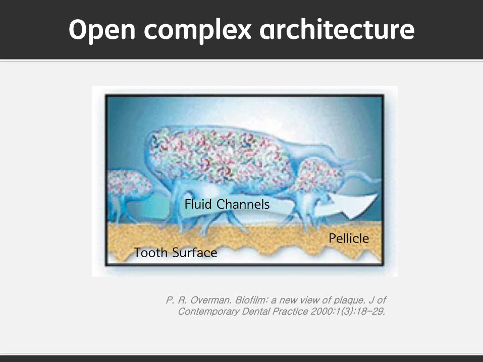
Open complex architecture
Tooth Surface Pellicle
Fluid Channels
P. R. Overman. Biofilm: a new view of plaque. J of Contemporary Dental Practice 2000:1(3):18-29.

우식학에 있어서 우식유발 균주에 대한 패러다임의 변화

Specific pathogens identified for many
dieases
Seardch begins for oral pathogens in
plaque
Non-specific plaque
hypothesis
Diseases linked to
constitutional defects
Specific plaque hypothesis
Treatment aimed at
causative agent
B I O F L M
The Golden Age of Micro biology
Plaque Control
1880 1900 1960 1930 1990 2000
P. R. Overman. Biofilm: a new view of plaque. J of Contemporary Dental Practice 2000:1(3):18-29.
The Changing Views of Plaque (1880 to 1930)
미생물학의 황금기로 많은 질병에 대한 특정 병원균이 발견됨 plaque 내에서 구강 내 질병의 특정 병원균을 탐색함
Biofilm
Specific pathogens identified for many
dieases
Seardch begins for oral pathogens in
plaque
Non-specific plaque
hypothesis
Diseases linked to
constitutional defects
Specific plaque hypothesis
Treatment aimed at
causative agent
B I O F L M
The Golden Age of Micro biology
Plaque Control
1880 1900 1960 1930 1990 2000
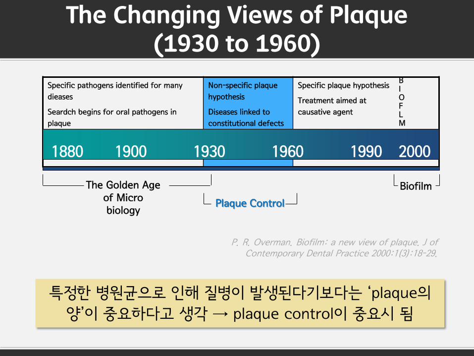
The Changing Views of Plaque (1930 to 1960)
특정한 병원균으로 인해 질병이 발생된다기보다는 ‘plaque의
양’이 중요하다고 생각 → plaque control이 중요시 됨
Biofilm
P. R. Overman. Biofilm: a new view of plaque. J of Contemporary Dental Practice 2000:1(3):18-29.

The Changing Views of Plaque (1960 to 1990)
Specific pathogens identified for many
dieases
Seardch begins for oral pathogens in
plaque
Non-specific plaque
hypothesis
Diseases linked to
constitutional defects
Specific plaque hypothesis
Treatment aimed at
causative agent
B I O F L M
The Golden Age of Micro biology
Plaque Control
1880 1900 1960 1930 1990 2000
치주질환의 원인에 대한 활발한 연구
Spirochete를 분리하는데 성공
Biofilm
P. R. Overman. Biofilm: a new view of plaque. J of Contemporary Dental Practice 2000:1(3):18-29.

The Changing Views of Plaque (1990 to 2000s)
Specific pathogens identified for many
dieases
Seardch begins for oral pathogens in
plaque
Non-specific plaque
hypothesis
Diseases linked to
constitutional defects
Specific plaque hypothesis
Treatment aimed at
causative agent
B I O F L M
The Golden Age of Micro biology
Plaque Control
Biofilm
1880 1900 1960 1930 1990 2000
plaque이 환경과 상호적으로 영향을 주고 받으며 하나의
생태계를 이룸 : Ecological plaque hypothesis
P. R. Overman. Biofilm: a new view of plaque. J of Contemporary Dental Practice 2000:1(3):18-29.
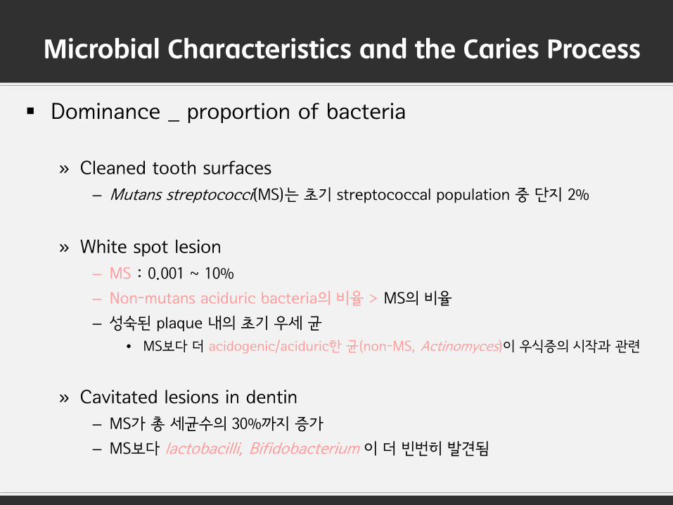
Microbial Characteristics and the Caries Process
Dominance _ proportion of bacteria
» Cleaned tooth surfaces
– Mutans streptococci(MS)는 초기 streptococcal population 중 단지 2%
» White spot lesion
– MS : 0.001 ~ 10%
– Non-mutans aciduric bacteria의 비율 > MS의 비율
– 성숙된 plaque 내의 초기 우세 균
• MS보다 더 acidogenic/aciduric한 균(non-MS, Actinomyces)이 우식증의 시작과 관련
» Cavitated lesions in dentin
– MS가 총 세균수의 30%까지 증가
– MS보다 lactobacilli, Bifidobacterium 이 더 빈번히 발견됨

Mutans streptococci(MS)와 치아우식증 간의 관계가
절대적이지 않음
S.mutans 외에도 다른 균종(Lactobacillus, Bifidobacterium,
Propionibacterium, non-mutans streptococci,
Actinomyces)이 우식 진행 과정에서
중요한 역할을 할 것임을 제안함.

Major Pathogen of Dental Caries, Mutans?

Extended Caries Ecological Hypothesis
J Dent Res 90(3):294-303, 2011
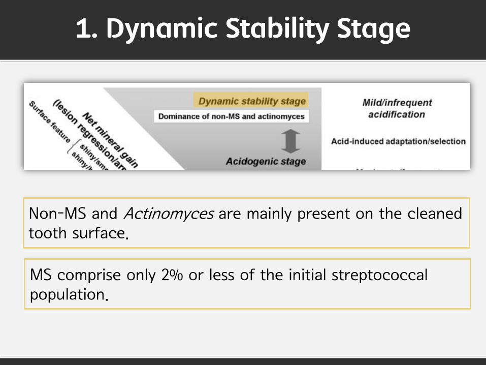
1. Dynamic Stability Stage
Non-MS and Actinomyces are mainly present on the cleaned tooth surface.
MS comprise only 2% or less of the initial streptococcal population.

2. Acidogenic Stage
When the frequency of intake of sugar increases, bacterial metabolism results in the biofilm spending more time at a low pH.
These changes of the microflora may shift the de- /remineralization balance from ‘net mineral gain’ to ‘net mineral loss’ and initiate lesion development.

3. Aciduric Stage
In prolonged acidic environments, more aciduric bacteria
such as MS and lactobacilli may replace the ‘low-pH’ non-
MS bacteria and further accelerate the caries process.

Take Home Message
Dental caries is a classical infectious disease.
Prevention and control of this condition by elimination of a specific bacteria is unwise.
The caries process could be prevented not only by inhibiting the causative bacteria directly but also
by interfering with the environmental changes that drive the deleterious shifts in the composition and
metabolic activity of the biofilm.