Embed Size (px)
Citation preview
181
Polymer(Korea), Vol. 40, No. 2, pp. 181-187 (2016)
http://dx.doi.org/10.7317/pk.2016.40.2.181
ISSN 0379-153X(Print)
ISSN 2234-8077(Online)
조직공학적 인공각막내피를 위한 실크필름의 정제시간이 각막내피세포의
성장에 미치는 영향
이선의·차세롬·장나금·김수영·김은영·송정은·박찬흠*·강길선†
전북대학교, BIN융합공학과, 고분자나노공학과, 고분자융합소재연구소
*한림대학교 의과대학 춘천성심병원 이비인후과
(2015년 8월 18일 접수, 2015년 9월 30일 수정, 2015년 9월 30일 채택)
Effect of Degumming Time of Silk Films on Growth of Corneal Endothelial Cells
for Tissue Engineered Endothelialized Neo-Corneas
Seon Eui Lee, Se Rom Cha, Na Keum Jang, Su Young Kim, Eun Young Kim, Jeong Eun Song,
Chan Hum Park*, and Gilson Khang†
Dept. of BIN Fusion Tech & Dept of PolymerNanoSci Tech, Chonbuk National University, 567 Baekje-daero, Jeonju 54898, Korea
*Dept. of Otorhinolaryngology-Head and Neck Surgery, Chuncheon Sacred Heart Hospital, College of Medicine,
Hallym University, 1-1 Okcheon, Chuncheon, Gangwon 24253, Korea
(Received August 18, 2015; Revised September 30, 2015; Accepted September 30, 2015)
초록: 실크는 생분해성 천연고분자로써 이식 후 낮은 면역 거부반응을 일으킨다. 뿐만 아니라, 실크는 다양한 형태
로 제조할 수 있으며 제조 방법에 따라 수 마이크로 미터 두께로 조정될 수 있다. 필름 형태의 실크 박막은 매우 투
명하며, 높은 수분 및 산소 투과도를 나타낸다. 그러나 실크 단백질인 세리신에 의해 염증반응이 일어난다는 보고가
있으며, 이식 후 분해에 장시간을 필요로 한다. 이 연구에서는 생체 적합성 및 염증반응 등에 세리신 정제시간의 영
향을 알기 위해 다양한 정제시간(0, 20, 40, 60분)을 가진 실크로 필름을 제조한 후 접촉각과 세포 생존율 및 RT-
PCR을 이용한 염증인자발현 실험을 시행하였다. 정제시간에 따라 세리신 함량을 달리한 실크 필름에 각막내피세포
파종 후 필름에서의 세포 부착도와 증식률, 특정 mRNA의 발현을 평가하였다. 그 결과 실크의 정제시간은 각막내
피세포의 성장에 영향을 주었으며 실크필름의 생명공학 신 각막으로써의 가능성을 보여주었다.
Abstract: Silk is a biodegradable natural polymer with low immunological rejection after transplantation. As well, silk
can be manufactured in various forms and adjusted to the thickness of several micrometers depending on the man-
ufacturing method. Silk shows high transparency in the form of film, high water and oxygen permeability. However, stud-
ies have cited some drawbacks of silk, such as inflammatory response due to presence of silk protein (sericin) and its
another disadvantage of silk requires a long period of time to decomposition after transplantation. In this study, silk films
with various degumming time (0, 20, 40 and 60 min) were fabricated in order to evaluate the effect of sericin in learns
of biocompatibility, inflammatory responses, etc. The experiments such as the contact angle, cell viability and inflam-
matory factor expression using RT-PCR were performed. Corneal endothelial cells were seeded on silk films and exam-
ined to evaluate the degree of adhesion in films, cell’s proliferation and specific mRNA expression. This study showed
degumming time of silk film fabrication which is a factor for growth of corneal endothelial cells.
Keywords: silk, sericine, film, degumming time, corneal endothelial cells.
Introduction
Silk is a natural protein composed of fibroin and sericin. Silk
is a biodegradable natural polymer with the characteristics of
bioresorbable, supporting cell growth and good mechanical
properties.1-4 The fibroin is a structured protein and a fibrous
protein. The fibroin with β-sheet structure is useful for appli-
cation to many biomedical and biotechnology as biomaterials
because of the ability of biodegradable, good mechanical
strength with little immune response.5,6 The sericin is a sticky
protein that surrounds the fibroin fibers. The sericin protects
damage, microbial decomposition and digestion of fibroin.7
†To whom correspondence should be addressed.E-mail: [email protected]
©2016 The Polymer Society of Korea. All rights reserved.
182 S. E. Lee et al.
폴리머, 제40권 제2호, 2016년
Degumming is the pre-treatment process of obtaining only
fibroin fiber structure by removing the sericin.8 According to
the related study of degumming, there was a change in the
characteristics of silk fibers such as molecular weight, vis-
cosity of the fibroin, packing density and tensile strength.9 The
cause of these changes is molecular changes, bonding break-
age and structural changes due to the removal of sericin. In
addition, hydrophilic sericin reduces the efficiency of stress
transfer at the fiber-matrix composite formation and results in
the effect to prevent binding of polymer surface to reduce
mechanical properties of composite substantially.10,11
Corneal transplantation is the most common transplant sur-
gery to replace a healthy donated cornea to improve vision
problems caused by a damaged cornea.12-14 However, corneal
transplantation has limitations due to the shortage of healthy
cornea that can be implanted.15 Therefore the development of
suitable scaffolds for regeneration of corneal endothelial cells
could be an alternative that overcomes the limitations of insuf-
ficient supply healthy donated cornea.16-18
In this study, we prepared films by using silk with a unique
mechanical properties, and biocompatibility for regeneration of
corneal endothelium.19,20 On the basis of reproduction of dam-
aged corneal endothelium to in vitro studies, we conducted to
evaluate the effect of different sericin contents to inflammation
and growth of corneal endothelial cells.21
Experimental
Materials. Four types of silk fibroin from silkworm cocoons
(Kyebong Farm, Korea) were used: Different sericin contents
on degumming process, boiling time was varied with 20, 40
and 60 min. The cocoons were cut into small pieces and boiled
in 2 L of distilled water containing 0.02 M of Na2CO3 (Showa
Chemical, Japan) to degumming treatments. 7 g of cocoons
were heated in a hot water bath for 20, 40 and 60 min, respec-
tively. Raw silk fibers were referred to as 0 min. Afterward
hot water treated fibers were washed with cold distilled water
and dried at 60 oC for 24 h. Dried silk fibroin was dissolved in
9.3 M LiBr (Kanto Chem., Japan) at 60 oC for 3 h. To remove
residual LiBr, the solution was dialyzed using dialysis tube
(SnakeSkin® Dialysis Tubing 3500 MWCO, Thermo Science,
USA) during 62 h. After dialyzation, the solution was lyo-
philized by freeze drying at -60 oC, 5 mTorr for 5 days. After
degumming, silk films were prepared using solvent evapo-
ration method. The 100 mg of silk was dissolved in 3 mL of
hexafluoroisopropanol (HFIP, Sigma, USA) at 60 oC for 3 h.
After all of the solution was poured into a glass dish (50 mm
in diameter), dried films were immersed in 99.5% methanol
(Samchun chemical, Korea) to make a semi-crystallized water-
insoluble film at room temperature for 1 h.22
Cell Culture. This study used NIH/3T3 (mouse embryo
fibroblast cell line, KCLB21658) (Korean Cell Line Bank,
Korea). The NIH/3T3 cells were cultured in RPMI 1640
(Rosewell Park Memorial Institute Medium) containing 10%
fetal bovine serum (FBS, Gibco, USA), and 1% antibiotics PS
(100 units/mL Penicillin and 100 μg/mL streptomycin).23 The
RAW 264.7 cells (Mouse leukaemic monocyte macrophage
cell line, KCLB40071) (Korean Cell Line Bank, Korea) were
cultured in DMEM (Dulbecco’s Modified Eagle Medium,
High Glucose, Gibco) containing 10% fetal bovine serum
(FBS, Gibco, USA), and 1% antibiotics (100 units/mL Pen-
icillin and 100 μg/mL streptomycin).24 Corneal endothelial
cells (rCEnCs) were collected from a New Zealand white rab-
bit (4 weeks old female, Hanil LabAnimal Co., Seoul, Korea).
Rabbit eyeballs were extracted, then washed with PBS 1X
containing 1.5% PS. After gaining cut cornea from eyeballs,
Descemet’s membrane (DM) with corneal endothelium was
digested with 0.3% collagenase type A (Roche) for 1 day.
Then, cells were cultured in EGM-2 (Endothelial Cell Growth
Medium, Lonza, USA) with 10% FBS containing 1% PS.25
The medium was changed every 3 days and all cells were cul-
tured in an incubator at 37 oC with 5% CO2 conditions.
Scanning Electron Microscopy (SEM). In order to observe
the morphology of silk fibers and surface of films with various
contents of sericin, scanning electron microscopy measure-
ment (SEM; Hitachi Co. Model S-2250N, Japan) was carried
out. Films were gold coated using plasma sputter (Emscope,
SC500L, UK) under argon gas.
Optical Transparency Test. Transparency of silk fibroin
films was evaluated by visual observation and spectrum anal-
ysis using a SYNERGY Mx spectrophotometer (BioTek®,
USA) at a wavelength range from 380 to 780 nm. Samples of
transparency were prepared without/with cells on films and
measured.26
Fourier Transformed Infrared Spectroscopy (FTIR).
The physicochemical characterization of silk films was eval-
uated by Fourier transformed infrared spectroscopy (Perkin
Elmer, USA). The samples were analyzed in the spectra range
of wave numbers from 500 to 4000 cm-1.
Contact Angle Measurement. To evaluate wettability of
fabricated silk films, contact angle expressed from 2 μL water
droplet volume on random area of specimens was measured by
Effect of Degumming Time of Silk Films on Growth of Corneal Endothelial Cells 183
Polymer(Korea), Vol. 40, No. 2, 2016
contact angle goniometer (CAM-PLUS, ChemInstrument,
USA) at 0, 1, 2, 3, 4 and 5 min. Average of measured value
was used.
Measurement of Cell Viability. Viability were observed by
MTT (3-(4,5-dimethylthiazole-2-yl)-2,5-diphenyltetrazolium-
bromide, Sigma) assay. The cells were seeded on 24-well plate
at a density of 1×10⁴ cells/plate. 1, 3, and 5 days after the
medium of all samples were removed and 100 μL of MTT
solution was added to samples for 4 h at 37 oC, 5% of CO2.
The samples were dissolved in 1 mL of dimethylsulfoxide
(DMSO, Sigma) until the violet-color crystal was totally dis-
solved in solution. The solution was pipetted into a 96-well
plate and absorbance was measured using an ELISA reader
(Emax, Molecular Device, USA) in the wavelength at 570 nm
to yield absorbance as a function of viable cell number.27
Reverse Transcriptase Polymerase Chain Reaction
(RT-PCR) Analysis. After seeding of RAW 264.7 cells in 24-
well plates with a density of 1×10⁴ cells/plate, the mRNA such
as GAPDH, TNF-α, COX-2, IL-1β and IL-6 were confirmed
in order to determine the inflammatory cytokines.28 Then the
culture medium was removed and total RNA was extracted
from the cells using 500 μL of RNAiso Plus (Takara, Japan)
and chloroform (1/5 of RNAiso Plus). After centrifugation at
4 oC, 12000 rpm for 15 min, the supernatants were precipitated
with iso-propanol (Sigma) and 5 μL of Polyacryl CarrierTM
(Molecular Res Center, USA). Each primer was extended
using TOPscriptTM One-step RT PCR DryMIX (Enzynomics,
Korea). All PCR products were separated via electrophoresis
on 1% (w/v) agarose gel containing EtBr (Ethidium Bromide,
Sigma) and visualized under UV light (Vilber Lourmat ETX-
20.M, France) at 360 nm.29 And to confirm the phenotype
maintenance of rCEnCs from a genetic perspective, RT-PCR
conducted based on previous steps. The primers related
rCEnCs (β-actin, Aquaporin-1, Na+/K+ ATPase and VDAC 3)
were purchased from Genotec (Korea).
Proliferation Assay of Corneal Endothelial Cells. To
study how well cells grow on films, cultured rabbit CEnCs
were seeded on fabricated each silk films with a density of
2×10⁴ cells/plate. To evaluate the initial attachment, the sam-
ples were fixed with 10% neutral formalin for 20 min after 30
min of seeding. And cells stained with DAPI (Santa Cruz Bio-
technology, USA). Images were obtained with a fluorescence
microscope and nuclear number was counted (cells/mm2)
using Image J program. At 1, 3, and 5 days after seeding, cell
proliferation was tested by MTT assay (3-(4,5-dimethylthi-
azol-2-yl)-2,5-diphenyl-tetrazolium bromide, Sigma, USA).30
Statistical Analysis. Statistical analysis was carried out
using the student's t-test. Differences were considered sig-
nificant when p*<0.05, p**<0.005 and p***<0.001.
Results and Discussion
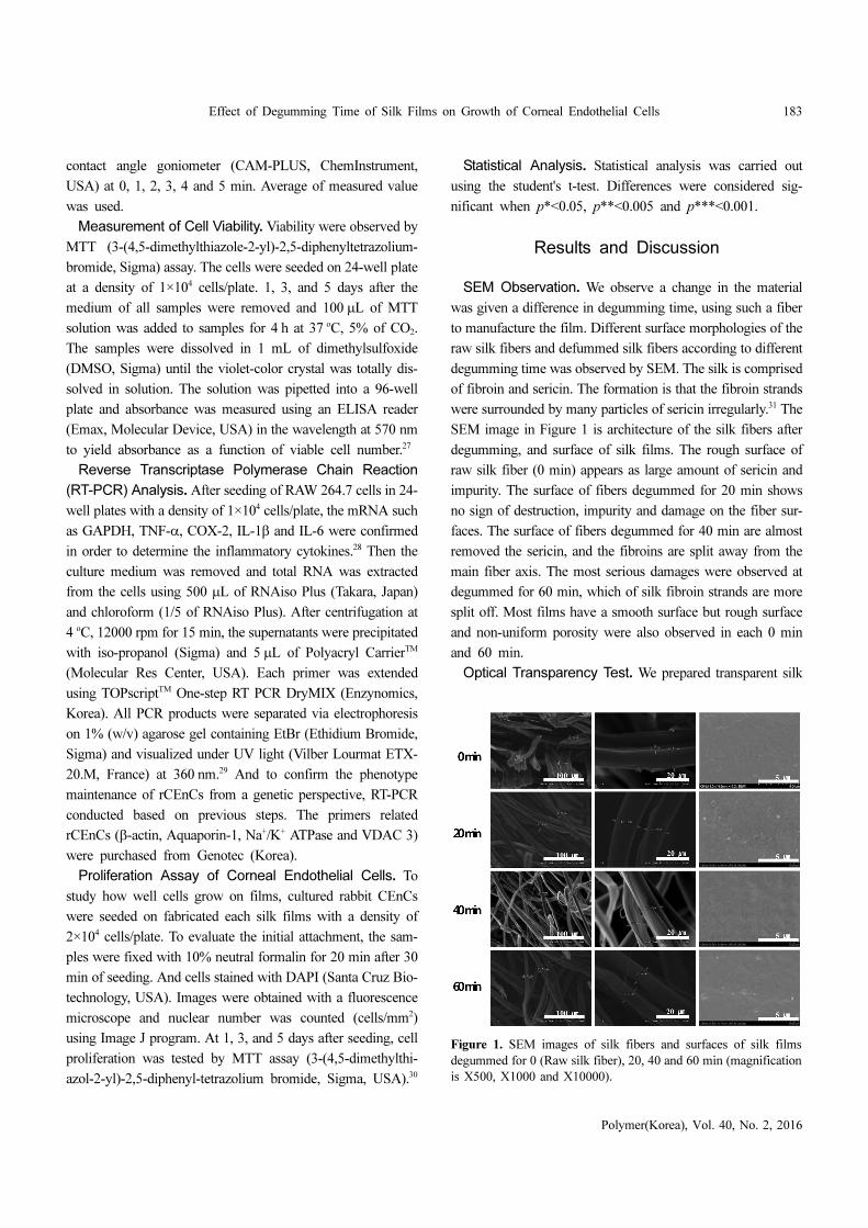
SEM Observation. We observe a change in the material
was given a difference in degumming time, using such a fiber
to manufacture the film. Different surface morphologies of the
raw silk fibers and defummed silk fibers according to different
degumming time was observed by SEM. The silk is comprised
of fibroin and sericin. The formation is that the fibroin strands
were surrounded by many particles of sericin irregularly.31 The
SEM image in Figure 1 is architecture of the silk fibers after
degumming, and surface of silk films. The rough surface of
raw silk fiber (0 min) appears as large amount of sericin and
impurity. The surface of fibers degummed for 20 min shows
no sign of destruction, impurity and damage on the fiber sur-
faces. The surface of fibers degummed for 40 min are almost
removed the sericin, and the fibroins are split away from the
main fiber axis. The most serious damages were observed at
degummed for 60 min, which of silk fibroin strands are more
split off. Most films have a smooth surface but rough surface
and non-uniform porosity were also observed in each 0 min
and 60 min.
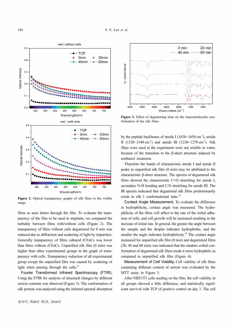
Optical Transparency Test. We prepared transparent silk
Figure 1. SEM images of silk fibers and surfaces of silk films
degummed for 0 (Raw silk fiber), 20, 40 and 60 min (magnification
is X500, X1000 and X10000).
184 S. E. Lee et al.
폴리머, 제40권 제2호, 2016년
films as seen letters through the film. To evaluate the trans-
parency of the film to be used in implants, we compared the
turbidity between films with/without cells (Figure 2). The
transparency of films without cells degummed for 0 min was
reduced due to diffraction and scattering of light by impurities.
Generally transparency of films cultured rCEnCs was lower
than films without rCEnCs. Unpurified silk film (0 min) was
higher than other experimental groups in the graph of trans-
parency with cells. Transparency reduction of all experimental
group except the unpurified film was caused by scattering of
light when passing through the cells.32
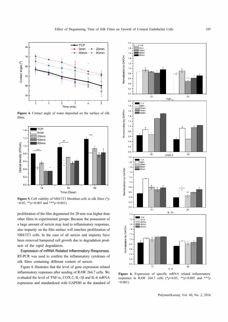
Fourier Transformed Infrared Spectroscopy (FTIR).
Using the FTIR for analysis of structural changes by different
sericin contents was observed (Figure 3). The conformation of
silk protein was analyzed using the infrared spectral absorption
by the peptide backbones of amide I (1630~1650 cm-1), amide
II (1520~1540 cm-1) and amide III (1230~1270 cm-1). Silk
films were used in the experiment were not soluble in water,
because of the transition to the β-sheet structure induced by
methanol treatment.
Therefore the bands of characteristic amide I and amide II
peaks in unpurified silk film (0 min) may be attributed to the
characteristic β-sheet structure. The spectra of degummed silk
films showed the characteristic C=O stretching for amide I,
secondary N-H bending and C-N stretching for amide III. The
IR spectra indicated that degummed silk films predominantly
exists in silk I conformational state.33
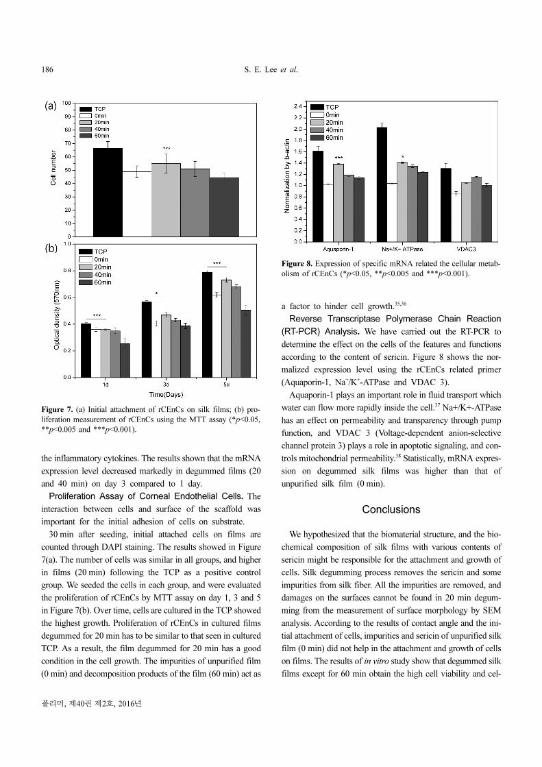
Contact Angle Measurement. To evaluate the difference
in hydrophilicity, contact angle was measured. The hydro-
philicity of the films will affect to the rate of the initial adhe-
sion of cells, and cell growth will be increased resulting in the
increase of initial rate. In general, the greater the angle between
the sample and the droplet indicates hydrophobic, and the
smaller the angle indicates hydrophilicity.34 The contact angle
measured for unpurified silk film (0 min) and degummed films
(20, 40 and 60 min) was indicated that the random coiled con-
formation of degummed silk films made it more hydrophilic as
compared to unpurified silk film (Figure 4).
Measurement of Cell Viability. Cell viability of silk films
containing different content of sericin was evaluated by the
MTT assay in Figure 5.
After NIH/3T3 cells seeding on the film, the cell viability in
all groups showed a little difference, and statistically signif-
icant survival with TCP of positive control on day 3. The cell
Figure 2. Optical transparency graphs of silk films in the visible
range.
Figure 3. Effect of degumming time on the macromolecular con-
formation of the silk films.
Effect of Degumming Time of Silk Films on Growth of Corneal Endothelial Cells 185
Polymer(Korea), Vol. 40, No. 2, 2016
proliferation of the film degummed for 20 min was higher than
other films in experimental groups. Because the possession of
a large amount of sericin may lead to inflammatory responses,
also impurity on the film surface will interfere proliferation of
NIH/3T3 cells. In the case of all sericin and impurity have
been removed hampered cell growth due to degradation prod-
ucts of the rapid degradation.
Expression of mRNA Related Inflammatory Responses.
RT-PCR was used to confirm the inflammatory cytokines of
silk films containing different content of sericin.
Figure 6 illustrates that the level of gene expression related
inflammatory responses after seeding of RAW 264.7 cells. We
evaluated the level of TNF-α, COX-2, IL-1β and IL-6 mRNA
expression and standardized with GAPDH as the standard of
Figure 4. Contact angle of water deposited on the surface of silk
films.
Figure 5. Cell viability of NIH/3T3 fibroblast cells in silk films (*p
<0.05, **p<0.005 and ***p<0.001).
Figure 6. Expression of specific mRNA related inflammatory
responses in RAW 264.7 cells (*p<0.05, **p<0.005 and ***p
<0.001).
186 S. E. Lee et al.
폴리머, 제40권 제2호, 2016년
the inflammatory cytokines. The results shown that the mRNA
expression level decreased markedly in degummed films (20
and 40 min) on day 3 compared to 1 day.
Proliferation Assay of Corneal Endothelial Cells. The
interaction between cells and surface of the scaffold was
important for the initial adhesion of cells on substrate.
30 min after seeding, initial attached cells on films are
counted through DAPI staining. The results showed in Figure
7(a). The number of cells was similar in all groups, and higher
in films (20 min) following the TCP as a positive control
group. We seeded the cells in each group, and were evaluated
the proliferation of rCEnCs by MTT assay on day 1, 3 and 5
in Figure 7(b). Over time, cells are cultured in the TCP showed
the highest growth. Proliferation of rCEnCs in cultured films
degummed for 20 min has to be similar to that seen in cultured
TCP. As a result, the film degummed for 20 min has a good
condition in the cell growth. The impurities of unpurified film
(0 min) and decomposition products of the film (60 min) act as
a factor to hinder cell growth.35,36
Reverse Transcriptase Polymerase Chain Reaction
(RT-PCR) Analysis. We have carried out the RT-PCR to
determine the effect on the cells of the features and functions
according to the content of sericin. Figure 8 shows the nor-
malized expression level using the rCEnCs related primer
(Aquaporin-1, Na+/K+-ATPase and VDAC 3).
Aquaporin-1 plays an important role in fluid transport which
water can flow more rapidly inside the cell.37 Na+/K+-ATPase
has an effect on permeability and transparency through pump
function, and VDAC 3 (Voltage-dependent anion-selective
channel protein 3) plays a role in apoptotic signaling, and con-
trols mitochondrial permeability.38 Statistically, mRNA expres-
sion on degummed silk films was higher than that of
unpurified silk film (0 min).
Conclusions
We hypothesized that the biomaterial structure, and the bio-
chemical composition of silk films with various contents of
sericin might be responsible for the attachment and growth of
cells. Silk degumming process removes the sericin and some
impurities from silk fiber. All the impurities are removed, and
damages on the surfaces cannot be found in 20 min degum-
ming from the measurement of surface morphology by SEM
analysis. According to the results of contact angle and the ini-
tial attachment of cells, impurities and sericin of unpurified silk
film (0 min) did not help in the attachment and growth of cells
on films. The results of in vitro study show that degummed silk
films except for 60 min obtain the high cell viability and cel-
Figure 7. (a) Initial attachment of rCEnCs on silk films; (b) pro-
liferation measurement of rCEnCs using the MTT assay (*p<0.05,
**p<0.005 and ***p<0.001).
Figure 8. Expression of specific mRNA related the cellular metab-
olism of rCEnCs (*p<0.05, **p<0.005 and ***p<0.001).
Effect of Degumming Time of Silk Films on Growth of Corneal Endothelial Cells 187
Polymer(Korea), Vol. 40, No. 2, 2016
lular metabolism. Growth of rCEnCs on degummed (20 and
40 min) silk films was not interfering by the impurities seen in
the unpurified film and degradation products seen in the
degummed film for 60 min, because proper degumming pre-
processing of silk prevents the rapid biodegradation of film.
Therefore, degummed for 20 min silk film has a positive effect
on maintenance of rCEnCs function than unpurified silk film,
and suggests that cell maintain a healthier state.
Acknowledgments: This research was supported by Brain
Korea 21 PLUS Project, the Bio & Medical Technology
Development Program of the National Research Foundation
(NRF) funded by the Korean government (MEST) (NRF-
2012M3A9C6050204).
References
1. Z. Shao and Vollrath, Nature, 418, 741 (2002).
2. Y. Y. Ha, Y. W. Park, H. Y. Kweon, Y. Y. Jo, and S. G. Kim,
Macromol. Res., 22, 9 (2014).
3. B. Panilaitis, G. H. Altman, J. S. Chen, H. J. Jin, V. Karageorgiou,
and D. L. Kaplan, Biomaterials, 24, 3079 (2003).
4. H. L. Kim, H. Yoo, H. J. Park, Y. G. Kim, D. Lee, Y. S. Kang,
and G. Khang, Polym. Korea, 35, 7 (2010).
5. G. H. Altman, F. Diaz, C. Jakuba, T. Calabro, R. L. Horan, J.
Chen, H. Lu, J. Richmond, and D. L. Kaplan, Biomaterials, 24,
401 (2003).
6. M. K. Kim and K. H. Lee, Polym. Korea, 38, 1 (2014).
7. L. Meinel, S. Hofmann, V. Karageorgiou, C. K. Head, J. McCool,
G. Gronowicz, L. Zichner, R. Langer, G. V. Novakovic, and D.
Kaplan, Biomaterials, 26, 147 (2005).
8. H. Oh, M. K. Kim, and K. H. Lee, Macromol. Res., 19, 3 (2011).
9. L. Uebersax, M. Mattotti, M. Papaloizos, P. M. Hans, and B.
Gander, Biomaterials, 28, 4449 (2007).
10. E. P. X. Hu, V. Finley, C. Kue, and D. Kaplan, Macromol. Biosci.,
13, 3 (2013).
11. M. Lee, Y. G. Ko, J. B. Lee, W. H. Park, D. Cho, and O. H.
Kwon, Macromol. Res., 22, 7 (2014).
12. N. C. Joyce, Prog. Retin. Eye Res., 22, 359 (2003).
13. J. Slingsby and S. Forstot, Arch. Ophthalmol., 99, 1041 (1981).
14. M. Terry and P. Ousley, Ophthalmology, 110, 755 (2003).
15. W. Wencan, Y. Mao, Y. Wentao, L. Fan, Q. Jia, W. Qinmei, and
Z. Xianqtian, Curr. Eye Res., 32, 199 (2007).
16. N. Koizumi, Y. Sakamoto, N. Okumura, N. Okagara, H.
Tsuchiya, R. Torii, L. Cooper, U. Ban, H. Tanioka, and S.
Kinoshita, Invest. Iphthalmol. Vis. Sci., 48, 4519 (2007).
17. F. Price and M. Price, J. Cataract. Refract. Surg., 32, 411 (2006).
18. P. Madden, J. Lai, K. George, T. Giovenco, D. Harkin, and T.
Chirila, Biomaterials, 32, 4076 (2011).
19. C. Vepari and D. L. Kaplan, Prog. Polym. Sci., 32, 991 (2007).
20. N. Minoura, M. Tsukada, and M. Nagura, Biomaterials, 11, 430
(1990).
21. T. Wang, I. Wang, S. Chen, Y. Chen, and T. Young, Colloid Surf.
B: Biointerfaces, 90, 236 (2012).
22. E. Y. Kim, C. H. Lee, S. Y. Kim, N. K. Jang, J. E. Song, C. K.
Joo, and G. Khang, Int. J. Tissue Reg., 5, 3 (2014).
23. S. E. Lee, S. R. Cha, N. K. Jang, S. M. Kim, E. Y. Kim, J. E.
Song, C. H. Park, and G. Khang, Int. J. Tissue Reg., 5, 103
(2014).
24. Y. Lee, J. Kwon, G. Khang, and D. Lee, Tissue Eng. Part A, 18,
19 (2012).
25. J. H. Lee, E. Y. Kim, C. J. Lee, C. K. Joo, and G. Khang, Int. J.
Tissue Reg., 4, 2 (2013).
26. H. Yoon, E. Y. Kim, H. Kim, C. H. Park, C. K. Joo, and G.
Khang, Macromol. Res., 22, 3 (2014).
27. S. A. Cho, S. R. Cha, C. M. Kim, E. Y. Kim, J. E. Song, J. Gao,
B. Min, and G. Khang, Int. J. Tissue Reg., 5, 46 (2014).
28. Y. J. Lee, J. Kwon, G. Khang, and D. Lee, Tissue Eng. Part A, 18,
1967 (2012).
29. S. J. Lee, H. Y. Kim, S. J. Kim, J. Y. Yang, S. U. Lee, C. H. Park,
C. K. Joo, and G. Khang, Polym. Korea, 38, 3 (2014).
30. E. Y. Kim, N. Tripathy, J. Y. Park, S. E. Lee, C. K. Joo, and G.
Khang, Macromol. Res., 23, 2 (2015).
31. M. Ho, H. Wang, and K. Lau, Appl. Surf. Sci., 258, 3948 (2012).
32. B. D. Lawrence, M. Cronin-Golomb, I. Georgakoudi, D. L.
Kaplan, and F. G. Omenetto, Biomacromolecules, 9, 1214 (2008).
33. M. Bhattacharjee, E. S. Thater, E. Trella, S. Das, M. Loparic, A.
R. Ray, I. M. Artin, G. C. Spaqnoli, and S. Ghosh, Biomaterials,
34, 8161 (2013).
34. S. W. Park and I. K. Kwon, Int. J. Tissue Reg., 6, 9 (2015).
35. T. Arai, G. Freddi, R. Innocenti, and M. Tsukada, J. Appl. Polym.
Sci., 91, 2383 (2004).
36. Y. Wang, D. D. Rudym, A. Walsh, L. Abrahamsen, H. J. Kim, H.
S. Kim, C. Kriker-Head, and D. L. Kaplan, Biomaterials, 29,
3415 (2008).
37. P. Barfort and D. Maurice, Exp. Eye Res., 19, 11 (1974).
38. S. Hatou, M. Yamada, H. Mochizuki, A. Shiraishi, T. Joko, and
T. Nishida, Curr. Eye Res., 34, 347 (2009).