Embed Size (px)
Citation preview

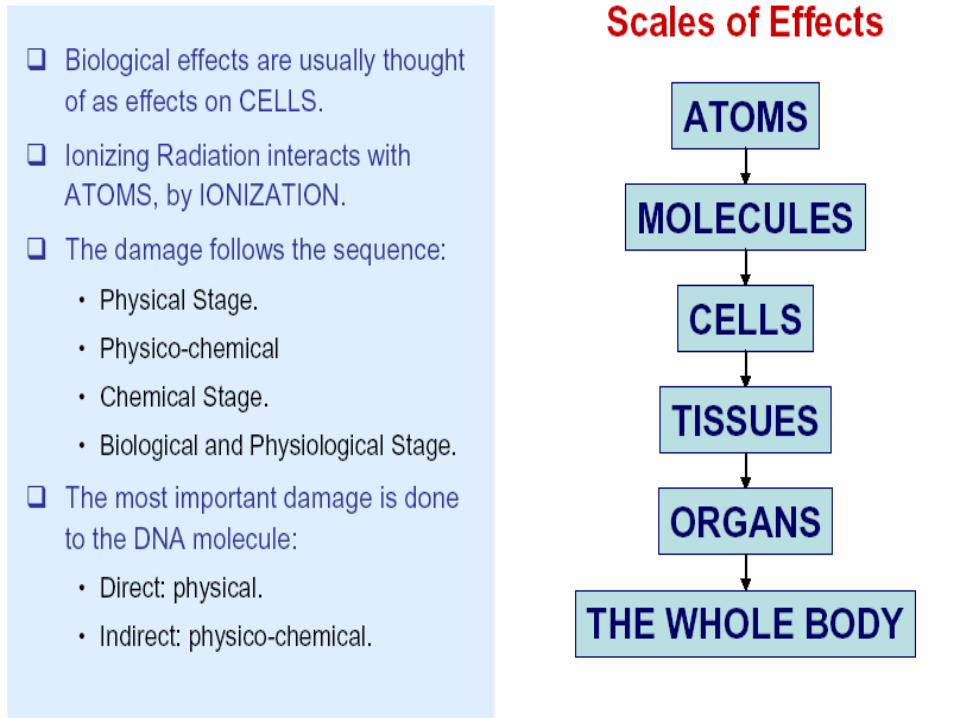
EFFECTS OF RADIATION
S.BAGYARAJTeaching Research AssociateDepartment of ECE,Anna University Chennai
ENERGY
Energy
Energy that is transferred through space .
Examples Radio waves Nuclear radiation (Alpha, Beta, Gamma) X-Radiation
Travels in WavesHigh Speed Particles
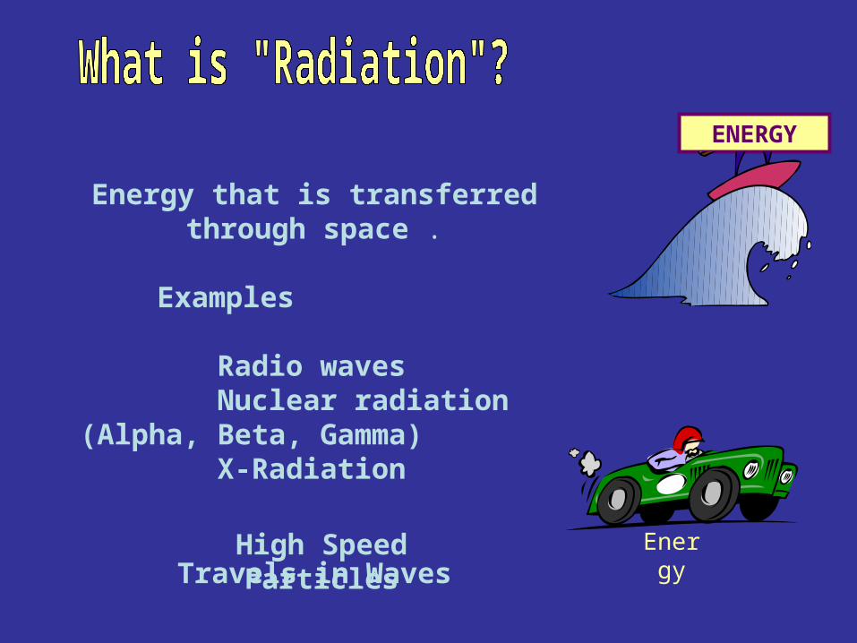
Radon
X-Rays
ConsumerProducts
NuclearPower
RadioactiveWaste
Nuclear Medicine
Solar Radiation Cosmic Rays
TerrestrialRadiation
Food &Drink
Each Other
Radiation in LifeRadiation in Life
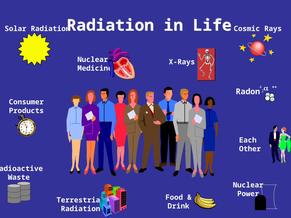
Sources of Annual Radiation Dose
Radon 55%
Cosmic8%
Terrestrial8%
Internal11%
MedicalX-Rays11%
NuclearMedicine4%
ConsumerProducts (3%)
Other (<1%)Occupational 0.3%Fallout <0.3%Nuclear Fuel Cycle 0.1%Miscellaneous 0.1%
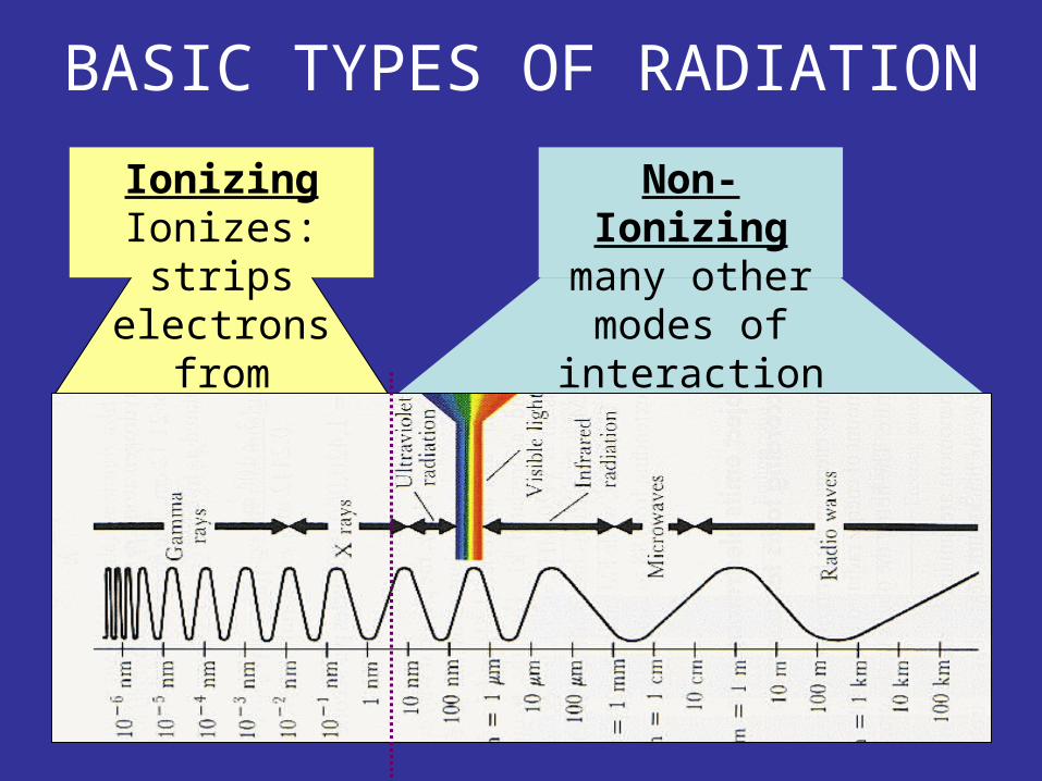
IonizingIonizes:strips
electrons fromatoms
Non-Ionizingmany other modes
of interaction
BASIC TYPES OF RADIATION
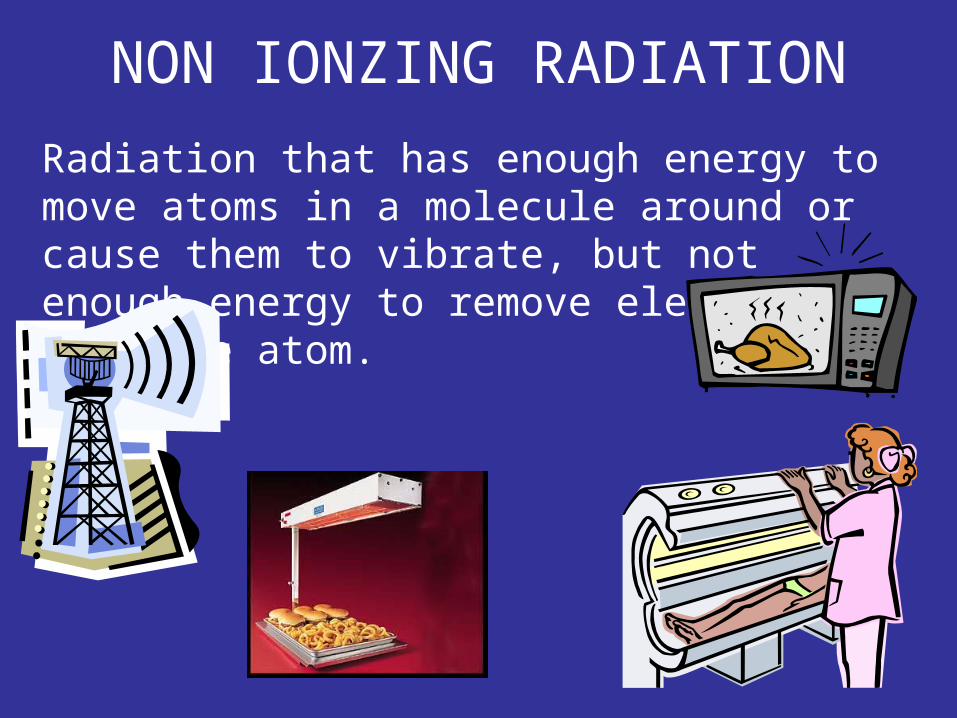
Radiation that has enough energy to move atoms in a molecule around or cause them to vibrate, but not enough energy to remove electrons from the atom.
NON IONZING RADIATION
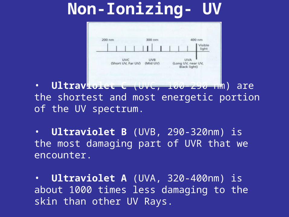
Non-Ionizing- UV
• Ultraviolet C (UVC, 100-290 nm) are the shortest and most energetic portion of the UV spectrum. • Ultraviolet B (UVB, 290-320nm) is the most damaging part of UVR that we encounter. • Ultraviolet A (UVA, 320-400nm) is about 1000 times less damaging to the skin than other UV Rays.
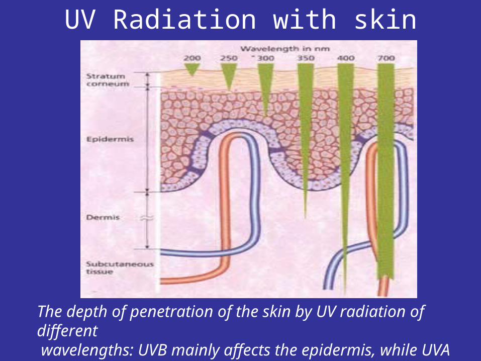
UV Radiation with skin
The depth of penetration of the skin by UV radiation of different wavelengths: UVB mainly affects the epidermis, while UVA penetrates deeper into the dermis.
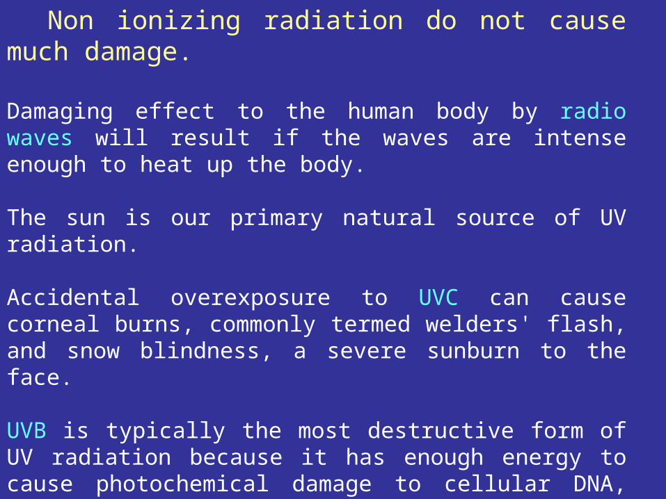
Non ionizing radiation do not cause much damage.
Damaging effect to the human body by radio waves will result if the waves are intense enough to heat up the body. The sun is our primary natural source of UV radiation.
Accidental overexposure to UVC can cause corneal burns, commonly termed welders' flash, and snow blindness, a severe sunburn to the face.
UVB is typically the most destructive form of UV radiation because it has enough energy to cause photochemical damage to cellular DNA, yet not enough to be completely absorbed by the atmosphere. UVB effects can include erythema (sunburn), cataracts, and development of skin cancer.
UVA exposure has an initial pigment-darkening effect (tanning) followed by erythema if the exposure is excessive.
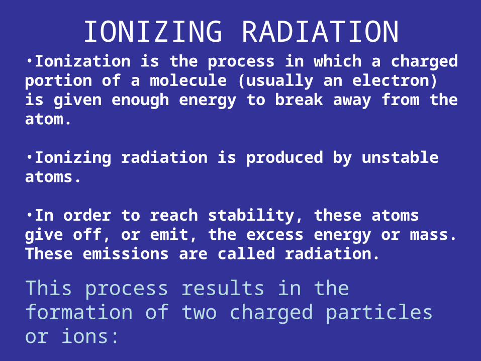
•Ionization is the process in which a charged portion of a molecule (usually an electron) is given enough energy to break away from the atom.
•Ionizing radiation is produced by unstable atoms.
•In order to reach stability, these atoms give off, or emit, the excess energy or mass. These emissions are called radiation.
This process results in the formation of two charged particles or ions:
• molecule with a net positive charge
• free electron with a negative charge
IONIZING RADIATION
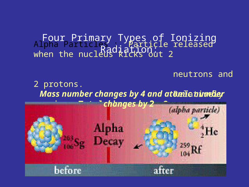
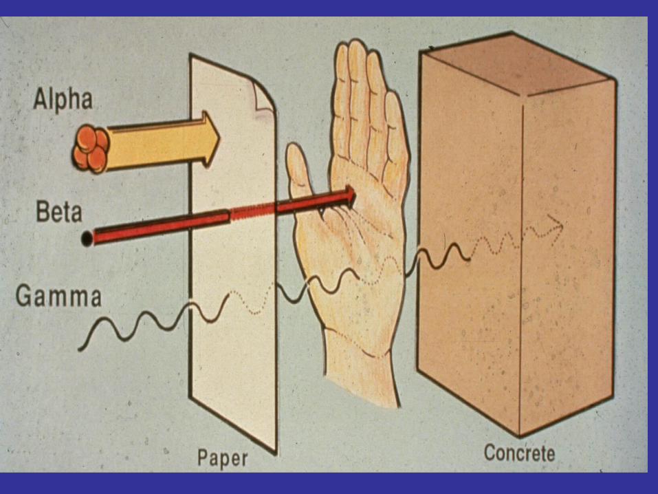
Alpha Particles : Particle released when the nucleus kicks out 2 neutrons and 2 protons. Relatively massive. Total charge of +2. Relatively slow ,They travel short distances . Only a hazard when inhaled
Four Primary Types of Ionizing Radiation:
Mass number changes by 4 and atomic number changes by 2
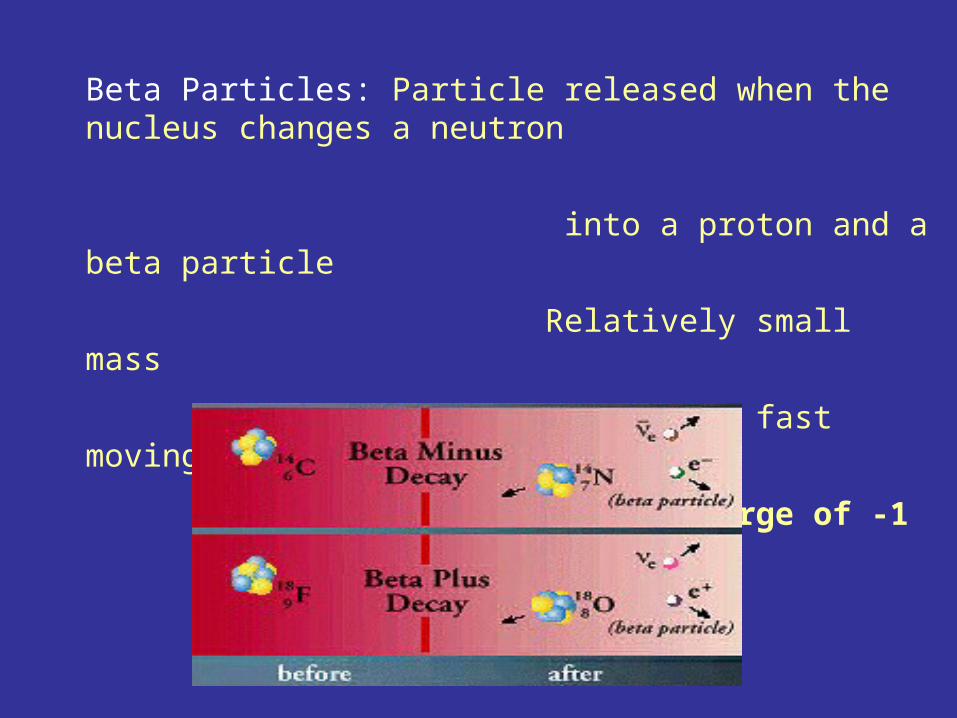
Beta Particles: Particle released when the nucleus changes a neutron
into a proton and a beta particle
Relatively small mass
Relatively fast moving
Total charge of -1
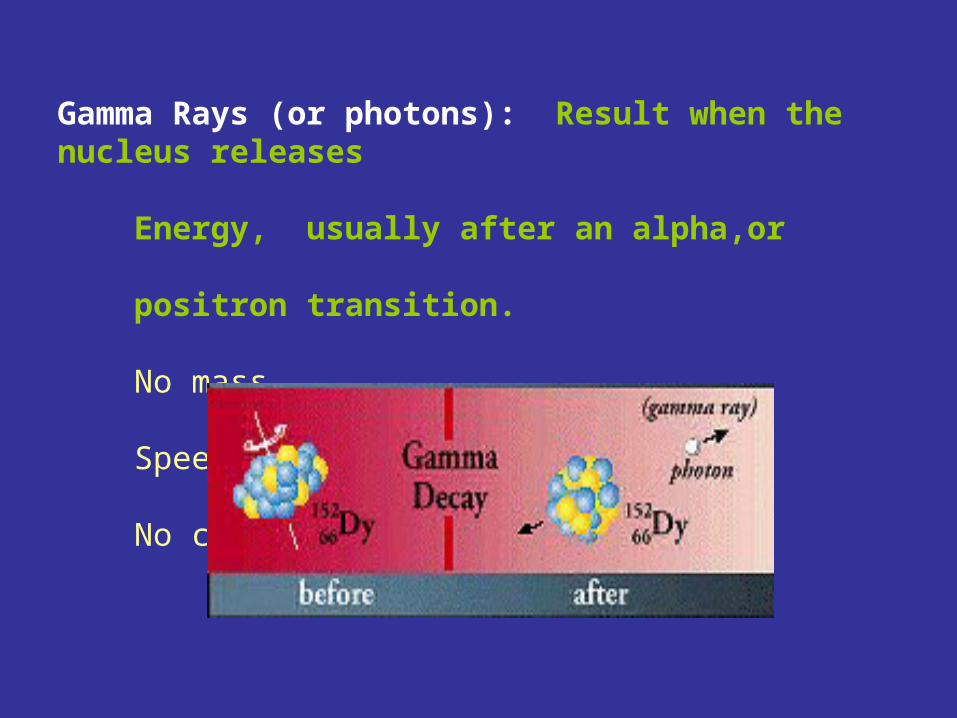
Gamma Rays (or photons): Result when the nucleus releases Energy, usually after an alpha,or positron transition. No mass Speed of light No charge
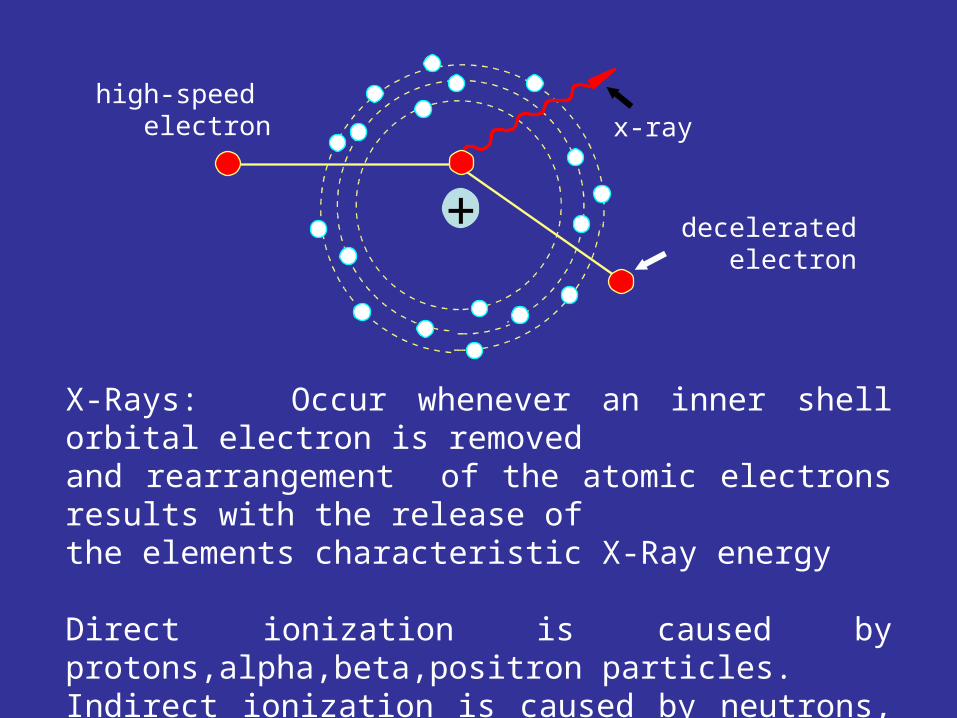
X-Rays: Occur whenever an inner shell orbital electron is removed and rearrangement of the atomic electrons results with the release ofthe elements characteristic X-Ray energy
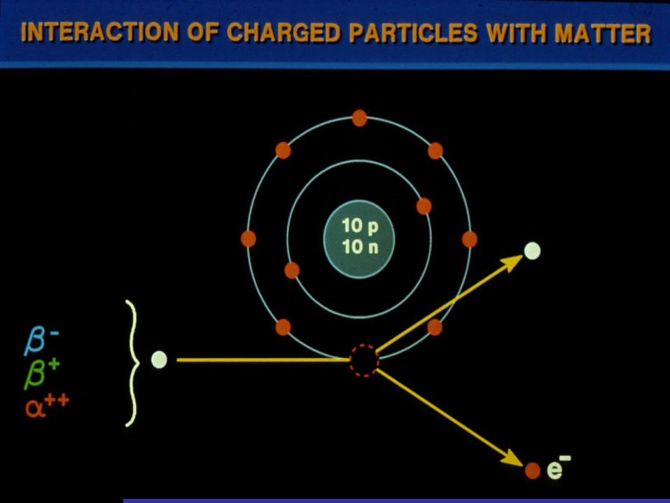
Direct ionization is caused by protons,alpha,beta,positron particles.Indirect ionization is caused by neutrons, gamma and X-rays
decelerated electron
high-speed electron x-ray
+
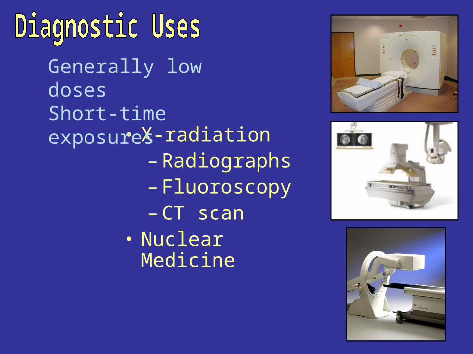
• X-radiation– Radiographs– Fluoroscopy– CT scan
• Nuclear Medicine
Generally low dosesShort-time exposures
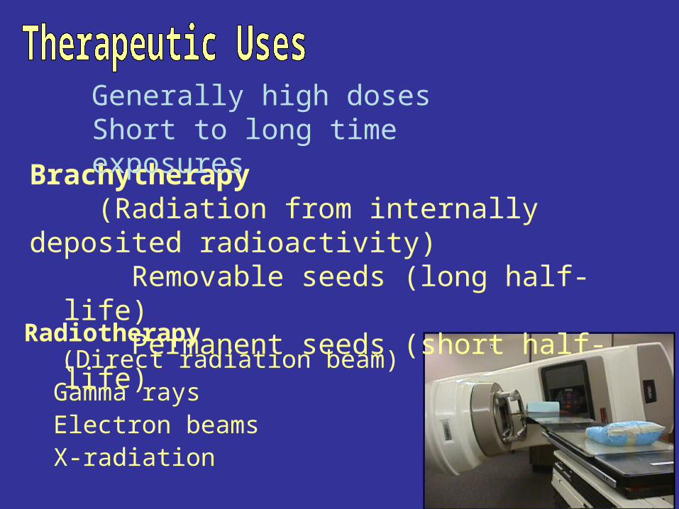
Radiotherapy (Direct radiation beam)Gamma raysElectron beamsX-radiation
Brachytherapy(Radiation from internally deposited radioactivity)
Removable seeds (long half-life)Permanent seeds (short half-life)
Generally high dosesShort to long time exposures
Units of radiation
Pronounced rent’gen with a hard “g”
Limitations– only applies to photons– only applies in air– only applies to energies less than 3 MeV
Rad:Radiation Absorbed Dose
1 rad = 1 Roentgen
oRoentgen : Named after Wilhelm C. Roentgen (thus the abbr... is capital “R”)
Dose in health record is in units of rem
1 rem = 1 Roentgen
Curie (Ci) : Named in honor of Pierre Curie
rem : Roentgen Equivalent Man
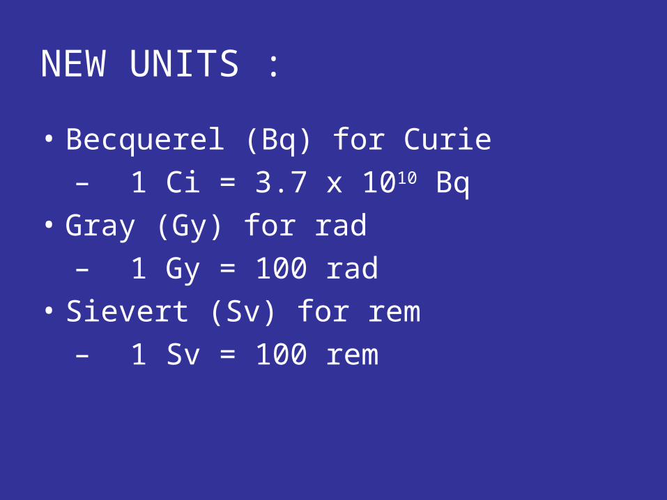
NEW UNITS :
• Becquerel (Bq) for Curie
– 1 Ci = 3.7 x 10¹⁰ Bq
• Gray (Gy) for rad
– 1 Gy = 100 rad
• Sievert (Sv) for rem
– 1 Sv = 100 rem
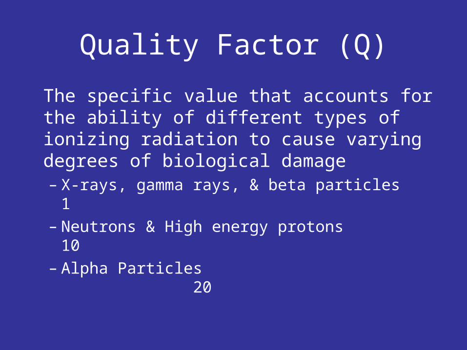
Quality Factor (Q)
The specific value that accounts for the ability of different types of ionizing radiation to cause varying degrees of biological damage– X-rays, gamma rays, & beta particles 1– Neutrons & High energy protons 10– Alpha Particles 20
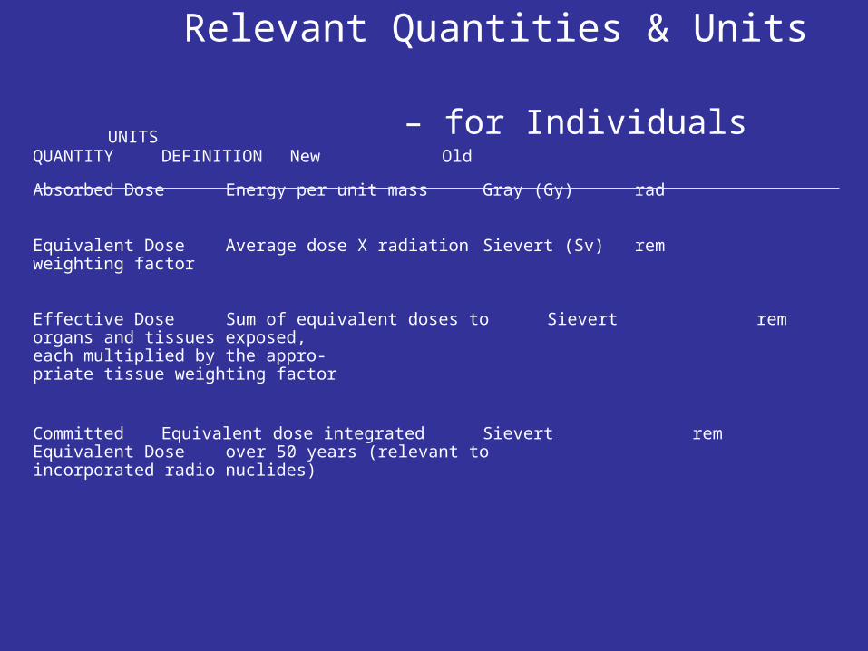
Relevant Quantities & Units – for Individuals
UNITSQUANTITY DEFINITION New Old
Absorbed Dose Energy per unit mass Gray (Gy) rad
Equivalent Dose Average dose X radiation Sievert (Sv) rem weighting factor
Effective Dose Sum of equivalent doses to Sievert remorgans and tissues exposed, each multiplied by the appro-priate tissue weighting factor
Committed Equivalent dose integrated Sievert remEquivalent Dose over 50 years (relevant to
incorporated radio nuclides)
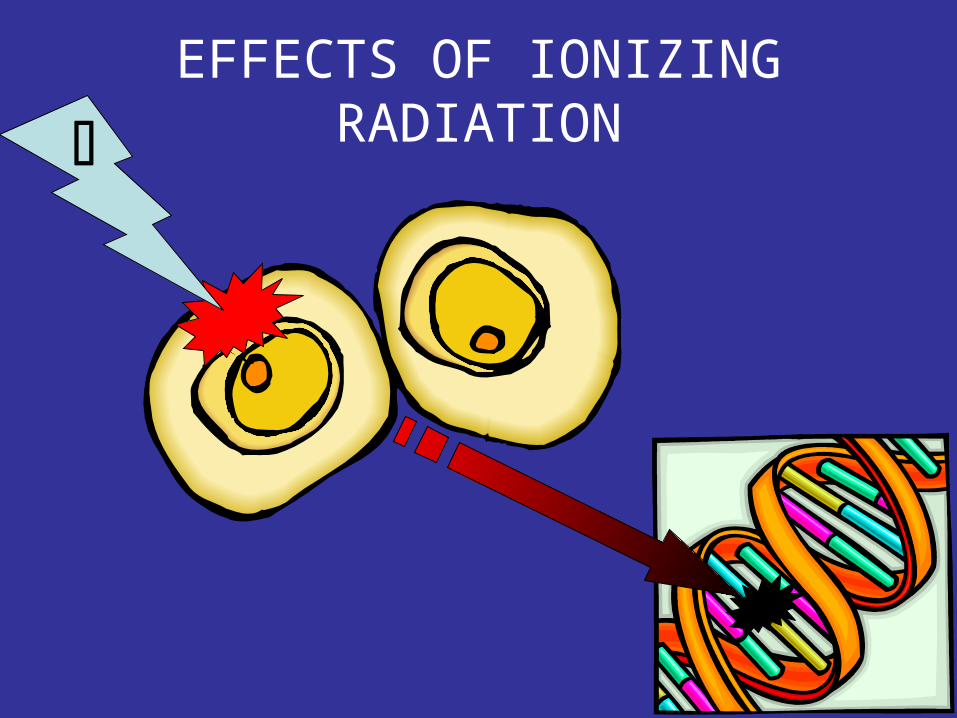
EFFECTS OF IONIZING
RADIATION
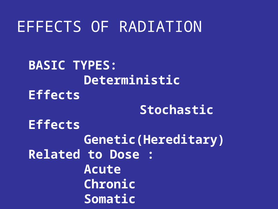
BASIC TYPES: Deterministic Effects Stochastic Effects
Genetic(Hereditary)Related to Dose :
AcuteChronicSomaticIn-utero
EFFECTS OF RADIATION
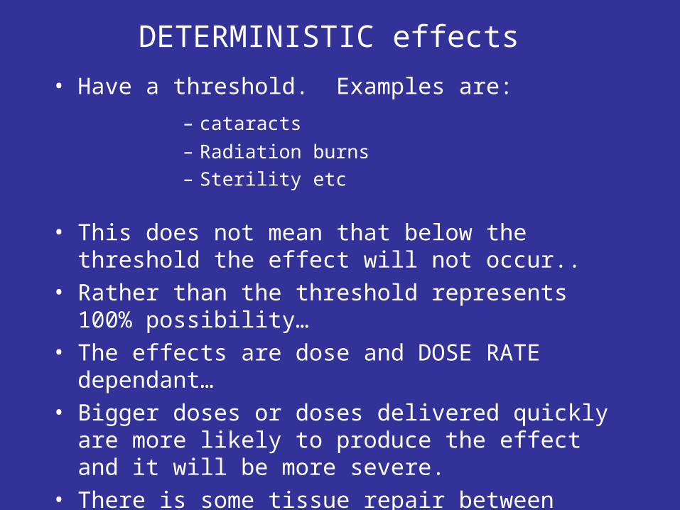
• Have a threshold. Examples are:
DETERMINISTIC effects
– cataracts
– Radiation burns– Sterility etc
• This does not mean that below the threshold the effect will not occur..
• Rather than the threshold represents 100% possibility…
• The effects are dose and DOSE RATE dependant…• Bigger doses or doses delivered quickly are more
likely to produce the effect and it will be more severe.• There is some tissue repair between exposures
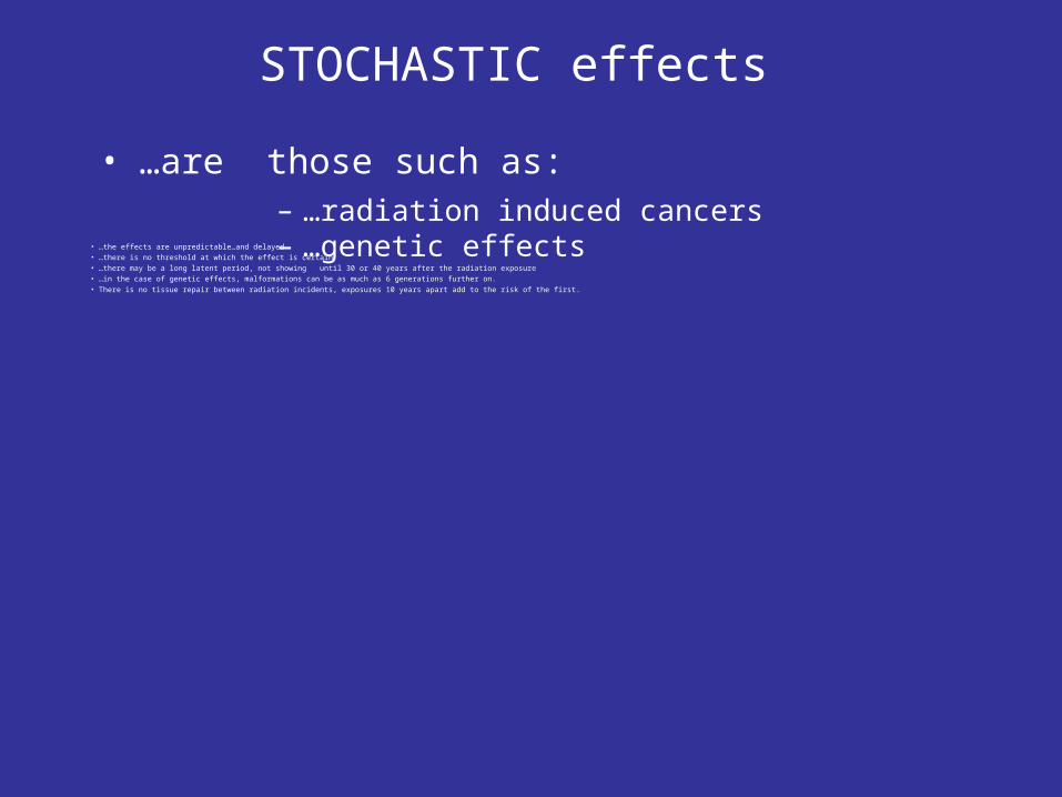
STOCHASTIC effects
• …the effects are unpredictable…and delayed• …there is no threshold at which the effect is certain• …there may be a long latent period, not showing until 30 or 40 years after the radiation exposure• …in the case of genetic effects, malformations can be as much as 6 generations further on.• There is no tissue repair between radiation incidents, exposures 10 years apart add to the risk of the first.
• …are those such as:– …radiation induced cancers – …genetic effects
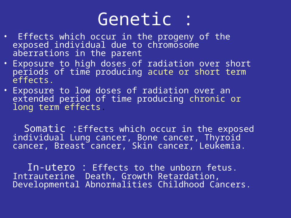
Genetic :• Effects which occur in the progeny of the exposed
individual due to chromosome aberrations in the parent• Exposure to high doses of radiation over short periods of
time producing acute or short term effects. • Exposure to low doses of radiation over an extended
period of time producing chronic or long term effects.
Somatic :Effects which occur in the exposed individual Lung cancer, Bone cancer, Thyroid cancer, Breast cancer, Skin cancer, Leukemia.
In-utero : Effects to the unborn fetus. Intrauterine Death, Growth Retardation, Developmental Abnormalities Childhood Cancers.
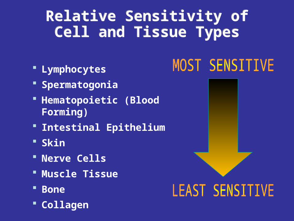
Relative Sensitivity of Cell and Tissue Types Relative Sensitivity of Cell and Tissue Types
Lymphocytes Spermatogonia Hematopoietic (Blood Forming) Intestinal Epithelium Skin Nerve Cells Muscle Tissue Bone Collagen
Symptoms of Radiation Sickness
• Nausea and vomiting • Diarrhoea • Skin burns (redness, blistering) • Weakness, fatigue, exhaustion, fainting • Dehydration • Inflammation of exposed areas (redness, tenderness, swelling, bleeding) • Hair Loss
Symptoms of Radiation Sickness (cont’) • Ulceration of the oral mucosa • Ulceration of the esophagus, stomach or intestines • Vomiting blood • Bloody stool • Bleeding from the nose, mouth, gums, and rectum • Bruising • Sloughing of skin • Open sores on the skin
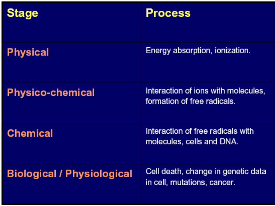
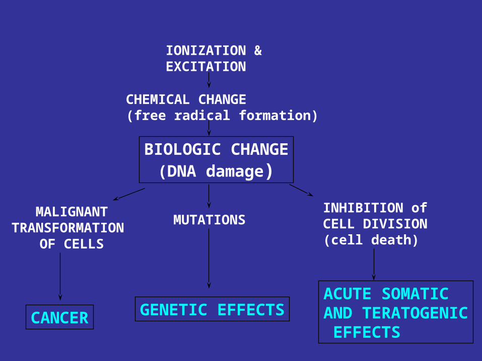
IONIZATION &EXCITATION
CHEMICAL CHANGE(free radical formation)
BIOLOGIC CHANGE(DNA damage)
MALIGNANTTRANSFORMATION
OF CELLS
CANCER
MUTATIONS
GENETIC EFFECTS
INHIBITION of CELL DIVISION(cell death)
ACUTE SOMATICAND TERATOGENIC EFFECTS
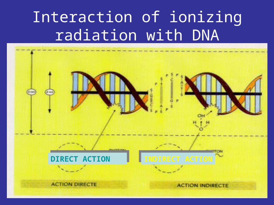
Interaction of ionizing radiation with DNA
DIRECT ACTIONDIRECT ACTION INDIRECT ACTIONINDIRECT ACTION
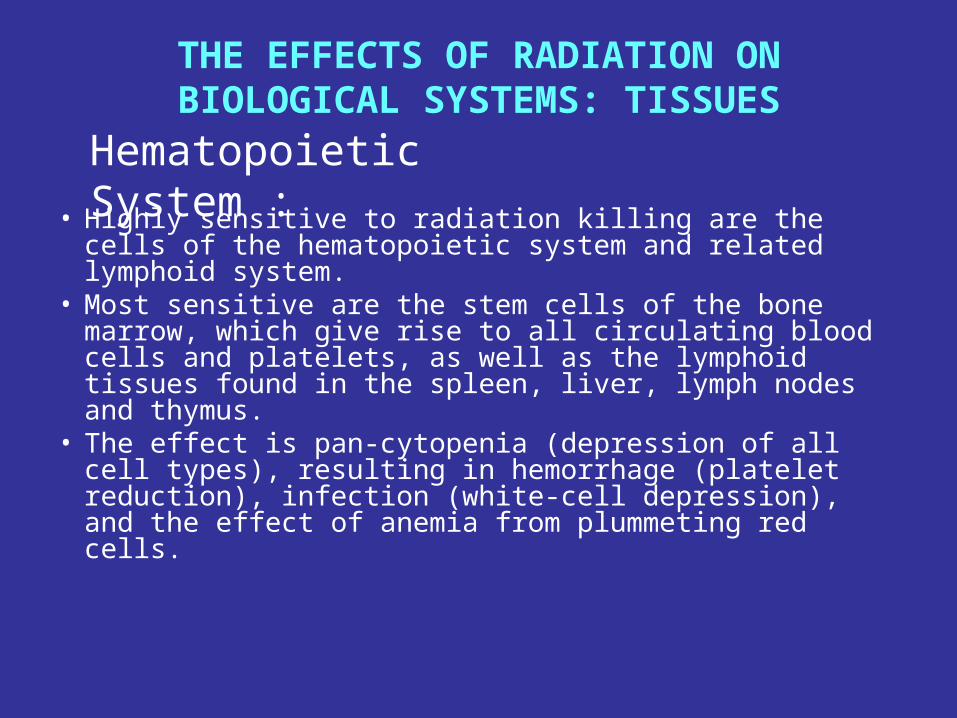
THE EFFECTS OF RADIATION ON BIOLOGICAL SYSTEMS: TISSUES
• Highly sensitive to radiation killing are the cells of the hematopoietic system and related lymphoid system.
• Most sensitive are the stem cells of the bone marrow, which give rise to all circulating blood cells and platelets, as well as the lymphoid tissues found in the spleen, liver, lymph nodes and thymus.
• The effect is pan-cytopenia (depression of all cell types), resulting in hemorrhage (platelet reduction), infection (white-cell depression), and the effect of anemia from plummeting red cells.
Hematopoietic System :
Reproductive System:• The cells of the reproductive system are
highly sensitive to radiation effects• In the human male, stem cells and
proliferating spermatogonia are highly sensitive. However, spermatids and mature sperm show considerable resistance.
• Also resistant are the interstitial cells of the testis, which control hormone production and secondary sexual characteristics.
• Therefore at sterilizing doses of 6 Gy, potency, fluid production of the prostate and seminal vesicles, as well as voice, beard and male social behavior are not affected.
• With a turnover time for spermatogenesis (stem cell to mature sperm) of 64 to 72 days, sterility is never seen immediately after the radiation dose, because mature sperm are resistant to the killing effects of radiation. They can sustain heritable genetic damage, however.
• Doses of about 6 Gy are required to permanently sterilize males (sterility occurs after several months). Although lower doses can also cause sterility after several months, the effect is temporary. Fertility and near-normal sperm counts return after 1 to 2 years.
• Radiation destroys both ovum and maturing follicules. This reduces hormone production. Therefore radiogenic sterility in females can be accompanied by artificial menopause, with significant effects on sexual characteristics and secondary genitalia.
• Total dose, dose rate, and age are important factors in the final effect. Younger women seems better able to recover fertility than do older women.
• A dose of 2 Gy permanently sterilizes women over 40 but causes temporary sterility in women age 35 and under.
• Menopause was caused in 50% of younger women exposed to doses of 1.5 to 5 Gy. Women over 40 showed 90% menopause at 1,5 Gy.
Gastrointestinal System:
• The gastrointestinal (GI) tract is highly sensitive to radiation. Following irradiation, the first changes seen occur in the epithelium lining of the small intestine containing millions of convolutions called villi.
• The crypt cells of the villi are highly proliferative,
supplying cells that continue to differentiate and migrate to the terminal villus. There they eventually slough off into the intestinal contents.
• Radiation causes mitotic arrest of the crypt cells followed by eventual denudation of the villi, ulceration of the wall, and septic infiltration.
• Effects on the large intestine cause functional impairment resulting in fluid and electrolyte loss, and diarrhea.
• Effects on the upper GI tract include vomiting, depression of acid, and pepsin secretion. Destruction of the epithelium lining of the pharynx and esophagus results in dryness, soreness, and petechia (capillary rupture).
Skin:• Skin is relatively radiosensitive.• The radio biologic end-points in skin are dependent
on the total dose, the dose rate, and the radiation quality.
• Radio biologic effects in skin include erythema (skin reddening), and temporary epilation (hair loss).
• At very high doses, permanent epilation and destruction of sub organs, including the vasculature, sebaceous and sweat gland, occur.
• The response of the skin to ionizing radiation is called radiation dermatitis. This effect follows a temporal as well as dose response depending on damage to the sub organs and connective tissue.
Skin responses include:
1. Initial erythema: Redness occurs within days due to capillary dilatation caused by histamine releases. Threshold dose is 2 Gy from beta radiation or 1000 R from x-ray radiation.
2. Dry desquamation. After several days the epidermis scales and peels as a result of reduction in sebaceous and sweat gland secretion, and vascular damage.
3. Erythema proper: After the third or fourth week redness with soreness and burning and edema results. This is caused by obstructive changes in the fine vasculature in the dermis.
4. Moist desquamation: At high doses of 2000 R, blisters form in the epidermis, permanent epilation results and edema with macrophage infiltration occurs. Severe damage to the vasculature and connective tissue is the cause.
5. Necrosis: At very high doses, dermal necrosis may result after erythema proper as a result of dermis destruction, or later because of obstructive changes in arterioles, infection, and subcutaneous-fat-cell destruction.
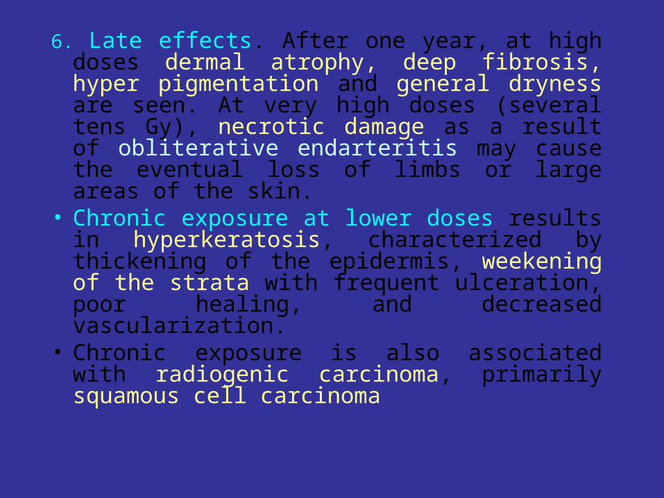
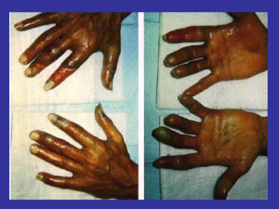
6. Late effects. After one year, at high doses dermal atrophy, deep fibrosis, hyper pigmentation and general dryness are seen. At very high doses (several tens Gy), necrotic damage as a result of obliterative endarteritis may cause the eventual loss of limbs or large areas of the skin.
• Chronic exposure at lower doses results in hyperkeratosis, characterized by thickening of the epidermis, weekening of the strata with frequent ulceration, poor healing, and decreased vascularization.
• Chronic exposure is also associated with radiogenic carcinoma, primarily squamous cell carcinoma
Mucous Membranes:• Mucous membranes are also radiosensitive,
particularly those in the mouth, pharynx, and esophagus.
• After considerable doses, dryness, soreness, and petechial ulceration of the mouth occur within 2 weeks.
• In the third week this progresses to swelling of the tongue with hyper secretion of the mucus, which eventually becomes a thick pseudomembrane that covers the buccal area, throat, and tongue.
• Later, fibrosis, ulceration, and poor vasculature accompanies skin effects.
Central Nervous System
• Generally, the CNS is resistant to radiation effects. • Very high doses are required to cause substantial effects
on the brain and nervous system.• The vasculature is the limiting factor in radiation effects
to the CNS. • Effects on the vessels cause breakdown of the capillary
circulation with rupture of the walls, interstitial edema, meningitis, encephalitis, and the breakdown of the blood-brain barrier.
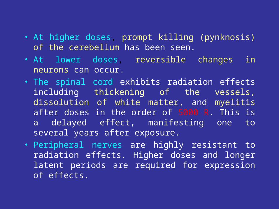
• At higher doses, prompt killing (pynknosis) of the cerebellum has been seen.
• At lower doses, reversible changes in neurons can occur.
• The spinal cord exhibits radiation effects including thickening of the vessels, dissolution of white matter, and myelitis after doses in the order of 5000 R. This is a delayed effect, manifesting one to several years after exposure.
• Peripheral nerves are highly resistant to radiation effects. Higher doses and longer latent periods are required for expression of effects.
The Fetus• Fetal effects are seen at relatively low doses of
radiation.• The fetus is a highly proliferative system with many
undifferentiated cells. Therefore it is extremely sensitive to radiation effects.
• The classic triad of effects of radiation upon the embryo are :
1. Intrauterine growth retardation (IUGR)
2. Embryonic, fetal, or neonatal death
3. Congenital malformation
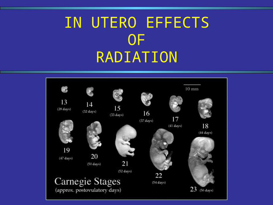
IN UTERO EFFECTSOF
RADIATION
EFFECTS OF RADIATION ACCORDING TO GESTATIONAL STAGE
• Preimplantation - “All or none”
In the human, implantation of the zygote in the wall of the uterus occurs at approximately days 10 to 12 following conception.
Radiation delivered exclusively during this stage may cause prenatal death with failure of implantation; otherwise a normal pregnancy ensues.
• Implantation - Transient Intrauterine Growth Retardation; threshold 10-20 Gy
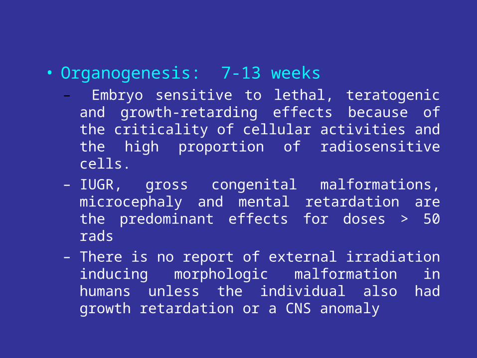
• Organogenesis: 7-13 weeks– Embryo sensitive to lethal, teratogenic and
growth-retarding effects because of the criticality of cellular activities and the high proportion of radiosensitive cells.
– IUGR, gross congenital malformations, microcephaly and mental retardation are the predominant effects for doses > 50 rads
– There is no report of external irradiation inducing morphologic malformation in humans unless the individual also had growth retardation or a CNS anomaly
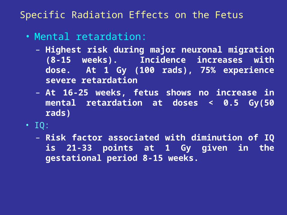
Specific Radiation Effects on the Fetus
• Mental retardation:– Highest risk during major neuronal migration (8-
15 weeks). Incidence increases with dose. At 1 Gy (100 rads), 75% experience severe retardation
– At 16-25 weeks, fetus shows no increase in mental retardation at doses < 0.5 Gy(50 rads)
• IQ:– Risk factor associated with diminution of IQ is
21-33 points at 1 Gy given in the gestational period 8-15 weeks.
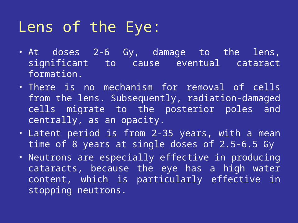
Lens of the Eye:
• At doses 2-6 Gy, damage to the lens, significant to cause eventual cataract formation.
• There is no mechanism for removal of cells from the lens. Subsequently, radiation-damaged cells migrate to the posterior poles and centrally, as an opacity.
• Latent period is from 2-35 years, with a mean time of 8 years at single doses of 2.5-6.5 Gy
• Neutrons are especially effective in producing cataracts, because the eye has a high water content, which is particularly effective in stopping neutrons.
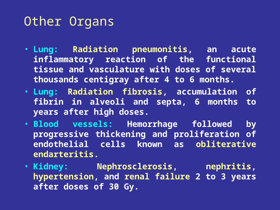
Other Organs
• Lung: Radiation pneumonitis, an acute inflammatory reaction of the functional tissue and vasculature with doses of several thousands centigray after 4 to 6 months.
• Lung: Radiation fibrosis, accumulation of fibrin in alveoli and septa, 6 months to years after high doses.
• Blood vessels: Hemorrhage followed by progressive thickening and proliferation of endothelial cells known as obliterative endarteritis.
• Kidney: Nephrosclerosis, nephritis, hypertension, and renal failure 2 to 3 years after doses of 30 Gy.
Radiation and Cancer
Ionizing radiation is quite efficient at inducing chromosomal aberrations such as
deletions and translocations
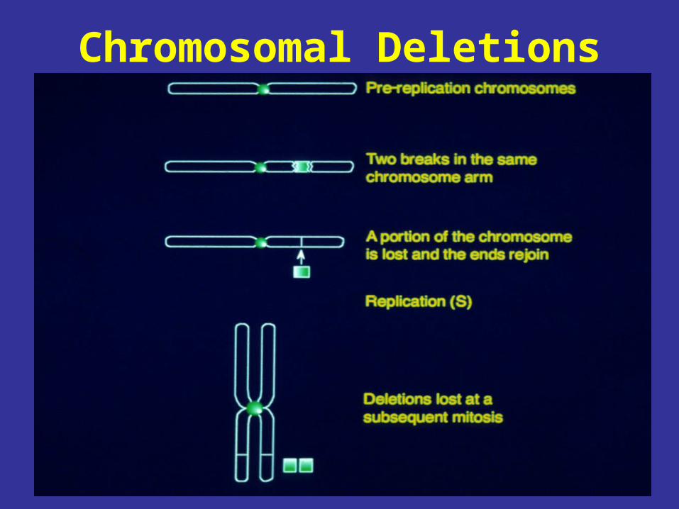
Chromosomal Deletions
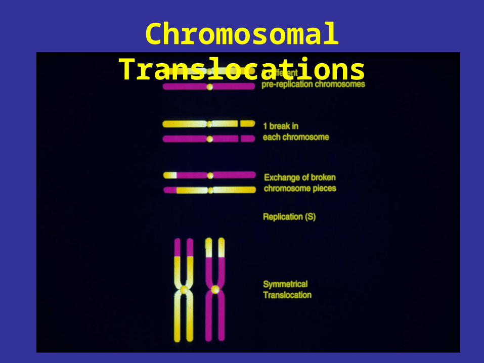
Chromosomal Translocations
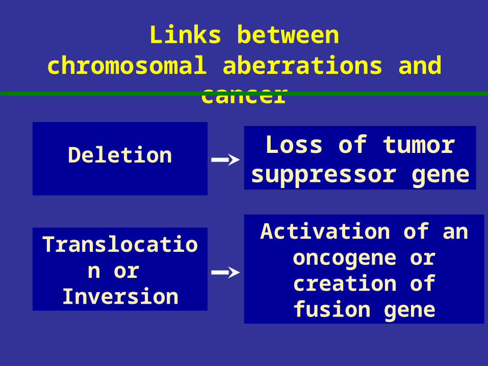
Links betweenchromosomal aberrations and
cancer
Translocation or
Inversion
Loss of tumor suppressor
gene
Activation of an oncogene or
creation of fusion gene
Deletion
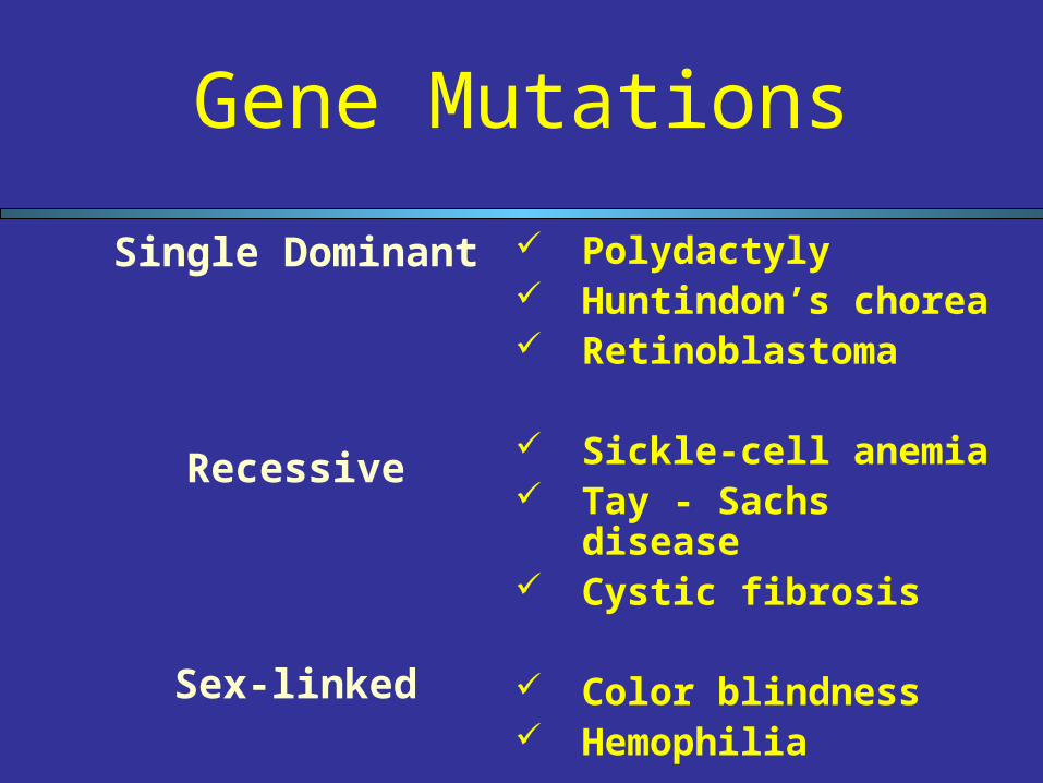
Gene Mutations
Polydactyly Huntindon’s chorea Retinoblastoma
Sickle-cell anemia Tay - Sachs disease Cystic fibrosis
Color blindness Hemophilia
Single Dominant
Recessive
Sex-linked
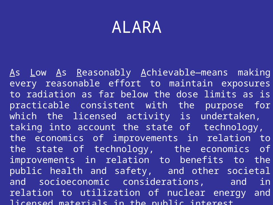
ALARA
As Low As Reasonably Achievable—means making every reasonable effort to maintain exposures to radiation as far below the dose limits as is practicable consistent with the purpose for which the licensed activity is undertaken, taking into account the state of technology, the economics of improvements in relation to the state of technology, the economics of improvements in relation to benefits to the public health and safety, and other societal and socioeconomic considerations, and in relation to utilization of nuclear energy and licensed materials in the public interest.
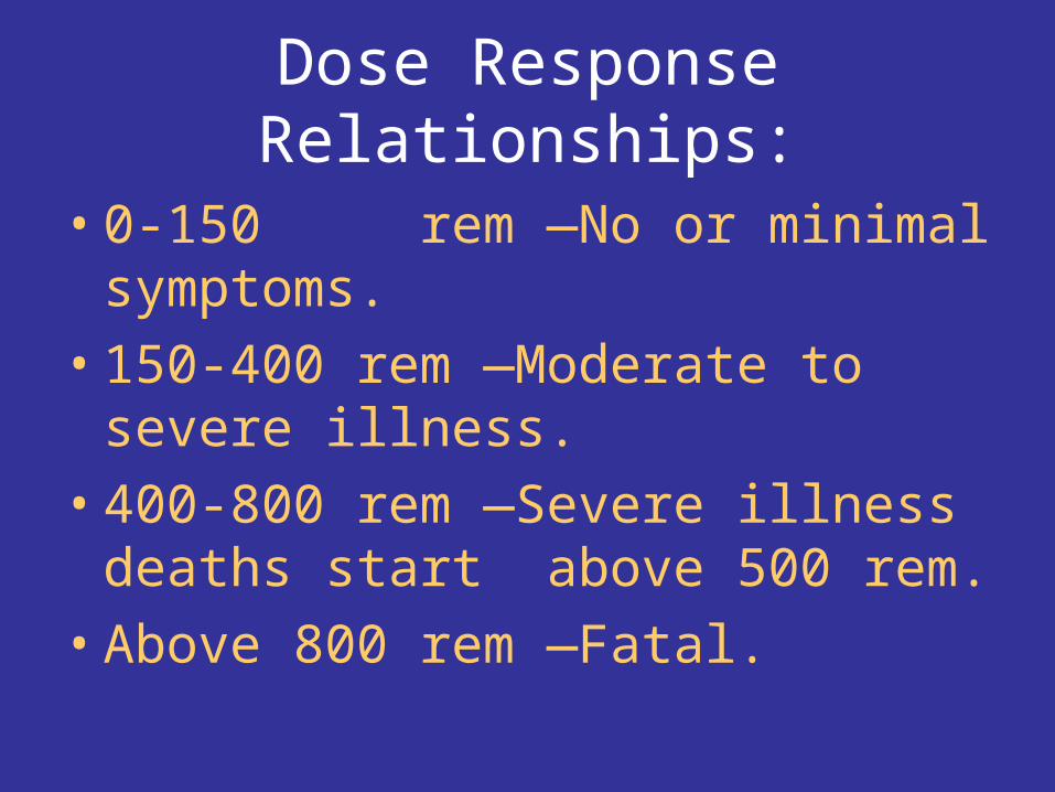
Dose Response Relationships:
• 0-150 rem —No or minimal symptoms.
• 150-400 rem —Moderate to severe illness.
• 400-800 rem —Severe illness deaths start above 500 rem.
• Above 800 rem —Fatal.
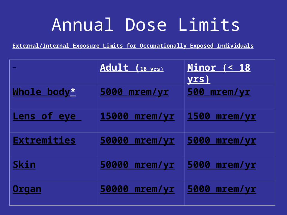
Annual Dose LimitsExternal/Internal Exposure Limits for Occupationally Exposed Individuals
Adult (18 yrs) Minor (< 18 yrs)
Whole body* 5000 mrem/yr 500 mrem/yr
Lens of eye 15000 mrem/yr
1500 mrem/yr
Extremities 50000 mrem/yr
5000 mrem/yr
Skin 50000 mrem/yr
5000 mrem/yr
Organ 50000 mrem/yr
5000 mrem/yr
*Effective dose equivalent
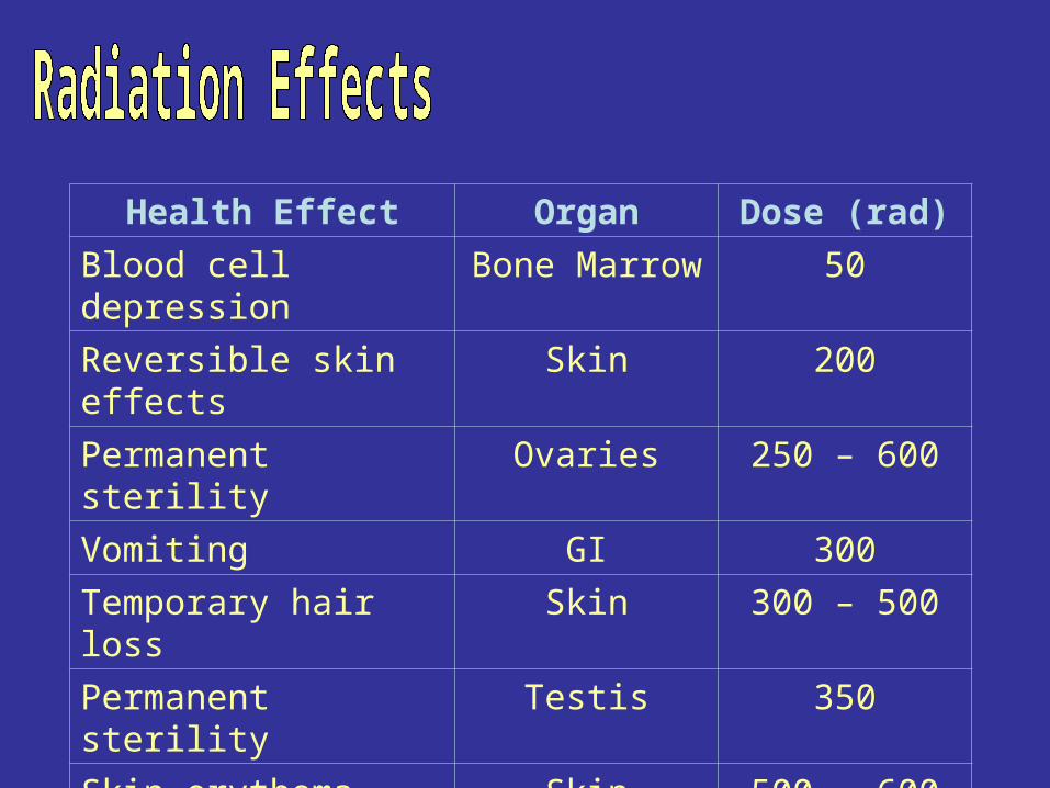
Health Effect Organ Dose (rad)
Blood cell depression Bone Marrow 50
Reversible skin effects Skin 200
Permanent sterility Ovaries 250 – 600
Vomiting GI 300
Temporary hair loss Skin 300 – 500
Permanent sterility Testis 350
Skin erythema Skin 500 – 600
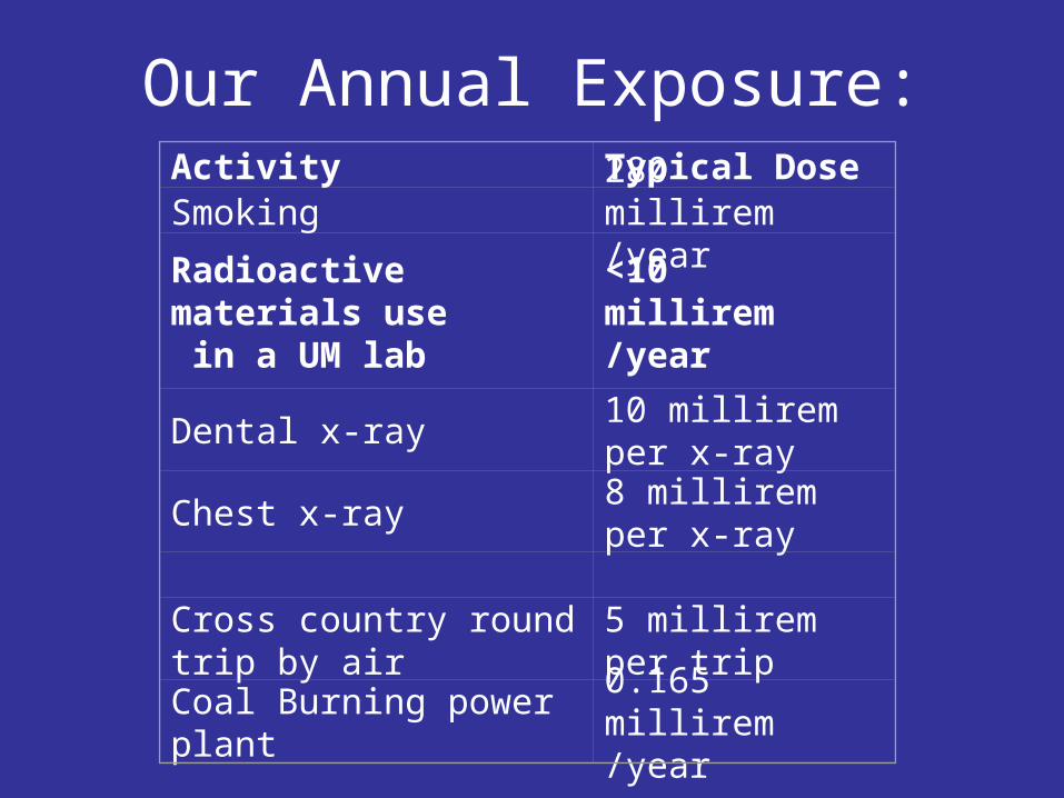
Our Annual Exposure:Activity Typical DoseSmoking 280 millirem /year
Radioactive materials use in a UM lab
<10 millirem /year
Dental x-ray 10 millirem per x-ray
Chest x-ray 8 millirem per x-ray
Cross country round trip by air 5 millirem per trip
Coal Burning power plant 0.165 millirem /year
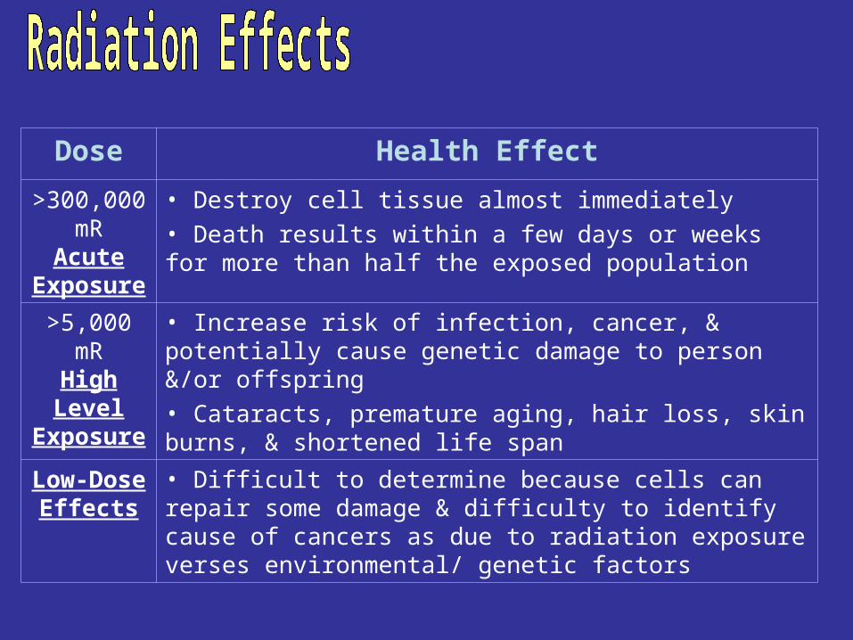
Dose Health Effect
>300,000 mR
Acute Exposure
• Destroy cell tissue almost immediately• Death results within a few days or weeks for more than half the exposed population
>5,000 mRHigh Level
Exposure
• Increase risk of infection, cancer, & potentially cause genetic damage to person &/or offspring• Cataracts, premature aging, hair loss, skin burns, & shortened life span
Low-Dose Effects
• Difficult to determine because cells can repair some damage & difficulty to identify cause of cancers as due to radiation exposure verses environmental/ genetic factors
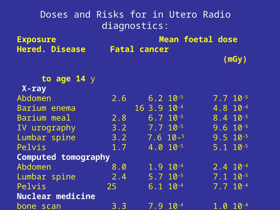
Doses and Risks for in Utero Radio diagnostics:
Exposure Mean foetal dose Hered. Disease Fatal cancer (mGy) to age 14 y X-ray Abdomen 2.6 6.2 10-5 7.7 10-5
Barium enema 16 3.9 10-4 4.8 10-4
Barium meal 2.8 6.7 10-5 8.4 10-5
IV urography 3.2 7.7 10-5 9.6 10-5
Lumbar spine 3.2 7.6 10-5 9.5 10-5
Pelvis 1.7 4.0 10-5 5.1 10-5
Computed tomographyAbdomen 8.0 1.9 10-4 2.4 10-4
Lumbar spine 2.4 5.7 10-5 7.1 10-5
Pelvis 25 6.1 10-4 7.7 10-4
Nuclear medicinebone scan 3.3 7.9 10-4 1.0 10-4
brain scan 4.3 1.0 10-5 1.3 10-4