Embed Size (px)
Citation preview

Faculty of Technology and ScienceChemistry
Anna Smedja Bäcklund
Electron transport in microbial chlorate respiration
Karlstad University Studies2009:18

Karlstad University Studies2009:18
Anna Smedja Bäcklund
Electron transport in microbial chlorate respiration
Anna Smedja Bäcklund. Electron transport in microbial chlorate respiration
Licentiate thesis
Karlstad University Studies 2009:18ISSN 1403-8099 ISBN 978-91-7063-241-9
© The Author
Distribution:Faculty of Technology and ScienceChemistrySE-651 88 Karlstad+46 54 700 10 00
www.kau.se
Printed at: Universitetstryckeriet, Karlstad 2009

”[…] Vi ska vårda vår längtan efter att förstå samtidigt
som vi bevarar vår ödmjukhet inför de oceaner av okunskap
som sträcker ut sig framför, bredvid och bakom oss”
(Citat ur ”Den sjunde dagen” av Stefan Einhorn)

i
Abstract
Several bacterial species are capable to use perchlorate and/or chlorate as an
alternative electron acceptor in absence of oxygen. Microbial respiration of
oxochlorates is important for biotreatment of effluent from industries where
oxochlorates are produced or handled. One of these species, the Gram-negative
Ideonella dechloratans, is able to reduce chlorate but not perchlorate. Two soluble
enzymes, chlorate reductase and chlorite dismutase, participate in the
conversion of chlorate into chloride and molecular oxygen. The present study
deals with the electron transport from the membrane-bound components to the
periplasmic chlorate reductase. Soluble c cytochromes were investigated for
their ability to serve as electron donors to chlorate reductase. The results show
that a 6 kDa c cytochrome serves as electron donor for chlorate reductase. This
cytochrome also serves as electron donor for a terminal oxidase in the
reduction of oxygen that is produced in the course of chlorate respiration. A
gene encoding a soluble c cytochrome was found in close proximity to the gene
cluster for chlorate reduction. This gene was cloned and expressed
heterologously, and the resulting protein was investigated as a candidate
electron donor for chlorate reductase. Electron transfer from this protein could
not be demonstrated, suggesting that the gene product does not serve as
immediate electron donor for chlorate reductase.

ii
List of papers
Paper I
Smedja Bäcklund A, Bohlin J, Gustavsson N, Nilsson T, (2009) Periplasmic c
Cytochromes and Chlorate Reduction in Ideonella dechloratans, Appl Environ
Microbiol, 75(8), In press
Paper II
Bohlin J, Smedja Bäcklund A, Gustavsson N, Wahlberg S, Nilsson T, (2008)
Characterization of a candidate cytochrome c gene associated with the gene
cluster for chlorate respiration in Ideonella dechloratans, Manuscript.

iii
Abbreviations
Ddh Dimethyl sulfide dehydrogenase
Edh Ethylbenzene dehydrogenase
DMSO Dimethyl sulfoxide
TMAO Trimethylamine-N-oxide
Ser Selenate reductase
Clr Chlorate reductase
PCR Polymerase chain reaction
SDS Sodium dodecyl sulfate
PAGE Polyacrylamide gel electrophoresis
ClO2- Chlorite
ClO3- Chlorate
ClO4- Perchlorate
Nap Periplasmic respiratory nitrate reductase
Pcr Perchlorate reductase
Nir Nitrate reductase
NO3- Nitrate
NO2- Nitrite
SeO4- Selenate
SeO3- Selenite
Q/QH2 Quinone pool
TMBZ 3-, 3’-, 5-, 5’- tetramethylbenzidine
DTT Dithiothreitol
MALDI Matrix assisted laser desorption ionization
TOF Time of flight
TFA Trifluoroacetic acid
MS Mass spectrometry
MS/MS Tandem mass spectrometry
LC Liquid chromatography
ccm cytochrome c maturation system
IMAC Immobilized metal ion affinity chromatography
mobB Gene encoding a protein participating in molybdenum cofactor
synthesis

Table of contents Abstract……………………………………………………….................................. .i
List of papers…………………………………………………………………… ii
Abbreviations…………………………………………………………………....iii
1. Introduction…………………………………………………………………...2 1.2 Oxyanions of chlorine ...............................................................................................2
1.3 Perchlorate and chlorate in the environment .....................................................2 1.3.1 Sources ................................................................................................... 2 1.3.2 Environmental effects................................................................................ 3 1.3.3 Treatments of perchlorate- and chlorate containing effluents ............................. 4
1.4 Bacterial perchlorate- and chlorate respiration ..................................................4 1.4.1 Mechanism and enzymes ........................................................................... 4 1.4.2 Perchlorate- and chlorate respiring bacteria...................................................5 1.4.3 Electron transfer pathway in chlorate respiration........................................... 6
1.5 c-type cytochromes .....................................................................................................8
2. Methods used in present study……………………………………………9 2.1 Analysis of periplasmic c cytochromes .................................................................9 2.1.1 Separation ................................................................................................................9 2.1.2 Detection ................................................................................................................10
2.2 Peptide mass fingerprinting by in-gel digestion and mass spectrometry ..11
3. Results and discussion……………………………………………………..12 3.1 Nativ periplasmic c cytochromes in Ideonella dechloratans [Paper I] .........12
3.2 Characterization of a candidate cytochrome c gene associated with the gene cluster for chlorate respiration [Paper IΙ] .......................................................16
4. Conclusions………………………………………………………………… 17 5. Tack……………...………………………………………………………........19 6. References……………………………………………………………………20

2
1. Introduction Sustainable development of industrial processes includes the minimization of the release of waste products in the environment. In many industrial operations, biological treatment of effluent is used to reduce their content of harmful substances. Knowledge about the organisms involved, and their physiological and biochemical properties, is important for the design, operation and optimization of waste-treatment plants. Chlorate and perchlorate are examples of toxic industrial by-products, which need to be removed from waste streams. The present work deals with the chemistry of microbial respiration of chlorate and perchlorate. Here, chlorate and/or perchlorate are used as alternative respiratory electron acceptors in absence of oxygen. The specific aim of this work was to elucidate the electron transport pathway in chlorate respiration.
1.2 Oxyanions of chlorine
Chlorine can combine with oxygen to produce four different oxyanions, shown in table 1. The most oxidized form is perchlorate. Most salts of chlorate and perchlorate are highly soluble and chemically inert under ambient conditions (Kang & Jackson, 2006; Urbansky & Schock, 1999). In contrast, chlorite and hypochlorite are highly reactive in different reactions. Table 1. Oxyanions of chlorine Name Formula Oxidation State
of chlorine
Hypochlorite ClO- I Chlorite ClO2
- III Chlorate ClO3
- V Perchlorate ClO4
- VII
1.3 Perchlorate and chlorate in the environment
1.3.1 Sources
For long, the only known occurrence of perchlorate in the environment was deposits in Chile (Urbansky et al., 2001). Recently it has been shown that perchlorate is readily formed by atmospheric processes when chloride is exposed to high concentrations of ozone. This suggests that some natural perchlorate background from the atmosphere should exist (Dasgupta et al., 2005). Also, Kang et al. (2006) were demonstrated that perchlorate can be generated by photochemical transformations of aqueous chlorine anions, as hypochlorite, chlorite and chlorate, upon exposure to UV-radiation. In the latter case there is a combination of anthropogenic and natural origins since the

3
chlorine oxyanions are introduced to the environment by human activities. A major anthropogenic origin is the manufacture of perchlorate compounds. Perchlorate salts are used in the military industries as oxidants in the production of rockets and missiles (Urbansky & Schock, 1999). Also, perchlorate is used in rocket fuel and most of the perchlorate contamination in the ground and surface water is a result of discharge from rocket fuel manufacturing plants, decades ago (Urbansky, 1998). At the present, there is no evidence for any natural sources of chlorate in the environment, but there are several anthropogenic origins. In agriculture, chlorate is applied as weed controller (herbicides) or as defoliant (Logan, 1998). When hypochlorite is used as disinfectant in drinking water, chlorate results as a by-product (van Ginkel et al., 1995). Another considerable source of chlorate is the pulp- and paper industry. Pulp is bleached with chorine dioxide and chlorate is formed both by decomposition of chlorine dioxide and in the bleaching process (Germgård et al., 1981; Rosemarin et al., 1994). Furthermore, chlorate can be formed during the manufacture and storage of hypochlorite solutions (Siddiqui, 1996).
1.3.2 Environmental effects
In Kalmar Strait, in the Baltic Sea, it was recorded in 1992 that the bladder wrack had disappeared from an area of 12 km2 caused by the effluent from Mönsterås pulp mill (Lehtinen et al., 1988; Rosemarine et al., 1994). Studies of the marine environment have shown that groups of marine algae are sensitive to high concentration of the chlorate anions, especially the benthic brown macro algae (van Wijk and Hutchinson, 1995). The mechanism of toxicity is not clear and appears to differ among species. It has been suggested that chlorate can be reduced by nitrate reductase and the resulting chlorite ion inhibits nitrate reduction, with nitrogen starvation as the result (Åberg, 1947; Stauber, 1998). Further support for this hypothesis is the finding that the cyanobacterium Nostoc muscorum, which is lacking the nitrate reduction system, is chlorate resistant (Singh et al., 1977). In conflict with this hypothesis, chlorate toxicity in plants and bacteria lacking nitrate reduction systems have also been reported (Siddiqi et al., 1992; Prieto & Fernadez, 1993). This suggests that other metabolic pathways are affected by chlorate. For example, in the microalgae Nitzschia closterium chlorate has no effect on the nitrate reductase activity, but the alga is still chlorate sensitive. Stauber (1998) suggests that the toxicity can be a result of decreasing flow of electrons in the electron transport chain, caused by chlorate-dependent altering of membrane-bound components, or inactivation of cytochromes. Furthermore, the oxidizing properties of chlorate may leads to oxidation of essential cell components.

4
1.3.3 Treatments of perchlorate- and chlorate containing effluents
The toxicity of chlorate is suggested to be coupled to reduction of chlorate to the highly reactive chlorite by the nitrate reduction systems, as was described in the above section. However, non-toxic decomposition of chlorate by microbial respiration has been known since the beginning of the 20th century (Åslander, 1928). The difference between toxic and non-toxic decomposition of perchlorate and chlorate depends on the bacterial enzyme systems, as will be discussed in the next section. Several bacterial species, able to grow with perchlorate and/or chlorate as an alternative electron acceptor under anoxic conditions, have been isolated (Korenkov et al., 1976; Malmqvist et al., 1994; Rikken et al., 1996; Wallace et al., 1996; Bruce et al., 1999; Achenbach et al., 2001; Wolterink et al., 2002). These strains can be utilized for anaerobic biotreatment of water and wastewater. One method, developed by Ødegaard et al. 1994, is a process where biomass is grown on small carrier elements that move along with waste water in reactor tanks. The carriers are made by polyethylene and are shaped like small cylinders (Johnson et al., 2000). This technique was introduced into Swedish paper mill industries in the 1990’ies (Kindh & Johansson, 1999). Another method, described by Kroon and van Ginkel (2004), is chlorate reduction in a gas-lift reactor, where microorganisms are attached to pumice particles in the reactor.
1.4 Bacterial perchlorate- and chlorate respiration
1.4.1 Mechanism and enzymes
Bacterial perchlorate or chlorate reduction is coupled to cell growth, and therefore a part of a respiratory chain that generates an electrochemical gradient, which can serve as driving force for ATP synthesis. Many bacteria use several types of acceptors like nitrate, sulfate, manganese (IV), iron (III), selenate, iodate, bromate and DMSO. Respiration of perchlorate is unique because it involves formation of molecular oxygen. The complete reaction takes place in three steps: ClO4- → ClO3- → ClO2- → O2 + Cl- (Kengen et al., 1999) and is catalyzed by two soluble enzymes, (per)chlorate reductase and chlorite dismutase (Bender et al., 2002; Stenklo et al., 2001; van Ginkel et al., 1996). In bacteria able to reduce perchlorate, the first and second step is catalyzed by the same enzyme, perchlorate reductase (Kengen et al., 1999). However, some bacteria are not able to reduce perchlorate and the first step, reduction of chlorate into chlorite, is catalyzed by chlorate reductase (Kengen et al., 1999; Wolterink et al., 2003). The last step, where chlorite is decomposed to chloride ion and oxygen, is catalyzed by chlorite dismutase.

5
1.4.2 Perchlorate- and chlorate respiring bacteria
During the last two decades many different species and strains of bacteria, capable of growth with chlorate or perchlorate as the sole electron acceptor, have been isolated and further investigated. A selection of these is listed in table 2. Table 2. Examples of isolated perchlorate and/or chlorate respiring bacteria
Bacteria strain Electron acceptor Reference Ideonella dechloratans
ClO3-, NO3
-*, O2
Malmqvist et al. 1994
Azospira oryzae strain Gr-1
ClO4
-, ClO3-, NO3,
Mn(IV), O2, IO3-, BrO3
-
Rikken et al. 1996 Wolterink et al. 2005 Kengen et al. 1999
Dechloromonas agitata Strain CKB
ClO4-, ClO3
-, O2
Bruce et al. 1999 Achenbach et al. 2001
Wolinella succinogenes
ClO4-, ClO3
-, NO3, O2
Wallace et al. 1996
Dechloromonas aromatica strain RCB
ClO4-, ClO3
-, NO3, O2
Bender et al. 2005
Pseudomonas chloritidismutans (DSM 13592T)
ClO3-, O2
Wolterink et al. 2003
Strain ASK-1
ClO3
-, O2
Wolterink et al. 2005
Dechloromonas hortensis
ClO4-, ClO3
-, NO3, O2
Wolterink et al. 2005
* After several subcultivations on chlorate, Ideonella dechloratans loses the ability to use nitrate as electron acceptor. The subject of the present study is chlorate respiration in the Gram-negative Ideonella dechloratans. It belongs to the beta subgroup of proteobacteria and was isolated at the environmental biotechnical company ANOX AB in 1994 (Malmqvist et al.). This bacterium can grow by reduction of chlorate, but is not capable to reduce perchlorate. The enzyme chlorite dismutase was isolated and purified by Stenklo et al. 2001. Also, the gene encoding chlorite dismutase was cloned, characterized and expressed by Danielsson Thorell et al. 2002. This enzyme, located in the periplasm, is a homotetrameric heme b- containing protein with the molecular weight of 100 kDa.

6
Chlorate reductase was isolated and the gene cluster for the chlorate metabolism was investigated by Danielsson Thorell and coworkers, 2003. Also this enzyme was found to be located in the periplasm. The protein is heterotrimeric and consists of molybdenum, iron-sulfur cluster and heme b, ordered in a αβγ subunit structure. The molecular weight of the native protein was estimated by gel filtration to 160 kDa. The enzyme belongs to the type II subgroup of the DMSO reductase family and I. dechloratans chlorate reductase shows strong sequence similarity (84 %) to another member of the family, Thauera selenatis selenate reductase (Ser). However, selenate is a poor substrate for chlorate reductase. Other related enzymes, based on sequence similarities and cofactor contents, are found in the marine photosynthetic bacterium Rhodovulum sulfidophilum (dimethyl sulfide dehydrogenase, Ddh) and in Azoarcus sp. Strain EbN1 (ethylbenzene dehydrogenase, Edh) (Danielsson Thorell et al., 2002). The A-subunit, containing molybdopterin, is similar to subunits in other oxidoreductases, e.g. in DMSO reductase, TMAO reductase, and in periplasmic nitrate reductase (NapAB) (Karlsson & Nilsson, 2005). The C-subunit of chlorate reductase was expressed, purified, refolded, and heme reconstituted. The results confirmed the C subunit as the cytochrome b moiety of chlorate reductase in I. dechloratans (Karlsson & Nilsson, 2005).
1.4.3 Electron transfer pathway in chlorate respiration
Although many perchlorate- and chlorate respiring species have been isolated and investigated, electron transport between the inner membrane components and the periplasmic oxidoreductases is not well understood. In the case of nitrate respiration relying on the periplasmic Nap system, electrons are mediated from the quinone pool to the soluble periplasmic NapAB by a membrane-anchored multi-heam c-type cytochrome, belonging to the NapC/NirT family (Berks et al., 1995; Roldan et al., 1998). In other cases the electron transfer agent is soluble and located in the periplasm. The oxidation of quinol is performed by the membrane-bound cytochrome bc1 complex, followed by electron transfer to a soluble c-type cytochrome, which further acts as electron donor to the periplasmic reductase. In T. selenatis, electron transfer to Ser has been suggested to be organized in this way. SerC is reduced by a soluble c cytochrome, which receives electrons from the bc1 complex (McEwan et al., 2002). In R. sulfidophilum a soluble c cytochrome is utilized for transfer of electrons, but in the reverse direction. The β subunit in Ddh donates electrons to the membrane-bound photochemical center, mediated by the soluble cytochrome c2 (McDevitt et al., 2002). Figure 1 illustrates different electron transfer pathways in anaerobic respiration.

7
Figure 1. Electron transport in anaerobic respiration, proposed for D. agitata, D. aromatica, P. denitrificans and T. selenatis. The gray arrows show the direction of electron transfer in D. agitata, D. aromatica, and P. denitrificans. In T. selenatis it is predicted that electrons are transferred through the bc1 complex to selenate reductase via a soluble cytochrome c, as illustrates by dashed arrows. In the perchlorate-reducing bacteria D. agitata and D. aromatica a NapC/NirT-type cytochrome gene was found, suggesting that a membrane-bound cytochrome acts as the link between the quinone pool and the periplasmic perchlorate reductase (Bender et al., 2005). In an earlier investigation we tried to identify a gene for a NapC/NirT-like protein in I. dechloratans by touchdown PCR, using degenerate primers based on conserved features of the NapC/NirT proteins. Such a gene, however, was not found (unpublished, 2004). Using the same primers we searched for a candidate in Azospira oryzae strain Gr-1 and obtained a gene product which showed nucleotide sequence similarity (69 %) to a NapC/NirT-type cytochrome gene in D. aromatica (unpublished, 2007/2008.) I. dechloratans is a facultative anaerobe, but does in fact never grow on chlorate in strictly absence of oxygen. Decomposition of chlorite results in formation of molecular oxygen, which further can be utilized as electron acceptor by terminal cytochrome c oxidase. Malmqvist et al. have shown that I. dechloratans is cytochrome c oxidase positive (1994), suggesting an electron transport chain containing a bc1-type complex transferring electrons from the quinone pool to terminal cytochrome c oxidase mediated by a soluble cytochrome c. Several varies of the latter occur in bacterial respiration. One classic heme-copper oxidase is the aa3-type. Another oxidase is the cbb3-type, which has an exceptional high affinity for oxygen (Preisig et al., 1996) and is therefore more often expressed in bacteria growing in microaerophilic environment. From the characteristic spectral changes, induced by CO-binding, cbb3 oxidase can be identified by spectroscopy analysis of reduced and CO-treated membrane fraction (Pitcher et al., 2002). This has been done with I. dechloratans membrane, and the result strongly indicated presence of cbb3 oxidase (unpublished, 2004).
Q/QH2 bc1 Cyt
c
NapC/NirT
ClO4-, ClO3
-,
NO3-,SeO4
2-
ClrABC, PcrABC,
NapAB, SerABC
ClO2-,
NO2-
SeO32-
dehydrogenases
Formate
Lactate
NADH
H2
Succinate

8
-0,02
0
0,02
0,04
0,06
0,08
0,1
0,12
0,14
450 500 550 600 650
1.5 c-type cytochromes
Many different types of c cytochromes occur in bacteria as electron carriers. They are characterized by covalently bound heme as a prosthetic group. Heme consists of a Fe-atom coordinated to four nitrogen atoms of a porhyrin ring. The heme-group is attached to the protein by two thioether linkages. These are formed by reaction of the vinyl groups of heme with two cysteine residues within a CXXCH sequence motif (Kranz et al., 1998) of the polypeptide chain, as shown in figure 2a. Cytochromes of c-type are often found in the periplasmic space where they are connected to the respiratory components of the cytoplasmic membrane, acting as electron carriers. They can either be parts of multisubunit enzyme complexes or occur as mobile electron shuttles (Thöny-Meyer, 1997). Cytochromes exhibit three characteristic absorption bands, designated α, β and Soret (γ)-band. In oxidized form α- and β- bands are broad whereas reduced cytochromes show three distinct peaks, as shown in figure 2b. Different types of cytochromes can be classified on the basis of their α-peak in the region of 550 – 610 nm (Nicholls & Ferguson, 2002). For c-type cytochromes the maximum of the α-peak is usually found between 550 – 557 nm, whereas the maximum for b-type is between 555 – 565 nm (Thöny-Meyer, 1997).
a) b) Figure 2. (a) The prosthetic group of heme c consists of a central Fe ion, coordinated to four nitrogen atoms of a cyclic porhyrin ring. The Fe atom is the redox component, either in the reduced ferrous [Fe (II)] or in the oxidized ferric [Fe (III)] state. (b) The characteristic spectra of cytochrome c in oxidized (dashed) and reduced (solid) state. The reduced spectrum shows the soret peak at 450 nm, the β-peak at 521 nm and the α-peak at 551 nm. The present work investigates the scenario that electron transfer in I. dechloratans is mediated by a soluble c-type cytochrome. In this case, electrons are passed from the quinone pool to soluble c cytochromes by a membrane-bound bc1

9
complex. This hypothesis is based on the apparent absence of a NapC/NirT-gene in I. dechloratans and the similarity between chlorate reductase and Ddh in R. sulfidophilum and Ser in T. selenatis. Several c-type cytochromes were extracted from the periplasm of I. dechloratans (paper I). Also, a gene encoding a candidate c cytochrome has been expressed heterologously (paper II). This gene is associated with the gene cluster for chlorate reduction. We can demonstrate that at least one of the native soluble c-type cytochromes is able to mediate the electron transfer between the membrane bound components and the soluble chlorate reductase. This ability could not be demonstrated for the recombinant c cytochrome. The gene product (paper II) was not detected in the periplasmic extract and its role in the chlorate metabolism remains to be clarified.
2. Methods used in present study
2.1 Analysis of periplasmic c cytochromes
2.1.1 Separation
A common technique for separation of proteins, almost exclusively depending on their molecular weights, is sodium dodecyl sulphate polyacrylamide gel electrophoresis, SDS-PAGE. Proteins are separated in a polyacrylamide gel, made by co-polymerization of acrylamide and N, N1-methylene- bis-acrylamide. The bis-acrylamide is a cross-linking agent for the gel. SDS is an anionic denaturing detergent forming mixed micelles containing detergent and denatured protein with a large negative net charge. For reduction of inter- or intramolecular disulphide bridges, detergents such β-mercapto ethanol can be added. This step needs to be avoid if the propose of the analysis is to identify proteins containing covalently bound heme, as will be discussed in next section. When SDS is used, the migration of the protein is a function of its size only. In continuous gel system the gel, sample and running buffer have the same ionic composition, and the same pH. The sample is loaded directly onto a resolving gel. In a discontinuous system, introduced by Ornstein, L. and Davis, B.J. (1964) and adapted by Laemmli (1970), the gel and running buffer consists of different ions and, most commonly, a discontinuous pH. The sample is loaded onto a large-pore stacking gel on top of the resolving gel. The advantage of the discontinuous system is that the proteins are concentrated into narrow zones by the process of isotachophoresis, before entering the small-pore resolving gel. In the gel and sample buffer, a leading ion with high effective mobility moves towards the anode at a given pH, usually pH 6.8. The running buffer contains a trailing ion with much lower effective mobility, due to its

10
small net charge at the current pH, usually pH 8.3. When the effective mobility of leading and trailing ions is extremely high respectively low, the mobility of the proteins will be intermediate. When the voltage is applied, the leading anion will migrate faster towards the anode, producing a zone of lower conductivity behind. As a result, the voltage gradient increases and anions with lower mobility speed up and migrate at the same velocity in a zone behind the leading anion. The trailing ions accelerate until a steady state is established, with high conductivity in front of the leading ion and low conductivity behind the trailing ions. The proteins will be stacked as thin bands delimited by sharp boundaries. When the moving boundary system reaches the resolving gel with higher pH, the effective mobility of the trailing ions increases and the proteins will migrate behind both leading- and trailing ions. The resolving gel will retard the proteins according to their molecular masses (Hames, 1990). In the Laemmli buffer system the stacking-gel buffer, (pH 6.8), contains chloride ions as the leading ion. The running buffer in the upper reservoir consists of glycine, which has a low net charge and low effective mobility at pH 6.8. NuPAGE® Bis-Tris Discontinuous Buffer System (Invitrogen) is a modified system where the gel buffer (pH 6.4) contains chlorine as the leading ion and the running buffer (pH 7.3-7.7) contains MES- (2-(N-morpholino) ethanesulfonic acid) as the trailing ion. Bis-tris is the common counter-ion present in both gel- and running buffer. The combination of pH results in an operating pH of 7 during electrophoresis which improves the protein stability, resulting in sharper bands and better resolution. MES buffer is optimal for resolving small molecular weight proteins, due to its fast ion migration. When using 4-12 % bis-tris gels and MES SDS running buffer, the separation range for proteins is between 2.5-200 kDa compared to the Laemmli tris-glycine system which needs 18 % gels to resolve low molecular weight proteins.
2.1.2 Detection
Proteins containing heme can be detected after SDS-PAGE by using a staining procedure relying on the peroxidase activity of the heme group (Thomas et al., 1976). The heme-catalyzed reaction between 3-,3’-,5-,5’-tetramethylbenzidine (TMBZ) and hydrogen peroxide results in the formation of a deep blue product. Cytochrome c, in which the heme group is bound covalently by thioether bounds, is readily detected on SDS-PAGE gels using this method. Agents used for reduction of disulphides (such 2-mercaptoethanol or DTT) may affect the coordination state of the heme and either stimulate or inhibit peroxidase activity (Dorward et al., 1992). Therefore, reducing agents are specifically omitted from the samples in the electrophoresis. In most cases, non-covalently bound heme is released from proteins by the denaturation with SDS, but incomplete denaturation can occur. Consequently, one complication is that other cytochromes than the c-types may be visualized by heme-staining.

11
Another problem is the possibility of non-specific binding of released heme to other proteins, resulting in a false signal (Thomas et al., 1976).
2.2 Peptide mass fingerprinting by in-gel digestion and mass spectrometry
Peptide mass fingerprinting is a method for identification of proteins. First, the protein is cleaved into peptides and second, the peptides are analyzed by mass spectrometry (MS). In the present case the ionization of peptides was preformed by Matrix Assisted Laser Desorption/Ionization (MALDI) and the analysis was performed by Time-of-Flight (TOF). Fore more specific information of the peptides, fragmentation of the ions and analysis of the fragments was performed by tandem mass spectrometry (MS/MS). Peptides can be generated by the enzyme trypsin, which catalyzes the hydrolysis of peptide bounds. Trypsin cleaves peptide bounds in which the carbonyl group is contributed by either an arginine (R) or lysine (K) residue. In-gel digestion is a method for enzymatic cleavage of proteins in polyacrylamide gels, after SDS-PAGE (Rosenfeld et al., 1992). After protein identification by staining, the protein band is excised from the gel and destained by ethanol/NH4HCO3. Small gel pieces are incubated with trypsin and peptides are extracted from the gel pieces by trifluoroacetic acid (TFA). The resulting peptide mixture can be used for MS peptide mapping. Mass spectrometers separate ions according to their mass-to-charge ratio (m/z). MALDI is a method for ionization where the sample is mixed with a matrix compound, capable of absorbing UV-light (Karas & Hillenkamp, 1987). The matrix will absorb energy from a laser beam and the embedded peptides are ionized and transferred to the gas phase. TOF is an analysis method where strong electric field is applied and the ions accelerate, followed by travel in a field-free flight tube. The flight time to reach the MS detector is proportional to the square root of the m/z ratio (Guilhaus et al., 2000). The masses obtained from MALDI-TOF can be compared to expected tryptic masses of known proteins, by data base search. The combination of liquid chromatography (LC) and MS produces efficient separation and sensitive identification of peptides and proteins, especially when the sample is a complex mixture of several proteins (Mann & Pandey, 2001) To obtain more specific information of a single peptide, tandem mass spectrometry (MS/MS) can be used. This is an instrument consisting of two MS analyzers where a single peptide ion can be selected out of the spectrum obtained from the first mass spectrometer. This peptide ion is further fragmented by collision with inert gas and the fragments are separated by the second mass spectrometer. Cleavage at the peptide bond is the most common mode of fragmentation and the resulting ions, in this case called b- and y- ions, retain their charge on the N or C terminus respectively. Fragmentation is

12
random and the process generates a series of fragments, differing by a single amino acid residue (Mann & Pandey, 2001). In case of unknown proteins, with no corresponding sequences in data bases, fragmentation is the only possibility to obtain information about the protein. It is possible to reconstruct the sequence of a single peptide by studying its fragmentation pattern. This method, called de novo peptide sequencing, is based on knowledge about peptide decomposition mechanisms (Cañas et al., 2006).
3. Results and discussion
The aim of the present study was to elucidate the route of electron transfer between the inner membrane and the periplasmic chlorate reductase. The report is divided in two parts, both dealing with soluble c cytochromes as candidates for the electron transport. First, soluble c cytochromes were isolated from I. dechloratans by extraction of periplasm from the cells (paper I). Second, a gene encoding a candidate soluble c-type cytochrome, located in association with the gene cluster for chlorate reduction, was cloned and expressed heterologously (paper II).
3.1 Native periplasmic c cytochromes in Ideonella dechloratans [Paper I]
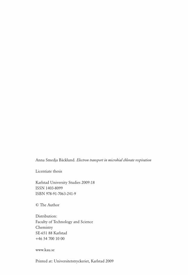
Periplasmic proteins were extracted from chlorate-grown I. dechloratans by osmotic lysis. The periplasmic extract was first reduced by dithionite and then exposed to chlorate in the presence of chlorate reductase. Spectrophotometric analysis showed a reduction peak at 551 nm, indicating reduction of c-type cytochromes. After addition of chlorate a partial reoxidation of the cytochromes was observed, which demonstrated the presence of at least one soluble c cytochrome capable of supplying electrons for chlorate reduction. In order to identify c cytochromes the extracted proteins were separated by SDS-PAGE (NuPAGE™) and stained by the heme-detecting procedure, described in section 2.1.2. Several bands were observable and the result is shown in figure 3. Since the soluble forms of c-type cytochromes, also called class I cytochrome c, usually occurs in the range 8-14 kDa (Thöny-Meyer, 1997) further attention was focused on proteins with molecular weights of 20 kDa and below. Five candidate c cytochromes (20, 10, 9, 8, and 6kDa), were observed. The ones most abundant, according to the heme staining, were the 10- and 6-kDa cytochromes. The bands at 8 and 9 kDa were not always observed as separate bands. As discussed below, it appears that the 8-kDa protein derives from the 9 kDa cytochrome as a degradation product.
13
kDa
188
98
62
49
38
28
17
14
6
3
20
109/86
kDa
188
98
62
49
38
28
17
14
6
3
20
109/86
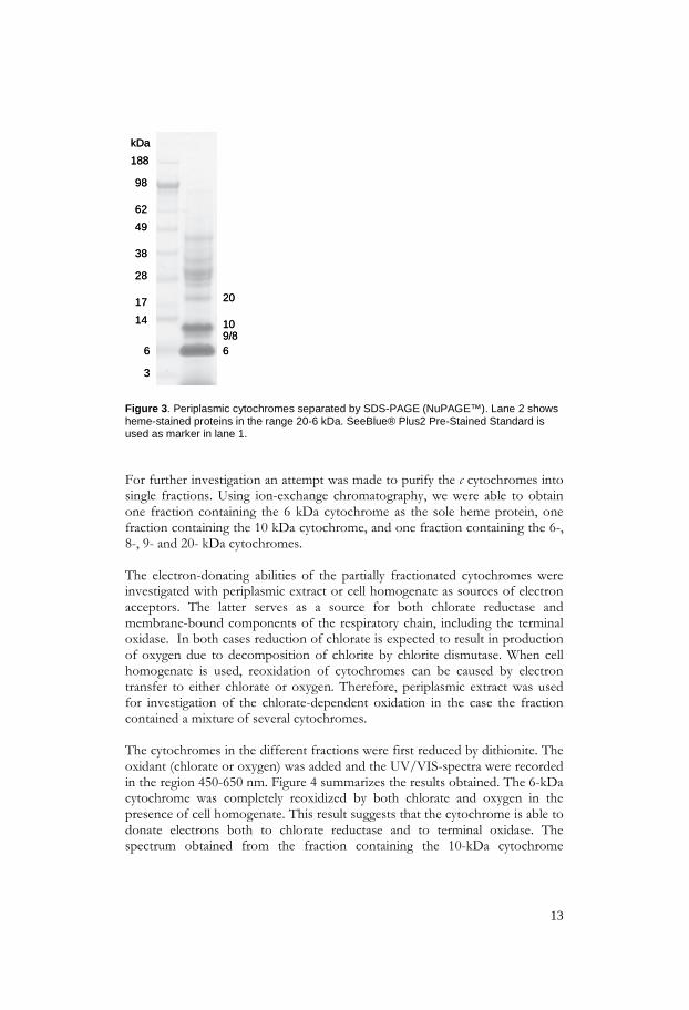
Figure 3. Periplasmic cytochromes separated by SDS-PAGE (NuPAGE™). Lane 2 shows heme-stained proteins in the range 20-6 kDa. SeeBlue® Plus2 Pre-Stained Standard is used as marker in lane 1. For further investigation an attempt was made to purify the c cytochromes into single fractions. Using ion-exchange chromatography, we were able to obtain one fraction containing the 6 kDa cytochrome as the sole heme protein, one fraction containing the 10 kDa cytochrome, and one fraction containing the 6-, 8-, 9- and 20- kDa cytochromes. The electron-donating abilities of the partially fractionated cytochromes were investigated with periplasmic extract or cell homogenate as sources of electron acceptors. The latter serves as a source for both chlorate reductase and membrane-bound components of the respiratory chain, including the terminal oxidase. In both cases reduction of chlorate is expected to result in production of oxygen due to decomposition of chlorite by chlorite dismutase. When cell homogenate is used, reoxidation of cytochromes can be caused by electron transfer to either chlorate or oxygen. Therefore, periplasmic extract was used for investigation of the chlorate-dependent oxidation in the case the fraction contained a mixture of several cytochromes. The cytochromes in the different fractions were first reduced by dithionite. The oxidant (chlorate or oxygen) was added and the UV/VIS-spectra were recorded in the region 450-650 nm. Figure 4 summarizes the results obtained. The 6-kDa cytochrome was completely reoxidized by both chlorate and oxygen in the presence of cell homogenate. This result suggests that the cytochrome is able to donate electrons both to chlorate reductase and to terminal oxidase. The spectrum obtained from the fraction containing the 10-kDa cytochrome

14
showed two peaks at 552 and 558 nm respectively. The 558-nm component was probably originated from heme b rather than heme c as a result of contamination, as discussed below. When oxygen was added, partial reoxidation was observed. As the spectral change appears dominated by the 558-nm component, it is not possible to assess the reoxidation of the 552-cytochrome c component. None of these components were reoxidized by the addition of chlorate. In the fraction containing 6-, 8-, 9-, and 20-kDa cytochromes partial reoxidation was observed after the addition of chlorate. In present case, a catalytic amount of periplasmic extract was used and thus no terminal oxidase was present. The extent of reoxidation was about 40%, which can be accounted for by the 6-kDa cytochrome since this component was shown to donate electrons to chlorate reductase. In presence of cell homogenate, the same fraction was completely reoxidized both by addition of chlorate and oxygen. This suggests that the components which were not oxidized by chlorate in absence of terminal oxidase are able to be oxidized when oxygen is produced as the result of chlorite dismutase activity. Taken together, these results suggest that 8-, 9-, and 20-kDa cytochromes are electron donors to terminal oxidase but most likely not to chlorate reductase. However, the individual roles of these cytochromes can not be assigned.
Figure 4. The diagram shows the percentage spectral changes when chlorate or oxygen is added to dithionite-reduced c cytochromes in presence of chlorate reductase and/or terminal oxidase. The gray color represents the extent of reoxidation and the black color represents the remaining reduction.
0 10 20 30 40 50 60 70 80 90 100
Extent of reoxidation %
6 kDa, chlorate
6 kDa, oxygen
10 kDa, chlorate
6,8,9,20 kDa, chlorate
6,8,9,20 kDa, oxygen

15
The identified c cytochromes of 6, 8, 9, and 10 kDa were further investigated by peptide mass fingerprinting, using MALDI-TOF MS and MS/MS. Peptides were generated by trypsin in-gel digestion of bands excised from SDS-PAGE gels. One of the peptides obtained from the 6 kDa cytochrome was identical in mass-to-charge ratio (m/z = 1094.56) to that predicted for the sequence YAGQKDAVDK from Acidovorax avenae subsp. citrulli AAC00-1, [YP_969867]. A majority of the theoretical b- and y-ions, expected from this fragment, could be matched to signals in the MS/MS-spectrum of the 1094.56-peptide from I. dechloratans. The sequence KLVGPSQDVAAR has been obtained earlier by de novo sequencing of a peptide (m/z =1403.76), generated by digestion of a mixture of cytochromes. A peptide with the same m/z was found at present in the digest of the 6 kDa cytochrome. MS/MS-data confirmed the amino acid sequence above. Further database searches for these two sequences produced the result shown in table 3. The sequence KLVGPSQDVAAR (1403.73) shows similarity to sequences in three different cytochrome c (class I), of the chlorate reducer Dechloromonas aromatica. Also, this sequence can be matched to Acidovorax avenae subsp. citrulli by a sequence located next to YAGQKDAVDK (1094.56).
I. dechloratans (mass 1403.76)
KLVGPSYQDVAAR
---------------------
I. dechloratans (mass 1094.56)
--------------------------- YAGQKDAVDK
A. ave sub.citr (YP 269867)
KLVGPSYKDVAAK
YAGQKDAVDK
D. aromatica (YP283918)
KLVGPAYKDVAAK
YKGDKGAVDK
D. aromatica (YP286520)
KLVGPAYKDVAAK
YKGDAGAVDK
D. aromatica (YP283533)
KLVGPAYKDVAAK
YKGDAKAPAM
Table 3. Alignments of the sequences obtained for the 6 kDa c-cytochrome by MALDI-TOF MS/MS and the matched sequences from database search. The identical amino acids are shown in gray. For the 8- and 9-kDa proteins, no matches were obtained from database searches. However, the presence of same peptide masses in digest obtained from these proteins supports the suggestion that the 8-kDa is a degradation product of the 9-kDa cytochrome. Several of the peptides from the 10-kDa band were, unexpectedly, identified as originating from chlorite dismutase. The residue coverage was 40%, with regard to the amino acid sequence. The presence of chlorite dismutase is probably a

16
result of proteolytic degradation. This finding accounts for the absorption maximum at 558 nm in the spectrophotometric study.
3.2 Characterization of a candidate cytochrome c gene associated with the gene cluster for chlorate respiration [Paper II]
In close proximity to the gene cluster for the chlorate reduction, downstream from the C subunit of chlorate reductase, two open reading frames were found by further sequencing of a clone from the GenomeWalker™ library (Karlsson & Nilsson, 2005). In the ORF closest to the C subunit, the highly conserved heme-binding motif CXXCH was found in the corresponding translated amino acid sequence. This finding suggests a gene encoding c-type cytochrome. The sequence showed similarity to soluble c cytochromes, class I, in Pfam database and by BLASTP search. The hydrophobic N-terminal sequence is suggested by SignalP to be a 19-residue signal peptide, and the molecular weight of the whole apoprotein is predicted to 10,9 kDa. The second ORF further downstream was identified as a mobB gene encoding a protein, functional for the maturation of molybdopterin as a co-factor for chlorate reductase A subunit. The results presented in paper I suggest an electron transfer route where soluble c-type cytochromes serve as mediators. A 6-kDa c cytochrome was shown to serve as electron donor to chlorate reductase, whereas a 10-kDa protein did not. However, none of the peptide masses or sequences obtained from the soluble periplasmic c cytochromes in paper I matched the predicted sequence of the gene above. In order to assess the role of this gene in chlorate reduction, the gene was expressed heterologously and the resulting protein was investigated. The gene, excluding the part encoding the 19-residue signal peptide, was amplified by PCR and the product was ligated into the expression vector pET-26b. This vector contributes a pelB leader for export to the periplasm and a C-terminal six-residue His-tag for subsequent purification by Immobilized Metal Ion Affinity Chromatography (IMAC). The construct was cotransformed with plasmid pEC86, which includes the cytochrome c maturation system (ccm), into the E. coli expression host BL21(DE3). The use of the ccm genes was based on the finding that E. coli expression hosts normally not express c cytochromes under aerobic conditions (Thöny-Meyer et al., 1996). For the purpose of synthesizing active holo-c-type- cytochromes the heme needs to be attached to the apo-protein after export to the periplasm, which is done by the ccm system. In transformant BL21(DE3) the target protein was produced at high level but was not exported as apo-protein into the periplasm. Instead, insoluble inclusion bodies were found in the cytosol, lacking the heme-group. In E. coli NovaBlue(DE3) the construct generated a very low yield of the recombinant c cytochrome. From these results we decided to produce inclusion bodies for purification, refolding, and heme reconstitution. The inclusion bodies were

17
solubilized with urea and the protein was purified in the denatured state by IMAC. After refolding, reduced heme was added to the protein in the ratio 1:1. The covalent incorporation of heme was detected spectrophotometrically. At first it was detected by the appearance of a distinct peak in the α-band at 556.5 nm, indicating a b-type cytochrome, but within seven days the α-peak was shifted to 553 nm. The formation of covalently bound heme, with two thioether bonds produced, was confirmed by pyridine hemochrome analysis (Berry & Trumpower, 1987). The pyridine hemochrome showed a peak in the α-band at 551 nm, which is characteristic for c-type cytochromes. The recombinant cytochrome was investigated by peptide mass fingerprinting using MALDI-TOF MS, as in the previous paper I. Peptides from the recombinant c cytochrome could be identified and verified (by MS/MS) with a coverage of 56 % of the amino acid sequence. Spectrophotometric studies of the redox activity were performed using the same approach as for the native proteins, described in paper I. The reduced form of the recombinant cytochrome c did not reoxidize after addition of chlorate in presence of chlorate reductase. This result suggests that the gene product does not serve as an immediate electron donor for chlorate reductase. However, we cannot exclude that the inability of donating electrons for chlorate reduction is a result of incorrect refolding or incorporation of heme. Since the protein was very sensitive to air, we were not able to test enzyme-dependent reoxidation by oxygen.
4. Conclusions
The aim of this study was to clarify the electron transport pathway in the chlorate reduction in I. dechloratans, in particular the route between the membrane-bound components and the periplasmic chlorate reductase. We have demonstrated that electron transport is mediated by soluble electron carriers. A 6-kDa c-type cytochrome is the major, and possibly the sole, donor to chlorate reductase when cells are grown anaerobically in presence of chlorate. The 6-kDa cytochrome can also serve as donor in the reduction of oxygen. Additional c cytochromes ((8- ), 9-, and 20-kDa) probably donate electrons to terminal cytochrome c oxidase, as inferred from their reoxidation on addition of oxygen. Since the anaerobic respiration involves production of molecular oxygen, the overall reduction of both chlorate and the produced oxygen (ClO3- → Cl- + H2O), requires transfer of six electrons. The reduction of chlorate to chlorite is a two-electron reaction whereas reduction of oxygen, produced by the subsequent decomposition of chlorite, requires four electrons. From the result presented here we suggest a branched electron transport chain as shown in Figure 5.

18
Furthermore, a gene found downstream from the gene encoding the C subunit of chlorate reductase has been characterized. The gene was identified as a soluble cytochrome c (class I) which is defined as a soluble electron transfer protein. The gene was expressed heterologously for the purpose to investigate its role as electron carrier in the chlorate reduction mechanism. However, the reconstituted protein was not able to serve as electron donor to chlorate reductase. The significance for the presence of the gene, located in the gene cluster for chlorate reduction, still remains to be elucidated.
Figure 5. The proposed electron-transfer pathways in Ideonella dechloratans. The arrows show the suggested electron route between the membrane-bound cytochrome bc1-complex and the chlorate reductase and the terminal oxidase, respectively.
e- (NADH)
ClO3 ClO2
III
IV
6
I
2e-
O2
Cl-
(8),9 20
cbb3 oxidase
Chlorate reductase
Chlorite dismutase
Q
periplasmm
cytoplasm
cyt.c
cyt.c
4e-
H2O

19
5. Tack
Jag vill börja med att tacka min handledare Prof. Thomas Nilsson som på alla sätt har stöttat mig i mitt arbete. I såväl medgång som i motgång har din entusiasm hjälpt mig framåt. Jag vill även tacka mina biträdande handledare Birgitta Sundström och Maria Rova. Ni har alltid ställt upp och gett tips och råd när jag behövt er hjälp. En person som jag har mycket att tacka för är min forne doktorandkollega Jan Bohlin. Tack för alla roliga stunder på labbet och på kontoret där vi har diskuterat film, musik och böcker men naturligtvis även jobbat. Det var du som introducerade mig för vårt gemensamma skötebarn Ideonella dechloratans. Det känns tomt utan dig. Jag vill rikta ett stort tack till alla övriga kollegor på avdelningen. Tack vare er är det roligt att gå till jobbet. Ett särskilt stort tack till Sandra, Gunilla C, Cecilia, Christina, Sofia, Maria, Tina, Jeanette, Rozbeh, Björn och Patrik för allt roligt vi har haft tillsammans (även utanför universitetet). Jag vill säga tack till de ex-jobbare som gjort sitt examensarbete inom projektet under den här tiden. Tack Kajsa, Malin, Eva-Lotta, Carin, Fredrik B, Fredrik P och Veronica för er arbetsinsats och för att ni bidrog till en trevlig stämning på labbet. Tack mamma och pappa för att ni lärde mig att tro på mig själv. Det krävs mod att sadla om mitt i livet, ska ni veta. Det modet har jag nog fått från er eftersom ni har varit så bra förebilder. Till mina barn Johan, Linda och Felicia; tack för att ni finns, jag älskar er så! Rimella och Olivia, den här boken är tillägnad er. Forskning är ju till för framtiden och det är Ni som är framtiden. Tack för att ni finns! Sist men inte minst, min älskade Bosse; utan dig hade detta inte varit möjligt.

20
6. References
Achenbach, L. A., Michaelidou, U., Bruce, R. A., Fryman, J., & Coates, J. D. (2001). Dechloromonas agitata gen. nov., sp. nov. and Dechlorosoma suillum gen. nov., sp. nov., two novel environmentally dominant (per)chlorate-reducing bacteria and their phylogenetic position. Int J Syst Evol Microbiol, 51(Pt 2), 527-533. Bender, K. S., O'Connor, S. M., Chakraborty, R., Coates, J. D., & Achenbach, L. A. (2002). Sequencing and transcriptional analysis of the chlorite dismutase gene of Dechloromonas agitata and its use as a metabolic probe. Appl Environ Microbiol, 68(10), 4820-4826. Bender, K. S., Shang, C., Chakraborty, R., Belchik, S. M., Coates, J. D., & Achenbach, L. A. (2005). Identification, Characterization, and Classification of Genes Encoding Perchlorate Reductase. Journal of Bacteriology, 187(15), 5090-5096. Berks, B. C., Richardson, D. J., Reilly, A., Willis, A. C., & Ferguson, S. J. (1995). The napEDABC gene cluster encoding the periplasmic nitrate reductase system of Thiosphaera pantotropha. Biochem J, 309(Pt 3), 983-992. Berry, E. A., & Trumpower, B. L. (1987). Simultaneous determination of hemes a, b, and c from pyridine hemochrome spectra. Anal Biochem, 161(1), 1-15. Bruce, R. A., Achenbach, L. A., & Coates, J. D. (1999). Reduction of (per)chlorate by a novel organism isolated from paper mill waste. Environ Microbiol, 1(4), 319-329. Canas, B., Lopez-Ferrer, D., Ramos-Fernandez, A., Camafeita, E., & Calvo, E. (2006). Mass spectrometry technologies for proteomics. Briefings in Functional Genomics and Proteomics, 4(4), 295-320. Danielsson Thorell, H., Karlsson, J., Portelius, E., & Nilsson, T. (2002). Cloning, characterisation, and expression of a novel gene encoding chlorite dismutase from Ideonella dechloratans. Biochim Biophys Acta, 1577(3), 445-451. Danielsson Thorell, H., Stenklo, K., Karlsson, J., & Nilsson, T. (2003). A gene cluster for chlorate metabolism in Ideonella dechloratans. Appl Environ Microbiol, 69(9), 5585-5592. Dasgupta, P. K., Martinelango, P. K., Jackson, W. A., Anderson, T. A., Tian, K., Tock, R. W., et al. (2005). The Origin of Naturally Occurring Perchlorate: The Role of Atmospheric Processes. Environmental Science & Technology, 39(6), 1569-1575.

21
Dorward, D. W., Huguenel, E. D., Davis, G., & Garon, C. F. (1992). Interactions between extracellular Borrelia burgdorferi proteins and non-Borrelia-directed immunoglobulin M antibodies. Infection and Immunity, 60(3), 838-844. Germgård, U., Teder, A., & Tormund, D. (1981). Chlorate formation during chlorine dioxide bleaching of softwood kraft pulp. Pap Puu, 63(3), 127-133. Guilhaus, M., Selby, D., & Mlynski, V. (2000). Orthogonal acceleration time-of-flight mass spectrometry. Mass Spectrometry Reviews, 19, 65-107. Hames B.D. 1990. One-dimensional polyacrylamide gel electrophoresis. In Gel electrophoresis of proteins: A practical approach, 2nd edition (ed. B.D. Hames and D. Rickwood). Oxford University Press, New York. Johnson, C. H., Page, M. W., & Blaha, L. (2000). Full scale moving bed biofilm reactor results from refinery and slaughter house treatment facilities. Water Science & Technology, 41(4), 401-407. Kang, N., Anderson, T. A., & Andrew Jackson, W. (2006). Photochemical formation of perchlorate from aqueous oxychlorine anions. Analytica Chimica Acta, 567(1), 48-56. Karas, M., & Hillenkamp, F. (1988). Laser desorption ionization of proteins with molecular masses exceeding 10,000 daltons. Anal Chem, 60(20), 2299-2301. Karlsson, J., & Nilsson, T. (2005). The C subunit of Ideonella dechloratans chlorate reductase: Expression, purification, refolding, and heme reconstitution. Protein Expression and Purification, 41(2), 306-312. Kengen, S. W., Rikken, G. B., Hagen, W. R., van Ginkel, C. G., & Stams, A. J. (1999). Purification and characterization of (per)chlorate reductase from the chlorate-respiring strain GR-1. J Bacteriol, 181(21), 6706-6711. Kindh, T., & Johansson, M. (1999). Slamflykt i aktivslamanläggningen-ett minne blott? Svensk Papperstidning, 102, 28-31. Korenkov, V. N., Romanenko, V. I., Kuznetsov, S. I., & Voronov, J. V. (1976). Process for purification of industrial waste waters from perchlorates and chlorates, United States Patent. United States. Kranz, R., Lill, R., Goldman, B., Bonnard, G., & Merchant, S. (1998). Molecular mechanisms of cytochrome c biogenesis: three distinct systems. Molecular Microbiology, 29(2), 383-396.

22
Kroon, A. G. M., & van Ginkel, C. G. (2004). Biological Reduction of Chlorate in a Gas-Lift Reactor Using Hydrogen as an Energy Source: Am Soc Agronom. Lehtinen, K.-J., Notini, M., Mattson, J., & Lander, L. (1988). Disappeareance of Bladder-Wrack (Fucus vesiculosus L.) in the Baltic Sea: Relation to Pulp-Mill Chlorate. Ambio, 17(6), 387-393. Logan, B. E. (1998). A review of chlorate- and perchlorate-respiring microorganisms. Biorem J, 2(2), 69-79. Malmqvist, Å., Welander, T., Moore, E., Ternström, A., Molin, G., & Stenström, I. (1994). Ideonella dechloratans gen.nov., sp.nov., a new bacterium capable of growing anaerobically with chlorate as an electron acceptor. System Appl Microbiol, 17, 58-64. Mann, M., Hendrickson, R. C., & Pandey, A. (2001).Analysis of proteins and proteomes by mass spectrometry. Annual Reviews in Biochemistry, 70(1), 437-473. McDevitt, C. A., Hugenholtz, P., Hanson, G. R., & McEwan, A. G. (2002). Molecular analysis of dimethyl sulphide dehydrogenase from Rhodovulum sulfidophilum: its place in the dimethyl sulphoxide reductase family of microbial molybdopterin-containing enzymes. Mol Microbiol, 44(6), 1575-1587. McEwan, A. G., Ridge, J. P., McDevitt, C. A., & Hugenholtz, P. (2002). The DMSO Reductase Family of Microbial Molybdenum Enzymes; Molecular Properties and Role in the Dissimilatory Reduction of Toxic Elements. Geomicrobiology Journal, 19(1), 3-19. Nicholls, D.G. & Ferguson, S.J (2002) Bioenergetics 3, Academic Press, ISBN 0-12-518121-3, page 92-93 Pitcher, R. S., Brittain, T., & Watmough, N. J. (2002). Cytochrome cbb~ 3 oxidase and bacterial microaerobic metabolism. Biochemical Society Transactions, 30(4), 653-657. Preisig, O., Zufferey, R., Thony-Meyer, L., Appleby, C. A., & Hennecke, H. (1996). A high-affinity cbb3-type cytochrome oxidase terminates the symbiosis-specific respiratory chain of Bradyrhizobium japonicum. Journal of Bacteriology, 178(6), 1532-1538. Prieto, R., & Fernandez, E. (1993). Toxicity of and mutagenesis by chlorate are independent of nitrate reductase activity in Chlamydomonas reinhardtii. Mol Gen Genet, 237(3), 429-438.

23
Rikken, G. B., Kroon, A. G., & van Ginkel, C. G. (1996). Transformation of (per)chlorate into chloride by a newly isolated bacterium: reduction and dismutation. Appl Microbiol Biotech, 45, 420-426. Roldan, M. D., Sears, H. J., Cheesman, M. R., Ferguson, S. J., Thomson, A. J., Berks, B. C., et al. (1998). Spectroscopic Characterization of a Novel Multiheme c-Type Cytochrome Widely Implicated in Bacterial Electron Transport. Journal of Biological Chemistry, 273(44), 28785-28790. Rosemarin, A., Lehtinen, K.-J., Notini, M., & Mattson, J. (1994). Effects of pulp mill chlorate on baltic sea algae. Environ Pollut, 85, 3-13. Rosenfeld, J., Capdevielle, J., Guillemot, J. C., & Ferrara, P. (1992). In-gel digestion of proteins for internal sequence analysis after one-or two-dimensional gel electrophoresis. Analytical Biochemistry, 203(1), 173-179. Siddiqi, M. Y., King, B. J., & Glass, A. D. M. (1992). Effects of Nitrite, Chlorate, and Chlorite on Nitrate Uptake and Nitrate Reductase Activity 1. Plant Physiology, 100(2), 644-650. Siddiqui, M. S. (1996). Chlorine-ozone interactions: formation of chlorate. Wat Res, 30(9), 2160-2170. Singh, H. N., Sonie, K. C., & Singh, H. R. (1977). Nitrate regulation of heterocyst differentiation and nitrogen fixation in a chlorate-resistant mutant of the blue-green alga, Nostoc muscorum. Mutation Research, 42(3), 447-452. Stauber, J. L. (1998). Toxicity of chlorate to marine microalgae. Aquatic Toxicology, Volume 41( Issue 3), 213-227. Stenklo, K., Danielsson Thorell, H., Bergius, H., Aasa, R., & Nilsson, T. (2001). Chlorite dismutase from Ideonella dechloratans. J. Biol. Inorg. Chem., 6(5-6), 601-607. Thomas, P. E., Ryan, D., & Levin, W. (1976). An improved staining procedure for the detection of the peroxidase activity of cytochrome P-450 on sodium dodecyl sulfate polyacrylamide gels. Anal Biochem, 75(1), 168-176. Thöny-Meyer, L., Kunzler, P., & Hennecke, H. (1996). Requirements for Maturation of Bradyrhizobium japonicum Cytochrome c550 in Escherichia coli. European Journal of Biochemistry, 235(3), 754-761. Thöny-Meyer, L. (1997). Biogenesis of respiratory cytochromes in bacteria. Microbiol Mol Biol Rev, 61(3), 337-376. Urbansky, E. T. (1998). Perchlorate Chemistry: Implications for Analysis and Remediation. Bioremediation Journal, 2(2), 81-95.

24
Urbansky, E. T., & Schock, M. R. (1999). Issues in managing the risks associated with perchlorate in drinking water. Journal of Environmental Management, 56(2), 79-95. Urbansky, E. T., Brown, S. K., Magnuson, M. L., & Kelty, C. A. (2001). Perchlorate levels in samples of sodium nitrate fertilizer derived from Chilean caliche. Environmental Pollution, 112(3), 299-302. van Ginkel, C. G., Plugge, C. M., & Stroo, C. A. (1995). Reduction of chlorate with various energy substrates and inocula under anaerobic conditions. Chemosphere, 31(9), 4057-4066. van Ginkel, C. G., Rikken, G. B., Kroon, A. G., & Kengen, S. W. (1996). Purification and characterization of chlorite dismutase: a novel oxygen-generating enzyme. Arch Microbiol, 166(5), 321-326. van Wijk, D. J., & Hutchinson, T. H. (1995). The ecotoxicity of chlorate to aquatic organisms: a critical review. Ecotoxicol Environ Saf, 32(3), 244-253. Wallace, W., Ward, T., Breen, A., & Attaway, H. (1996). Identification of an anaerobic bacterium which reduces perchlorate and chlorate as Wollinella succinogenes. J Ind Microbiol, 16, 68-72. Wolterink, A. F., Jonker, A. B., Kengen, S. W., & Stams, A. J. (2002). Pseudomonas chloritidismutans sp. nov., a non-denitrifying, chlorate-reducing bacterium. Int J Syst Evol Microbiol, 52(Pt 6), 2183-2190. Wolterink, A. F., Schiltz, E., Hagedoorn, P. L., Hagen, W. R., Kengen, S. W., & Stams, A. J. (2003). Characterization of the chlorate reductase from Pseudomonas chloritidismutans. J Bacteriol, 185(10), 3210-3213. Wolterink, A., Kim, S., Muusse, M., Kim, I. S., Roholl, P. J. M., van Ginkel, C. G., et al. (2005). Dechloromonas hortensis sp. nov. and strain ASK-1, two novel (per) chlorate-reducing bacteria, and taxonomic description of strain GR-1 (Vol. 55, pp. 2063-2068): Soc General Microbiol. Åberg, B. (1947). On the mechanism of the toxic action of chlorates and some related substances upon young wheat plants. The Annals of the Royal Agricultural College of Sweden, 15, 37-107. Åslander, A. (1928). Experiments on the eradication of canada thistle, Cirsum arvense, with chlorates and other herbicides. J Agricult Res, 36(11), 915-934.

Karlstad University StudiesISSN 1403-8099
ISBN 978-91-7063-241-9
Electron transport in microbial chlorate respiration
Several bacterial species are capable to use perchlorate and/or chlorate as an alternative electron acceptor in absence of oxygen. Microbial respiration of oxochlorates is important for biotreatment of effluent from industries where oxochlorates are produced or handled. One of these species, the Gram-negative Ideonella dechloratans, is able to reduce chlorate but not perchlorate. Two soluble enzymes, chlorate reductase and chlorite dismutase, participate in the conversion of chlorate into chloride and molecular oxygen. The present study deals with the electron transport from the membrane-bound components to the periplasmic chlorate reductase. Soluble c cytochromes were investigated for their ability to serve as electron donors to chlorate reductase. The results show that a 6 kDa c cytochrome serves as electron donor for chlorate reductase. This cytochrome also serves as electron donor for a terminal oxidase in the reduction of oxygen that is produced in the course of chlorate respiration. A gene encoding a soluble c cytochrome was found in close proximity to the gene cluster for chlorate reduction. This gene was cloned and expressed heterologously, and the resulting protein was investigated as a candidate electron donor for chlorate reductase. Electron transfer from this protein could not be demonstrated, suggesting that the gene product does not serve as immediate electron donor for chlorate reductase.