Embed Size (px)
Citation preview
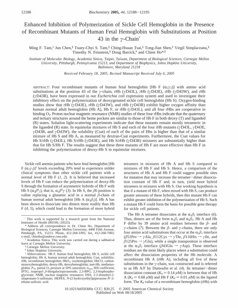
Enhanced Inhibition of Polymerization of Sickle Cell Hemoglobin in the Presenceof Recombinant Mutants of Human Fetal Hemoglobin with Substitutions at Position
43 in theγ-Chain†
Ming F. Tam,‡ Jun Chen,§ Tsuey-Chyi S. Tam,§ Ching-Hsuan Tsai,§ Tong-Jian Shen,# Virgil Simplaceanu,§
Timothy N. Feinstein,§ Doug Barrick,# and Chien Ho*,§
Institute of Molecular Biology, Academia Sinica, Taipei, Taiwan, Department of Biological Sciences, Carnegie MellonUniVersity, Pittsburgh, PennsylVania 15213, and Department of Biophysics, Johns Hopkins UniVersity,
Baltimore, Maryland 21218
ReceiVed February 18, 2005; ReVised Manuscript ReceiVed July 6, 2005
ABSTRACT: Four recombinant mutants of human fetal hemoglobin [Hb F (R2γ2)] with amino acidsubstitutions at the position 43 of theγ-chain, rHb (γD43L), rHb (γD43E), rHb (γD43W), and rHb(γD43R), have been expressed in ourEscherichia coliexpression system and used to investigate theirinhibitory effect on the polymerization of deoxygenated sickle cell hemoglobin (Hb S). Oxygen-bindingstudies show that rHb (γD43E), rHb (γD43W), and rHb (γD43R) exhibit higher oxygen affinity thanhuman normal adult hemoglobin (Hb A), Hb F, or rHb (γD43L), and all four rHbs are cooperative inbinding O2. Proton nuclear magnetic resonance (NMR) studies of these four rHbs indicate that the quaternaryand tertiary structures around the heme pockets are similar to those of Hb F in both deoxy (T) and liganded(R) states. Solution light-scattering experiments indicate that these mutants remain mostly tetrameric inthe liganded (R) state. In equimolar mixtures of Hb S and each of the four rHb mutants (γD43L, γD43E,γD43R, andγD43W), the solubility (Csat) of each of the pairs of Hbs is higher than that of a similarmixture of Hb S and Hb A, as measured by dextran-Csat experiments. Furthermore, the Csat values forHb S/rHb (γD43L), Hb S/rHb (γD43E), and Hb S/rHb (γD43R) mixtures are substantially higher thanthat for Hb S/Hb F. The results suggest that these three mutants of Hb F are more effective than Hb F ininhibiting the polymerization of deoxy-Hb S in equimolar mixtures.
Sickle cell anemia patients who have fetal hemoglobin [HbF (R2γ2)]1 levels exceeding 20% tend to experience milderclinical symptoms than other sickle cell patients with anormal level of Hb F (1, 2). It is believed that increasedlevels of Hb F can reduce the polymerization of deoxy-HbS through the formation of asymmetric hybrids of Hb F withHb S (R2âS
2), that is,R2âSγ (3). In Hb S, theâ6 position isvaline replacing a glutamic acid in a normalâ-chain ofhuman normal adult hemoglobin [Hb A (R2â2)]. Hb A hasbeen shown to dissociate into dimers more readily than HbF (4, 5), which could lead to the formation of more hybrid
tetramers in mixtures of Hb A and Hb S compared tomixtures of Hb F and Hb S. Hence, a comparison of thestructures of Hb A and Hb F could suggest possible sitesfor mutation that may increase the tetramer-dimer dissocia-tion constant of Hb F and, in turn, yield more hybridtetramers in mixtures with Hb S. Our working hypothesis isthat if a mutant of Hb F, when mixed with Hb S, can producegreater amounts of these hybrids, then this mutant Hb F canexhibit greater inhibition of the polymerization of Hb S. Sucha mutant Hb F could form the basis for possible gene therapyfor sickle cell patients.
The Hb A tetramer dissociates at theR1â2 interface (6).Thus, dimers are of the formR1â1 andR2â2. Hb A and HbF differ by 39 amino acid residues between theâ- andγ-chains (7). Between theâ- and γ-chains, there are onlyfour amino acid substitutions that occur at theR1â1 interface(â51Prof γAla, â112Cysf γThr, â116Hisf γIle, andâ125Prof γGlu), while a single transposition is observedat the R1â2 interface (â43Glu f γAsp). These interfaceresidues are the most likely places where a substitution couldaffect the dissociation properties of the Hb molecule. Arecombinant Hb A (rHb A), including all five of thesesubstitutions in theγ-chain, was constructed and is referredto as Hb A/F by Dumoulin et al. (4). Its tetramer-dimerdissociation constant (Kd ) 0.14µM) is between that of HbA (Kd ) 0.68µM) and Hb F (Kd ) 0.01µM) in the ligatedform. TheKd value of a recombinant hemoglobin (rHb) with
† This work is supported by a research grant from the NationalInstitutes of Health (R01HL-24525).
* Address all correspondence to Dr. Chien Ho, Department ofBiological Sciences, Carnegie Mellon University, 4400 Fifth Avenue,Pittsburgh, PA, 15213. Phone, 412-268-3395; fax, 412-268-7083;e-mail, [email protected].
‡ Academia Sinica. This work was carried out during a sabbaticalleave at Carnegie Mellon University.
§ Carnegie Mellon University.# Johns Hopkins University.1 Abbreviations: Hb F, human fetal hemoglobin; Hb S, sickle cell
hemoglobin; Hb A, human normal adult hemoglobin; Csat, solubility;rHb, recombinant hemoglobin; HbO2, oxyhemoglobin; HbCO, carbon-monoxyhemoglobin; deoxy-Hb, deoxyhemoglobin; met-Hb, methemo-globin;P50, partial O2 pressure at 50% saturation;nmax, Hill coefficient;IPTG, isopropylâ-thiogalactopyranoside; 2,3-BPG, 2,3-bisphospho-glycerate; NMR, nuclear magnetic resonance; DSS, 2-2-dimethyl-2-silapentane-5-sulfonate; HEPES,N-(2-hydroxyethyl)piperazine-N′-2-ethane-sulfonic acid.
12188 Biochemistry2005,44, 12188-12195
10.1021/bi050300z CCC: $30.25 © 2005 American Chemical SocietyPublished on Web 08/18/2005
a â43Gluf γAsp mutation is 0.21µM, which accounts formost of the tetramer-dimer strength of the penta-substitutedHb A mutant (8).
In this study, we have used ourEscherichia coliexpressionsystem (9) to construct four rHb F with mutations atγ43,namely rHb (γD43L), rHb (γD43E), rHb (γD43W), and rHb(γD43R), in the hope that these Hb F mutants could exhibitstronger inhibition of the polymerization when mixed withHb S. Oxygen-binding and proton nuclear magnetic reso-nance (NMR) studies were then conducted to characterizethe functional and structural changes brought about by thesesubstitutions in Hb F. The solubility (Csat) as measured bya modified dextran-Csat method (10) was used to investigatethe inhibitory effect of these mutations on the polymerizationof deoxy-Hb S.
MATERIALS AND METHODS
Construction of Plasmids.An expression plasmid (pHE8)containing bothR- andγ-globin genes in addition to theE.coli methionine aminopeptidase (MAP) gene was constructedin our laboratory (9). The 1.0-kb SmaI-NsiI fragment ofpHE8, which contains theR- and γ-globin genes, wasinserted into plasmid pTZ18U (Bio-Rad). The resultantplasmid (pTH8) was used in the mutagenesis experiments.Two synthetic oligonucleotides, 5′-ACTCAGCGTTTCT-TCTTAAGTTTCGGCAACCTGTC-3′ and 5′-CAGCGTT-TCTTTGAAAGCTTCGGCAACCTGTC-3′, were used asmutation primers to generate Aspf Leu and Aspf Glusubstitutions, respectively, at residue 43 of theγ-globin geneon plasmid pTH8. The wild-typeγ-globin gene of pHE8 wasthen replaced by the mutatedγ-globin genes in pTH8. TheQuikChange Site-Directed Mutagenesis Kit (Stratagene) wasused to make theγ43Asp f Trp substitution, using theprimers 5′-CCCGTGGACTCAGCGTTTCTTTTGGTCCT-TCGGCAACCTGTCTTC-3′ and 5′-GAAGACAGGTTGC-CGAAGGACCAAAAGAAACGCTGAGTCCACGGG-3′ inthe PCR experiment. Theγ43Aspf Arg mutant was gen-erated similarly using the primers 5′-CCCGTGGACT-CAGCGTTTCTTTCGATCGTTCGGCAACCTGTCTTC-3′ and 5′-GAAGACAGGTTGCCGAACGATCGAAAGAAA-CGCTGAGTCCACGGG-3′. The mutations on theγ-globingene were confirmed by DNA sequencing.
Production and Purification of Hemoglobins.The expres-sion plasmids were transformed intoE. coli JM109 cells andgrown in TB medium (11) in a Microferm fermentor (NewBrunswick Scientific, model MF20) or a Biostat fermentor(B. Braun Biotech, Inc., model Biostat-C-20) as describedpreviously (9, 12). Cells were grown to a density ofapproximately 1× 10⁹ cells/mL. Isopropylâ-thiogalacto-pyranoside (IPTG) was added to induce the expression ofthe Hb and MAP genes. Hemin and glucose were addedduring the induction period. Cell growth was continued foranother 4 h after IPTG induction, then harvested bycentrifugation and stored at-80 °C. Isolation and purifica-tion of the Hbs were performed according to the establishedprotocol developed in our laboratory (9, 12).
Edman degradation and electrospray ionization massspectrometric analyses of the four mutant rHbs were carriedout to assess the N-terminal processing of the proteins andconfirm the mutations on the recombinant Hbs (9, 12). Thefour mutant rHbs had the correct molecular weight andcontained∼1% methionine at the amino terminus.
Preparation of Other Hemoglobins.Human normal adultblood samples were obtained from the local blood bank, andHb A that was stripped of 2,3-bisphosphoglycerate (2,3-BPG)was isolated and purified by established methods in ourlaboratory (13). Hemolysates obtained from a blood sampleof an SS donor were prepared according to the sameprocedure as that for Hb A. They were equilibrated with 10mM phosphate/0.5 mM EDTA at pH 6.8 and 25°C. Thesamples were then passed through a Mono S column(Amersham Pharmacia cation exchanger 16/10) to separateHb S and Hb F from other components. Hb S samples werefree of 2,3-BPG. The Hb samples were then frozen in liquidnitrogen and stored in the CO-liganded form at-80 °C untilthey were used.
Oxygen-Binding Studies.Oxygen dissociation curves forthe Hb samples were obtained using a Hemox-Analyzer (TCSMedical Products, Huntington Valley, PA) at 29°C in 0.1M sodium phosphate buffer and pH range 5.6-8.2. Amethemoglobin (met-Hb) reductase system was utilized (14)to reduce the amount of met-Hb to less than 5% in allsamples tested. Partial pressure at 50% oxygenation (P50)and the Hill coefficient (nmax) were determined from eachcurve. The accuracy ofP50 measurements (in mmHg) is(8%, while that of nmax is (10%.
1H NMR Spectroscopy InVestigation.1H NMR spectra ofthe four rHb F mutants as well as Hb A and Hb F wereobtained at 29°C on a Bruker AVANCE DRX-300spectrometer operating at 300-MHz. All Hb samples werein 0.1 M sodium phosphate buffer at pH 7.0 and 100% water.The Hb concentration was about 4% (2.5 mM in terms ofheme). The water signal was suppressed by using a jump-and-return pulse sequence (15). Proton chemical shifts arereferenced to the methyl proton resonance of 2,2-dimethyl-2-silapentane-5-sulfonate (DSS) indirectly by using the watersignal, which occurs at 4.76 ppm downfield from that ofDSS at 29°C, as the internal reference.
Dextran-Csat Assay.This assay was performed as reported(10) with modifications for micro-sample handling. Thedextran-Csat developed by Bookchin et al. (10) is aconvenient procedure to investigate effects of Hb S modi-fications or mixtures with non-S Hbs on polymerizationunder conditions that avoid problems associated with highionic strength buffers during Csat measurements (16). Ourprotocol was developed using a total working volume of 100µL, which is 5-7 times smaller than that reported in theoriginal procedure, reducing the total Hb needed per mea-surement to about 8-14 mg per tube. Hb samples used wereconverted from the CO-liganded to the O2-liganded formusing a procedure developed in our laboratory (13). Theywere then concentrated to>200 mg/mL and changed into50 mM potassium phosphate (pH 7.5) using a Centriconconcentrator (Amicon). A stock solution of dextran (averagemass of 66 700 Da, Sigma), 320 mg/mL, was prepared in50 mM potassium phosphate at pH 7.5 using a volumetricflask. To a 200-µL PCR tube, 37.5µL of the stock dextransolution was added with a M50 Microman Pipet (Gilson,Inc., Middleton, WI). All other solutions were delivered withgastight glass syringes. Hb S alone or 50% Hb S plus 50%non-Hb S sample was deposited into the tube to give thedesired protein concentration. Buffer (50 mM potassiumphosphate, pH 7.5) was then added to yield a volume of
Sparing Effect of rHb F Mutants on Polymerization of Hb S Biochemistry, Vol. 44, No. 36, 200512189
95µL. The sample was mixed thoroughly then covered withmineral oil. Finally, 5µL of a 1 M stock solution of sodiumdithionite (oxygen free) dissolved in 50 mM potassiumphosphate (oxygen free) was delivered to the bottom of thePCR tube and stirred with a gastight syringe. The presenceof sodium dithionite ensured anaerobic condition for thehemoglobins and brought the total assay volume to 100µL.After incubation for 30 min at 37°C in a water bath, theresulting gel under the oil layer was carefully disrupted witha metal plunger, and the tubes were spun at room temperaturein a tabletop microfuge (Eppendorf Model 5415) at 14 000rpm for 20 min. The gel disruption was repeated twice more,followed by a final 30-min spin, and the supernatant withoutthe mineral oil was collected. There is a sufficient volumeof the soluble phase after centrifugation to permit triplicatemeasurements of the Hb concentrations over a range of initialHb concentrations from 80 to 140 mg/mL. Samples beforeand after dextran-Csat assay were converted to cyanomet-Hb using Drabkin’s solution, and the absorbance wasmeasured at 540 and 700 nm (17). In handling these highlyconcentrated and viscous protein solutions for spectropho-tometric measurements, samples were prepared by weightdilution in an analytical balance. Each data-point reportedin this paper represents the mean of at least two parallelexperiments and three replicate measurements each.
Light Scattering Measurements.To determine averagemolecular weights, Hb samples were run on a size-exclusionchromatography (SEC) column, and multi-angle static light-scattering measurements of the samples were performed. Hbsamples were first dialyzed extensively against 50 mMsodium phosphate buffer (pH 7.5) at concentrations of 5 mg/mL. Samples (50-µL) were injected onto a TosoHaasG3000PWXL SEC column equilibrated in 50 mM sodiumphosphate at a flow rate of 0.5 mL/min. Following chro-matographic separation, multi-angle light-scattering measure-ments were made on an in-line MiniDAWN multi-anglelight-scattering detector (Wyatt Technologies, Santa BarbaraCA). Concentrations were monitored using an in-line OptilabDSP differential refractive index detector (Wyatt Technolo-gies). In addition, dynamic light scattering was measuredby taking one of the 90° signals from the Minidawn andcomputing the autocorrelation function in a QELS detector(Wyatt Technologies). Weight-averaged molecular weights(Mw) and hydrated radii (determined from the translationaldiffusion constant using the Stokes-Einstein equation) werecalculated using the ASTRA 4.0 software package (WyattTechnologies).
RESULTS AND DISCUSSION
Oxygen-Binding Studies.We have constructed four re-combinant mutants of Hb F with amino acid substitutions atthe 43 position of theγ-chain, which is located at theR1γ2
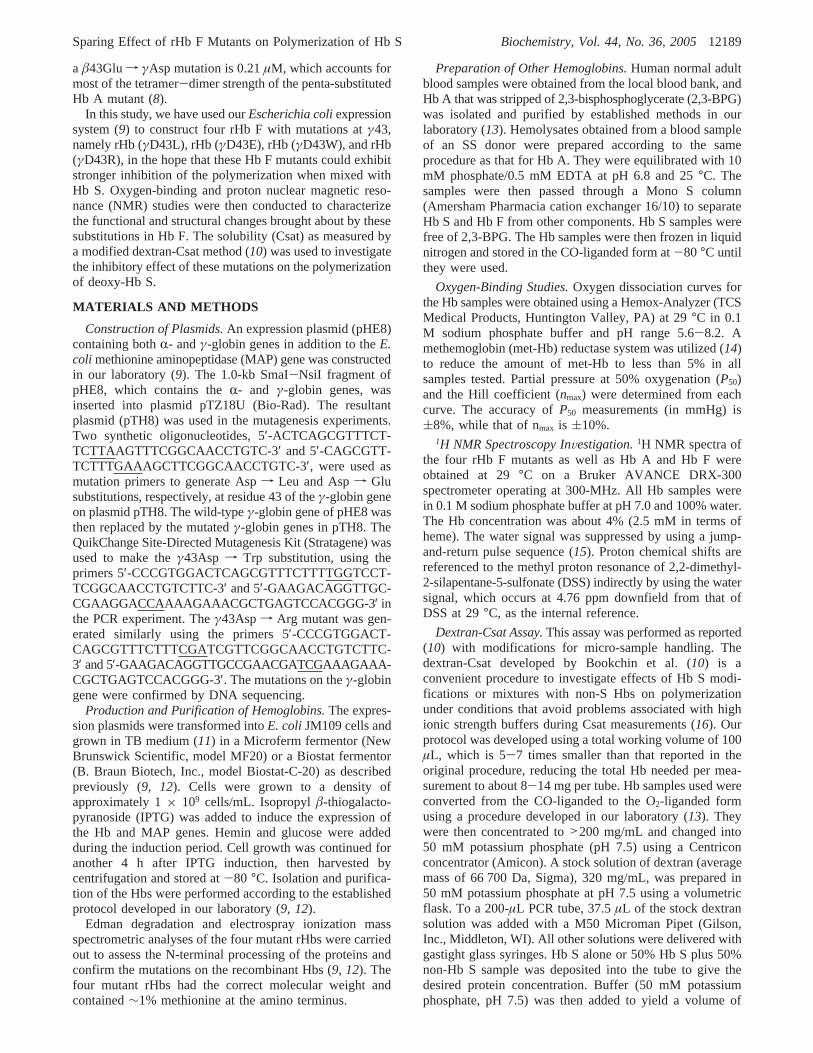
subunit interface, namely rHb (γD43L), rHb (γD43E), rHb(γD43W), and rHb (γD43R). Figure 1 shows the O2-bindingproperties of Hb A, Hb F, and the four mutants of Hb F in0.1 M sodium phosphate at 29°C. Plots ofP50 versus pH(whereP50 is the oxygen pressure at 50% saturation) indicateno significant difference in the oxygen-binding affinityamong Hb A, Hb F, and rHb (D43L) from pH 6.8 to 8.2(Figure 1A). However, these plots suggest a possibledifference in the properties of these three Hbs below pH 6.5.rHb (γD43E), rHb (γD43W), and rHb (γD43R) exhibit
higher oxygen affinity than Hb A, Hb F, and rHb (γD43L)at all pH values below 8.0. All four rHbs are cooperative inbinding O2 with Hill coefficients (nmax) ranging from 2.1 to3.2 (Figure 1B).
The Bohr effect, an important functional property, can beexpressed as the number of hydrogen ions released uponoxygenation and measured as∆H+ ) -∆log P50/∆pH (18).Table 1 summarizes the number of H+ ions released per hemeover the pH range from 6.5 to 8.2 for Hb A, Hb F, and thefour Hb F mutants. All four Hb F mutants have a noticeabledecrease in the number of H+ ions released compared to HbF over the specified pH range, suggesting that they possessa slightly lower alkaline Bohr effect than Hb F. rHb (γD43L)and rHb (γD43E) exhibit a Bohr effect only slightly lessthan that of Hb A. In spite of these differences in the oxygen-binding properties among these mutants and wild-type Hb
FIGURE 1: Oxygen-binding properties of Hb A (b), Hb F (0), rHb(γD43L) (4), rHb (γD43E) (O), rHb (γD43W) ([), and rHb(γD43R) (9) in 0.1 M sodium phosphate buffer at 29°C as afunction of pH: (A) oxygen affinity; and (B) Hill coefficient.
Table 1: Bohr Effects of Hb A, Hb F, and Four Hb F Mutants in0.1 M Sodium Phosphate Buffer (pH 6.5-8.3) at 29°Ca
hemoglobin -∆log P50/∆pHb % reductionc
Hb A 0.42 (pH 6.52-8.21) -Hb F 0.56 (pH 6.52-7.99) -rHb (γD43L) 0.41 (pH 6.50-8.33) 26.8rHb (γD43E) 0.39 (pH 6.50-8.16) 30.4rHb (γD43W) 0.36 (pH 6.52-8.17) 35.7rHb (γD43R) 0.37 (pH 6.82-8.14) 33.9a All rHb data are taken in the presence of a methemoglobin reductase
system (14). Met-Hb formation did not exceed 4.3% in any sample,either during sample preparation or measurement.b The Bohr effect ismeasured by-∆log P50/∆pH, which gives the number of H+ ionsreleased upon ligand binding over the pH range specified.c Thereduction of Bohr effects of the four Hb F mutants is relative to that ofHb F.
12190 Biochemistry, Vol. 44, No. 36, 2005 Tam et al.
F, our results show clearly that these mutants can functionas oxygen carriers.
It is of interest to note that the mutant rHb (γD43L) hasoxygen affinity close to that of Hb F. In contrast, rHb(γD43E), rHb (γD43W), and rHb (γD43R) all possess higheroxygen affinity than that of Hb F. Moreover, the oxygenaffinity of Hb F and of these four mutants of Hb F isincreased in accordance with the size of the substituted aminoacid residue: Asp< Leu < Glu < Arg < Trp. In Hb F andrHb (γD43L), the presence ofγ43Asp orγ43Leu at theR1γ2
subunit interface permits maximal oxygen release, but thereplacement of Asp byγ43Glu, γ43Trp, andγ43Arg mayintroduce some conformational rearrangements around theheme pocket as suggested from the ring-current-shiftedproton resonances (see below) resulting in perturbation ofO2 binding.
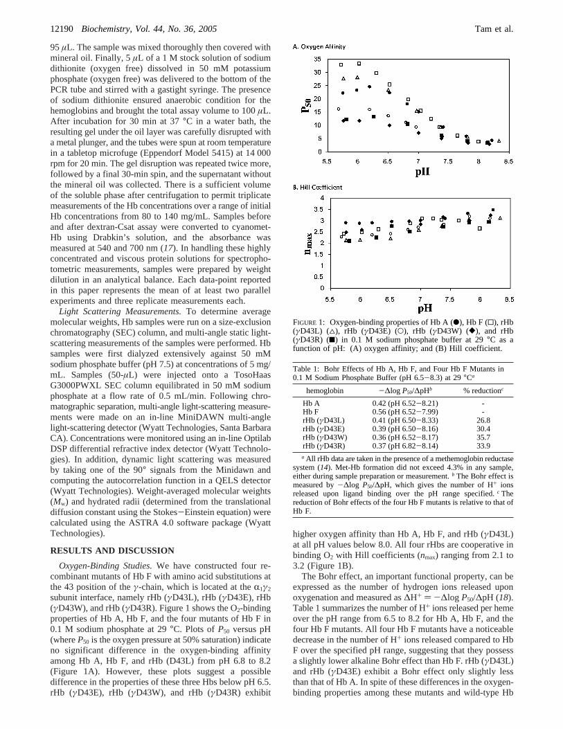
1H NMR Studies of rHb F Samples.The exchangeableproton resonances and ring-current-shifted proton resonancesof Hb A, Hb F, and the four mutants of rHb F in the COform are shown in Figure 2. The ring-current-shifted protonresonances are an excellent indication of the tertiary structureof the heme pocket (19). The resonances at∼-1.75 and∼-1.85 ppm have been assigned to theγ2-CH3 of theE11Val of theR-chain andâ-chain of HbCO A, respectively(20, 21). Since theR-chain of Hb F is the same as that ofHb A, we can assume that the resonance at∼-2.0 ppm isdue to theγ2-CH3 of the E11Val of theγ-chain of HbCO F.These two resonances of rHbCO (γD43L) are essentiallyidentical to those of HbCO F. The∼-1.8-ppm resonanceof rHbCO (γD43E) is shifted slightly upfield, and that at-2.0-ppm peak is shifted about 0.1 ppm upfield compared
to those of HbCO F. For rHbCO (γD43W), the resonanceat ∼-1.8 ppm is shifted about 0.1 ppm downfield and thatat -2.0 ppm is shifted upfield by about 0.1 ppm comparedto those of HbCO F. The-1.8-ppm-resonance of rHbCO(γD43R) is shifted slightly downfield, and that at-2.0-ppmsignal is shifted slightly upfield relative to those of HbCOF. These results indicate that, upon mutatingγ43Asp, theheme pocket of theγ-chain is more perturbed than that ofthe R-chain, as expected, but the changes are minimal.
The resonances in the downfield portion of the1H NMRspectrum of HbCO arise from the exchangeable protons inthe subunit interfaces (19). The resonances at∼12.8 ppmand∼12.0 ppm have been assigned to the H-bonds betweenR122His andâ35Tyr andR103His andâ131Gln in theR1â1
subunit interface (22, 23), respectively. On the basis of thesimilarity of the crystal structures of Hb A and Hb F (24),we can assume that the H-bond pattern in theR1â1 andR1γ1
interfaces should be very similar as shown in Figure 2A.These two resonances of rHbCO (γD43L) are essentiallyidentical to those of Hb F, indicating no significant perturba-tions around theR1γ1 interface of this mutant. The resonancefor the H-bond betweenR122His andγ35Tyr of rHbCO(γD43E) is shifted downfield by about 0.1 ppm relative tothat of HbCO F, but the resonance at 12.0 ppm of rHbCO(γD43E) remains the same as that of HbCO F. For rHbCO(γD43W), the resonance at 12.8 ppm is shifted about 0.1ppm upfield and that at 12.0 ppm is shifted downfield byabout 0.1 ppm compared to those of HbCO F. The 12.8-ppm resonance of rHbCO (γD43R) is shifted slightly upfield,and that at the 12.0-ppm signal is shifted downfield by about0.1 ppm relative to those of HbCO F.
FIGURE 2: The 300-MHz1H NMR spectra of 4-6% solutions of Hb A, Hb F, rHb (γD43L), rHb (γD43E), rHb (γD43W), and rHb(γD43R) in the CO form in 0.1 M sodium phosphate buffer at pH 7.0 and 29°C: (A) exchangeable proton resonances; and (B) ring-current-shifted proton resonances.
Sparing Effect of rHb F Mutants on Polymerization of Hb S Biochemistry, Vol. 44, No. 36, 200512191
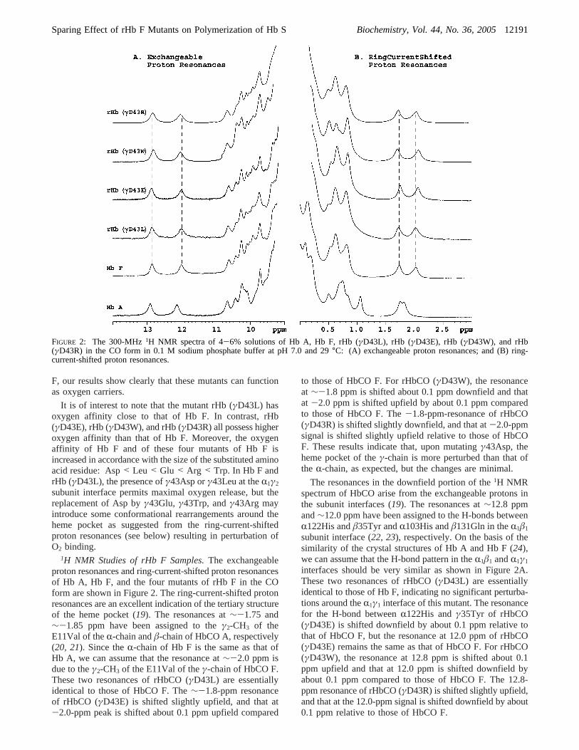
Figure 3 shows the hyperfine-shifted resonances andexchangeable proton resonances of the four mutants in thedeoxy form. The resonance at 63 ppm has been assigned tothe hyperfine-shifted NH-exchangeable proton of the proxi-mal histidine residueR87His of theR-chain of deoxy-HbA, and the peak at 74 ppm has been assigned to thecorrespondingâ92His of theâ-chain of deoxy-Hb A (25,26). As shown in Figure 3A, the resonance at 63 ppm is thesame in both deoxy-Hb A and deoxy-Hb F as expected. Thehyperfine-shifted NH-exchangeable proton resonance of theproximal histidine of theγ-chain of deoxy-Hb F is shiftedupfield by 1 ppm compared to that of theâ-chain of deoxy-Hb A, reflecting a slight difference in the heme environmentbetween theâ- andγ-chains (24, 27). These two resonancesof rHb (γD43E), rHb (γD43W), and rHb (γD43R) areindistinguishable from those of Hb F, suggesting that noperturbation has been introduced into the heme pocket atthe proximal histidyl residues by the mutation. However,these two resonances of rHb (γD43L) are shifted upfield by1 ppm relative to those of Hb F, indicating a slight differencein the conformation between the proximal histidines andheme group of the mutant compared to that of Hb F. Theresonances in the spectral region from 13 to 24 ppm arisefrom the hyperfine-shifted resonances of the porphyrin ringand the amino acid residues located in proximity to the hemepockets as well as the exchangeable proton resonances(Figure 3B) (19). There are no noticeable differences in theresonances from 13 to 24 ppm between deoxy-Hb F and thefour mutants in the deoxy form.
Taking all these data together, we can conclude that,whether the negatively chargedγ43Asp of Hb F is exchangedfor a neutral, negative, or positive residue, the overallstructures of these mutants are similar to those of Hb F inboth deoxy (T) and liganded (R) states.
Dextran-Csat Measurements. We have shown previouslywith delay-time gelation and Csat measurements that Hb Swith amino acid substitutions atR114 andâ87 can inhibitthe formation of the Hb S polymer (28). In this study, weused a modified version of the method of Bookchin et al.(10) to measure the solubilities of Hb S and of equimolarmixtures of Hb S and either Hb A, Hb F, or each of the fourmutants of Hb F. This method provides a convenient wayto screen the effects of amino acid substitutions on thepolymerization of Hb S under conditions that avoid problemsassociated with high ionic strength buffers (10). With ourmodified protocol, a Csat value can be obtained with lessthan 50 mg of proteins for each trial. However, it should benoted that there are limitations in using the dextran-Csatmethod; that is, it is not under physiological conditions.
The dextran-Csat results are shown in Figure 4 and listedin Table 2. A greater increase in dextran-Csat occurs uponmixing equimolar quantities of Hb S with Hb F than withHb A. Our data for Hb S alone, 50%Hb S/50%Hb A, and50%Hb S/50%Hb F are similar to the published results (10),indicating that our procedures and data are reliable. However,within the range of 80-140 mg/mL, we have observed agradual increase in Csat values for all samples tested. Thedextran-Csat value of an equimolar mixture of Hb S withrHb (γD43W) is similar to that of Hb S/Hb F. The Csatvalues obtained for Hb S/rHb (γD43L), Hb S/rHb (γD43E),and Hb S/rHb (γD43R) are similar at either 80 or 100 mg/mL initial Hb concentration and significantly higher thanthat for Hb S/Hb F (Table 2). At higher initial proteinconcentrations (120 and 140 mg/mL), Hb S/rHb (γD43E)and Hb S/rHb (γD43R) have higher Csat values than that ofHb S/rHb F (γD43L). Even thoughγD43 of Hb F is locatedat theR1γ2 interface, it is also solvent-accessible. Whetherthe charged guanidino or glutamyl side chain has better
FIGURE 3: The 300-MHz1H NMR spectra of 4-6% solutions of Hb A, Hb F, rHb (γD43L), rHb (γD43E), rHb (γD43W), and rHb(γD43R) in the deoxy form in 0.1 M sodium phosphate buffer at pH 7.0 and 29°C: (A) hyperfine-shifted NδH resonances of proximalhistidines; and (B) hyperfine-shifted and exchangeable proton resonances.
12192 Biochemistry, Vol. 44, No. 36, 2005 Tam et al.
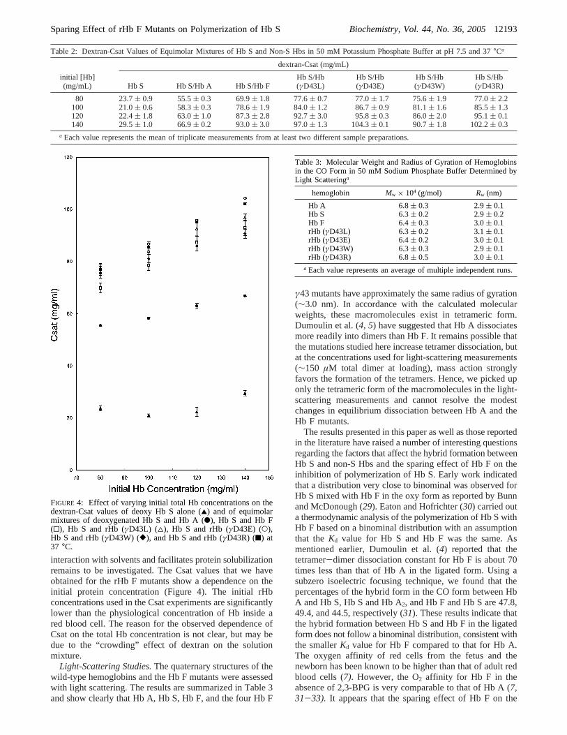
interaction with solvents and facilitates protein solubilizationremains to be investigated. The Csat values that we haveobtained for the rHb F mutants show a dependence on theinitial protein concentration (Figure 4). The initial rHbconcentrations used in the Csat experiments are significantlylower than the physiological concentration of Hb inside ared blood cell. The reason for the observed dependence ofCsat on the total Hb concentration is not clear, but may bedue to the “crowding” effect of dextran on the solutionmixture.
Light-Scattering Studies.The quaternary structures of thewild-type hemoglobins and the Hb F mutants were assessedwith light scattering. The results are summarized in Table 3and show clearly that Hb A, Hb S, Hb F, and the four Hb F
γ43 mutants have approximately the same radius of gyration(∼3.0 nm). In accordance with the calculated molecularweights, these macromolecules exist in tetrameric form.Dumoulin et al. (4, 5) have suggested that Hb A dissociatesmore readily into dimers than Hb F. It remains possible thatthe mutations studied here increase tetramer dissociation, butat the concentrations used for light-scattering measurements(∼150 µM total dimer at loading), mass action stronglyfavors the formation of the tetramers. Hence, we picked uponly the tetrameric form of the macromolecules in the light-scattering measurements and cannot resolve the modestchanges in equilibrium dissociation between Hb A and theHb F mutants.
The results presented in this paper as well as those reportedin the literature have raised a number of interesting questionsregarding the factors that affect the hybrid formation betweenHb S and non-S Hbs and the sparing effect of Hb F on theinhibition of polymerization of Hb S. Early work indicatedthat a distribution very close to binominal was observed forHb S mixed with Hb F in the oxy form as reported by Bunnand McDonough (29). Eaton and Hofrichter (30) carried outa thermodynamic analysis of the polymerization of Hb S withHb F based on a binominal distribution with an assumptionthat theKd value for Hb S and Hb F was the same. Asmentioned earlier, Dumoulin et al. (4) reported that thetetramer-dimer dissociation constant for Hb F is about 70times less than that of Hb A in the ligated form. Using asubzero isoelectric focusing technique, we found that thepercentages of the hybrid form in the CO form between HbA and Hb S, Hb S and Hb A2, and Hb F and Hb S are 47.8,49.4, and 44.5, respectively (31). These results indicate thatthe hybrid formation between Hb S and Hb F in the ligatedform does not follow a binominal distribution, consistent withthe smallerKd value for Hb F compared to that for Hb A.The oxygen affinity of red cells from the fetus and thenewborn has been known to be higher than that of adult redblood cells (7). However, the O2 affinity for Hb F in theabsence of 2,3-BPG is very comparable to that of Hb A (7,31-33). It appears that the sparing effect of Hb F on the
Table 2: Dextran-Csat Values of Equimolar Mixtures of Hb S and Non-S Hbs in 50 mM Potassium Phosphate Buffer at pH 7.5 and 37°Ca
dextran-Csat (mg/mL)
initial [Hb](mg/mL) Hb S Hb S/Hb A Hb S/Hb F
Hb S/Hb(γD43L)
Hb S/Hb(γD43E)
Hb S/Hb(γD43W)
Hb S/Hb(γD43R)
80 23.7( 0.9 55.5( 0.3 69.9( 1.8 77.6( 0.7 77.0( 1.7 75.6( 1.9 77.0( 2.2100 21.0( 0.6 58.3( 0.3 78.6( 1.9 84.0( 1.2 86.7( 0.9 81.1( 1.6 85.5( 1.3120 22.4( 1.8 63.0( 1.0 87.3( 2.8 92.7( 3.0 95.8( 0.3 86.0( 2.0 95.1( 0.1140 29.5( 1.0 66.9( 0.2 93.0( 3.0 97.0( 1.3 104.3( 0.1 90.7( 1.8 102.2( 0.3
a Each value represents the mean of triplicate measurements from at least two different sample preparations.
FIGURE 4: Effect of varying initial total Hb concentrations on thedextran-Csat values of deoxy Hb S alone (2) and of equimolarmixtures of deoxygenated Hb S and Hb A (b), Hb S and Hb F(0), Hb S and rHb (γD43L) (4), Hb S and rHb (γD43E) (O),Hb S and rHb (γD43W) ([), and Hb S and rHb (γD43R) (9) at37 °C.
Table 3: Molecular Weight and Radius of Gyration of Hemoglobinsin the CO Form in 50 mM Sodium Phosphate Buffer Determined byLight Scatteringa
hemoglobin Mw × 104 (g/mol) Rw (nm)
Hb A 6.8( 0.3 2.9( 0.1Hb S 6.3( 0.2 2.9( 0.2Hb F 6.4( 0.3 3.0( 0.1rHb (γD43L) 6.3( 0.2 3.1( 0.1rHb (γD43E) 6.4( 0.2 3.0( 0.1rHb (γD43W) 6.3( 0.3 2.9( 0.1rHb (γD43R) 6.8( 0.5 3.0( 0.1
a Each value represents an average of multiple independent runs.
Sparing Effect of rHb F Mutants on Polymerization of Hb S Biochemistry, Vol. 44, No. 36, 200512193
polymerization of Hb S inside red blood cells could beaffected by a number of factors, for example, the extent andnature of hybridization of Hb F with Hb S, 2,3-BPG, degreesof oxygenation of Hb A, Hb S, and Hb F, and so forth.Hence, additional studies are clearly needed to elucidate thefactors that affect the hybrid formation and the sparing effectof Hb F on the polymerization of Hb S.
In conclusion, it is known that when two hemoglobins(such as Hb S-Hb A or Hb S-Hb F) are mixed in the oxystate, the tetramers are in fairly rapid dissociation equilibriumwith dimers, which reassociate randomly to form hybridtetramers (Hb AS or Hb FS) (7). Kinetic and thermodynamicstudies of the polymerization of cross-linked Hb AS hybridhemoglobin suggested that Hb AS polymerized similarly oridentically to Hb S (34). Thus, Hb A does not inhibit thepolymerization of Hb S through the formation of Hb ASbut can prevent the aggregation of Hb AS by acting as aninert component which simply dilutes the Hb S molecules.In contrast, kinetic and thermodynamic studies of cross-linked Hb FS hybrid hemoglobin have shown that Hb FSdoes not polymerize (35). The effect of cross-linked Hb FSon the aggregation of Hb S has shown that Hb FS couldinhibit the polymerization of deoxy-Hb S, but to a lesserextent than does Hb F. These results thus explain the strongerinhibitory effect of Hb F on the polymerization of deoxy-Hb S than that shown by Hb A. We report here three Hb Fmutants that have an even stronger inhibitory effect on thepolymerization of deoxy-Hb S than that of the wild-type HbF. The mechanism of this inhibition needs to be furtherinvestigated.
REFERENCES
1. Perrine, R. P., Pembrey, M. E., John, P., Perrine, S., and Shoup,F. (1978) Natural history of sickle cell anemia in Saudi Arabs. Astudy of 270 subjects,Ann. Intern. Med. 88, 1-6.
2. Powars, D. R., Weiss, J. N., Chan, L. S., and Schroeder, W. A.(1984) Is there a threshold level of fetal hemoglobin thatameliorates morbidity in sickle cell anemia?,Blood 63, 921-926.
3. Bookchin, R. M., Nagel, R. L., and Balazs, T. (1975) Role ofhybrid tetramer formation in gelation of haemoglobin S,Nature256, 667-668.
4. Dumoulin, A., Manning, L. R., Jenkins, W. T., Winslow, R. M.,and Manning, J. M. (1997) Exchange of subunit interfaces betweenrecombinant adult and fetal hemoglobins. Evidence for a functionalinter-relationship among regions of the tetramer,J. Biol. Chem.272, 31326-31332.
5. Dumoulin, A., Padovan, J. C., Manning, L. R., Popowicz, A.,Winslow, R. M., Chait, B. T., and Manning, J. M. (1998) TheN-terminal sequence affects distant helix interactions in hemo-globin. Implications for mutant proteins from studies on recom-binant hemoglobin felix,J. Biol. Chem. 273, 35032-35038.
6. Park, C. M. (1973) Isoelectric focusing and the study of interactingprotein systems: ligand binding, phosphate binding, and subunitexchange in hemoglobin,Ann. N.Y. Acad. Sci. 209, 237-257.
7. Bunn, H. F., and Forget, B. G. (1986)Hemoglobin: Molecular,Genetic and Clinical Aspects, W. B. Saunders Co., Philadelphia,PA.
8. Chen, W., Dumoulin, A., Li, X., Padovan, J. C., Chait, B. T.,Buonopane, R., Platt, O. S., Manning, L. R., and Manning, J. M.(2000) Transposing sequences between fetal and adult hemoglo-bins indicates which subunits and regulatory molecule interfacesare functionally related,Biochemistry 39, 3774-3781.
9. Shen, T.-J., Ho, N. T., Zou, M., Sun, D. P., Cottam, P. F.,Simplaceanu, V., Tam, M. F., Bell, D. A., Jr., and Ho, C. (1997)Production of human normal adult and fetal hemoglobins inEscherichia coli, Protein Eng. 10, 1085-1097.
10. Bookchin, R. M., Balazs, T., Wang, Z. P., Josephs, R., and Lew,V. L. (1999) Polymer structure and solubility of deoxyhemoglobinS in the presence of high concentrations of volume-excluding 70-
kDa dextran. Effects of non-S hemoglobins and inhibitors,J. Biol.Chem. 274, 6689-6697.
11. Lech, K., and Brent, R. (1987)Escherichia coli, Plasmids andBacteriophages, inCurrent Protocols in Molecular Biology(Ausubel, F. M., Brent, R., Kingston, R. E., Moore, O. D.,Seidman, J. G., Smith, J. A., and Struhl, K., Eds.), Vol. I, p 1.0.1-1.2.2, Wiley, New York.
12. Shen, T.-J., Ho, N. T., Simplaceanu, V., Zou, M., Green, B. N.,Tam, M. F., and Ho, C. (1993) Production of unmodified humanadult hemoglobin inEscherichia coli, Proc. Natl. Acad. Sci. U.S.A.90, 8108-8112.
13. Lindstrom, T. R., and Ho, C. (1972) Functional nonequivalenceof R andâ hemes in human adult hemoglobin,Proc. Natl. Acad.Sci. U.S.A. 69, 1707-1710.
14. Hayashi, A., Suzuki, T., and Shin, M. (1973) An enzymic reductionsystem for metmyoglobin and methemoglobin, and its applicationto functional studies of oxygen carriers,Biochim. Biophys. Acta310, 309-316.
15. Plateau, P., and Gue´ron, M. (1982) Exchangeable proton NMRwithout base-line distorsion, using new strong-pulse sequences,J. Am. Chem. Soc. 104, 7310-7311.
16. Magdoff-Fairchild, B., Poillon, W. N., and Bertles, J. F. (1976)Thermodynamic studies of polymerization of deoxygenated sicklecell hemoglobin,Proc. Natl. Acad. Sci. U.S.A. 73, 990-994.
17. Drabkin, D. L. (1946) Spectrophotometric studies XIV. Thecrystallographic and optical properties of the hemoglobin of manin comparison with those of other species,J. Biol. Chem. 164,703-723.
18. Wyman, J. (1964) Linked functions and reciprocal effects inhemoglobin: a second look,AdV. Protein Chem. 19, 223-286.
19. Ho, C. (1992) Proton nuclear magnetic resonance studies onhemoglobin: cooperative interactions and partially ligated inter-mediates,AdV. Protein Chem. 43, 153-312.
20. Lindstrom, T. R., Nore´n, I. B. Charache, S., Lehmann, H., andHo, C. (1972) Nuclear magnetic resonance studies of hemoglobins.VII. Tertiary structure around ligand binding site in carbonmon-oxyhemoglobin,Biochemistry 11, 1677-1681.
21. Dalvit, C., and Ho, C. (1985) Proton nuclear Overhauser effectinvestigation of the heme pockets in ligated hemoglobin: con-formational differences between oxy and carbonmonoxy forms,Biochemistry 24, 3398-3407.
22. Simplaceanu, V., Lukin, J. A., Fang, T. Y., Zou, M., Ho, N. T.,and Ho, C. (2000) Chain-selective isotopic labeling for NMRstudies of large multimeric proteins: application to hemoglobin,Biophys. J. 79, 1146-1154.
23. Chang, C. K., Simplaceanu, V., and Ho. C. (2002) Effects of aminoacid substitutions at beta 131 on the structure and properties ofhemoglobin: evidence for communication between alpha 1 beta1- and alpha 1 beta 2-subunit interfaces,Biochemistry 41, 5644-5655.
24. Frier, J. A., and Perutz, M. F. (1977) Structure of human foetaldeoxyhaemoglobin,J. Mol. Biol. 112, 97-112.
25. Takahashi, S., Lin, A.-K. C., and Ho, C. (1980) Proton nuclearmagnetic resonance studies of hemoglobins M Boston (R58E7His f Tyr) and M Milwaukee (â67E11 Valf Glu): spectralassignments of hyperfine-shifted proton resonances and of proxi-mal histidine (E7) NH resonances to theR andâ chains of normalhuman adult hemoglobin,Biochemistry 19, 5196-5202.
26. La Mar, G. N., Nagai, K., Jue, T., Budd, D. L., Gersonde, K.,Sick, H., Kagimoto, T., Hayashi, A., and Taketa, F. (1980)Assignment of proximal histidyl imidazole exchangeable protonNMR resonances to individual subunits in hemoglobins A, Boston,Iwate and Milwaukee,Biochem. Biophys. Res. Commun. 96,1172-1177.
27. Davis, D. G., Charache, S., and Ho, C. (1969) Nuclear magneticresonance studies of hemoglobins. III. Evidence for the non-equivalence ofR- andâ-chains in azide derivatives of methemo-globins,Proc. Natl. Acad. Sci. U.S.A. 63, 1403-1409.
28. Ho, C., Willis, B. F., Shen, T.-J., Ho, N. T., Philip Sun, D.-Z.,Tam, M. F., Suzuka, S. M., Fabry, M. E., and Nagel, R. L. (1996)Roles of alpha 114 and beta 87 amino acid residues in thepolymerization of hemoglobin S: implications for gene therapy,J. Mol. Biol 263, 475-485.
29. Bunn, H. F., and McDonough, M. (1974) Asymmetric hemoglobinhybrids. An approach to the study of subunit interactions,Biochemistry 13,988-993.
30. Eaton, W. A., and Hofrichter, J. (1990) Sickle cell hemoglobinpolymerization,AdV. Protein Chem. 40,63-279.
12194 Biochemistry, Vol. 44, No. 36, 2005 Tam et al.
31. Larson, S. C., Fisher, G. W., Ho, N. T., Shen, T.-J., and Ho, C.(1999) A biochemical and biophysical characterization of recom-binant mutants of fetal hemoglobin and their interaction with sicklecell hemoglobin,Biochemistry 38,9549-9555.
32. Allen, D. W., Wyman, J., and Smith, C. A. (1953) The oxygenequilibrium of foetal and adult hemoglobin,J. Biol. Chem. 203,81-97.
33. Fang, T.-Y., Zou, M., Simplaceanu, V., Ho, N. T., and Ho, C.(1999) Assessment of roles of surface histidyl residues in themolecular basis of the Bohr effect and ofâ143 histidine in the
binding of 2,3-bisphosphoglycerate in human normal adulthemoglobin,Biochemistry 38,13423-13432.
34. Adachi, K., and Asakura, T. (1984) Polymerization of AS hybridhemoglobin. Potent inhibitory effect of hemoglobin A on thepolymerization of AS hybrid hemoglobin,J. Biol. Chem. 259,2108-2112.
35. Nibu, K., and Adachi, K. (1985) Effect of FS (R2γâS) hybridhemoglobin on Hb S nucleation and aggregation,Biochim.Biophys. Acta 829, 97-102.
BI050300Z
Sparing Effect of rHb F Mutants on Polymerization of Hb S Biochemistry, Vol. 44, No. 36, 200512195