Embed Size (px)
Citation preview

Enzymatyczne układy antyoksydacyjnedysmutazy nadtlenkowe, katalazy,
peroksydazy
Wykład 11
17.01.2011

Enzymy tworzące i unnieczynniające reaktywne formy tlenu
Forstermann U, Nature Rev Cardiol 2008

Dysmutazy ponadtlenkowe
Dysmutazy nadtlenkowe – (SOD EC1.15.1.1) – klasa enzymów katalizujących dysmutację anionorodnika nadtlenkowego do tlenu oraz nadtlenku wodoru.
Dysmutazy tworzą istotny system antyoksydacyjny w komórkach niemal wszystkich żywych organizmów
Jednym z wyjątków są bakterie z rodzaju Lactobacillus (Lactobacillus plantarum)-używają innego systemu - także kilka gatunków Leptospira oraz trzy wirulentne szczepy gonokoków Neisseria gonorrhoeae (bogate w katalazę oraz peroksydazy)

Dysmutazy ponadtlenkowe
Reakcja :
M(n+1)+-SOD + O2− → Mn+-SOD + O2
Mn+-SOD + O2− + 2H+ → M(n+1)+-SOD + H2O
Gdzie: M = Cu (n=1) ; Mn (n=2) ; Fe (n=2) ; Ni (n=2).
W reakcji tej stan oksydacyjny metalu oscyluje pomiędzy n a n+1
Całościowo:
O2-. + O2
-.+ 2H+ H2O2 + O2

Figure 18.22Superoxide Dismutase MechanismThe oxidized form of superoxide dismutase (Mox) reacts with one superoxide ion to form O2 and generate the reduced form of the enzyme (Mred). The reduced form then reacts with a second superoxide and two protons to form hydrogen peroxide and regenerate the oxidized form of the enzyme.
Dysmutazy ponadtlenkowe
Stryer, Biochemistry,

Discovered by Irwin Fridovich and Joe McCord, SOD enzymes were previously thought to be several metalloproteins with unknown function (for example, CuZnSOD was known as erythrocuprein).[2] Several common forms of SOD exist: they are proteins cofactored with copper and zinc, or manganese, iron, or nickel. For example, Brewer (1967) identified a protein that became known as superoxide dismutase as an indophenol oxidase by protein analysis of starch gels using the phenazine-tetrazolium technique.
Dysmutazy ponadtlenkowe
Wikipedia

Różne formy dysmutaz ponadtlenkowych
1. SOD1 – CuZn-SOD - cytoplazmatyczna, zawiera Cu & Zn w centrum aktywnym;
gen zlokalizowany w chromosomie 21 (21q22.1)2. SOD2 – MnSOD- mitochondrialna – zawiera Mn
gen w chromosomie 6 - 6q25.33. SOD3 –EC-SOD (CuZn-SOD) - pozakomórkowa –
zawiera CuZn(gen w chromosomie 4 – 4p15.3-p15.1)
1. SOD1 występuje poza cytozolem w jądrze komórkowym, peroksysomach, przestrzeni między-błonowej w mitochondriach ludzkich komórek
Ssaki (i większość strunowców)

Dysmutaza miedziowo-cynkowa
Valnetine et al., Ann Rev Biochem 2005
Homodimer32kDajedno miejsce wiążące Zn oraz jedno Cu

Różne formy dysmutaz ponadtlenkowych
Rośliny Mn-SOD – mitochondria i peroksysomyFe-SOD – chloroplasty; także peroksysomyCuZn-SOD – cytosol, chloroplasty, peroksysomy, apoplasty
Bakterie E. coli – cztery rodzaje SOD
1. CuZnSOD – periplazmatyczna 2. MnSOD – wewnątrzkomórkowa 3. FeSOD 4. Hybrydowa – zawiera podjednostkę enzymu Mn oraz podjednostkę
Fe w tej samej dimerycznej cząsteczce

Copper and zinc – most commonly used by eukaryotes. The cytosols of virtually all eukaryoticcells contain an SOD enzyme with copper and zinc (Cu-Zn-SOD). For example, Cu-Zn-SOD available commercially is normally purified from the bovine erythrocytes: The Cu-Zn enzyme is a homodimer of molecular weight 32,500. The bovine Cu-Zn protein was the first SOD structure to be solved, in 1975.[5]
Iron or manganese – used by prokaryotes and protists, and in mitochondria
Manganese – Chicken liver (and nearly all other) mitochondria, and many bacteria (such as E. coli), contain a form with manganese (Mn-SOD): for example, the Mn-SOD found in human mitochondria. \
Nickel – prokaryotic.
Dysmutazy eukariotyczne i prokariotyczne

Faraci and Didion, ATVB 2004
Lokalizacja subkomórkowa dysmutaz
W normalnych warunkach mitochondrialny łańcuch oddechowyjest głównym źródłem anionorodnika ponadtlenkowego, przekształcając około 5% cząsteczkowego tlenu do O2
-.

HZ Szeto, AAPS Journal. 2006; 8(3): E521-E531. DOI:
Lokalizacja SOD względem łańcucha oddechowegow mitochondriach

Inhibitory dysmutaz ponadtlenkowych
1. CuZnSOD - a) cyjanek b) DTC – diethydithiocarbamate
2. FeSOD oraz MnSOD nie są hamowane przez cyjanek
3. CuZnSOD oraz FeSOD są hamowane przez dlugą inkubację z nadtlenkiem wodoru, podczas gdy MnSOD nie jest
3. FeSOD – hamowana przez DTC
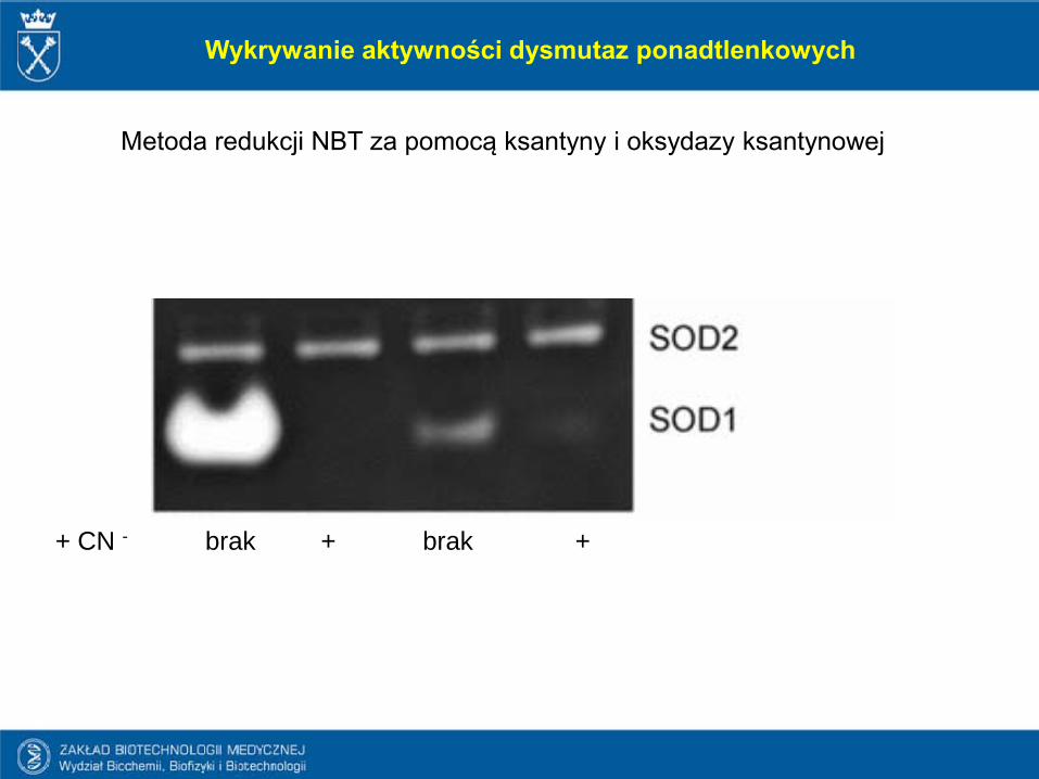
Wykrywanie aktywności dysmutaz ponadtlenkowych
Metoda redukcji NBT za pomocą ksantyny i oksydazy ksantynowej
+ CN - brak + brak +

a- mózg b- sercec- wątroba d- płuca
Mysz Kurczak
Bez CN-
2 mM CN-
Wykrywanie aktywności dysmutaz ponadtlenkowych
MnSOD
CuZnSOD
Halliwell & Gutteridge , 2001

SOD & nadtlenoazotyn

Powstawanie nadtlenoazotynu
Guzik i wsp J Physiol Pharmacol 2005

Powstawanie nadtlenoazotynu

HZ Szeto, AAPS Journal. 2006; 8(3): E521-E531. DOI:
Uszkodzenie mitochondriów przez ROS oraz NO.

Enzymy unieczynniające nadtlenek wodoru

(EC 1.11.1.6) - enzym występujący u niemal wwszystkich organizmów
Rozkłada nadtlenek wodoru do wody i tlenu
Enzym charakteryzujący się jedną z najwyższych liczb obrotu – jedna cząsteczka katalazy może przeksztalcicc w ciągu sekundy 40 mln cząsteczek nadtlenku wodoru do wody oraz tlenu
Jest tetramerem – każdy z łańcuchów składa się z 500 aminokwasów
Zawiera 4 grupy hemowe umożliwiające enzymowi reakcję z nadtlenkiem wodoru
Ludzie – chromosom 11, myszy – chromosom 2
KatalazaBiałko hemowe
Zlokalizowana w peroksysomach

HistoryCatalase was first noticed as a substance in 1818 when Louis Jacques Thénard, who discovered H2O2 (hydrogen peroxide), suggested that its breakdown is caused by a substance. In 1900, Oscar Loew was the first to give it the name catalase, and found its presence in many plants and animals.[8] In 1937 catalase from beef liver was crystallised by James B. Sumner[9] and the molecular weight was worked out in 1938.[10]
In 1969, the amino acid sequence of bovine catalase was worked out.[11] Then in 1981, the 3D structure of the protein was revealed.[12]
Historia badań katalazy
Wikipedia

2 H2O2 → 2 H2O + O2
While the complete mechanism of catalase is not currently known, the reaction is believed to occur in two stages:H2O2 + Fe(III)-E → H2O + O=Fe(IV)-E(.+)
H2O2 + O=Fe(IV)-E(.+) → H2O + Fe(III)-E + O2[
Here Fe()-E represents the iron center of the heme group attached to the enzyme. Fe(IV)-E(.+) is a mesomeric form of Fe(V)-E, meaning that iron is not completely oxidized to +V but receives some "supporting electron" from the heme ligand. This heme has to be drawn then as radical cation (.+).
Reakcje katalizowane przez katalazę (1)

H2O2 + H2R → 2H2O + R
Reakcje katalizowana przez katalazę (2)
Katalaza może utleniać także różne toksyny, takie jakie formaldehyd, kwas mrówkowy, fenole i alkohole. W reakcjach takich zużywa nadtlenku wodoru w następujący sposób

According to recent scientific studies, low levels of catalase may play a role in the graying process of human hair. Hydrogen peroxide is naturally produced by the body and catalase breaks it down. If there is a dip in catalase levels, hydrogen peroxide cannot be broken down. This causes the hydrogen peroxide to bleach the hair from the inside out. Scientists believe this finding may someday be incorporated into anti-greying treatments for aging hair
The true biological significance of catalase is not always straightforward to assess: Mice genetically engineered to lack catalase are phenotypically normal, indicating that this enzyme is dispensable in animals under some conditions.
Catalase deficiency may increase the likelihood of developing Type II Diabetes
Some human beings have very low levels of catalase (acatalasia), yet show few ill effects. It is likely that the predominant scavengers of H2O2 in normal mammalian cells are peroxiredoxins rather than catalase
Znaczenie katalazy w procesach chorobowych

Reaktywne formy tlenu a siwienie
Wood et al., FASEB J 2009
MSR – methionine sulfoxide reductase

Sigma-Aldrich
Komórkowe układy antyoksydacyjne

Peroksydazy glutationowe
-U ludzi w postaci 8 izoform-Używają glutationu jako dawcy elektronu i reagują z nadtlenkiem wodoru oraz
organicznymi nadtlenkami -Gpx1, Gpx2, Gpx3 oraz Gpx4 – enzymy zawierające selen -Gpx6 – także bialko selenowe u ludzi,

Atherogenesis
Emphysema; bronchitis
Parkinson disease
Duchenne muscular dystrophy
Cancer
Alcoholic liver disease
Diabetes
Acute renal failure
Down syndrome
Cerebrovascular disorders
Ischemia; reperfusion injury
Choroby spowodowane zaburzeniami w produkcji/eliminacji ROS lub związane z takimi zaburzeniami

Izoformy NOX w komórkach ściany naczyń krwionośnych

Yellon & Housenloy, NEJM 2007
Znaczenie uszkodzenia ischemiczo-reperfuzyjnegow zawale mięśnia sercowego

Niedobór reaktywnych form tlenu Choroba ziarniniakowa – chronic granulomatous disease
Chronic granulomatous disease (CGD) is a rare inherited immunodeficiencycharacterized by recurrent, often life threatening bacterial and fungal infections due to a functional defect in the microbial-killing activity of phagocytic neutrophils. It occurs as a result of mutations in genes encoding a multicomponent enzymecomplex, the NADPH oxidase, that catalyses the respiratory burst. Themajority of patients have an X-linked form of the disease which is associated withmutations in a membrane-bound component gp91phox. HLA-matched allogeneichematopoietic stem cell (HSC) transplantation can be curative, but for patients withoutsuitable donors, genetic modification of autologous hematopoietic stem cells is an attractive alternative.

Możliwości leczenia choroby ziarniniakowej – terapia genowa
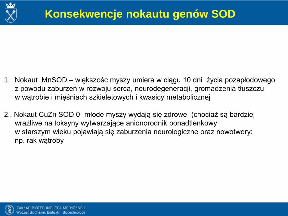
Konsekwencje nokautu genów SOD
1. Nokaut MnSOD – większośc myszy umiera w ciągu 10 dni życia pozapłodowego z powodu zaburzeń w rozwoju serca, neurodegeneracji, gromadzenia tłuszczu w wątrobie i mięśniach szkieletowych i kwasicy metabolicznej
2,. Nokaut CuZn SOD 0- młode myszy wydają się zdrowe (chociaż są bardziej wrażliwe na toksyny wytwarzające anionorodnik ponadtlenkowy w starszym wieku pojawiają się zaburzenia neurologiczne oraz nowotwory: np. rak wątroby

Mutations in the first SOD enzyme (SOD1) can cause familial amyotrophic lateral sclerosis(ALS, a form of motor neuron disease).The most common mutation in the U.S. is A4V, while the most intensely studied is G93A.
The other two isoforms of SOD have not been linked to any human diseases,
Overexpression of SOD1 has been linked to the neural disorders seen in Down's syndrome.
Nadmierna aktywność dysmutazy nadtlenkowej

Over 100 different mutations have been identified in the SOD1 gene of patients diagnosed with the familial form of amyotrophic lateral sclerosis (fALS). These mutations result in a highly diverse group of mutant proteins, some of them very similar to and others enormously different from wild-type SOD1. Despite their differences in properties, each member of this diverse set of mutant proteins causes the same clinical disease, presenting a challenge in formulating hypotheses as to what causes SOD1-associated fALS.
SOD a stwardnienie zanikowe boczne (ALS)
Valnetine et al., Ann Rev Biochem 2005
1903-1941
Lou Gehring
1942 –Diagnosed at age of 21
Stephen Hawking
stwardnienie zanikowe boczne (ALS)

Stwardnienie zanikowe boczne (ALS)
1869 – Jean Martin Charcot
Famous patients:
-Lou Gehring- Dimitir Shostakovich - Mao Zedong (infamous patient...) - Stephen Hawking – suffers from unusually slowly progressing
form of a disease

stwardnienie zanikowe boczne (ALS)
Incidence – 2-3:100 000
Onset at 50-60 years
Sporadic (SALS) – most instances (90-95%)Familial (FALS) – 5-10% - of these 20-25% are mapped to CuZnSOD
gene
Degeneration of motor neurons – progressive loss of the ability to move, speak,
Usually fatal within 1-5 years of onset
No treatment available

stwardnienie zanikowe boczne (ALS)90-95 % of cases – no apparent genetic linkage 5-10 % - familial ALS – mutation in SOD1 gene are responsible
for 10-20 % of cases of FALS
-about 100 different mutations in SOD1 gene
-mutations do not cause the lost of functions, but rather gaining of a toxic phenotype
-among others, the formation of hydroxyl radicals and peroxy-nitrite has been suggested

Yuan J & Yankner BA, Nature 407, 802 - 809 (2000)
Stwardnienie zanikowe boczne (ALS)

Reaktywne formy tlenu regulują syntezę czynników angiogennych

Mole et al., IUBM, 2001
HIF-1β &
np. VEGF
Czynnik aktywowany przez hipoksję

Nadtlenek wodoru indukuje syntezę VEGF
Grzenkowicz-Wydra i wsp. Mol Cel Biochem 2004

Nadtlenek wodoru aktywuje HIF
Grzenkowicz-Wydra i wsp. Mol Cel Biochem 2004
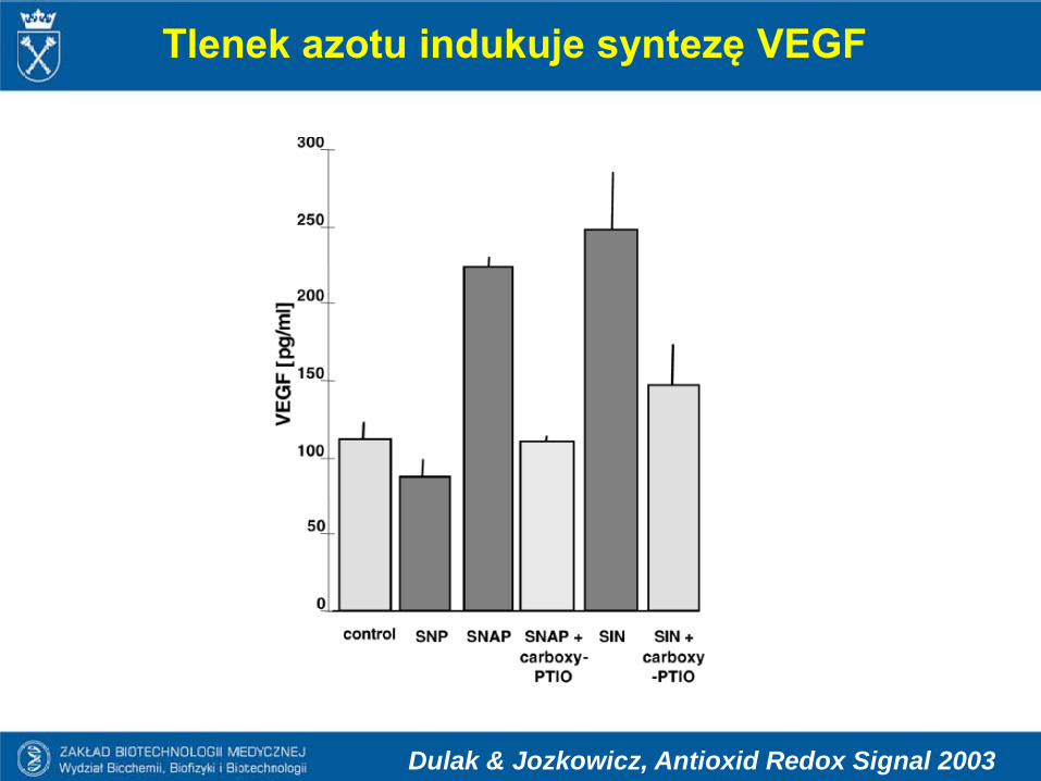
Tlenek azotu indukuje syntezę VEGF
Dulak & Jozkowicz, Antioxid Redox Signal 2003

Tlenek azotu aktywuje czynnik transkrypcyjny HIF-1
Dulak & Jozkowicz, Antioxid Redox Signal 2003

Indukcja produkcji VEGF w hipoksji zależy od HO-1
Inhibition of HO-1 attenuates
hypoxia-inducedVEGF production
(Dulak et al., Antioxid Redox Signal 2002)
PNAS, March 2007 , vol 104, 5100-5114

Dulak & Jozkowicz, Acta Biochimica Polonica 2003
Biologiczny efekt CO może zależeć od aktywacji MnSOD

Wpływ ROS na regulację ekspresji genów związanych z angiogenezą

Termin egzaminu….?

Kinnula and Crapo, Free Radicals Biology & Medicine, 2004