Embed Size (px)
Citation preview
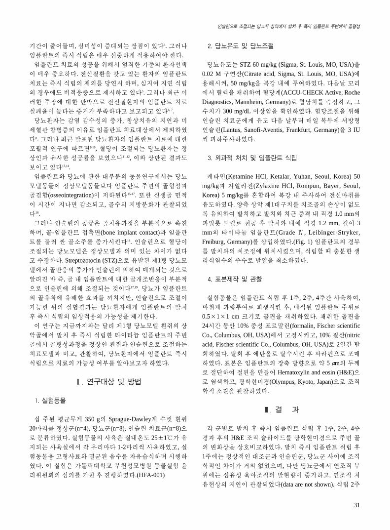
DOI:10.5125/jkaoms.2011.37.1.30
30
Ⅰ. 서 론
사회의 발달과 함께 의학발전과 수명연장이 이루어지고
있으나, 여전히 고혈압, 당뇨와 같은 만성질환의 발병은 높
다. 이러한 만성질환이 현 사회의 발달과 함께 날로 증가
하는 추세여서 당뇨환자에서의 일반적인 치과치료의 안전
성 확보와 더불어 임플란트 치료 또한 추가적인 안전성이
필요하다1. 특히 당뇨질환은 사성 질환으로 전 세계인구
의 약 2% 정도가 당뇨병에 이환되었으며, 우리나라에서도
연간 당뇨병 발생률이 2.5%에 이르고 있어 중요한 문제가
되고있다.
전반적인 치과질환을 통해 알 수 있듯이, 당뇨는 상처치
유의 지연, 감염의 감수성 증가, 면역체계 저하 등을 가져
오지만2, 당뇨와 구강 내 질환과의 상호연관성과 기전 등은
아주 복잡하고 다양한 요소와 관련되어 아직 완전하게 이
해되지않고 있다.
전통적으로 치과 임플란트를 이용한 구강기능의 수복은
장기간의 무치악 상태의 잔존 치조제에 식립하거나 또는
발치 후 일정기간의 치유 후에 식립함을 전제로 하 다. 그
러나 치과 임플란트 주위골의 치유과정의 생물학적 원리
에 한 이해가 증진됨에 따라 신선한 발치와에 즉시 식립
하는 기법을 도입하여 높은 성공률을 보이고 있다3. 치과
임플란트의 즉시 식립은 외과적 처치 횟수의 감소와, 치유
표 성 운420-717 경기도부천시원미구소사동 2가톨릭 학교부천성모병원구강악안면외과Sung-Woon PyoDepartment of Oral and Maxillofacial Surgery, Bucheon St. Mary’s Hospital, 2 Sosa-dong, Wonmi-gu, Bucheon, 420-717, KoreaTEL: +82-32-340-2134 FAX: +82-32-340-2255 E-mail: [email protected]
인슐린으로 조절되는 당뇨쥐 상악에서 발치 후 즉시 임플란트 주변에서 골형성
김 원1∙허현아2∙임상규2∙이 원1,2∙김 실3∙표성운1,2
1가톨릭 학교 임상치과학 학원 구강악안면외과학, 2가톨릭 학교 의과 학 치과학교실 구강악안면외과, 3제주 학교 의과 학 병리학교실
Bone response around immediately placed titanium implant in the extraction socket of diabetic and insulin-treated rat maxilla
Dae-Won Kim1, Hyun-A Heo2, Sang-Gyu Lim2, Won Lee1,2, Young-Sil Kim3, Sung-Woon Pyo1,2
1Department of Oral and Maxillofacial Surgery, Graduate School of Clinical Dental Science, The Catholic University of Korea,2Division of Oral and Maxillofacial Surgery, Department of Dentistry, School of Medicine, The Catholic University of Korea, Bucheon,
3Department of Pathology, College of Medicine, Jeju National University, Jeju, Korea
Introduction: Dental implants are used routinely with high success rates in generally healthy individuals. By contrast, their use in patients with dia-
betes mellitus is controversial because altered bone healing around implants has been reported. This study examined the bone healing response around
titanium implants placed immediately in rats with controlled and uncontrolled diabetes.
Materials and Methods: Twenty rats were divided into the control, insulin-treated and diabetic groups. The rats received streptozotocin (60 mg/kg)
to induce diabetes; animals in the insulin-treated group also received three units of subcutaneous slow-release insulin. A titanium implant (1.2×3
mm) was placed in the extraction socket of the maxillary first molar and bone block was harvested at 1, 2 and 4 weeks.
Results: Bone formation around the implants was consistently (from 1 to 4 week post-implantation) slower for the diabetic group than the control and
insulin-treated group. Bone morphogenesis in the diabetic rats was characterized by fragmented bone tissues and extensive soft tissue intervention.
Conclusion: The immediate placement of titanium implants in the maxilla of diabetic rats led to an unwanted bone healing response. These results
suggest that immediate implant insertion in patients with poorly controlled diabetes might be contraindicated.
Key words: Diabetes mellitus, Dental implants, Insulin, Bone remodeling, Osseointegration
[paper submitted 2010. 9. 28 / revised 2010. 12. 17 / accepted 2011. 1. 20]
*위 논문은 2009년도 가톨릭 학교 부천성모병원 임상의학연구비지원으로 이루어졌음.
Abstract (J Korean Assoc Oral Maxillofac Surg 2011;37:30-5)
인슐린으로 조절되는 당뇨쥐 상악에서 발치 후 즉시 임플란트 주변에서 골형성
31
기간이 줄어들며, 심미성이 증 되는 장점이 있다4. 그러나
임플란트의즉시 식립은매우 신중하게적용하여야 한다.
임플란트 치료의 성공을 위해서 엄격한 기준의 환자선택
이 매우 중요하다. 전신질환을 갖고 있는 환자의 임플란트
치료는 즉시 식립의 제외를 당연시 하며, 심지어 지연 식립
의 경우에도 비적응증으로 제시하고 있다5. 그러나 최근 이
러한 주장에 한 반박으로 전신질환자의 임플란트 치료
실패율이높다는 증거가부족하다고 보고되고있다6, 7.
당뇨환자는 감염 감수성의 증가, 창상치유의 지연과 미
세혈관 합병증의 이유로 임플란트 치료 상에서 제외하
다8. 그러나 최근 발표된 당뇨환자의 임플란트 치료에 한
포괄적 연구에 따르면9,10, 혈당이 조절되는 당뇨환자는 정
상인과 유사한 성공률을 보 으나11,12, 이와 상반된 결과도
보이고있다13,14.
임플란트와 당뇨에 관한 부분의 동물연구에서는 당뇨
모델동물이 정상모델동물보다 임플란트 주변의 골형성과
골결합(osseointegration)이 저하된다15-17. 또한 신생골 면적
이 시간이 지나면 감소되고, 골수의 지방분화가 관찰되었
다16.
그러나 인슐린의 공급은 골치유과정을 부분적으로 촉진
하며, 골-임플란트 접촉면(bone implant contact)과 임플란
트를 둘러 싼 골소주를 증가시킨다18. 인슐린으로 혈당이
조절되는 당뇨모델은 정상모델과 의미 있는 차이가 없다
고 주장한다. Streptozotocin (STZ)으로 유발된 제1형 당뇨모
델에서 골반응의 증가가 인슐린에 의하여 매개되는 것으로
알려진 바 즉, 골 내 임플란트에 한 골개조반응이 부분적
으로 인슐린에 의해 조절되는 것이다17,19. 당뇨가 임플란트
의 골유착에 유해한 효과를 끼치지만, 인슐린으로 조절이
가능한 위의 실험결과는 당뇨환자에게 임플란트의 발치
후즉시 식립의임상적용의 가능성을제기한다.
이 연구는 지금까지와는 달리 제1형 당뇨모델 흰쥐의 상
악골에서 발치 후 즉시 식립한 타이타늄 임플란트의 주변
골에서 골형성과정을 정상인 흰쥐와 인슐린으로 조절하는
치료모델과 비교, 관찰하여, 당뇨환자에서 임플란트 즉시
식립으로치료의 가능성여부를 알아보고자하 다.
Ⅱ. 연구 상 및 방법
1. 실험동물
십 주된 평균무게 350 g의 Sprague-Dawley계 수컷 흰쥐
20마리를 정상군(n=4), 당뇨군(n=8), 인슐린 치료군(n=8)으
로 분류하 다. 실험동물의 사육은 실내온도 25±1℃가 유
지되는 사육실에서 각 우리마다 1-2마리씩 사육하 고, 실
험동물용 고형사료와 멸균된 음수를 자유습식하며 시행하
다. 이 실험은 가톨릭 학교 부천성모병원 동물실험 윤
리위원회의심의를 거친후 진행하 다.(HFA-001)
2. 당뇨유도 및 당뇨조절
당뇨유도는 STZ 60 mg/kg (Sigma, St. Louis, MO, USA)을
0.02 M 구연산(Citrate acid, Sigma, St. Louis, MO, USA)에
용해시켜, 50 mg/kg을 복강 내에 투여하 다. 다음날 꼬리
에서혈액을채취하여혈당계(ACCU-CHECK Active, Roche
Diagnostics, Mannheim, Germany)로 혈당치를 측정하고, 그
수치가 300 mg/dL 이상임을 확인하 다. 혈당조절을 위해
인슐린 치료군에게 유도 다음 날부터 매일 복부에 서방형
인슐린(Lantus, Sanofi-Aventis, Frankfurt, Germany)을 3 IU
씩 피하주사하 다.
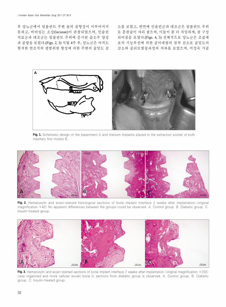
3. 외과적 처치 및 임플란트 식립
케타민(Ketamine HCl, Ketalar, Yuhan, Seoul, Korea) 50
mg/kg과 자일라진(Zylaxine HCl, Rompun, Bayer, Seoul,
Korea) 5 mg/kg를 혼합하여 복강 내 주사하여 전신마취를
유도하 다. 양측 상악 제1 구치를 치조골의 손상이 없도
록 유의하여 발치하고 발치와 치근 중격 내 직경 1.0 mm의
파일롯 드릴로 천공 후 발치와 내에 직경 1.2 mm, 길이 3
mm의 타이타늄 임플란트(Grade Ⅳ, Leibinger-Stryker,
Freiburg, Germany)를 삽입하 다.(Fig. 1) 임플란트의 경부
를 발치와의 치조정에 위치시켰으며, 식립할 때 충분한 생
리식염수의 주수로발열을 최소하 다.
4. 표본제작 및 관찰
실험동물은 임플란트 식립 후 1주, 2주, 4주간 사육하여,
마취제 과량투여로 희생시킨 후, 매식된 임플란트 주위로
0.5×1×1 cm 크기로 골편을 채취하 다. 채취한 골편을
24시간 동안 10% 중성 포르말린(formalin, Fischer scientific
Co., Columbus, OH, USA)에서 고정시키고, 10% 질산(nitric
acid, Fischer scientific Co., Columbus, OH, USA)로 2일간 탈
회하 다. 탈회 후 에탄올로 탈수시킨 후 파라핀으로 포매
하 다. 표본은 임플란트의 장축 방향으로 약 5 μm의 두께
로 절단하여 절편을 만들어 Hematoxylin and eosin (H&E)으
로 염색하고, 광학현미경(Olympus, Kyoto, Japan)으로 조직
학적 소견을관찰하 다.
Ⅲ. 결 과
각 군별로 발치 후 즉시 임플란트 식립 후 1주, 2주, 4주
경과 후의 H&E 조직 슬라이드를 광학현미경으로 주변 골
의 변화상을 상호비교하 다. 발치 즉시 임플란트 식립 후
1주에는 정상적인 조군과 인슐린군, 당뇨군 사이에 조직
학적인 차이가 거의 없었으며, 다만 당뇨군에서 연조직 부
위에는 섬유성 육아조직의 발현량이 증가하고, 연조직 치
유현상의 지연이 관찰되었다(data are not shown). 식립 2주
J Korean Assoc Oral Maxillofac Surg 2011;37:30-5
32
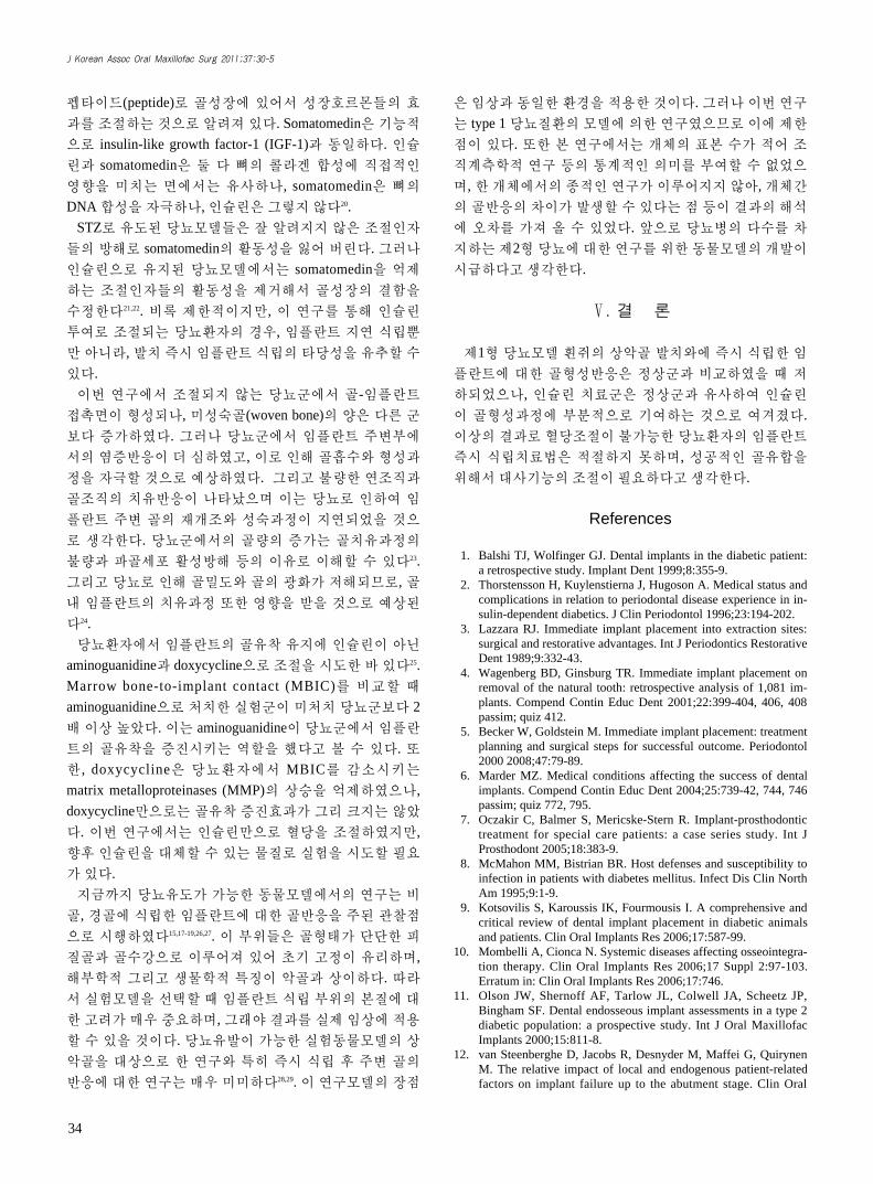
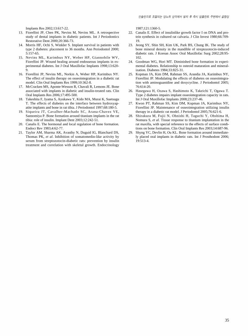
후 당뇨군에서 임플란트 주변 골의 골형성이 이루어지지
못하고, 비어있는 소강(lacunae)이 관찰되었으며, 인슐린
치료군과 조군는 임플란트 주위에 증가된 골소주 양상
과 골량을 보 다.(Figs. 2, 3) 식립 4주 후, 당뇨군은 아직도
현저한 연조직의 광범위한 형성에 의한 주변의 골 도 감
소를 보 고, 반면에 인슐린군과 조군은 임플란트 주위
로 층판골이 자리 잡으며, 이들이 좀 더 치 하게, 잘 구성
되어짐을 보 다.(Figs. 4, 5) 전체적으로 당뇨군은 조골세
포의 기능부전에 의한 골미네랄의 침착 감소로 골 도의
감소와 골리모델링과정의 저하를 보 으며, 미성숙 가골
Fig. 1. Schematic design of the experiment A and titanium implants placed in the extraction socket of both
maxillary first molars B.
A B
Fig. 2. Hematoxylin and eosin-stained histological sections of bone implant interface 2 weeks after implantation.(original
magnification ×40) No apparent differences between the groups could be observed. A. Control group. B. Diabetic group. C.
Insulin-treated group.
Fig. 3. Hematoxylin and eosin-stained sections of bone implant interface 2 weeks after implantation.(original magnification ×200)
Less organized and more cellular woven bone in sections from diabetic group is observed. A. Control group. B. Diabetic
group. C. Insulin-treated group.

인슐린으로 조절되는 당뇨쥐 상악에서 발치 후 즉시 임플란트 주변에서 골형성
33
의 형성으로 임플란트의 골유착 감소를 보 다. 이에 반해
인슐린 치료군과 조군은 조골세포판의 형성으로 골수강
이 포함된 성숙된 형태의 층판골이 임플란트 주변부에 형
성되었다.
Ⅳ. 고 찰
당뇨는 확장혈관 합병증으로 심근경색, 뇌혈관질환, 말
초동맥질환 등의 전신적 합병증뿐만 아니라, 치주질환과
구강질환과 같은 미세혈관 합병증도 일으킨다. 이러한 당
뇨병은 임상적으로 연조직과 경조직의 치유를 지연시키
고, 감염의 감수성을 증가시키는 등의 문제점을 야기하나,
일반적인 치과치료와 마찬가지로 임플란트 치료에서도,
술전 주의사항을 주지하여 임상적으로 우수한 결과를 보
이고 있다. 지금까지 전신질환이 없는 정상적인 환자들에
한 골-임플란트 접촉에 한 연구는 많았지만, 당뇨환자
에서의 임플란트 주변의 골반응에 한 연구는 그다지 많
지 않았으며, 특히 현재 임상에서 많이 시도되고 있는 발치
즉시 식립 후 골반응에 한 연구는 더욱 없다. 이 연구에
서 당뇨모델로 쥐를 택하고 STZ로 제1형 당뇨를 유발하고,
정상군과 인슐린 치료군 그리고 당뇨군의 상악골의 발치
와에서 임플란트식립 후의골반응을 관찰하고자하 다.
통상 당뇨환자에서 임플란트 식립에 의한 치료가 금기임
에도 불구하고, 당뇨환자의 임플란트 식립 후 골유착 성공
률도 높다고 보고되어 왔다14. Morris 등14은 제2형 당뇨환자
에서 임플란트 치료 성공률이 정상적인 환자에 비해 낮지
않음을 보고한 반면, Fiorellini 등13은 조절이 잘 되는 당뇨
환자라 할 지라도 임플란트 식립 성공률이 6.5년 후 평균
86%로 그다지높지 않다고보고 하 다.
이전 동물실험은 당뇨모델들이 비당뇨모델에 비해서 골-
임플란트 접촉이 감소됨을 입증해 왔다15. 그러나 인슐린으
로 조절된 당뇨군에서 골형성과 골융합이 증가됨을 보여
주었다17-19. 인슐린은 혈당조절뿐만 아니라, 골형성과 무기
질 침착, 골기질형성의 유도, 골 도 증가에 관여하는 인자
로 작용하는 것으로 보인다. 이번 연구에서, 인슐린으로 조
절되고 있는 당뇨군은 정상군과 유사한 골형성반응을 보
이고 있어기존 결과와동일함을 보 다.
조절되지 않는 당뇨쥐에서는 골융합과 골 사과정에 중
요한 somatomedin의 활동성이 소실되어 있다. Somatomedin
은 골과 연골에 성장촉진 활동성을 갖는 인슐린과 유사한
Fig. 4. Hematoxylin and eosin-stained sections of bone implant interface 4 weeks after implantation.(original magnification ×40)
Decreased bone apposition was observed in the diabetic group, which suggest an impaired osteoblastic function or mineralization
defect. A. Control group. B. Diabetic group. C. Insulin-treated group.
Fig. 5. Hematoxylin and eosin-stained sections of bone implant interface 4 weeks after implantation.(original magnification ×200)
Calcified mature woven bone with the marked restitution of marrow can be observed in the control and insulin-treated
groups. A. Control group. B. Diabetic group. C. Insulin-treated group.
J Korean Assoc Oral Maxillofac Surg 2011;37:30-5
34
펩타이드(peptide)로 골성장에 있어서 성장호르몬들의 효
과를 조절하는 것으로 알려져 있다. Somatomedin은 기능적
으로 insulin-like growth factor-1 (IGF-1)과 동일하다. 인슐
린과 somatomedin은 둘 다 뼈의 콜라겐 합성에 직접적인
향을 미치는 면에서는 유사하나, somatomedin은 뼈의
DNA 합성을 자극하나, 인슐린은그렇지 않다20.
STZ로 유도된 당뇨모델들은 잘 알려지지 않은 조절인자
들의 방해로 somatomedin의 활동성을 잃어 버린다. 그러나
인슐린으로 유지된 당뇨모델에서는 somatomedin을 억제
하는 조절인자들의 활동성을 제거해서 골성장의 결함을
수정한다21,22. 비록 제한적이지만, 이 연구를 통해 인슐린
투여로 조절되는 당뇨환자의 경우, 임플란트 지연 식립뿐
만 아니라, 발치 즉시 임플란트 식립의 타당성을 유추할 수
있다.
이번 연구에서 조절되지 않는 당뇨군에서 골-임플란트
접촉면이 형성되나, 미성숙골(woven bone)의 양은 다른 군
보다 증가하 다. 그러나 당뇨군에서 임플란트 주변부에
서의 염증반응이 더 심하 고, 이로 인해 골흡수와 형성과
정을 자극할 것으로 예상하 다. 그리고 불량한 연조직과
골조직의 치유반응이 나타났으며 이는 당뇨로 인하여 임
플란트 주변 골의 재개조와 성숙과정이 지연되었을 것으
로 생각한다. 당뇨군에서의 골량의 증가는 골치유과정의
불량과 파골세포 활성방해 등의 이유로 이해할 수 있다23.
그리고 당뇨로 인해 골 도와 골의 광화가 저해되므로, 골
내 임플란트의 치유과정 또한 향을 받을 것으로 예상된
다24.
당뇨환자에서 임플란트의 골유착 유지에 인슐린이 아닌
aminoguanidine과 doxycycline으로 조절을 시도한 바 있다25.
Marrow bone-to-implant contact (MBIC)를 비교할 때
aminoguanidine으로 처치한 실험군이 미처치 당뇨군보다 2
배 이상 높았다. 이는 aminoguanidine이 당뇨군에서 임플란
트의 골유착을 증진시키는 역할을 했다고 볼 수 있다. 또
한, doxycycline은 당뇨환자에서 MBIC를 감소시키는
matrix metalloproteinases (MMP)의 상승을 억제하 으나,
doxycycline만으로는 골유착 증진효과가 그리 크지는 않았
다. 이번 연구에서는 인슐린만으로 혈당을 조절하 지만,
향후 인슐린을 체할 수 있는 물질로 실험을 시도할 필요
가 있다.
지금까지 당뇨유도가 가능한 동물모델에서의 연구는 비
골, 경골에 식립한 임플란트에 한 골반응을 주된 관찰점
으로 시행하 다15,17-19,26,27. 이 부위들은 골형태가 단단한 피
질골과 골수강으로 이루어져 있어 초기 고정이 유리하며,
해부학적 그리고 생물학적 특징이 악골과 상이하다. 따라
서 실험모델을 선택할 때 임플란트 식립 부위의 본질에
한 고려가 매우 중요하며, 그래야 결과를 실제 임상에 적용
할 수 있을 것이다. 당뇨유발이 가능한 실험동물모델의 상
악골을 상으로 한 연구와 특히 즉시 식립 후 주변 골의
반응에 한 연구는 매우 미미하다28,29. 이 연구모델의 장점
은 임상과 동일한 환경을 적용한 것이다. 그러나 이번 연구
는 type 1 당뇨질환의 모델에 의한 연구 으므로 이에 제한
점이 있다. 또한 본 연구에서는 개체의 표본 수가 적어 조
직계측학적 연구 등의 통계적인 의미를 부여할 수 없었으
며, 한 개체에서의 종적인 연구가 이루어지지 않아, 개체간
의 골반응의 차이가 발생할 수 있다는 점 등이 결과의 해석
에 오차를 가져 올 수 있었다. 앞으로 당뇨병의 다수를 차
지하는 제2형 당뇨에 한 연구를 위한 동물모델의 개발이
시급하다고생각한다.
Ⅴ. 결 론
제1형 당뇨모델 흰쥐의 상악골 발치와에 즉시 식립한 임
플란트에 한 골형성반응은 정상군과 비교하 을 때 저
하되었으나, 인슐린 치료군은 정상군과 유사하여 인슐린
이 골형성과정에 부분적으로 기여하는 것으로 여겨졌다.
이상의 결과로 혈당조절이 불가능한 당뇨환자의 임플란트
즉시 식립치료법은 적절하지 못하며, 성공적인 골유합을
위해서 사기능의 조절이필요하다고 생각한다.
References
1. Balshi TJ, Wolfinger GJ. Dental implants in the diabetic patient:a retrospective study. Implant Dent 1999;8:355-9.
2. Thorstensson H, Kuylenstierna J, Hugoson A. Medical status andcomplications in relation to periodontal disease experience in in-sulin-dependent diabetics. J Clin Periodontol 1996;23:194-202.
3. Lazzara RJ. Immediate implant placement into extraction sites:surgical and restorative advantages. Int J Periodontics RestorativeDent 1989;9:332-43.
4. Wagenberg BD, Ginsburg TR. Immediate implant placement onremoval of the natural tooth: retrospective analysis of 1,081 im-plants. Compend Contin Educ Dent 2001;22:399-404, 406, 408passim; quiz 412.
5. Becker W, Goldstein M. Immediate implant placement: treatmentplanning and surgical steps for successful outcome. Periodontol2000 2008;47:79-89.
6. Marder MZ. Medical conditions affecting the success of dentalimplants. Compend Contin Educ Dent 2004;25:739-42, 744, 746passim; quiz 772, 795.
7. Oczakir C, Balmer S, Mericske-Stern R. Implant-prosthodontictreatment for special care patients: a case series study. Int JProsthodont 2005;18:383-9.
8. McMahon MM, Bistrian BR. Host defenses and susceptibility toinfection in patients with diabetes mellitus. Infect Dis Clin NorthAm 1995;9:1-9.
9. Kotsovilis S, Karoussis IK, Fourmousis I. A comprehensive andcritical review of dental implant placement in diabetic animalsand patients. Clin Oral Implants Res 2006;17:587-99.
10. Mombelli A, Cionca N. Systemic diseases affecting osseointegra-tion therapy. Clin Oral Implants Res 2006;17 Suppl 2:97-103.Erratum in: Clin Oral Implants Res 2006;17:746.
11. Olson JW, Shernoff AF, Tarlow JL, Colwell JA, Scheetz JP,Bingham SF. Dental endosseous implant assessments in a type 2diabetic population: a prospective study. Int J Oral MaxillofacImplants 2000;15:811-8.
12. van Steenberghe D, Jacobs R, Desnyder M, Maffei G, QuirynenM. The relative impact of local and endogenous patient-relatedfactors on implant failure up to the abutment stage. Clin Oral
인슐린으로 조절되는 당뇨쥐 상악에서 발치 후 즉시 임플란트 주변에서 골형성
35
Implants Res 2002;13:617-22.13. Fiorellini JP, Chen PK, Nevins M, Nevins ML. A retrospective
study of dental implants in diabetic patients. Int J PeriodonticsRestorative Dent 2000;20:366-73.
14. Morris HF, Ochi S, Winkler S. Implant survival in patients withtype 2 diabetes: placement to 36 months. Ann Periodontol 2000;5:157-65.
15. Nevins ML, Karimbux NY, Weber HP, Giannobile WV,Fiorellini JP. Wound healing around endosseous implants in ex-perimental diabetes. Int J Oral Maxillofac Implants 1998;13:620-9.
16. Fiorellini JP, Nevins ML, Norkin A, Weber HP, Karimbux NY.The effect of insulin therapy on osseointegration in a diabetic ratmodel. Clin Oral Implants Res 1999;10:362-8.
17. McCracken MS, Aponte-Wesson R, Chavali R, Lemons JE. Boneassociated with implants in diabetic and insulin-treated rats. ClinOral Implants Res 2006;17:495-500.
18. Takeshita F, Iyama S, Ayukawa Y, Kido MA, Murai K, SuetsuguT. The effects of diabetes on the interface between hydroxyap-atite implants and bone in rat tibia. J Periodontol 1997;68:180-5.
19. Siqueira JT, Cavalher-Machado SC, Arana-Chavez VE,Sannomiya P. Bone formation around titanium implants in the rattibia: role of insulin. Implant Dent 2003;12:242-51.
20. Canalis E. The hormonal and local regulation of bone formation.Endocr Rev 1983;4:62-77.
21. Taylor AM, Sharma AK, Avasthy N, Duguid IG, Blanchard DS,Thomas PK, et al. Inhibition of somatomedin-like activity byserum from streptozotocin-diabetic rats: prevention by insulintreatment and correlation with skeletal growth. Endocrinology
1987;121:1360-5.22. Canalis E. Effect of insulinlike growth factor I on DNA and pro-
tein synthesis in cultured rat calvaria. J Clin Invest 1980;66:709-19.
23. Jeong SY, Shin SH, Kim UK, Park BS, Chung IK. The study ofbone mineral density in the mandible of streptozotocin-induceddiabetic rats. J Korean Assoc Oral Maxillofac Surg 2002;28:95-102.
24. Goodman WG, Hori MT. Diminished bone formation in experi-mental diabetes. Relationship to osteoid maturation and mineral-ization. Diabetes 1984;33:825-31.
25. Kopman JA, Kim DM, Rahman SS, Arandia JA, Karimbux NY,Fiorellini JP. Modulating the effects of diabetes on osseointegra-tion with aminoguanidine and doxycycline. J Periodontol 2005;76:614-20.
26. Hasegawa H, Ozawa S, Hashimoto K, Takeichi T, Ogawa T.Type 2 diabetes impairs implant osseointegration capacity in rats.Int J Oral Maxillofac Implants 2008;23:237-46.
27. Kwon PT, Rahman SS, Kim DM, Kopman JA, Karimbux NY,Fiorellini JP. Maintenance of osseointegration utilizing insulintherapy in a diabetic rat model. J Periodontol 2005;76:621-6.
28. Shirakura M, Fujii N, Ohnishi H, Taguchi Y, Ohshima H,Nomura S, et al. Tissue response to titanium implantation in therat maxilla, with special reference to the effects of surface condi-tions on bone formation. Clin Oral Implants Res 2003;14:687-96.
29. Shyng YC, Devlin H, Ou KL. Bone formation around immediate-ly placed oral implants in diabetic rats. Int J Prosthodont 2006;19:513-4.


![HPEXKDQ/XND*LQJLYD7LNXV Sprague Dawley · HQ]LP \DQJ SHQWLQJ GDODP SURVHV LQÀDPDVL GDQ nyeri, 11 sementara hesperidin mampu mengurangi pembengkakan dengan meregulasi mikrosirkulasi.12](https://img.pdfslide.tips/doc/110x75/60718a72a0dfa368fb0ea39e/hpexkdqxndlqjlyd7lnxv-sprague-dawley-hqlp-dqj-shqwlqj-gdodp-survhv-lqdpdvl.jpg)