Embed Size (px)
Citation preview
http://www.TurkJBiochem.com ISSN 1303–829X (electronic) 0250–4685 (printed) 291
Establishment and Biological characterization of a Large Yorkshire Pig fibroblast line
[Geniş yapılı Yorkshire domuz ırkına ait fibroblast hattının kurulması ve biyolojik karakterizasyonu]*
Research Article [Araştırma Makalesi] Yayın tarihi 30 Eylül, 2013 © TurkJBiochem.com
[Published online 30 September, 2013]
TÜR
K BİY
OKİMYA DERNEĞİ DER
GİSİTÜ
RK
BİY
OKİMYA DERNEĞİ DER
GİSİ
1976
TÜR
K BİY
OKİMYA DERNEĞİ DER
GİSİTÜ
RK
BİY
O
KİMYA DERNEĞ
İ DER
GİSİ
1976
ORJİNAL
1. ÖRNEK 2. ÖRNEK
Yuhua Gao1, 2, Changqing Liu3, Changli Li1, Weijun Guan1, Yuehui Ma 1
1Institute of Animal Science, Chinese Academy of Agricultural Sciences, Beijing 100193, 2College of Wildlife Resources, Northeast Forestry University, Harbin, 150040, 3Department of Life Science, Bengbu Medical College, Bengbu, 233030, China
Yazışma Adresi[Correspondence Address]
Yuehui Ma
Institute of Animal Science, Chinese Academy ofAgricultural Sciences, Beijing 100193Tel. +86-10- 6281-5992Fax. +86-10-6281-3463E-mail: [email protected]
Registered: 9 August 2012; Accepted: 8 May 2013[Kayıt tarihi: 9 Ağustos 2012; Kabul tarihi: 8 Mayıs 2013]
ABSTRACTAim: This study aimed to establish the fibroblast cell bank of the Large Yorkshire pig in the form of somatic cells, and to study its biological characteristics, to explore long-term conservation and sustainable use of this genetic resource.Material and methods: Ear marginal tissue of the Large Yorkshire pig was used to establish a fibroblast cell line by direct culture of explants and cryopreservation techniques, and the quality of the cell line was assessed.Results: Biological analyses showed that the cells were morphologically homogenous fibroblasts, and the estimated population doubling time (PDT) was 72 hours. The growth curve appeared as a typical “S” shape as the cell growth passed through a latent phase, a logarithmic phase and a plateau phase. Tests for bacteria, fungi, viruses and Mycoplasma were negative. Analysis of lactate dehydrogenase (LDH) and malate dehydrogenase (MDH) isoenzymes ruled out cross-contamination between cell lines. Karyotyping and G-banding indicated a total chromosome number of 2n = 36; the proportion of diploid cells in the population was 91–96%. The transfection efficiencies of pEGFP-N3, pEYFP-N1 and pDsRed1-N1 were between 5.14 and 12.13%. Bioindicators of the cell bank met all the standards of the American Type Culture Collection (ATCC).Conclusion: This study not only shows the effective preservation of the Large Yorkshire pig germplasm at the cellular level, but also provides a valuable experimental resource for fields including cell biology, medicine, genomics, post-genomics, genetic engineering and embryo engineering.Key Words: Large Yorkshire Pig, fFibroblast line; biological characterizationConflict of Interest: We declare that there is no conflict of interest.
ÖZETAmaç: Çalışmada geniş yapılı Yorkshire domuzlarından somatik hücre formunda fibroblast hücre bankasının kurulması, biyolojik karakterizasyonu, uzun dönem saklanması ve genetik kaynak olarak sürdürülebilirliği araştırılmıştır. Gereç ve Yöntem: Fibroblast hücre hattının kurulmasında kulak kenar dokusu doğrudan kültürü yapılarak kullanılmış, dondurularak saklanma yöntemleri ve hücre hattının kalitesi değerlendirilmiştir.Bulgular: Biyolojik analizler hücrelerin morfolojik olarak homojen fibroblastlar olduğunu göstermektedir. Populasyonun tahmini ikiye katlanma süresi (PDT) 72 saat olarak bulunmuştur. Büyüme eğrisi latent faz, logaritmik faz ve plato fazını kapsayan “S” şeklindedir. Bakteri, mantar, virus ve Myoplasma testleri negatiftir. Laktat dehidrogenaz (LDH) ve malat dehidrogenaz (MDH) analizleri hücre hatları arasındaki kontaminasyon olasılığını ortadan kaldırmaktadır. Karyotipleme G-bantlama, toplam kromozom sayısının 2n = 36 olduğunu, populasyondaki diploid hücre oranının % 91-96 olduğunu göstermektedir. pEGFP-N3, pEYFP-N1 ve pDsRed1-N1’nin transfeksiyon verimleri % 5.14 ile % 12.13 arasında değişmektedir. Hücre bankasının biyobelirteçleri Amerikan Tipi Kültür Koleksiyonu (ATCC)’nun bütün standartlarını karşılamaktadır.Sonuç: Çalışma sadece geniş yapılı Yorkshire domuzlarının üreme hücrelerinin hücresel düzeyde etkili bir biçimde saklanmasını göstermesi bakımından değil, aynı zamanda hücre biyolojisi, tıp, genomiks, post-genomiks, genetic mühendislik ve embriyo mühendisliği gibi alanlar için deneysel kaynak sağlaması bakımından önemlidir.Anahtar Kelimeler: Geniş yapılı Yorkshire domuzu, fibroblast hattı, biyolojik karakterizasyonÇıkar Çatışması: Çıkar çatışması bulunmamaktadır.
doi: 10.5505/tjb.2013.58076
Turk J Biochem, 2013; 38 (3) ; 291–298 Goa et al.292
IntroductionAnimal Genetic Resources are vital resources that constitute an important component of biological and genetic diversity. Livestock and poultry genetic resource conservation is an important area of research, as it is essential for the sustainable development of animal husbandry and biodiversity. The United States Department of Agriculture has emphasized the protection of animal genetic resources, launching projects for the effective use and preservation of national animal genetic resources at the beginning of this century [1]. Major culture collections around the world include the American Type Culture Collection (ATCC), the European Collection of Animal Cell Culture (ECACC) and the German Collection of Microorganisms and Cell Culture (DSMZ) [2]. Animal genetic resource conservation in China has currently fallen behind the United States and Europe. However, because of strong support from the government, it is developing rapidly, with the establishment of specialized conservation farms and a number of gene banks containing national genetic resources, for living animal reserves, semen and embryo preservation, and tissue preservation.Currently there are several ways to conserve animal genetic resources, including living animal reserves, semen preservation, embryo preservation, and genomic library and cDNA library preservation. With the development of somatic cell cloning technology, animal somatic cells are becoming of interest as a supplement for the preservation of animal genetic resources [3]. Therefore, building a cell bank of important livestock and poultry species can preserve and make use of their genetic resources effectively and permanently at the cellular level. This can also provide valuable experimental material for life science research on the cell biology, genomics, post-genomics and embryo engineering of livestock and poultry species [4]. Currently the cell bank of breeds such as the white ear lobe chicken, Luxi cattle, Simmental cattle, Landrace pigs, and the Qingyuan partridge chicken [5-9] have been established, but the available cell bank of the Large Yorkshire pig has not yet been established in China.The Large Yorkshire pig, also known as the Large White, originated in the English county of Yorkshire and is one of the world-famous lean meat breeds. Its appearance is characterized by a large physique, high weight by stature, erect ears, straight nose, slightly bowed back, long limbs, and white hair all over the body, and in livestock production this breed shows fast growth velocity, high feed use, good farrowing performance and a high carcass lean meat percentage. The Large Yorkshire pig was introduced to China in the early twentieth century and shows good adaptability after many years of domestication. In China, the Large Yorkshire pig is mainly used as the male parent in a two-way cross, a three-way cross or a multiple cross, with a local sow as
the female parent. The hybrid progenies show obvious hybrid vigor in terms of daily weight gain and feed use, as well as in reproductive ability, giving good economic value [10]. In this study we establish the fibroblast cell bank of the Large Yorkshire pig in the form of somatic cells, and study its biological characteristics, to explore long-term conservation and sustainable use of this genetic resource. This study has laid the foundation for genetic resource preservation of other breeds.
Materials and Methods
Cell cultureEar marginal tissue samples were taken from 30 Large Yorkshire pigs (10 males and 20 females) that were provided by the Pig Breeding Farm of The Chinese Academy of Agricultural Sciences, Beijing, PR China. The samples were collected into separate tubes which contained DMEM (Gibco, Carlsbad, CA, USA) supplemented with ampicillin (100 U/ml) and streptomycin (100 μg/ml) (Solarbio, China, Beijing). The samples were then rinsed and chopped into 1-mm3 pieces, which were seeded onto the surface of a tissue culture flask containing 90% DMEM and 10% fetal bovine serum (FBS) in a incubator at 37°C with 5% CO2 [11-13]. When the cells reached 80–90% confluency, they were harvested and divided into prepared culture flasks at 1:2 or 1:3 ratios [11].
Cryopreservation and recoveryThe cells were taken in the logarithmic growth phase and counted with a hemocytometer, and their viability was checked using the CellTiter-Blue Cell Viability Assay (Promega, Madison, WI, USA) before freezing. The harvested cells were resuspended in freezing medium containing 40% DMEM, 10% dimethyl sulfoxide (DMSO) (Sigma-Aldrich, St. Louis, MO, USA) and 50% FBS, to a final concentration between 3×10⁶ and 4×10⁶ viable cells/ml. Single cells were dispensed into sterile plastic cryovials labeled with species, gender, freezing serial number and date. The sealed cryovials were initially kept at 4°C for 20–30 min to allow the DMSO to equilibrate. Then, the cryovials were transferred to a commercially available freezing kit (Nalgene, USA), stored at −80°C overnight (a process in which the temperature decreased at a rate of 1°C/min), and subsequently transferred to liquid nitrogen [12]. To recover the cells, the cryovials were removed from liquid nitrogen and immediately thawed in a 42°C water bath. They were then subsequently transferred with a straw to a culture flask containing 90% DMEM and 10% FBS. The suspension was mixed gently with a pipette to distribute the cells, and then cultured at 37°C with 5% CO2. The medium was refreshed after 24 h.
Growth dynamics and viabilitiesCells were seeded into 24-well plates at 1.5×10⁴ cells/ml
Turk J Biochem, 2013; 38 (3) ; 291–298 Goa et al.293
and cultured for 7 d, following Gu and Kong’s method [13-14]. Cell growth and density data were monitored daily by counting three wells every 24 h, until the cells reached the plateau phase. A cell growth curve was then plotted, and the population doubling time (PDT) was calculated from the curve. Cell viabilities, both before freezing and after recovery, were determined using a hemocytometer to enumerate 1000 cells by Butler’s dye exclusion method [15].
Microorganism contamination detection
Detection of bacteria and fungiCells were cultured in a medium free of antibiotics. Bacteria and fungi contamination was assessed within three days. The detailed procedure used for the contamination test was that described by Doyle [16].
Detection of MycoplasmaCells were stained with Hoechst 33258 (ATCC), according to the methods of Freshney and Masover & Becker [11,17], for fluorescent staining of deoxyribonucleic acid (DNA). An ELISA Mycoplasma Detection kit (Roche, Lewes, UK) was used to confirm the results of the DNA fluorescence staining.
Virus DetectionUnder normal culture conditions, the cells were selected randomly for cytopathogenic examination using phase-contrast microscopy following Hay’s hemadsorption protocol [18].
Karyotype analysisChromosomes were prepared, fixed and stained following standard methods [19]. After Giemsa staining, the chromosome numbers per spread were counted by oil immersion objective. Relative length, arm ratio, centromeric index and type were counted according to published protocols [20-21].
Isoenzyme analysisIsoenzyme patterns of lactate dehydrogenase (LDH) and malate dehydrogenase (MDH) were identified by non-continuous native polyacrylamide (Sigma-Aldrich, USA) gel electrophoresis (PAGE). The stacking gels were at an acrylamide concentration of 5% w/v, and the separating gel was at an acrylamide concentration of 13% w/v. The cells were harvested, and protein extraction solution (0.9% Triton X-100, 0.06 mmol NaCl:EDTA in a mass ratio of 1:15; pH 7.0) was added to cells at a density of 5×10⁷ cells/ml. The suspension was centrifuged and stored in aliquots at −70°C. Equal volumes of native gel sample loading buffer (Solarbio) were added to the samples. The the gel buffer was prepared which using Tris-citric acid buffer at two different buffer: 0.078 mol/L Tris-citric acid (stacking gels; pH 6.8), and 0.017 mol/L Tris-citric acid (separating gel; pH 8.9). The PAGE apparatus was assembled and run, the electrophoretic
buffer was changed to Tris-glycin (pH 8.7).The buffered staining mixture was 0.1 M Tris-HCL (pH 8.0) and 100 mM DL-lactate lithium salt (for LDH staining), or 50 mM L-malic acid (for MDH staining). Gel were stained for LDH and MDH by incubation in the dark in 100 ml of the appropriate buffered staining mixture containing 10 mg nicotinamide adenine dinucleotide, 10 mg 3-(4,5-dimethylthiazolyl-2)-2,5-diphenyltetrazolium bromide and 5 mg phenazine methosulfate. The gel was soaked in an enzymatic activity staining solution to locate isoenzyme subtypes by producing a water-insoluble blue color.
Expression of three fluorescent proteins in Large Yorkshire pig fibroblastsFollowing the Lipofectamine medium methods of Escriou and Tsuchiya [22-23], the cultured cells were transfected with the plasmid DNAs (Clontech) for three fluorescent proteins (pEGFP-N3, pEYFP-N1 and pDsRed1-N1), and with Lipofectamine 2000 (Invitrogen, USA), at an optimal concentration of 2 μg of plasmid DNA and 6 μl of Lipofectamine 2000. Six hours later, the medium was refreshed. The transfected cells were observed at 24 h, 48 h, 72 h, 96 h, 1 week, 2 weeks and 1 month after transfection for expression of the pEGFP-N3, pEYFP-N1 and pDsRed1-N1 genes using excitation wavelengths of 405, 488 and 543 nm, respectively. For each experimental group, images were captured from ten visual fields, and confocal microscopy was used to measure the total and positive cell counts in each field to determine the transfection efficiencies
Results
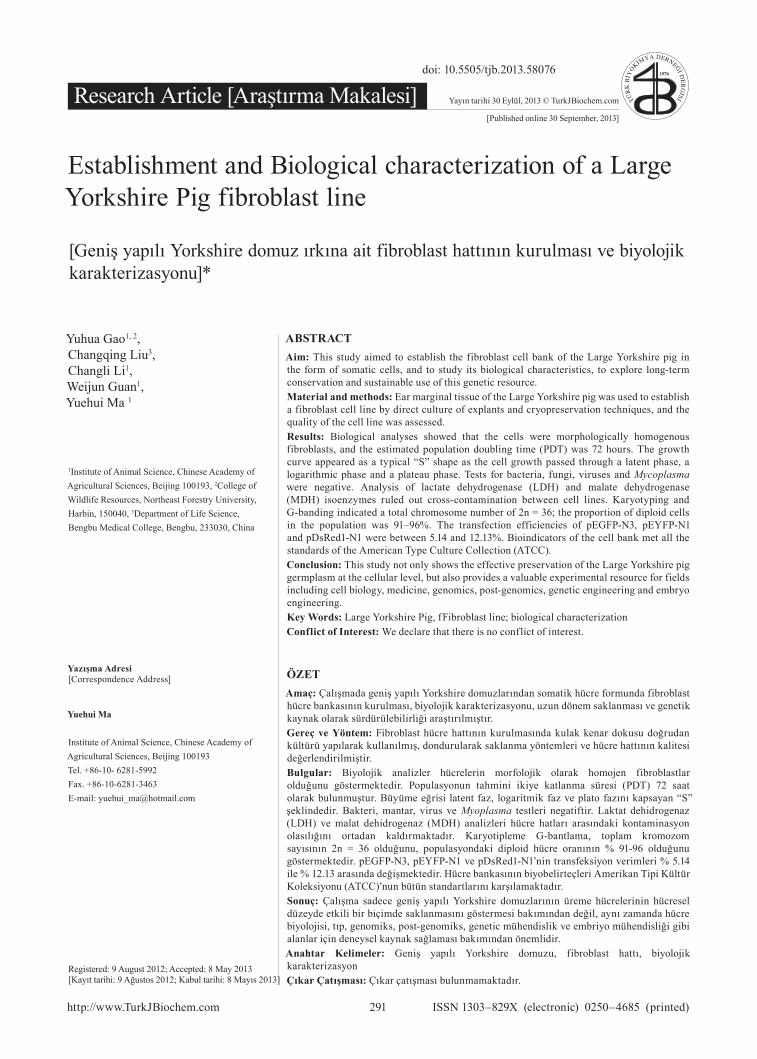
Morphology of cultured cellsFibroblast-like or epithelial-like cells could be seen migrating from the tissue pieces 5–12 days after explanting (Figure 1A). When the cultured cells reached 90% confluence they were subcultured (Figure 1B). After subculturing, the fibroblasts grew rapidly, and gradually outgrew and excluded other cells such as epithelial cells, this result was similar to Zhou’s reported [24]. After 3–4 passages, we obtained purified fibroblasts (Figures 1C-D). The viabilities of Large Yorkshire pig fibroblasts before freezing and after recovery, measured by Trypan Blue staining, were 98.3 and 92.5%, respectively.
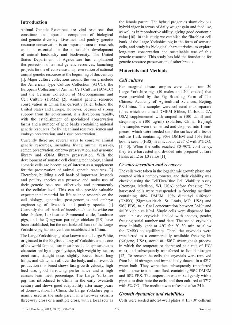
Growth dynamicsThe growth curve of ear marginal tissue fibroblasts of the Large Yorkshire pig had an obvious “S” shape (Figure 2A) and the PDT was about 48 h. There was a lag time or latency phase of about 48 h after seeding, corresponding to the adaptation and recovery of the cells from protease damage, after which the cells proliferated rapidly and entered exponential phase. As the cell density increased, cell proliferation was inhibited by contact inhibition. After the sixth day, the cells no longer proliferated and
Turk J Biochem, 2013; 38 (3) ; 291–298 Goa et al.294
entered into plateau phase. They subsequently began to degenerate and entered into a decline phase.
Microbial detectionAll the results of Streptococcus, fungal and yeast contamination assays were negative; no microorganisms were observed in the culture media by high power microscopy. No viruses were indicated by cytopathogenic evidence or by the hemadsorption test. After staining with Hoechst 33258, the most effective and frequently used method for detecting Mycoplasma contamination [19], fibroblast nuclei appeared as blue ellipses, showing that the established cell line was Mycoplasma negative (Figure 2B). All these results showed that the cultured cells were not subject to microbial contamination.
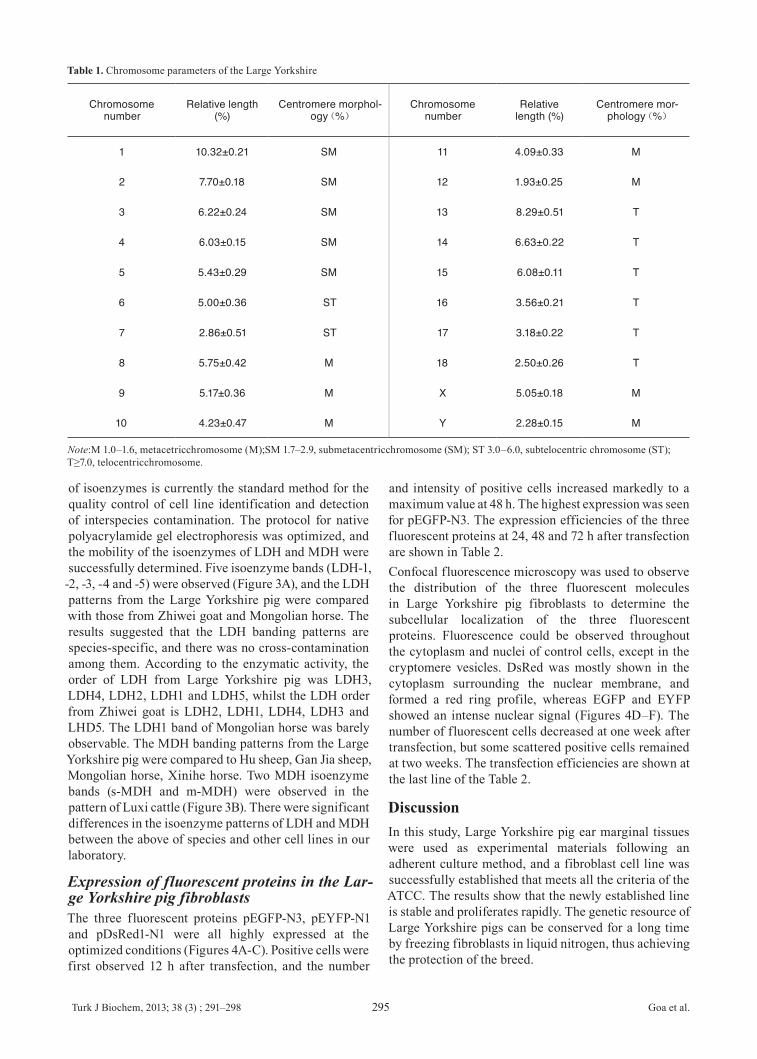
Karyotype analysisThe chromosome number of Large Yorkshire pig fibroblasts was 2n = 36, comprising 34 autosomes and two sex chromosomes, XY or XX (Figure 2D). The parameters, including relative length and centromere morphology are shown in Table 1. The chromosome numbers per spread were counted for 100 spreads of the first, second and fourth passages, and the frequencies of cells with 2n = 36 were 96, 94 and 91%, respectively.
Isoenzyme analysis of Large Yorkshire pig fibroblastsIsoenzyme polymorphism occurs among species, and sometimes even among breeds, individuals and tissues within a species [25]. Polymorphism analysis
22
430
Figure 1. Culture of Large Yorkshire pig fibroblasts. (A) Primary cells of Large 431
Yorkshire ear marginal explants (100×); (B) Subcultured fibroblasts (100×); (C) 432
Fibroblast cells before freezing (100×); (D) Fibroblast cells 24 h after recovery (100×). 433
434
Figure 2. Growth curve, detection of microbial contamination and karyotype of Large 435
Yorkshire pig fibroblasts. (A) Growth curve of Large Yorkshire pig fibroblasts, with 436
cell density on the vertical axis. The curve appeared as a typical “S” shape, including 437
latency phase, exponential growth phase and plateau phase; (B) Negative result of 438
Streptococcus detection for Large Yorkshire pig fibroblasts (100×); (C) Negative result 439
Figure 1. Culture of Large Yorkshire pig fibroblasts. (A) Primary cells of Large Yorkshire ear marginal explants (100×); (B) Subcultured fibroblasts (100×); (C) Fibroblast cells before freezing (100×); (D) Fibroblast cells 24 h after recovery (100×)
Figure 2. Growth curve, detection of microbial contamination and karyotype of Large Yorkshire pig fibroblasts. (A) Growth curve of Large Yorkshire pig fibroblasts, with cell density on the vertical axis. The curve appeared as a typical “S” shape, including latency phase, exponential growth phase and plateau phase; (B) Negative result of Streptococcus detection for Large Yorkshire pig fibroblasts (100×); (C) Negative result of Mycoplasma detection for Large Yorkshire pig fibroblasts (200×); (D) Chromosomes at metaphase (left) and karyotype (right) of Large Yorkshire pig fibroblast line (1000×). The chromosome number of Large Yorkshire pig fibroblasts was 2n = 36, comprising 34 autosomes and two sex chromosomes, whilst the sex chromosome type was XY (♂).
Turk J Biochem, 2013; 38 (3) ; 291–298 Goa et al.295
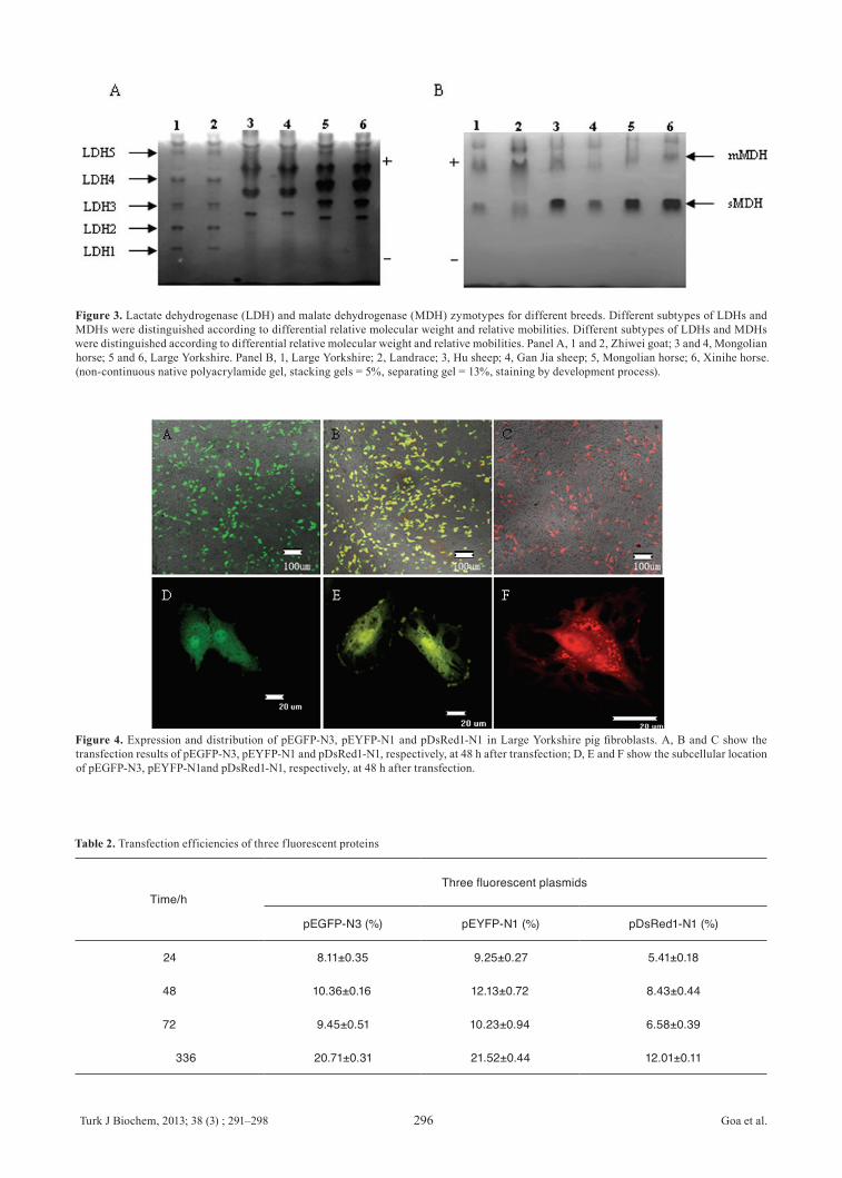
of isoenzymes is currently the standard method for the quality control of cell line identification and detection of interspecies contamination. The protocol for native polyacrylamide gel electrophoresis was optimized, and the mobility of the isoenzymes of LDH and MDH were successfully determined. Five isoenzyme bands (LDH-1,
-2, -3, -4 and -5) were observed (Figure 3A), and the LDH patterns from the Large Yorkshire pig were compared with those from Zhiwei goat and Mongolian horse. The results suggested that the LDH banding patterns are species-specific, and there was no cross-contamination among them. According to the enzymatic activity, the order of LDH from Large Yorkshire pig was LDH3, LDH4, LDH2, LDH1 and LDH5, whilst the LDH order from Zhiwei goat is LDH2, LDH1, LDH4, LDH3 and LHD5. The LDH1 band of Mongolian horse was barely observable. The MDH banding patterns from the Large Yorkshire pig were compared to Hu sheep, Gan Jia sheep, Mongolian horse, Xinihe horse. Two MDH isoenzyme bands (s-MDH and m-MDH) were observed in the pattern of Luxi cattle (Figure 3B). There were significant differences in the isoenzyme patterns of LDH and MDH between the above of species and other cell lines in our laboratory.
Expression of fluorescent proteins in the Lar-ge Yorkshire pig fibroblastsThe three fluorescent proteins pEGFP-N3, pEYFP-N1 and pDsRed1-N1 were all highly expressed at the optimized conditions (Figures 4A-C). Positive cells were first observed 12 h after transfection, and the number
and intensity of positive cells increased markedly to a maximum value at 48 h. The highest expression was seen for pEGFP-N3. The expression efficiencies of the three fluorescent proteins at 24, 48 and 72 h after transfection are shown in Table 2. Confocal fluorescence microscopy was used to observe the distribution of the three fluorescent molecules in Large Yorkshire pig fibroblasts to determine the subcellular localization of the three fluorescent proteins. Fluorescence could be observed throughout the cytoplasm and nuclei of control cells, except in the cryptomere vesicles. DsRed was mostly shown in the cytoplasm surrounding the nuclear membrane, and formed a red ring profile, whereas EGFP and EYFP showed an intense nuclear signal (Figures 4D–F). The number of fluorescent cells decreased at one week after transfection, but some scattered positive cells remained at two weeks. The transfection efficiencies are shown at the last line of the Table 2.
Discussion In this study, Large Yorkshire pig ear marginal tissues were used as experimental materials following an adherent culture method, and a fibroblast cell line was successfully established that meets all the criteria of the ATCC. The results show that the newly established line is stable and proliferates rapidly. The genetic resource of Large Yorkshire pigs can be conserved for a long time by freezing fibroblasts in liquid nitrogen, thus achieving the protection of the breed.
Table 1. Chromosome parameters of the Large Yorkshire
Chromosome number
Relative length (%)
Centromere morphol-ogy(%)
Chromosome number
Relative length (%)
Centromere mor-phology(%)
1 10.32±0.21 SM 11 4.09±0.33 M
2 7.70±0.18 SM 12 1.93±0.25 M
3 6.22±0.24 SM 13 8.29±0.51 T
4 6.03±0.15 SM 14 6.63±0.22 T
5 5.43±0.29 SM 15 6.08±0.11 T
6 5.00±0.36 ST 16 3.56±0.21 T
7 2.86±0.51 ST 17 3.18±0.22 T
8 5.75±0.42 M 18 2.50±0.26 T
9 5.17±0.36 M X 5.05±0.18 M
10 4.23±0.47 M Y 2.28±0.15 M
Note:M 1.0–1.6, metacetricchromosome (M);SM 1.7–2.9, submetacentricchromosome (SM); ST 3.0–6.0, subtelocentric chromosome (ST); T≥7.0, telocentricchromosome.
Turk J Biochem, 2013; 38 (3) ; 291–298 Goa et al.296
Figure 4. Expression and distribution of pEGFP-N3, pEYFP-N1 and pDsRed1-N1 in Large Yorkshire pig fibroblasts. A, B and C show the transfection results of pEGFP-N3, pEYFP-N1 and pDsRed1-N1, respectively, at 48 h after transfection; D, E and F show the subcellular location of pEGFP-N3, pEYFP-N1and pDsRed1-N1, respectively, at 48 h after transfection.
Table 2. Transfection efficiencies of three fluorescent proteins
Time/hThree fluorescent plasmids
pEGFP-N3 (%) pEYFP-N1 (%) pDsRed1-N1 (%)
24 8.11±0.35 9.25±0.27 5.41±0.18
48 10.36±0.16 12.13±0.72 8.43±0.44
72 9.45±0.51 10.23±0.94 6.58±0.39
336 20.71±0.31 21.52±0.44 12.01±0.11
23
of Mycoplasma detection for Large Yorkshire pig fibroblasts (200×); (D) Chromosomes 440
at metaphase (left) and karyotype (right) of Large Yorkshire pig fibroblast line (1000×). 441
The chromosome number of Large Yorkshire pig fibroblasts was 2n = 36, comprising 442
34 autosomes and two sex chromosomes, whilst the sex chromosome type was XY (♂). 443
444
Figure 3. Lactate dehydrogenase (LDH) and malate dehydrogenase (MDH) zymotypes 445
for different breeds. Different subtypes of LDHs and MDHs were distinguished 446
according to differential relative molecular weight and relative mobilities. Different 447
subtypes of LDHs and MDHs were distinguished according to differential relative molecular weight 448
and relative mobilities. Panel A, 1 and 2, Zhiwei goat; 3 and 4, Mongolian horse; 5 and 6, 449
Large Yorkshire. Panel B, 1, Large Yorkshire; 2, Landrace; 3, Hu sheep; 4, Gan Jia 450
sheep; 5, Mongolian horse; 6, Xinihe horse. (non-continuous native polyacrylamide gel, 451
stacking gels = 5%, separating gel = 13%, staining by development process). 452
Figure 3. Lactate dehydrogenase (LDH) and malate dehydrogenase (MDH) zymotypes for different breeds. Different subtypes of LDHs and MDHs were distinguished according to differential relative molecular weight and relative mobilities. Different subtypes of LDHs and MDHs were distinguished according to differential relative molecular weight and relative mobilities. Panel A, 1 and 2, Zhiwei goat; 3 and 4, Mongolian horse; 5 and 6, Large Yorkshire. Panel B, 1, Large Yorkshire; 2, Landrace; 3, Hu sheep; 4, Gan Jia sheep; 5, Mongolian horse; 6, Xinihe horse. (non-continuous native polyacrylamide gel, stacking gels = 5%, separating gel = 13%, staining by development process).
24
453
Figure 4. Expression and distribution of pEGFP-N3, pEYFP-N1 and pDsRed1-N1 in 454
Large Yorkshire pig fibroblasts. A, B and C show the transfection results of 455
pEGFP-N3, pEYFP-N1 and pDsRed1-N1, respectively, at 48 h after transfection; D, E 456
and F show the subcellular location of pEGFP-N3, pEYFP-N1and pDsRed1-N1, 457
respectively, at 48 h after transfection. 458
Turk J Biochem, 2013; 38 (3) ; 291–298 Goa et al.297
Microbial contamination is one of the most common phenomena in cell culture resulting in experimental failure. Potential sources of contamination include air, equipment, serum, and tissue samples [26]. Whilst contamination by bacteria, eumycetes, and mycetes can be detected by the naked eye because of the turbidity of the culture media, it is harder to detect Mycoplasma contamination. Mycoplasma have no nuclei and can grow and reproduce in cell culture media. They are hard to eliminate and can coexist with cultured cells for long periods of time. The methods used to detect Mycoplasma include direct solid agar culture, indirect fluorescence staining of DNA, and new DNA-style hybridization. Because fluorescent staining of Mycoplasma DNA is a ready-to-use technique, it is commonly used by some cell culture collections such as the ATCC. Our microbiological detection results showed that the Large Yorkshire pig fibroblast bank was purified and free of Mycoplasma contamination. The Streptococcus contamination-detection results showed that the Large Yorkshire pig fibroblast bank was also free of Streptococcus.Karyotype analysis is commonly used for detecting abnormal cells in a population because the chromosome number, shape and structure of normal cells remain very stable. Aberrations in chromosome numbers showed a tendency to increase with passage number, indicating that in vitro culture can have a slight effect on the stability of the cells, but suggesting that the cell line was reproducibly diploid. In addition, there were no chromosomal abnormalities, further indicating that the cell lines were stably diploid.LDH and MDH are pivotal enzymes involved in the glycolytic pathway and citric acid cycle, respectively. They are species-specific and evolutionarily conserved, but the subtypes and activities differ among species, providing a biochemical indicator of species classification by chromatography and electrophoresis. LDH and MDH were therefore selected to determine the species origin of the cells and to detect cross-contamination [27-28]. Biochemical analysis of isoenzyme polymorphism is currently considered the standard method for quality control of cell line identification and interspecies contamination, as demonstrated when Arai et al measured the LDH isoenzyme patterns in horse leucocytes and plasma, and in Debao pony fibroblasts [29]. MDH is a dimeric enzyme composed of cytosolic (s-MDH) and mitochondrial (m-MDH) subunits. The mobilities of MDH bands among poultry are essentially identical, and the same is true among those from livestock. However, MDH from livestock migrates more rapidly than that from poultry, and livestock contains a higher concentration of MDH.Research using fluorescent proteins mainly focuses on tumor, nerve and stem cells [30]. It has been shown in Vero cells, HeLa cells and other cell lines that transfection efficiencies can be affected by DNA
concentration, Lipofectamine concentration, incubation time, reagent combinations, and the presence of serum [30]. This study has shown the expression of fluorescent proteins in Large Yorkshire pig fibroblasts with no obvious effect on the growth and proliferation of the transfected cells. This indicates that fluorescent protein genes can be safely used as a marker of foreign genes in transgenic animals. In summary, the Large Yorkshire pig fibroblast bank has been successfully established, containing biologically normal and genetically stable fibroblasts. All aspects of the bank have met the standards for cell line quality supervision of major international culture collections and centers. Results from exogenous gene expression support the vitality of the cells, and their applications in transgenic therapy and genetics. The precious genetic resource of the Large Yorkshire pig has been effectively preserved at a cellular level. This bank can serve as a resource providing biological materials for further experimental work in fields including genetics, biomedical sciences, cell and molecular biology, and immunology.
AcknowledgementsThis research was supported by the the Ministry of Agriculture of China for Transgenic Research Program (2011ZX08009-003-006, 2011ZX08012-002-06), the project of National Infrastructure of Animal Germplasm Resources (2013 year and the central level, scientific research institutes for R&D special fund business (2011cj-9, 2012zl072), and the central level, scientific research institutes for R&D special fund business (2011cj-9) and Science and Technological Fund of Anhui Province for Outstanding Youth (#10040606Q43).
References[1] Rege JE. O, Gibson JP. Animal genetic resources and econo-
mic development: issues in relation to economic valuation. Ecol Econ 2003; 45(3): 319-330.
[2] Hartung T, Balls M, Bardouille C, Blanck O, Coecke S,et al. EC-VAM Good Cell Culture Practice Task Force report 1. ATLA 2002; 30: 407–414.
[3] Hiemstra S J, van der Lende T, Woelders H. The potential of cryopreservation and reproductive technologies for animal ge-netic resources conservation strategies. 2006; pp.45-59, FAO, Rome.
[4] Leon Quinto T, Simon MA, Cadenas R, Jones J, Martinez-Her-nandez FJ, et al. Developing biological resource banks as a sup-porting tool for wildlife reproduction and conservation: the Ibe-rian lynx bank as a model for other endangered species. Anim Reprod Sci 2009; 112 (3): 347-361.
[5] Wu HM, Guan W J, Li H, Ma YH. Establishment and charac-teristics of white ear lobe chicken embryo fibroblast line and expression of six fluorescent proteins in the cells. Cell Biol Int 2008; 32: 1478-85.
[6] Liu CQ, Guo Y, Guan WJ, Ma YH, Zhang HH, et al. Estab-lishment and biological characteristics of Luxi cattle fibroblast bank. Tissue Cell 2008; 40: 417-24.
Turk J Biochem, 2013; 38 (3) ; 291–298 Goa et al.298
[7] Li LF, Yue H, Ma JZ, Guan WJ, Ma YH. Establishment and cha-racterization of a fibroblast line from Simmental cattle. Cryobi-ology 2009; 59: 63-68.
[8] Bai CY, Li CY, Jin DP, Guo Y, Guan WJ, et al. Establishment and characterization of a fibroblast line from landrace. Aritf Cell Blood Sub 2010; 38: 129-35.
[9] Na RS, Bai CY, Jin DP, Su XH, Feng BG, et al. Establishment and biological characteristics of Qingyuan partridge chicken fibroblast line. Poultry Sci 2010; 89: 1207-16.
[10] Han XP, Wu HD , Pan JF, Fu H ,Liu J. The selection of the pro-ductive performance of large Yorkshire. Acta Agriculturae Uni-versitatis Jiangxiensis 2000;22(3): 447-50.
[11] Freshney RI. Culture of Animal Cells: A Manual of Basic Tech-nique. 2000; pp.149-175, Wiley-Liss,New York.
[12] Guan WJ, Ma YH, Zhou XY, Liu GL, Liu XD. The estab-lishment of fibroblast cell line and its biological characteristic research in Taihang black goat. Rev China Agric Sci Technol. 2005; 7: 25-33.
[13] Gu YP, Li HZ, Mik J. Phenotypic characterization of te-lomerase-immortalized primary non-malignant and malignant tumor-derived human prostate epithelial cell lines. Exp Cell Res 2006; 312: 841-843.
[14] Kong D, Nishino N, Shibusawa M, Kusano M. Establishment and characterization of human pancreatic adenocarcinoma cell line in tissue culture and the nude mouse. Tissue Cell 2007; 39: 217-223.
[15] Butler M. Cell counting and viability measurements. In: Jenkins Nigel, Editor, Animal Cell Biotechnology Methods Pro-tocols, 1999; pp. 131-144, Humana Press Inc, New Jersey.
[16] Doyle AR, Hay BE. Animal Cells, Living Resources for Biotechnology, 1990; pp. 81-100, Cambridge University Press, Cambridge, UK.
[17] Masover GK, Becker FA. Detection of mycoplasmas in cell cultures by cultural methods. In: Miles R.J. & Nicholas, R.A.J. ed, Methods in Molecular Biology, vol. 104, Mycoplasma Protocols, 1998; pp. 207-215, 217-226, Humana Press, Totawa.
[18] Hay RI. Cell line preservation and characterization. In: R.I. Freshney, ed, Animal Cell Culture: A Practical Approach, 1992.pp. 104-135, Oxford University Press, Oxford.
[19] Hirofumi S, Kentaro Y, Kouichi H, Tsuyoshi F, Norihiro T, et al. Efficient establishment of human embryonic stem cell lines and long-term maintenance with stable karyotype by enz-ymatic bulk passage. Biochem Bioph Res Com 2006; 345: 926-932.
[20] Sun YL, Linb CS, Chou YC. Establishment and characterization of a spontaneously immortalized porcine mammary epithelial cell line. Cell Bio Int 2006; 30: 970-976.
[21] Kawarai S, Hashizakia K, Kitaoc S, Naganoa S, Madarameb H, Neoa S, Ishikawaa T, Furuichia M, Hisasuea M, Tsuchiyaa R, Tsujimotod H, Yamada T. Establishment and characterization of primary canine hepatocellular carcinoma cell lines producing alpha-fetoprotein. Vet Immunol Immunop 2006; 113: 30-36.
[22] Escriou V, Carriere M, Bussone F, Wils P, Scherman D. Critical assessment of the nuclear import of plasmid during cationic li-pid-mediated gene transfer. J Gene Med 2001; 3: 179-187.
[23] Tsuchiya R, Yoshiki F, Kudo Y, Morita M. Cell type-selective expression of green fluorescent protein and the calcium indica-ting protein, yellow chameleon, in rat cortical primary cultures. Brain Res 2002; 956: 221-229.
[24] Zhou XM, Ma YH, Guan WJ, Zhao DM. Establishment and identification of Debao pony ear marginal tissue fibroblast cell line. Asian-Australian Journal of Animal Science 2004; 17: 1338-1343.
[25] O’Brien SJ, Kleiner G, Olson R, Shannon JE. Enzyme poly-
morphisms as genetic signatures in human cells cultures. Scien-ce 1977; 195: 1345-1348.
[26] Lincoln CK and Gabridge MG. Cell culture contamination: so-urces, consequences, prevention, and elimination. Method Cell Bio 1998; 57: 49-65.
[27] Nelson-Rees WA, Daniels DW, Flandermeyer RR. Crossconta-mination of cells in culture. Science 1998; 212: 446-452.
[28] Parodi B, Aresu O, Bini D, Lorenzini R, Schena F, et al. Species identification and confirmation of human and animal cell lines: a PCR-based method. Bio Techniques 2002; 32: 432-440.
[29] Arai T, Inoue A, Uematsu, Y, Sako T, Kimura N. Activities of enzymes in the malate aspartate shuttle and the isoenzyme pattern of lactate dehydrogenase in plasma and peripheral leu-kocytes of lactating Hostein cows and riding horses. Research in Veterinary Science 2003; 75: 15-19.
[30] Jung S, Ackerley C, Ivanchuk S. Tracking the invasiveness of human astrocytoma cells by using green fluorescent protein in an organotypical brain slice model. Neurosurgery 2001; 94: 80-89.