Embed Size (px)
Citation preview
Dr.Ban I.S. head & neck anatomy 2nd y.
جامعة تكريت
كلية طب االسنان
التشريحمادة
املرحلة الثانية
أ.م.د. بان امساعيل صديق
6102/6102
Dr.Ban I.S. head & neck anatomy 2nd y.
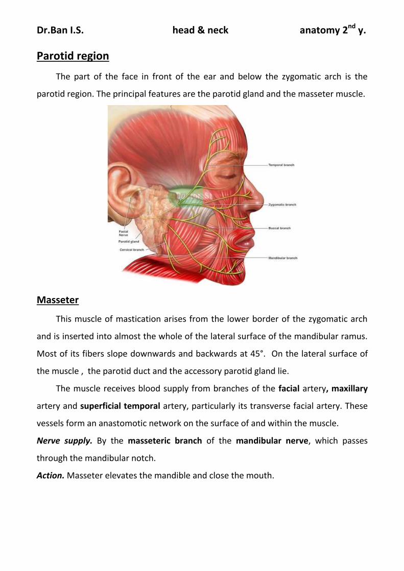
Parotid region
The part of the face in front of the ear and below the zygomatic arch is the
parotid region. The principal features are the parotid gland and the masseter muscle.
Masseter
This muscle of mastication arises from the lower border of the zygomatic arch
and is inserted into almost the whole of the lateral surface of the mandibular ramus.
Most of its fibers slope downwards and backwards at 45°. On the lateral surface of
the muscle , the parotid duct and the accessory parotid gland lie.
The muscle receives blood supply from branches of the facial artery, maxillary
artery and superficial temporal artery, particularly its transverse facial artery. These
vessels form an anastomotic network on the surface of and within the muscle.
Nerve supply. By the masseteric branch of the mandibular nerve, which passes
through the mandibular notch.
Action. Masseter elevates the mandible and close the mouth.
Dr.Ban I.S. head & neck anatomy 2nd y.
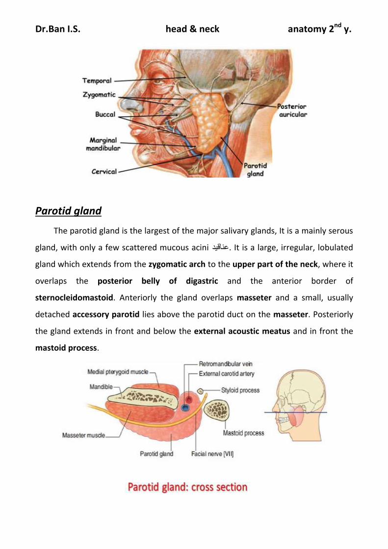
Parotid gland
The parotid gland is the largest of the major salivary glands, It is a mainly serous
gland, with only a few scattered mucous acini عناقيد. It is a large, irregular, lobulated
gland which extends from the zygomatic arch to the upper part of the neck, where it
overlaps the posterior belly of digastric and the anterior border of
sternocleidomastoid. Anteriorly the gland overlaps masseter and a small, usually
detached accessory parotid lies above the parotid duct on the masseter. Posteriorly
the gland extends in front and below the external acoustic meatus and in front the
mastoid process.
Dr.Ban I.S. head & neck anatomy 2nd y.
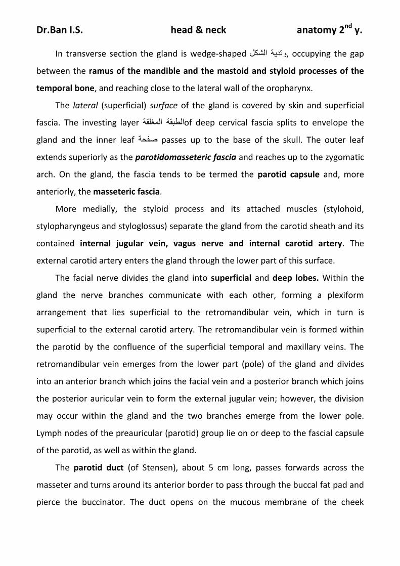
In transverse section the gland is wedge-shaped وتدية الشكل, occupying the gap
between the ramus of the mandible and the mastoid and styloid processes of the
temporal bone, and reaching close to the lateral wall of the oropharynx.
The lateral (superficial) surface of the gland is covered by skin and superficial
fascia. The investing layer الطبقة المغلقةof deep cervical fascia splits to envelope the
gland and the inner leaf صفحة passes up to the base of the skull. The outer leaf
extends superiorly as the parotidomasseteric fascia and reaches up to the zygomatic
arch. On the gland, the fascia tends to be termed the parotid capsule and, more
anteriorly, the masseteric fascia.
More medially, the styloid process and its attached muscles (stylohoid,
stylopharyngeus and styloglossus) separate the gland from the carotid sheath and its
contained internal jugular vein, vagus nerve and internal carotid artery. The
external carotid artery enters the gland through the lower part of this surface.
The facial nerve divides the gland into superficial and deep lobes. Within the
gland the nerve branches communicate with each other, forming a plexiform
arrangement that lies superficial to the retromandibular vein, which in turn is
superficial to the external carotid artery. The retromandibular vein is formed within
the parotid by the confluence of the superficial temporal and maxillary veins. The
retromandibular vein emerges from the lower part (pole) of the gland and divides
into an anterior branch which joins the facial vein and a posterior branch which joins
the posterior auricular vein to form the external jugular vein; however, the division
may occur within the gland and the two branches emerge from the lower pole.
Lymph nodes of the preauricular (parotid) group lie on or deep to the fascial capsule
of the parotid, as well as within the gland.
The parotid duct (of Stensen), about 5 cm long, passes forwards across the
masseter and turns around its anterior border to pass through the buccal fat pad and
pierce the buccinator. The duct opens on the mucous membrane of the cheek
Dr.Ban I.S. head & neck anatomy 2nd y.
opposite the second upper molar tooth; it pierces the buccinator further back and
runs forwards beneath the mucous membrane to its orifice.
Blood supply
Branches from the external carotid artery supply the gland. Venous return is to
the retromandibular vein.
Lymph drainage
Lymph drains to the preauricular (parotid) nodes and then to the deep cervical
nodes.
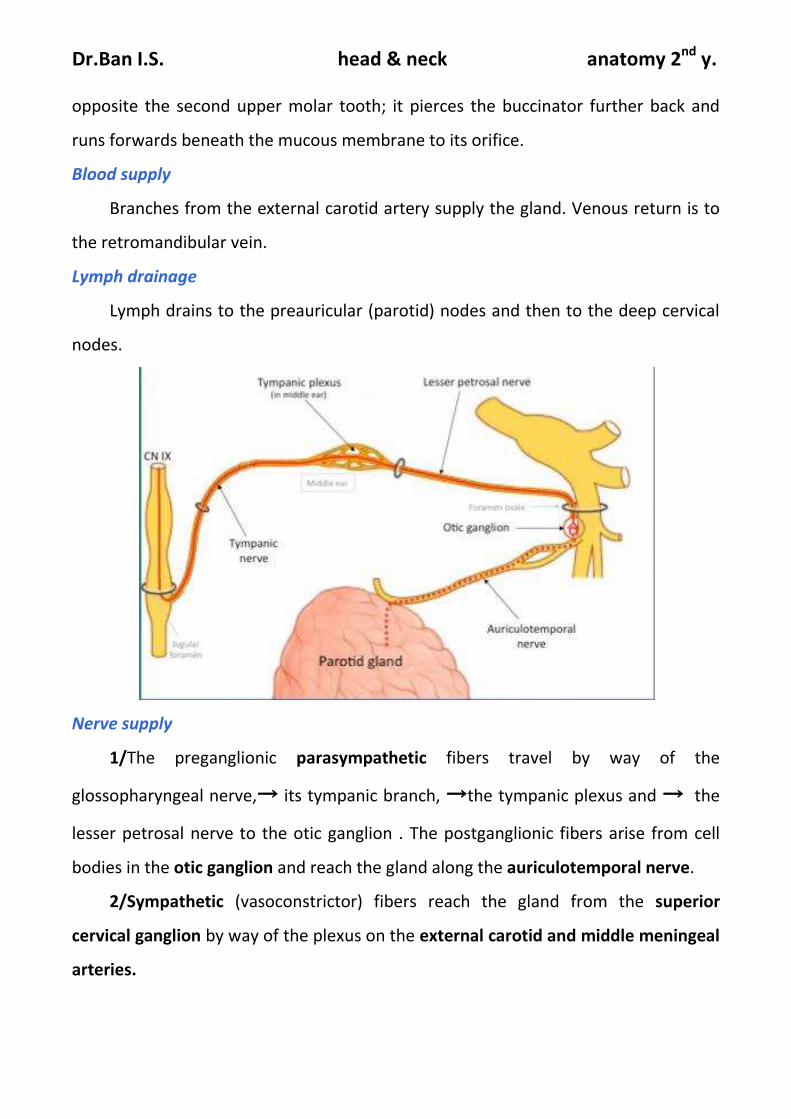
Nerve supply
1/The preganglionic parasympathetic fibers travel by way of the
glossopharyngeal nerve,→ its tympanic branch, →the tympanic plexus and → the
lesser petrosal nerve to the otic ganglion . The postganglionic fibers arise from cell
bodies in the otic ganglion and reach the gland along the auriculotemporal nerve.
2/Sympathetic (vasoconstrictor) fibers reach the gland from the superior
cervical ganglion by way of the plexus on the external carotid and middle meningeal
arteries.
Dr.Ban I.S. head & neck anatomy 2nd y.
3/Sensory innervation is supplied by the auriculotemporal nerve, a branch of
the mandibular nerve.
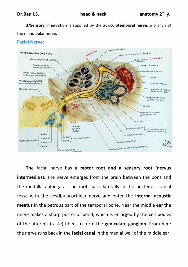
Facial Nerve:
The facial nerve has a motor root and a sensory root (nervus
intermedius). The nerve emerges from the brain between the pons and
the medulla oblongata. The roots pass laterally in the posterior cranial
fossa with the vestibulocochlear nerve and enter the internal acoustic
meatus in the petrous part of the temporal bone. Near the middle ear the
nerve makes a sharp posterior bend, which is enlarged by the cell bodies
of the afferent (taste) fibers to form the geniculate ganglion. From here
the nerve runs back in the facial canal in the medial wall of the middle ear.
Dr.Ban I.S. head & neck anatomy 2nd y.
It now curves downwards behind the middle ear, and passes vertically
down in the facial canal. After shedding all the nervus intermedius fibers,
the nerve emerges from the stylomastoid foramen as a motor nerve and
passes through the parotid gland to its distribution .
Important Branches of the Facial Nerve
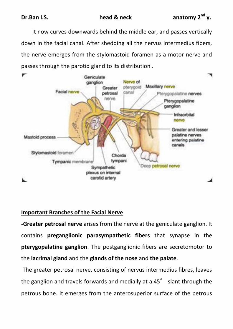
-Greater petrosal nerve arises from the nerve at the geniculate ganglion. It
contains preganglionic parasympathetic fibers that synapse in the
pterygopalatine ganglion. The postganglionic fibers are secretomotor to
the lacrimal gland and the glands of the nose and the palate.
The greater petrosal nerve, consisting of nervus intermedius fibres, leaves
the ganglion and travels forwards and medially at a 45° slant through the
petrous bone. It emerges from the anterosuperior surface of the petrous
Dr.Ban I.S. head & neck anatomy 2nd y.
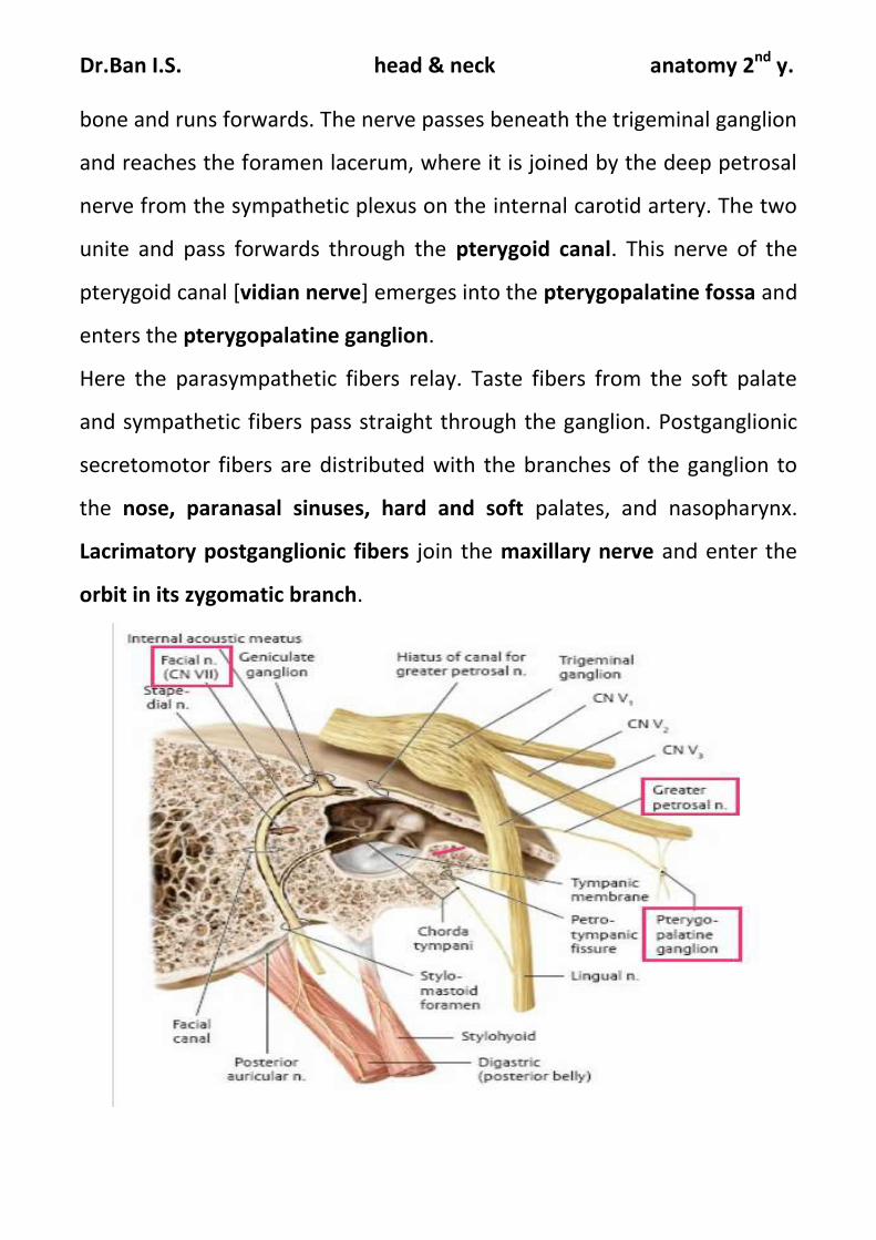
bone and runs forwards. The nerve passes beneath the trigeminal ganglion
and reaches the foramen lacerum, where it is joined by the deep petrosal
nerve from the sympathetic plexus on the internal carotid artery. The two
unite and pass forwards through the pterygoid canal. This nerve of the
pterygoid canal [vidian nerve] emerges into the pterygopalatine fossa and
enters the pterygopalatine ganglion.
Here the parasympathetic fibers relay. Taste fibers from the soft palate
and sympathetic fibers pass straight through the ganglion. Postganglionic
secretomotor fibers are distributed with the branches of the ganglion to
the nose, paranasal sinuses, hard and soft palates, and nasopharynx.
Lacrimatory postganglionic fibers join the maxillary nerve and enter the
orbit in its zygomatic branch.

Dr.Ban I.S. head & neck anatomy 2nd y.
■■ Nerve to stapedius supplies the stapedius muscle in the middle ear .
The nerve to stapedius is given off in the facial canal and reaches the
muscle by a minute canaliculus.
■■ Chorda tympani arises from the facial nerve in the facial canal in the
posterior wall of the middle ear. It runs forward over the medial surface of
the upper part of the tympanic membrane and leaves the middle ear
through the petrotympanic fissure, thus entering the infratemporal fossa
and joining the lingual nerve. The chorda tympani contains preganglionic
parasympathetic secretomotor fibers to the submandibular and the
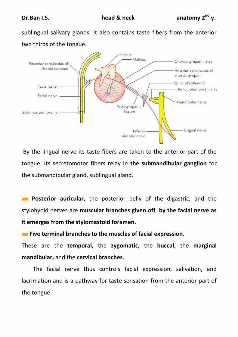
Dr.Ban I.S. head & neck anatomy 2nd y.
sublingual salivary glands. It also contains taste fibers from the anterior
two thirds of the tongue.
By the lingual nerve its taste fibers are taken to the anterior part of the
tongue. Its secretomotor fibers relay in the submandibular ganglion for
the submandibular gland, sublingual gland.
■■ Posterior auricular, the posterior belly of the digastric, and the
stylohyoid nerves are muscular branches given off by the facial nerve as
it emerges from the stylomastoid foramen.
■■ Five terminal branches to the muscles of facial expression.
These are the temporal, the zygomatic, the buccal, the marginal
mandibular, and the cervical branches.
The facial nerve thus controls facial expression, salivation, and
lacrimation and is a pathway for taste sensation from the anterior part of
the tongue.


![]یعلاط نیسحلادبعjep.emamat.ir/article_59929_4448c888fa0f903c07a83b4e10ff2729.pdfرد ار نآ قرط یر بط ریرج نب دمحم ماما .ت سا هدومرف](https://img.pdfslide.tips/doc/110x75/5e415ca09f0d32037a3e8bba/oe-oejep-oe-oe-.jpg)