Embed Size (px)
Citation preview

Instructions for use
Title FETAL ELECTROCARDIOGRAM IN DAIRY CATTLE : V FINDINGS AT PARTURITION
Author(s) TOO, Kimehiko; KANAGAWA, Hiroshi; KAWATA, Keiichiro; INOUE, Tadahiro; ODAJIMA, Takeshi
Citation Japanese Journal of Veterinary Research, 15(1), 21-30
Issue Date 1967-03
DOI 10.14943/jjvr.15.1.21
Doc URL http://hdl.handle.net/2115/1863
Type bulletin (article)
File Information KJ00002369279.pdf
Hokkaido University Collection of Scholarly and Academic Papers : HUSCAP
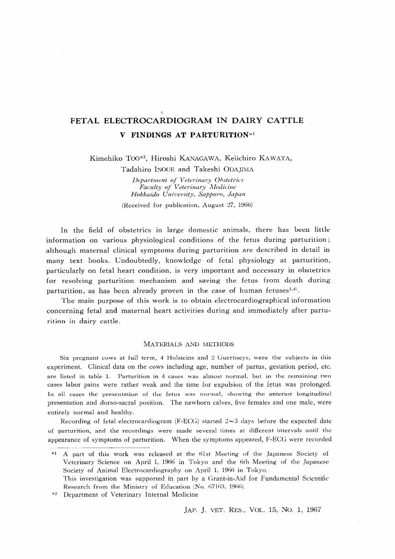
FETAL ELECTROCARDIOGRAM IN DAIRY CATTLE
V FINDINGS AT PARTURITION*l
Kimehiko TOO*2, Hiroshi KANAGAWA, Keiichiro KAWATA,
Tadahiro INOUE and Takeshi ODAJIMA
Department of Veterinary Ohstetrics Faculty of Veterinary Aledicine
Hokkaido Uni'uersity, Sapporo, Japan
(Received for publication, August '27, 1966)
In the field of obstetrics in large domestic animals, there has been little
information on various physiological conditions of the fetus during parturition;
although maternal clinical symptoms during parturition are described in detail in
many text books. Undoubtedly, knowledge of fetal physiology at parturition,
particularly on fetal heart condition, is very important and necessary in obstetrics
for resolving parturition mechanism and saving the fetus from death during
parturition, as has been already proven in the case of human fetuses2,4).
The main purpose of this work is to obtain electrocardiographical information
concerning fetal and maternal heart activities during and immediately after partu
rition in dairy cattle.
MATERIALS AND lV1ETHODS
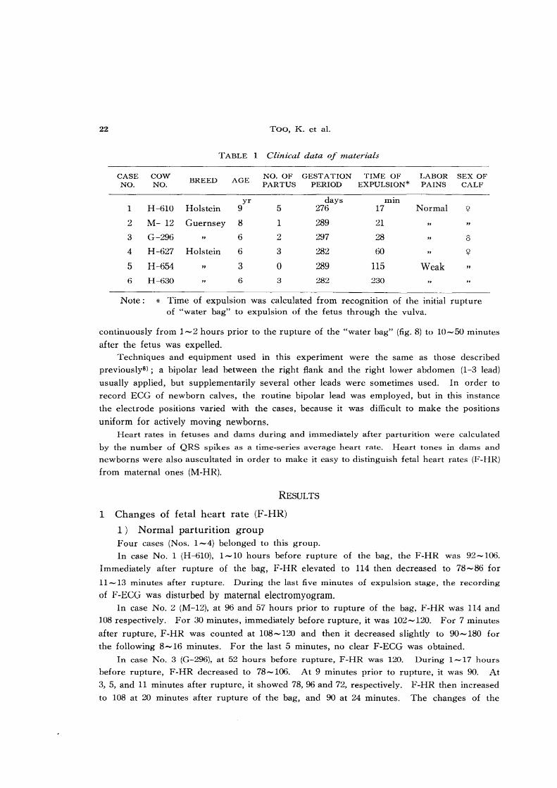
Six pregnant cows at full term, 4 Holsteins and '2 Guernseys, were the subjects in this
experiment. Clinical data on the cows including age, number of partus, gestation period, etc.
are listed in table 1. Parturition in 4 cases was almost normal, but in the remaining two cases labor pains were rather weak and the time for expulsion of the fetus was prolonged.
In all cases the presentation of the fetus was normal, showing the anterior longitudinal presentation and dorso-sacral position. The newborn calves, five females and one male, were
entirely normal and healthy.
Recording of fetal electrocardiogram (F-ECG) started 2-3 days before the expected date
of parturition, and the recordings were made several times at different intervals until the
appearance of symptoms of parturition. When the symptoms appeared, F-ECG were recorded
*1 A part of this work was released at the 61st Meeting of the Japanese Society of Veterinary Science on April 1, 1966 in Tokyo and the 6th Meeting of the Japanese Society of Animal Electrocardiography on April 1, 1%6 in Tokyo. This investigation was supported in part by a Grant-in-Aid for Fundamental Scientific Research from the Ministry of Education (No. h71h3, 19(6).
*2 Department of Veterinary Internal Medicine
JAP. J. VET. RES., VOL. 15, NO.1, 1967
22 ToO, K. et aL
TABLE 1 Clinical data of materials
CASE COW BREED AGE NO. OF GESTATION TIME OF LABOR SEX OF NO. NO. PARTUS PERIOD EXPULSION* PAINS CALF
yr days min 1 H-61O Holstein 9 5 276 17 Normal <;2
2 M- 12 Guernsey 8 1 289 21
3 G-296 6 2 297 28 0
4 H-627 Holstein 6 3 282 60 <;2
5 H-654 .. 3 0 289 115 Weak " 6 H-630 6 3 282 230
Note: * Time of expulsion was calculated from recogmtIOn of the initial rupture of "water bag" to expulsion of the fetus through the vulva.
continuously from 1-2 hours prior to the rupture of the "water bag" (fig. 8) to 10-50 minutes
after the fetus was expelled.
Techniques and equipment used in this experiment were the same as those described
previously8); a bipolar lead between the right flank and the right lower abdomen (1-3 lead)
usually applied, but supplementarily several other leads were sometimes used. In order to
record ECG of newborn calves, the routine bipolar lead was employed, but in this instance
the electrode positions varied with the cases, because it was difficult to make the positions
uniform for actively moving newborns.
Heart rates in fetuses and dams during and immediately after parturition were calculated
by the number of QRS spikes as a time-series average heart rate. Heart tones in dams and
newborns were also auscultated in order to make it easy to distinguish fetal heart rates (F-HR)
from maternal ones (M-HR).
RESULTS
1 Changes of fetal heart rate (F -HR)
1) Normal parturition group Four cases (Nos. 1-4) belonged to this group.
In case No.1 (H-61O), 1-10 hours before rupture of the bag, the F-HR was 92-106.
Immediately after rupture of the bag, F-HR elevated to 114 then decreased to 78-86 for
11-13 minutes after rupture. During the last five minutes of expulsion stage, the recording
of F-ECG was disturbed by maternal electromyogram.
In case No.2 (M-12), at 96 and 57 hours prior to rupture of the bag, F-HR was 114 and
108 respectively. For 30 minutes, immediately before rupture, it was 102-120. For 7 minutes
after rupture, F-HR was counted at 108-120 and then it decreased slightly to 90-180 for
the following 8-16 minutes. For the last 5 minutes, no clear F-ECG was obtained.
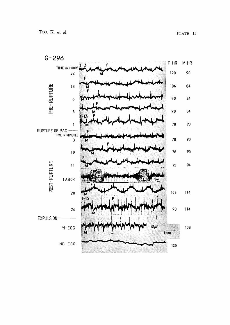
In case No.3 (G-296), at 52 hours before rupture, F-HR was 120. During 1-17 hours
before rupture, F-HR decreased to 78-106. At 9 minutes prior to rupture, it was 90. At
3, 5, and 11 minutes after rupture, it showed 78,96 and 72, respectively. F-HR then increased
to 108 at 20 minutes after rupture of the bag, and 90 at 24 minutes. The changes of the

Fetal electrocardiogram in cattle V 23
heart rates of the fetus and the dam in this case are in plate II.
In case No.4 (H-627), 47 hours before rupture, F-HR was 114, and then it decreased to
108 within 2 hours before rupture. At 60 minutes before rupture, F-HR showed 114-120,
but at 40 minutes it decreased to 84. For 25 minutes prior to rupture it returned to 114-
120 and immediately before rupture it decreased to 102. At 15, 20 and 30 minutes after
rupture, F-HR was 120, 96 and 120, respectively. At 35 minutes after rupture, it became 78,
being minimized throughout the whole course of parturition, and then it gradually increased
to 114 until the expulsion of the fetus.
Before rupture of the water bag, the F-HR generally maintained a level of 100 beats per
minute or showed a little decreasing tendency, but after rupture, at approximately the middle
of the stage of expulsion, a marked fall of F-HR was characteristic in all cases.
2) Group with weak labor pains
Two cases (Nos. 5 & 6) belonged to this group. The stage of expulsion in this group
was prolonged.
In No.5 (H-654), 61-62 hours before rupture, F-HR was 102-108. At 15 and 5 minutes
before rupture it showed 104 and 96 respectively. Immediately before rupture, it was
counted as 108. For 44 minutes after rupture, F-ECG could not be recorded clearly. At
45-57 minutes after rupture it was 104-108, but then it decreased to 85. The final F-HR,
in the stage of expulsion, at 105 minutes after rupture, was 120.
In case No.6 (H-630), at 7, 38 and 57 hours before rupture, F-HR showed 96, 108 and
128 respectively. For 2 hours prior to rupture, it maintained a rate of approximately 108.
hUITlediately after rupture, F-HR was 96. For 40 ITlinutes after rupture it was 96-108. Then,
F-HR showed an alternate increase and decrease within a range from 120 to 206 during the subsequent 150 ITlinutes.
Thus, these 2 cases showed a similar tendency in F-HR change as that of normal parturition cases before rupture, but after rupture, one of theITl showed a gradual increase
in F-HR until the middle of the stage of expulsion then alternate increase and decrease in
rate occurred until expulsion of the fetus.
2 Heart rate of newborn (NB-HR)
The heart rate of newborn calves was calculated by means of newborn electrocardiogram
and by direct auscultation within 1-11 minutes after expulsion. A comparison was made
TABLE 2 Final fetal heart rate (F-HR) and newborn's heart rate (NB-HR)
FINAL INITIAL TIME AFTER RANGE OF RECORDING CASE NO. F-HR NB-HR EXPULSION NB-HR LENGTH
mIn min 1 90 136 1 108-144 10
2 96 132 4 120-138 40
3 90 144 11 120-196 45 4 114 90 1 114-174 17
5 120 66 5 108-120 13
6 210 180 1 162-198 45
24 Too, K. et al.
between the NB-HR and the final F -HR. This data IS listed in table 2. In 3 cases with
normal labor pains, the NB-HR showed a higher level than the final F-HR. While, in the
remaining case of the normal parturition group and 2 cases of the other group, the NB-HR
was rather lower than the level of the final F-HR In case No. 5 the NB-HR maintained
a marked lower level than other cases for 13 minutes. In case No.4 the NB-HR elevated
within a short time. In case No.6, however, the NB-HR maintained a higher level but did
not exceed the level of the final F-HR for 45 minutes after expulsion.
In conclusion, when the final F-HR remained at rather lower levels, the initial NB-HR
showed a rapid elevation within a short time after delivery, while when the final F-HR was
high, the initial NB-HR generally decreased immediately after delivery. However, after 10
minutes or so, there was a tendency to maintain a high level of NB-HR in all cases.
3 Maternal heart rate (M-HR)
Respective values of M-HR before rupture of the bag, immediately after rupture, at term and after expulsion are shown in table 3. Variation of M-HR throughout the above course
was more slight than that of F -HR. It tended to increase from the middle phase to termi
nation of expulsion and also after expulsion. During rupture, in 2 cases, M-HR was higher
than before rupture, and in 1 case it was a little lower. In the remaining 3 cases M-HR
was almost the same as before rupture. Therefore, rupture of the bag did not seem to give
any significant effect on maternal heart rhythm.
CASE NO.
1
2
3
4
5
6
Notes: *1 *2 *3
TABLE 3 Changes of maternal heart rate
BEFORE IMMEDIA TEL Y TERMINA TION AFTER RUPTURE*l AFTER RUPTURE*2 OF EXPULSION*2 EXPULSION
98-108 90-95 98-102 104-110 72- 98 84-90 98-102 78-100
78- 84 90-94 108-114 108-132
84- 86 90-94 90-102 102*3
84- 90 78-96 96-120
78-102 90-96 96-114 136*3
Range before rupture Range within approximately 3 minutes after rupture or expulsion Values in short time recordings No maternal electrocardiogram could be recorded.
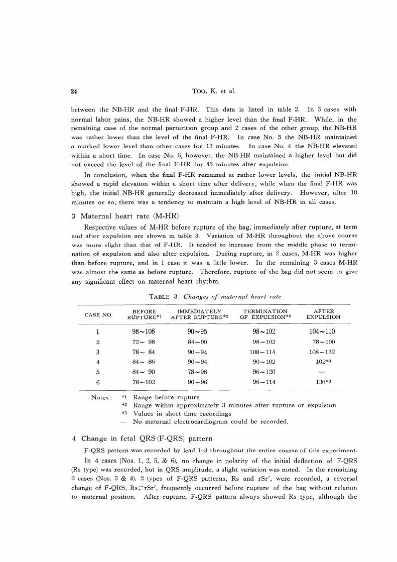
4 Change in fetal QRS (F-QRS) pattern
F-QRS pattern was recorded by lead 1-3 throughout the entire course of this experiment.
In 4 cases (Nos. 1, 2, 5, & 6), no change in polarity of the initial deRection of F -QRS
(Rs type) was recorded, but in QRS amplitude, a slight variation was noted. In the remaining
2 cases (Nos. 3 & 4), 2 types of F-QRS patterns, Rs and rSr', were recorded, a reversal
change of F-QRS, Rs~~rSr', frequently occurred before rupture of the bag without relation
to maternal position. After rupture, F-QRS pattern always showed Rs type, although the
Bemimin 2001 180~
16+ 140
120
100
Fetal electrocardiogram in cattle V
FIGURE 1 Changes in fetal QRS pattern (case No.8)
24 25 26 27 28 29
--~------
2 67 IQ illS 21 '-" ~ ..•... U.-'
'l'l •
RUPTURE
~
. ~:
~~~-11~1~-1~6~-13~-b~-5~~~-3~~z~o~-~m~o~-~oo~---~6o~!--~k~!--~w-L~~~W~-4~O-L~6~O~
HOURS MINUTES
22
Notes Each number in the above electrocardiogram (1-29) corresponds to that of the below figure, meaning each recording period. Nos. 1-22 are before rupture and Nos. 23-29 are after rupture. Underlined fetal QRS indicates that of recording at lying position of the dam and QRS without underline at standing position.
All fetal electrocardiogram were recorded by lead 1-3. F (dotted line): Fetal heart rate M (full line): Maternal heart rate X : Fetal arrhythmia
NB (wave line): Heart rate of newborn calf
25
amplitude varied. An example of F-QRS changes throughout the whole course examined IS
shown in figure 1.
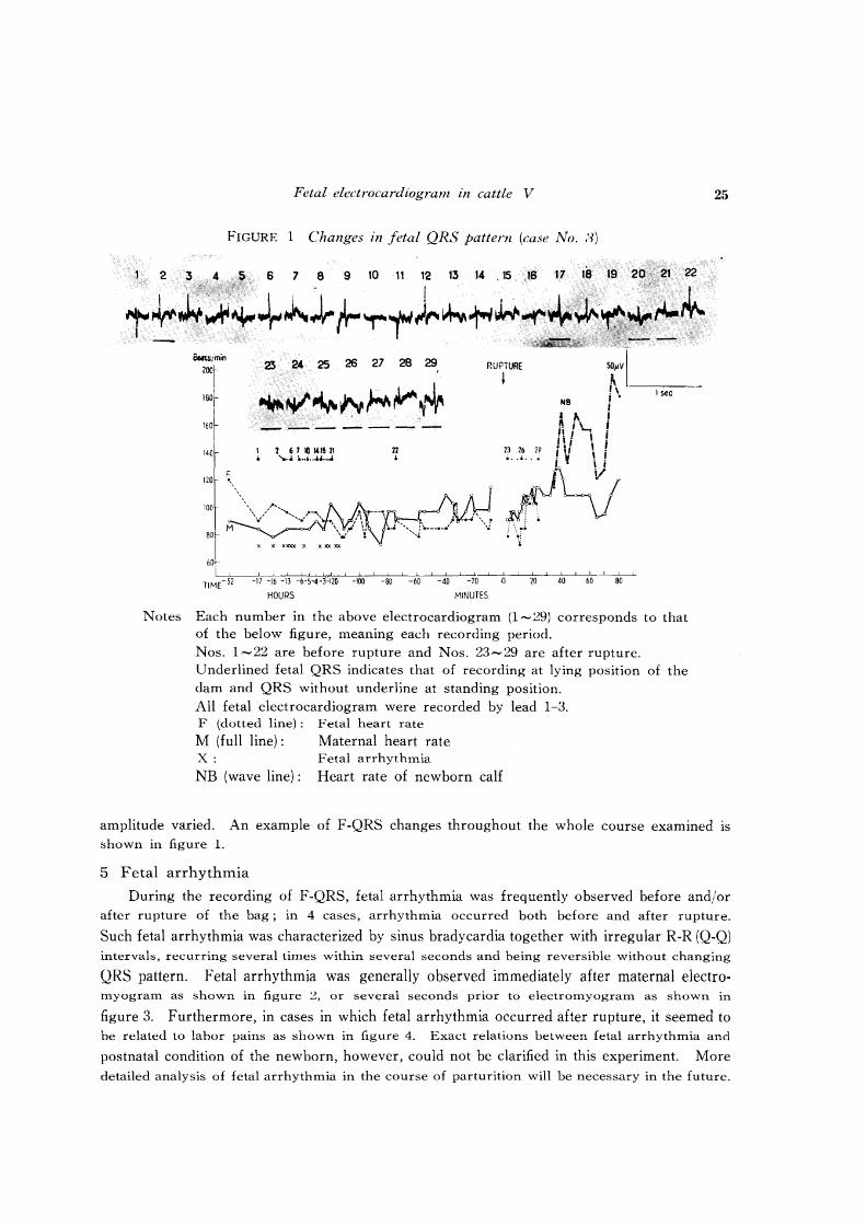
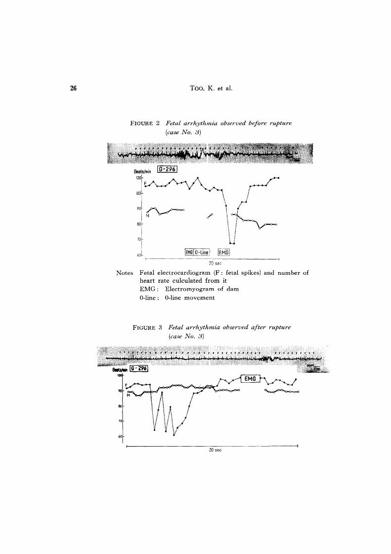
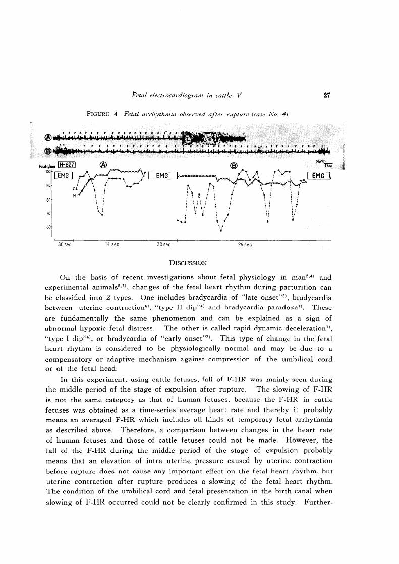
5 Fetal arrhythmia
During the recording of F-QRS, fetal arrhythmia was frequently observed before and/or
after rupture of the bag; in 4 cases, arrhythmia occurred both before and after rupture.
Such fetal arrhythmia was characterized by sinus bradycardia together with irregular R-R (Q-Q) intervals, recurring several times within several seconds and being reversible without changing
QRS pattern. Fetal arrhythmia was generally observed immediately after maternal electro-myogram as shown in figure 2, or several seconds prior to electromyogram as shown in
figure 3. Furthermore, in cases in which fetal arrhythmia occurred after rupture, it seemed to
be related to labor pains as shown in figure 4. Exact relations between fetal arrhythmia and
postnatal condition of the newborn, however, could not be clarified in this experiment. More
detailed analysis of fetal arrhythmia in the course of parturition will be necessary in the future.
26 Too, K. et al.
FIGURE 2 Fetal arrhythmia observed before rupture
(case No.3)
Beats/min I G-2961
100
90 /""'\ ~
80
70
I
60~
M \.-"'"
IEMGj O-Line I IEMGI
20 sec
Notes Fetal electrocardiogram (F: fetal spikes) and number of heart rate culculated from it EMG : Electromyogram of dam
O-line: O-line movement
FIGURE 3 Fetal arrhythmia observed after rupture
(case No.3)
30 sec
Petal electrocardiogram in cattle V 27
FIGURE 4 Fetal arrhythmia observed after rupture (case No.4)
BeaWmin IH-6271
100 I EMG I ,j\ 90
80
70
60
30 sec
i F' M
®
14 sec
EMG
30sec 26 sec
DISCUSSION
.. \
\1 ~
, ;/ ':~-" F' 'F' F ~f f f ~"
EMG t
On the basis of recent investigations about fetal physiology in man2,4) and
experimental animals5 •7), changes of the fetal heart rhythm during parturition can
be classified into 2 types. One includes bradycardia of "late onset"2), bradycardia
between uterine contraction6 ), "type II dip"4) and bradycardia paradoxal). These
are fundamentally the same phenomenon and can be explained as a sign of
abnormal hypoxic fetal distress. The other is called rapid dynamic deceler&tion1),
"type I dip"4), or bradycardia of "early onset"2). This type of change in the. fetal
heart rhythm is considered to be physiologically normal and may be due to a
compensatory or adaptive mechanism against compression of the umbilical cord
or of the fetal head.
In this experiment, using cattle fetuses, fall of F -HR was mainly seen during
the middle period of the stage of expulsion after rupture. The slowing of F -HR is not the same category as that of human fetuses, because the F-HR in cattle
fetuses was obtained as a time-series average heart rate and thereby it probably means an averaged F -HR which includes all kinds of temporary fetal arrhythmia
as described above. Therefore, a comparison between changes in the heart rate
of human fetuses and those of cattle fetuses could not be made. However, the
fall of the F -HR during the middle period of the stage of expulsion probably
means that an elevation of intra uterine pressure caused by uterine contraction
before rupture does not cause any important effect on the fetal heart rhythm, but
uterine contraction after rupture produces a slowing of the fetal heart rhythm.
The condition of the umbilical cord and fetal presentation in the birth canal when
slowing of F-HR occurred could not be clearly confirmed in this study. Further-
28 Too, K. et al.
more, III cases with weak labor pains, an increasing tendency of F-HR was
noticed instead of a falling of F-HR. This also means that weak pressure on
the fetus does not make the F-HR slow. Therefore, the following factors may
be suggested as causes: fetal hypoxia caused by disturbances in maternal-fetal
circulation, compression of the fetal head in the birth canal, vagal effect to the
fetal heart, and a fall of the POz value in the central nervous system of the
fetus. These factors may be interrelated and may act on the fetus simultaneously
or independently under certain maternal and fetal conditions during delivery. In
order to confirm the physiological significance of these factors ond changes of
the heart rhythm of bovine fetuses, further experimentation will be needed.
In the course of this study, fetal arrhythmia with temporary slowing of
F -HR was recorded. This type of slowing should not be considered an abnormal
feature, because these fetuses were born alive, the newborn calves were entirely
normal and healthy, and such arrhythmia disappeared immediately after delivery.
However, a marked descent of the F-HR and prolongation of fetal undoubtedly
be dangerous to fetal life, particularly in cases where these phenomena bradycardia
may are associated with severe dystocia, since such disorders would diminish the
cardiac output, thus reducing the blood supply to the fetal tissues.
A reversal change of F -QRS pattern, Rs~rSr', was frequently recorded before rupture in a limited number of cases. A similar change of F-QRS, Qrz:::!Rs, was
observed in a fetus at 180 days of pregnancy, and the possible cause in this case
was discussed in the previous paper9). In the former case, however, the mechanism
seems to be somewhat different from the present cases. The Rs~rSr' change may be due to some change in fetal presentation which may be caused principally labor
pains before rupture of the bag. It is assumed that when the forelegs of the fetus
pass through the cervical canal, the head portion enters tightly into the cervical
canal and rupture of the bag has already occurred, no reversal change is observed
and the QRS pattern always shows only the Rs type. In order to make this assumption decisive, however, much more information concerning changes of fetal
presentation during parturition will be required.
In human subjects, the level of the heart rate of the newborn is comparatively
low at birth but it elevates until the 4th day post-partum4). In cattle, on the other
hand, there hitherto was no information concerning the heart rate of the newborn,
although it was reported that the heart rate of calves of 2---60 days post-partum
ranged 110--134. In this experiment" it became certain that the heart rate of
newborn calves is more rapidly accelerated after delivery than that of human newborns.
The reasons for such acceleration of the heart rate of newborns both in man
and cattle may be due to the sudden commencement of pulmonary circulation,
Fetal electrocardiogram in cattle V 29
a sudden release from predominant vagus control during fetal life, respiratory
effect, gases in blood, exercise of newborns and abrupt enviromental change, etc.3 )
Slight elevations of M-HR were recorded during parturition; rupture 'of the
water bag and labor pains have little effect in elevating the M-HR. This gives
a good contrast in comparison to the changes of the maternal heart rhythm during
parturition in a mare, in which a clear A-V block was recordedlOl •
SUMMARY
This experiment was attempted to obtain electrocardiographical information
about fetal and maternal heart activities during and after parturition in dairy
cattle. Six pregnant cows were submitted to this experiment. The results are
summarized as follows:
1) In 4 cases of normal parturition, the change in fetal heart rate was not
so great before rupture of the bag, but after rupture a marked fall of fetal heart
rate was characteristic in all cases at approximately the middle of the stage of
expulsion. While, one of 2 cases with prolongation of the expulsion stage due
to weak labor pains, showed a gradual increasing tendency of the heart rate until
the middle of the stage of expulsion, and showed an alternate increase and decrease
until termination of expulsion.
2) In the majority of cases, the heart rate of newborns was rapidly accelerated
after expulsion and maintained a higher level for 10--50 minutes post-partum.
No irregular QRS of the newborn was observed.
3) A reversal change of the fetal QRS, Rs~rSr/, was observed before rupture
of the bag in 2 cases. After rupture, however, the pattern always showed Rs
type only.
4) Fetal arrhythmia such as smus bradycardia was recorded within several
seconds before and/or after rupture. Fetal arrhythmia was generally observed
immediately after or prior to maternal electromyograms, thus it seemed to be
related to attacks of labor.
5) Variation of the maternal heart rate was smaller than that of fetal one
throughout the whole course of parturition. Slight elevation of the maternal heart
rate was recorded at the terminal stage of parturition.
The authors wish to express their cordial gratitude to Dr. T. ISHIKAWA, Professor of
the Department of Veterinary Obstetrics and Dr. M. OHYA, Professor of the Department of
Veterinary Internal Medicine, for their kind guidance in this experiment.
30 Too, K. et a1.
REFERENCES
1) ASHITAKA, Y., KURACHI, K. & TAKEMURA, H. (1965): Jap. J. med. Electron., 3,
40 (in Japanese)
2} HON, E. H. (1962): Am. J. Obstet. Gynec., 83, 333 3 ) MATSUURA, M. (1954): Jap. J. med. Prog., 41, 421 (in Japanese)
4) MENDEZ-BAUER, C., POSEIRO, J. 1.. ARELLANO-HERNANDEZ, G., ZAMBRANA, R. &
CALDEYRO-BARCIA, R. (1963): Am. J. Obstet. Gynec., 85, 1033
5) PAUL, W. M. (1956): Bull. Johns Hopkins Hosp., 99, 357
6) QUILIGAN, E. J., KA TIGBANK, E. & HOBSCHILD, J. (1965): Am. J. Obstet. Gynec.,
91, 1123 7) REYNOLDS, S. R. M. (1954): .. l1m. J. Physiol., 176, 162
8) Too, K., KANAGA W A, H. & KA W AT A, K. (1965): Jap. J. vet. Res., 13, 71
9) Too, K., KANAGAWA, H. & KAWATA, K. (1966): Ibid., 14, 103
10) Too, K .• KANAGAWA, H. & KAWATA, K. (1967): Ibid., 15, 5
EXPLANATION OF PLATES
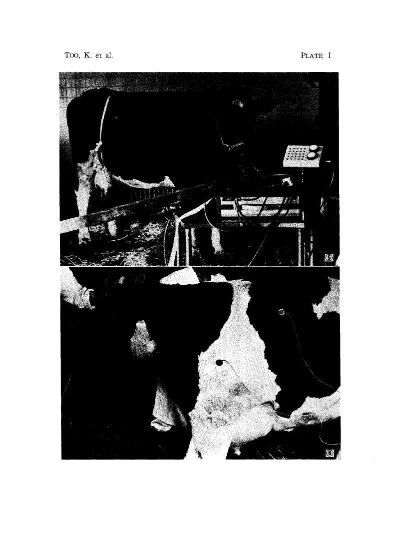
PLATE I
Fig. 5 General appearance of fetal electrocardiogram recording before delivery of a cow
Fig. 6 An example of skin electrode application during parturition
Too, K. et al. PLATE I
PLATE II
Fetal, maternal and newborn's electrocardiograms at parturition (case No.3)
1-3 and 1-13 indicate lead methods employed.
F: Fetal QRS
M: Maternal QRS
Labor: Labor pains
Expulsion: Expulsion of fetus
Rupture of bag: The first rupture of bag
M-ECG: Maternal electrocardiogram
NB-ECG: Electrocardiogram of newborn calf
Too, K. et al. PLATE II
G-296 TIME IN HOURS 1-3
F-HR M-HR
52 120 90
'1 '/ ~
W 13 106 84 a:::
~! ::J l- t:; a.. ..l :::> 6 :M 90 84 c:t::
I W 0:: 3 90 84 a..
78 90
78 90
10 78 90
L.U a: 11 72 94 :::> I-a.. :::> 0::: LABOR
I
t-(/) 0 Cl.. 20 114
24 114
EXPULSION---
M-ECG 108
N8-ECG

PLATE III Fetal, ITIaternal and newborn's electrocardiograITIs at parturition (case No.6)
1-3 indicates lead method.
F: Fetal QRS
M: Maternal QRS
Rupture of bag: The first rupture of bag Labor: Labor pains
Expulsion: Expulsion of fetus
M-ECG: Maternal electrocardiogram
NB-ECG: Electrocardiogram of newborn calf
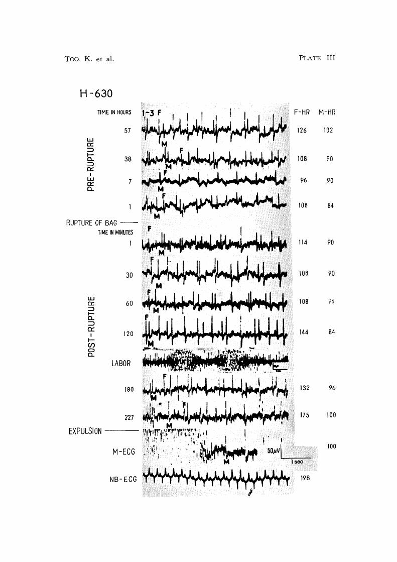
Too, K. et al.
H-630
TIME IN MINUTES
1
30 M
PLATE III
lOB 90
96 90
108 84
114 90
108 90
W 0:: ::J to..
60 ~.rM.~1 ~1~ 108 96
=> 0::
I tCJ) o a.
120 ~{~r~~~44 144
,....~:~-: .~- ~ ,,'. : ';" .
LABOR
M-ECG
198
B4
96
100
100