Embed Size (px)
Citation preview
J. Gen. Appl. Microbiol., 61, 203–210 (2015)doi 10.2323/jgam.61.2032015 Applied Microbiology, Molecular and Cellular Biosciences Research Foundation
Full Paper
*Corresponding author: Hiroyuki Yamaguchi, Department of Medical Laboratory Science, Faculty of Health Sciences, Hokkaido University, Nishi-5 Kita-12, Kita-ku, Sapporo, Hokkaido 060-0812, Japan.Tel: +81-11-706-3326 Fax: +81-11-706-3326 E-mail: [email protected]
None of the authors of this manuscript has any financial or personal relationship with other people or organizations that could inappropriatelyinfluence their work.
Introduction
The microbial community ubiquitously seen in naturalenvironments such as soil or pond water comprises a di-verse mixture of bacteria and protozoa such as ciliates oramoebae (Novarino et al., 1997; Rodríguez-Zaragoza etal., 1994). While such a microbiota plays a fundamentalrole in feeding bacteria as a nutrient source for predatorssuch as ciliates or amoebae, the bacteria presumably ex-ploit the microbiota for nutrient-rich environments to en-sure survival (Russell and Rychlik, 2001; Tyson et al.,
Protozoal ciliate promotes bacterial autoinducer-2 accumulationin mixed culture with Escherichia coli
(Received June 29, 2015; Accepted July 27, 2015)
Satoshi Oguri,1,3 Tomoko Hanawa,2 Junji Matsuo,3 Kasumi Ishida,3 Tomohiro Yamazaki,3,5
Shinji Nakamura,4 Torahiko Okubo,3 Tatsuya Fukumoto,1 Kouzi Akizawa,1
Chikara Shimizu,1 Shigeru Kamiya,2 and Hiroyuki Yamaguchi3,∗
1 Hokkaido University Hospital, Nishi-5 Kita-14, Kita-ku, Sapporo, Hokkaido 060-8648, Japan2 Department of Infectious Diseases, Kyorin University School of Medicine, Shinkawa, Mitaka, Tokyo 181-8611, Japan
3 Department of Medical Laboratory Science, Faculty of Health Sciences, Hokkaido University,Nishi-5 Kita-12, Kita-ku, Sapporo, Hokkaido 060-0812, Japan
4 Division of Biomedical Imaging Research, Juntendo University Graduate School of Medicine,2-1-1 Hongo, Bunkyo-ku, Tokyo 113-8421, Japan
5 Research Fellow of Japan Society for the Promotion of Science, Kojimachi Business Center Building, Chiyoda-ku, Tokyo, Japan
We have previously demonstrated conjugation ofEscherichia coli into vacuoles of the protozoal cili-ate (Tetrahymena thermophila). This indicated apossible role of ciliates in evoking bacterial quo-rum sensing, directly connecting bacterial survivalvia accumulation in the ciliate vacuoles. We there-fore assessed if cil iates promoted bacterialautoinducer (AI)-2 accumulation with vacuole for-mation, which controls quorum sensing. E. coli AI-2 accumulation was significantly enhanced in thesupernatants of a mixed culture of ciliates and bac-teria, likely depending on ciliate density rather thanbacterial concentration. As expected, AI-2 produc-tion was significantly correlated with vacuole for-mation. The experiment with E. coli luxS mutantsshowed that ciliates failed to enhance bacterial AI-2 accumulation, denying a nonspecific phenom-enon. Fluorescence microscopy revealed accumu-lation of fragmented bacteria in ciliate vacuoles,and, more importantly, expulsion of the vacuolescontaining disrupted bacteria into the culturesupernatant. There was no increase in the expres-sion of luxS (encoding AI-2) or ydgG (a transporterfor controlling bacterial export of AI-2). We con-clude that ciliates promote bacterial AI-2 accumu-lation in a mixed culture, via accumulation of dis-rupted bacteria in ciliate vacuoles followed by ex-
pulsion of the vacuoles, independently of luxS orydgG gene induction. This is believed to be the firstdemonstration of a relationship between E. coli AI-2 dynamics and ciliates. In the natural environ-ment, ciliate biotopes may provide a survival ad-vantage to bacteria inhabiting such biotopes, viaevoking quorum sensing.
Key Words: autoinducer-2; ciliates; Escherichiacoli; luxS; quorum sensing; Tetrahymenathermophila; vacuole; ydgG
Abbreviations: AI-2, autoinducer-2; CPFX,ciprofloxacin; CTX, cefotaxime; ESBL, extended-spectrum βββββ-lactamase
204 OGURI et al.
2004), and the acquisition of virulence factors or resist-ance against unpredictable harmful conditions throughgene exchange (Aminov, 2011). Highly packed bacteria,such as in biofilms or ciliate vacuoles, are directly respon-sible for phenotypic features, possibly with genetic ex-changes, against harmful environmental conditions, andalso evoke quorum sensing via autoinducer (AI)-2 pro-duction (Aminov, 2011; Hojo et al., 2012; Lang and Faure,2014).
We have previously found that a mixed culture of Es-cherichia coli with Tetrahymena ciliates could prove ben-eficial in gene exchange between the bacteria (Matsuo etal., 2010; Oguri et al., 2011). Since the gene exchangessignificantly decline in the presence of phagocytosis in-hibitor (latrunculin B), they appear to occur by conjuga-tion via bacterial uptake following accumulation in cili-ate vesicles (Matsuo et al., 2010). Thus, our data clearlyindicate that ciliate vacuoles provide benefits, via geneexchanges, to the engulfed bacteria. These findings raise
another, fundamental question: whether ciliates promotebacterial quorum sensing via AI-2 accumulation into cili-ate vacuoles or to their biotopes. However, it remains un-clear.
In the present study, we aimed to verify if ciliates evokebacterial AI-2 accumulation, using a mixed culture of Tet-rahymena thermophila and E. coli [cefotaxime (CTX)-re-sistant (CTX-M) extended-spectrum β-lactamase (ESBL)producing and/or ciprofloxacin (CPFX)-resistant clinicalisolates], or luxS mutants.
Materials and Methods
Bacteria and protozoa. CTX-resistant ESBL and/orCPFX-resistant (R) E. coli and T. thermophila IB wereused for the mixed culture. Both bacteria were originallyisolated from Hokkaido University Hospital, Japan (Oguriet al., 2011). The bacteria were statically cultured in LBbroth containing 1% NaCl, 1% peptone and 0.5% yeast
Bacterial strains and plasmids Characteristics Reference
E. coliCPFX-R Clinical isolate, CPFXr Oguri et al. (2011)CTX-M ESBL Clinical isolate, CTXr Oguri et al. (2011)CPFX-R ∆luxS CPFXr ∆luxS This studyCTX-M ESBL ∆luxS CTX-M ESBL ∆luxS This studySM10λpir thi thr leu tonA lacY supE recA::RP4-2-Tc::Mu λ pir R6K Kmr Miller and Mekalanos (1988)
PlasmidspYAK1 R6Kori, Cmr, sacB+ Kodama et al. (2002)pYAK1c pYAK1 cloned ∆luxS allele of CPFX-R E. coli strain This studypYAK1e pYAK1 cloned ∆luxS allele of CTX-M ESBL E. coli strain This study
Primer Sequence (5′ > 3′) Reference
For establishment of luxS mutantsEcluxS-F1 *ggccggatcc CGG CGA AGC GTA TCA GA This studyEcluxS-R1 tctctttcggcagtgcca CTG CAG GCG CTTC CAT CC This studyEcluxS-F2 ggatggaagcgcctgcag TGG CAC TGC CGA AAG AGA This studyEcluxS-R2 ggccgcatgc TTG CCA CCG GAA CGA CTG This studyEc luxS-R1-3 ttcttcgttgctgttgatgc CTG CAG GCG CTT CCA TCC This studyEcluxS-F3 ggatggaagcgcctgcag GCA TCA ACA GCA ACG AAG AA This studyEcluxS-R3 ggccgcatg ACC GGG TATG GGC AAA GA This studyEcluxS-CHF TTG CGG TGT GGC TGG AAA AAC This studyEcluxS-CHR GAC GCA ACA GCA GGC ATCA GG This study
For detection of luxS gene expressionluxS-F GAT CAT ACC CGG ATG GAA G Medellin-Pena et al. (2007)luxS-R AGA ATG CTA CGC GCA ATA TC
For detection of ydgG gene expressionydgG-F CCC TAT CGC TCA GGT ACT GG This studyydgG-R TGT TCA AGC GCA ATT TTG AC This study
House keeping genes (gst and groEL)gst-F CTT TGC CGT TAA CCC TAA GGG Mith et al. (2015)gst-R GCT GCA ATG TGC TCT AAC CCgroEL-F GGN GAC GGN ACN ACN ACN GCA ACN GT Teng et al. (2001)groEL-R TCN CCR AAN CCN GGY GCN TTN ACN GC
Table 1. Bacterial strains and plasmids used for this study.
Table 2. PCR primers used for this study.
*Bold sequence show linkers for cloning.§Cutting sites of restriction enzymes.
E. coli AI-2 dynamics and ciliates 205
extract at 37°C. The ciliates were also statically maintainedin peptone-yeast extract-glucose broth containing 0.75%peptone, 0.75% yeast extract, and 1.5% glucose at 30°C,as described previously (Matsuo et al., 2010). The lumi-nescent bacterium Vibrio harveyi BB170 was used for as-sessing AI-2 level in the culture. The luminescent bacte-ria were maintained in an Autoinducer Bioassay brothconsisting of 3.7% brain-heart infusion, 1.25% NaCl,1.23% MgSO4 and 0.1 M L-arginine (Turovskiy andChikindas, 2006). E. coli was also used for establishingluxS mutants (see below). Table 1 shows the bacteria usedfor this study.Construction of E. coli luxS mutants. DNA fragments (~1kbp) containing ∆luxS allele were obtained from CPFX-Rand CTX-M ESBL E. coli by polymerase chain reaction(PCR)-overlap extension, with two distinct primer sets foramplifying specific regions clipping the target sequence,luxS (Horton et al., 1989). The primer sets used for ampli-fying the ∆luxS allelic fragments were as follows: CPFX-R E. coli, EcluxS-F1/EcluxS-R1 and EcluxS-F2/EcluxS-R2; CTX-M ESBL E. coli, EcluxS-F1/EcluxS-R2 andEcluxS-F1/EcluxS-R3 (Supplementary Fig. S1, Table 2).The primer sets for the E. coli CPFX-R strain were de-signed using a sequence for reference, the luxS locus of E.coli K-12 (accession number: U00096). Second, each ofthe in-frame luxS deletion fragments was generated fromthe ∆luxS allelic fragments by PCR with the followingprimer sets: CPFX-R E. coli, EcluxS-F1/EcluxS-R2; CTX-M ESBL E. coli, and EcluxS-F1/EcluxS-R3 (Fig. S1, Ta-ble 2). After digestion with BamHI and SphI, each of the
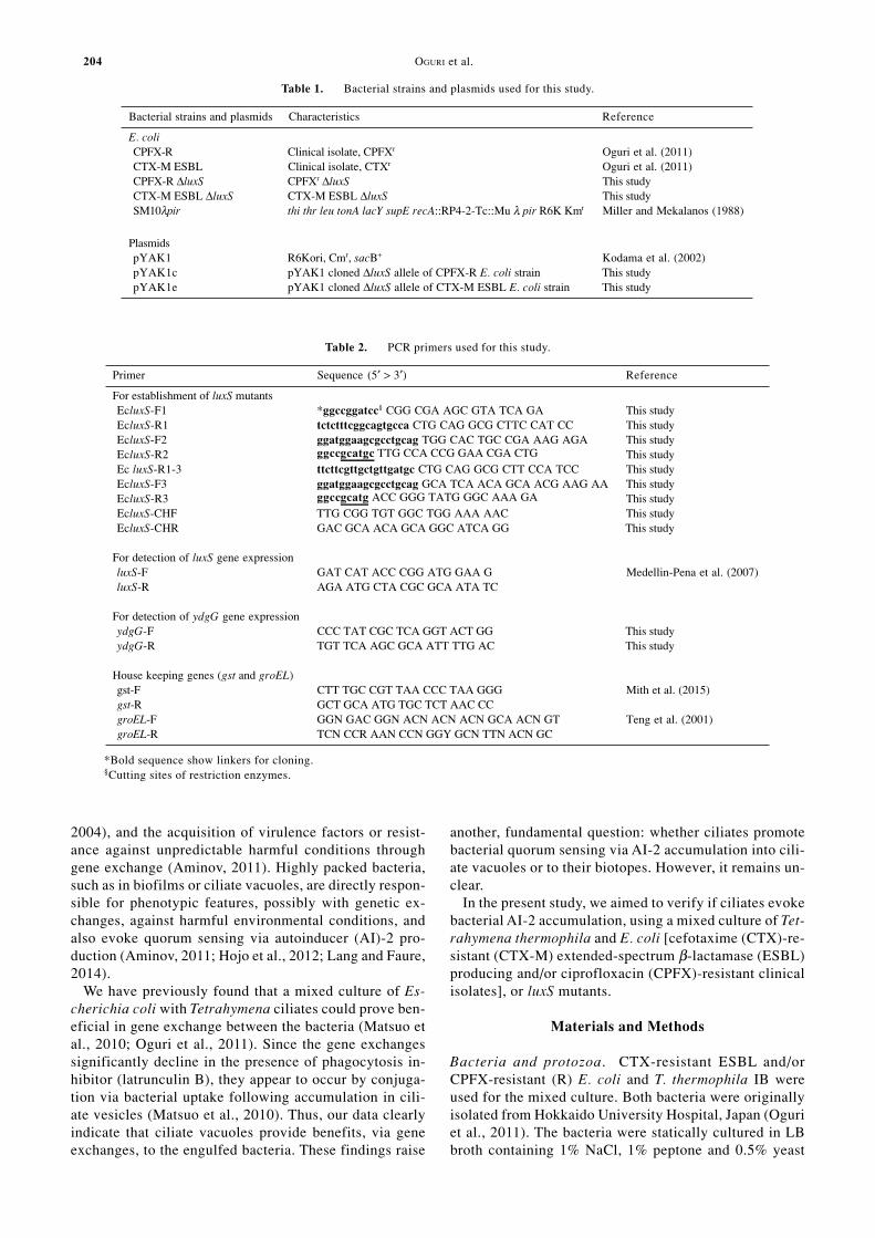
fragments was cloned into pYAK1 plasmid vector (Hortonet al., 1989) treated with BamHI and SphI [pYAK1c(CPFX-R E. coli) and pYAK1e (CTX-M ESBL E. coli)].We confirmed using direct sequencing that two distinctluxS allelic fragments were collectively identical to thetarget regions in the original sequences. After transfor-mation, E. coli SM10 λ pir (Miller et al., 1988) carryingpYAK1c or pYAK1e (Kodama et al., 2002) was used forconjugation with CPFX-R and CTX-M ESBL E. coli, re-spectively. The plasmid integrated trans-conjugants wereselected on LB agar plates containing 40 µg/ml chloram-phenicol, and the luxS mutants generated by second re-combination were selected on LB agar containing 5% su-crose. Finally, the luxS deletions in the obtained cloneswere confirmed by PCR using a primer set, EcluxS-CHFand EcluxS-CHR (Fig. S2). Tables 1 and 2 show theplasmids and primer sets used for this study, respectively.The construction was confirmed using PCR with directsequencing. Supplementary Fig. S3 summarizes the pro-tocol for establishing luxS mutant.Mixed culture of ciliates and bacteria. A mixed culturesystem for ciliates and bacteria was constructed as previ-ously reported (Matsuo et al., 2010). As shown in Fig. 1,CTX-resistant ESBL E. coli and/or CPFX-resistant E. coli
Fig. 1. Protocol with a mixed culture system for assessing vacuolenumbers in ciliates and AI-2 production.
The mixed cultures were constructed as described previously (Matsuoet al., 2010). Assessment of ciliate vacuoles was performed microscopi-cally, as previously described (Schlimme et al., 1995). CPFX-R and/orCTX-M ESBL E. coli ∆luxS mutants were used in some experiments.The ratio of the bacteria in the mixed culture was 1:1.
Fig. 2. Changes in AI-2 production in the mixed culture with or with-out ciliates.
Data show mean ±SD obtained from three experiments. *P < 0.05 ver-sus each of the values in mixed culture without ciliates.
206 OGURI et al.
were incubated with or without T. thermophila IB intoPage’s amoeba saline (Na2HPO4 0.142 g, KH2PO4 0.136g, NaCl 0.12 g, MgSO4·7H2O 0.04 g, CaCl2·6H2O 0.06 g,1L) (Page, 1988) for up to 24 h at 30°C. Adequate amountsof suspension in the mixed culture with or without ciliates(total 500 µl) were collected for assessing vacuole num-bers formed in ciliates (20–50 µl) and AI-2 production(15 µl). Each of the assessments was performed by thefollowing protocols.Assessment of AI-2 in the mixed culture. The amount ofAI-2 in the mixed culture was estimated as follows. Thesolution was passed through a filter with 0.22-µm poresize. The filtered solution (15 µl) was mixed with a di-luted solution of V. harveyi (135 µl). One hundredmicroliters of the solution was placed into the luminometerblack plate and incubated for 5 h at 30°C. After incuba-tion, the amount of luminescence produced in the solu-tion was measured by a luminometer (Luminescencer-JNRII AB-2300; ATTO, Tokyo, Japan). The remainingsolution was used for determining the number of V. harveyiorganisms. The luminescence showing AI-2 level was ex-pressed as relative light units (RLU) per bacterium (V.harveyi).
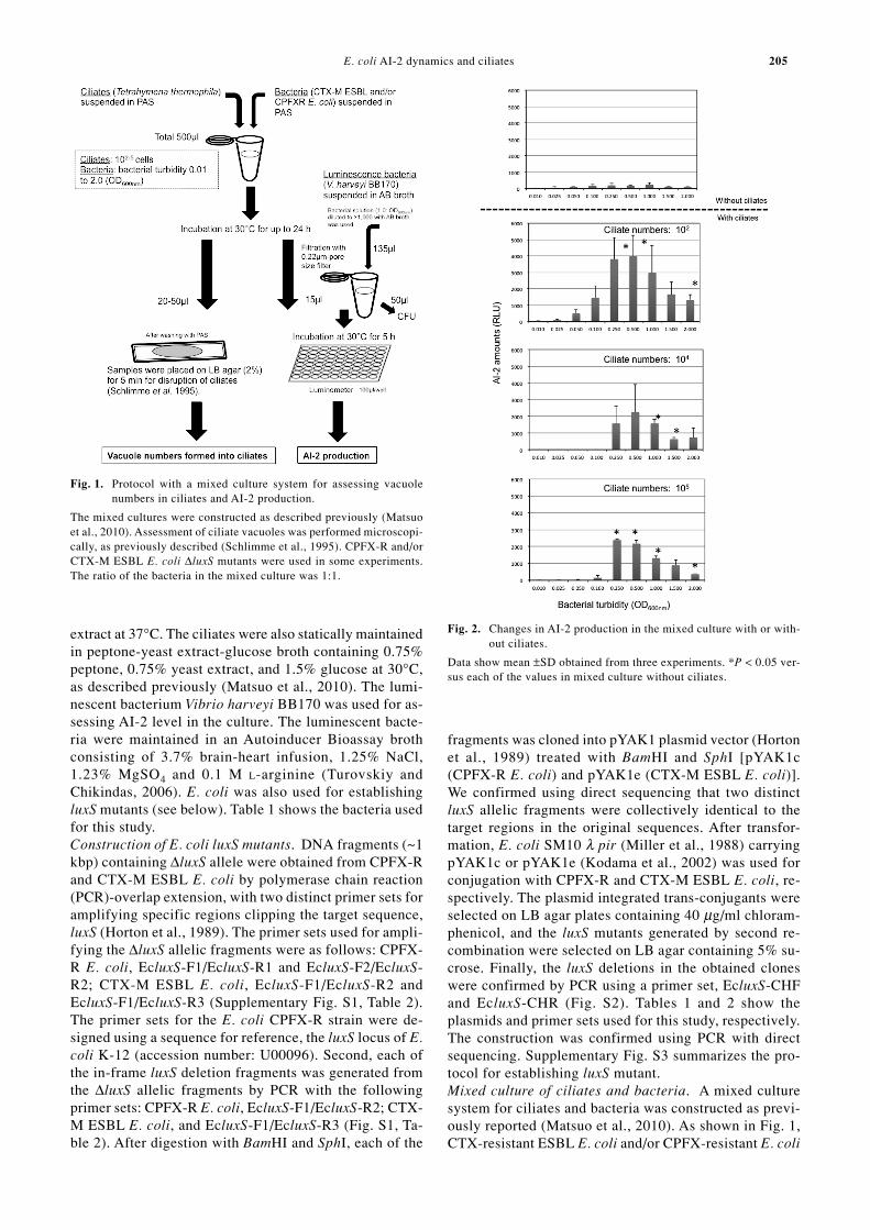
Assessment of vacuole numbers formed in ciliates. Thenumber of vacuoles formed in ciliates was estimated asdescribed previously (Schlimme et al., 1995). The mixedsolution was gently placed on 2% LB agar solidified ontoa glass slide, and incubated for 5 min at room tempera-ture. After incubation, the vacuoles remaining on the glassslide were observed by light microscopy. The number ofvacuoles per ciliate was estimated by observing more than100 ciliates under light microscopy.Assessment of bacterial localization in ciliate vacuoles.To confirm E. coli accumulation in ciliate vacuoles, ciliatesand vital-stained CTX-M ESBL E. coli (fluorescence color:
Fig. 3. Vacuole images and changes in vacuole numbers formed inciliates.
A. Representative images showing vacuoles formed in ciliates (24 hpost incubation). B. Changes in vacuole numbers formed in ciliates.Data show mean ±SD obtained from three experiments. *P < 0.05 ver-sus the values at each number of ciliates in the mixed cultures withoutbacteria (left column).
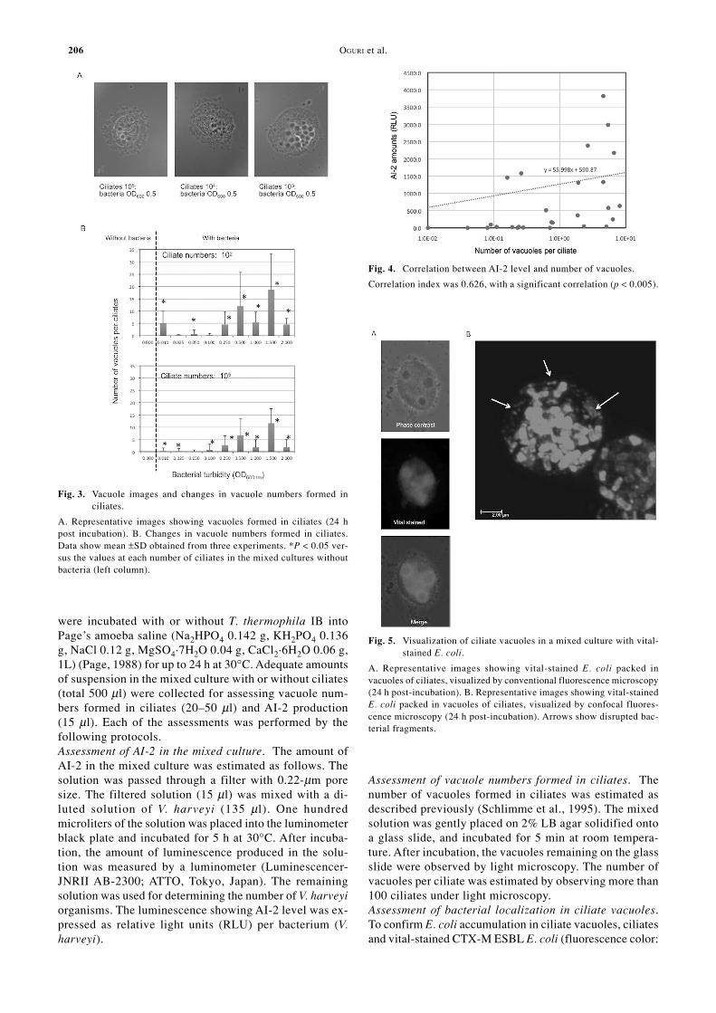
Fig. 4. Correlation between AI-2 level and number of vacuoles.
Correlation index was 0.626, with a significant correlation (p < 0.005).
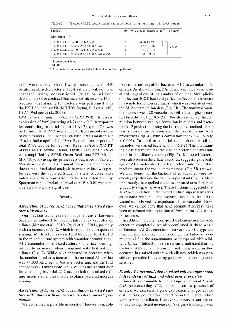
Fig. 5. Visualization of ciliate vacuoles in a mixed culture with vital-stained E. coli.
A. Representative images showing vital-stained E. coli packed invacuoles of ciliates, visualized by conventional fluorescence microscopy(24 h post-incubation). B. Representative images showing vital-stainedE. coli packed in vacuoles of ciliates, visualized by confocal fluores-cence microscopy (24 h post-incubation). Arrows show disrupted bac-terial fragments.
E. coli AI-2 dynamics and ciliates 207
red) were used. After fixing bacteria with 4%paraformaldehyde, bacterial localization in ciliates wasassessed using conventional (with or withoutdeconvolution) or confocal fluorescence microscopy. Fluo-rescence vital staining for bacteria was performed withthe PKH-26 labeling kit (MINI26; Sigma, St Louis, MO,USA) (Wallace et al., 2008).RNA extraction and quantitative (q)RT-PCR. To assessexpression of luxS (encoding AI-2) and ydgG (transporterfor controlling bacterial export of AI-2), qRT-PCR wasperformed. Total RNA was extracted from mixed cultureof ciliates and E. coli using High Pure RNA Isolation Kit(Roche, Indianapolis, IN, USA). Reverse transcription oftotal RNA was performed with ReverTraAce qPCR RTMaster Mix (Toyobo, Osaka, Japan). Resultant cDNAswere amplified by SYBR Green Real-time PCR MasterMix (Toyobo) using the primer sets described in Table 2.Statistical analysis. Experiments were repeated at leastthree times. Statistical analysis between values was per-formed with the unpaired Student’s t test. A correlationindex (r) with a regression curve was calculated bySpearman rank correlation. A value of P < 0.05 was con-sidered statistically significant.
Results
Association of E. coli AI-2 accumulation in mixed cul-ture with ciliates
Our previous study revealed that gene transfer betweenbacteria is induced by accumulation into vacuoles ofciliates (Matsuo et al., 2010; Oguri et al., 2011), possiblywith an increase of AI-2, which is responsible for quorumsensing. We therefore assessed if AI-2 could be detectedin the mixed culture system with vacuolar accumulation.AI-2 accumulation in mixed culture with ciliates was sig-nificantly increased when compared with that withoutciliates (Fig. 2). While AI-2 appeared to decrease whenthe number of ciliates increased, the maximal AI-2 valuewas ~4,000 RLU per V. harveyi bacterium, and the foldchange was 20 times more. Thus, ciliates are a stimulatorfor enhancing bacterial AI-2 accumulation in mixed cul-ture supernatants, presumably evoking bacterial quorumsensing.
Association of E. coli AI-2 accumulation in mixed cul-ture with ciliates with an increase in ciliate vacuole for-mation
We confirmed a possible association between vacuole
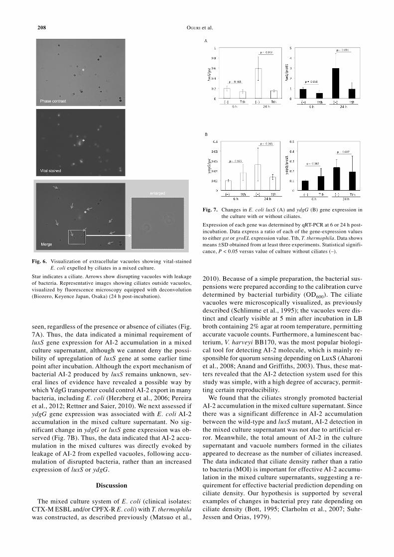
formation and engulfed bacterial AI-2 accumulation inciliates. As shown in Fig. 3A, ciliate vacuoles were visu-alized, regardless of the number of ciliates. Multiplicityof infection (MOI) had no significant effect on the increasein vacuole formation in ciliates, which was consistent withthe AI-2 accumulation data (Fig. 3B). The maximal vacu-ole number was ~20 vacuoles per ciliate at higher bacte-rial turbidity (OD600 0.5–2.0). We also estimated the cor-relation between vacuole formation in ciliates and bacte-rial AI-2 production, using the least squares method. Therewas a correlation between vacuole formation and AI-2production (Fig. 4), with a correlation index r = 0.626 (p< 0.005). To confirm bacterial accumulation in ciliatevacuoles, we stained bacteria with PKH-26. The vital stain-ing clearly revealed that the labeled bacteria had accumu-lated in the ciliate vacuoles (Fig. 5). Disrupted bacteriawere also seen in the ciliate vacuoles, suggesting the leak-age of AI-2 molecules from the bacteria into the culturemedium, across the vacuolar membranes (Fig. 5B, arrows).We also found that the bacteria-filled vacuoles were fre-quently expelled into the culture supernatant (Fig. 6). Moreimportantly, the expelled vacuoles appeared to be disruptedgradually (Fig. 6, arrows). These findings suggested thatAI-2 accumulation in the mixed culture supernatants wasassociated with bacterial accumulation in the ciliatevacuoles, followed by expulsion of the vacuoles. How-ever, we cannot deny that AI-2 accumulation may havebeen associated with induction of luxS and/or AI-2 trans-porter gene.
In addition, to deny a nonspecific phenomenon for AI-2detection completely, we also confirmed if there was adifference in AI-2 accumulation between the wild-type andluxS mutant. The luxS mutants completely failed to accu-mulate AI-2 in the supernatants, as compared with wild-type E. coli (Table 3). The data clearly indicated that thebacterial AI-2 accumulation, but not nonspecific matter,occurred in a mixed culture with ciliates, which was pos-sibly responsible for evoking peripheral bacterial quorumsensing.
E. coli AI-2 accumulation in mixed culture supernatantindependently of luxS and ydgG gene expression
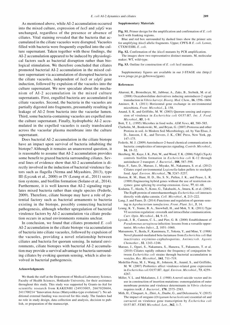
Since it is reasonable to predict upregulation of E. coliluxS gene encoding AI-2, depending on the presence ofciliates, we assessed if gene expression changed at twodistinct time points after incubation in the mixed culturewith or without ciliates. However, contrary to our expec-tation, no significant increase of luxS gene transcripts was
Table 3. Changes of AI-2 production into mixed culture system of ciliates with luxS mutant.
208 OGURI et al.
seen, regardless of the presence or absence of ciliates (Fig.7A). Thus, the data indicated a minimal requirement ofluxS gene expression for AI-2 accumulation in a mixedculture supernatant, although we cannot deny the possi-bility of upregulation of luxS gene at some earlier timepoint after incubation. Although the export mechanism ofbacterial AI-2 produced by luxS remains unknown, sev-eral lines of evidence have revealed a possible way bywhich YdgG transporter could control AI-2 export in manybacteria, including E. coli (Herzberg et al., 2006; Pereiraet al., 2012; Rettner and Saier, 2010). We next assessed ifydgG gene expression was associated with E. coli AI-2accumulation in the mixed culture supernatant. No sig-nificant change in ydgG or luxS gene expression was ob-served (Fig. 7B). Thus, the data indicated that AI-2 accu-mulation in the mixed cultures was directly evoked byleakage of AI-2 from expelled vacuoles, following accu-mulation of disrupted bacteria, rather than an increasedexpression of luxS or ydgG.
Discussion
The mixed culture system of E. coli (clinical isolates:CTX-M ESBL and/or CPFX-R E. coli) with T. thermophilawas constructed, as described previously (Matsuo et al.,
2010). Because of a simple preparation, the bacterial sus-pensions were prepared according to the calibration curvedetermined by bacterial turbidity (OD600). The ciliatevacuoles were microscopically visualized, as previouslydescribed (Schlimme et al., 1995); the vacuoles were dis-tinct and clearly visible at 5 min after incubation in LBbroth containing 2% agar at room temperature, permittingaccurate vacuole counts. Furthermore, a luminescent bac-terium, V. harveyi BB170, was the most popular biologi-cal tool for detecting AI-2 molecule, which is mainly re-sponsible for quorum sensing depending on LuxS (Aharoniet al., 2008; Anand and Griffiths, 2003). Thus, these mat-ters revealed that the AI-2 detection system used for thisstudy was simple, with a high degree of accuracy, permit-ting certain reproducibility.
We found that the ciliates strongly promoted bacterialAI-2 accumulation in the mixed culture supernatant. Sincethere was a significant difference in AI-2 accumulationbetween the wild-type and luxS mutant, AI-2 detection inthe mixed culture supernatant was not due to artificial er-ror. Meanwhile, the total amount of AI-2 in the culturesupernatant and vacuole numbers formed in the ciliatesappeared to decrease as the number of ciliates increased.The data indicated that ciliate density rather than a ratioto bacteria (MOI) is important for effective AI-2 accumu-lation in the mixed culture supernatants, suggesting a re-quirement for effective bacterial prediction depending onciliate density. Our hypothesis is supported by severalexamples of changes in bacterial prey rate depending onciliate density (Bott, 1995; Clarholm et al., 2007; Suhr-Jessen and Orias, 1979).
Fig. 7. Changes in E. coli luxS (A) and ydgG (B) gene expression inthe culture with or without ciliates.
Expression of each gene was determined by qRT-PCR at 6 or 24 h post-incubation. Data express a ratio of each of the gene-expression valuesto either gst or groEL expression value. Tth, T. thermophila. Data showsmeans ±SD obtained from at least three experiments. Statistical signifi-cance, P < 0.05 versus value of culture without ciliates (–).
Fig. 6. Visualization of extracellular vacuoles showing vital-stainedE. coli expelled by ciliates in a mixed culture.
Star indicates a ciliate. Arrows show disrupting vacuoles with leakageof bacteria. Representative images showing ciliates outside vacuoles,visualized by fluorescence microscopy equipped with deconvolution(Biozero, Keyence Japan, Osaka) (24 h post-incubation).
E. coli AI-2 dynamics and ciliates 209
As mentioned above, while AI-2 accumulation occurredinto the mixed culture, expression of luxS and ydgG wasunchanged, regardless of the presence or absence ofciliates. Vital staining revealed that the bacteria that ac-cumulated in the ciliate vacuoles were disrupted. Vacuolesfilled with bacteria were frequently expelled into the cul-ture supernatant. Taken together with these findings, theAI-2 accumulation appeared to be induced by physiologi-cal factors such as bacterial disruption rather than bio-logical stimulation. We therefore concluded that ciliatespromoted bacterial AI-2 accumulation in the mixed cul-ture supernatant via accumulation of disrupted bacteria inthe ciliate vacuoles, independent of luxS or ydgG geneinduction, followed by expulsion of the vacuoles into theculture supernatant. We now speculate about the mecha-nism of AI-2 accumulation in the mixed culturesupernatants. First, engulfed bacteria are accumulated inciliate vacuoles. Second, the bacteria in the vacuoles arepartially digested into fragments, presumably resulting inleakage of AI-2 from bacteria into the vacuolar lumen.Third, some bacteria-containing vacuoles are expelled intothe culture supernatant. Finally, hydrophobic AI-2 accu-mulated in the expelled vacuoles is easily translocatedacross the vacuolar plasma membrane into the culturesupernatant.
Does bacterial AI-2 accumulation in the ciliate biotopehave an impact upon survival of bacteria inhabiting thebiotope? Although it remains an unanswered question, itis reasonable to assume that AI-2 accumulation providessome benefit to grazed bacteria surrounding ciliates. Sev-eral lines of evidence show that AI-2 accumulation is di-rectly involved in the induction of bacterial virulence fac-tors such as flagella (Verma and Miyashiro, 2013), typeIII (Lyczak et al., 2000) or IV (Leung et al., 2011) secre-tion systems, and biofilm formation (Solano et al., 2014).Furthermore, it is well known that AI-2 signaling regu-lates mixed bacteria rather than single species (Federle,2009). Therefore, ciliate biotope may have a role of po-tential factory such as bacterial armaments to bacteriaexisting in the biotope, possibly connecting bacterialpathogenesis, although whether upregulation of bacterialvirulence factors by AI-2 accumulation via ciliate preda-tion occurs in actual environments remains unclear.
In conclusion, we found that ciliates promoted E. coliAI-2 accumulation in the ciliate biotope via accumulationof bacteria into ciliate vacuoles, followed by expulsion ofthe vacuoles, providing a novel relationship betweenciliates and bacteria for quorum sensing. In natural envi-ronments, ciliate biotopes with bacterial AI-2 accumula-tion may provide a survival advantage to bacteria surround-ing ciliates by evoking quorum sensing, which is also in-volved in bacterial pathogenesis.
Acknowledgments
We thank the staff at the Department of Medical Laboratory Science,Faculty of Health Sciences, Hokkaido University, for their assistancethroughout this study. This study was supported by Grants-in-Aid forscientif ic research from KAKENHI (24933005, 2667020604,2411700214 “Innovation Areas (Matryoshka-type evolution)”). No ad-ditional external funding was received for this study. The funders hadno role in study design, data collection and analysis, decision to pub-lish, or preparation of the manuscript.
Supplementary Materials
Fig. S1. Primer design for the amplification and confirmation of E. coliluxS with franking regions.
Blue and red box surrounded by dashed lines show the primer setsfor amplifying ∆luxS allelic fragments. Upper, CPFX-R E. coli. Lower,CTXM ESBL E. coli.
Fig. S2. Confirmation of the ∆luxS mutants by PCR amplification.The images show two representative distinct mutants. M, molecular
maker; WT, wild-type.
Fig. S3. Outline for construction of E. coli luxS mutants.
Supplementary figures are available in our J-STAGE site (http://www.jstage.jst.go.jp/browse/jgam).
References
Aharoni, R., Bronstheyn, M., Jabbour, A., Zaks, B., Srebnik, M. et al.(2008) Oxazaborolidine derivatives inducing autoinducer-2 signaltransduction in Vibrio harveyi. Bioorg. Med. Chem., 16, 1596–1604.
Aminov, R. I. (2011) Horizontal gene exchange in environmentalmicrobiota. Front. Microbiol., 2, 158.
Anand, S. K. and Griffiths, M. W. (2003) Quorum sensing and expres-sion of virulence in Escherichia coli O157:H7. Int. J. Food.Microbiol., 85, 1–9.
Bott, T. L. (1995) Microbes in food webs. ASM News, 61, 580–585.Clarholm, M., Bonkowski, M., and Bryan, G. (2007) Protozoa and other
Protista in soil. In Modern Soil Microbiology, ed. by Van Elsas, J.D., Jansson, J. K., and Trevors, J. K., CRC Press, New York, pp.147–175.
Federle, M. J. (2009) Autoinducer-2-based chemical communication inbacteria: complexities of interspecies signaling. Contrib. Microbiol.,16, 18–32.
Herzberg, M., Kaye, I. K., Peti, W., and Wood, T. K. (2006) YdgG (TqsA)controls biofilm formation in Escherichia coli K-12 throughautoinducer 2 transport. J. Bacteriol., 188, 587–598.
Hojo, F., Sato, D., Matsuo, J., Miyake, M., Nakamura, S. et al. (2012)Ciliates expel environmental Legionella-laden pellets to stockpilefood. Appl. Environ. Microbiol., 78, 5247–5257.
Horton, R. M., Hunt, H. D., Ho, S. N., Pullen, J. K., and Pease, L. R.(1989) Engineering hybrid genes without the use of restriction en-zymes: gene splicing by overlap extension. Gene, 77, 61–68.
Kodama, T., Akeda, Y., Kono, G., Takahashi, A., Imura, K. et al. (2002)The EspB protein of enterohaemorrhagic Escherichia coli interactsdirectly with alpha-catenin. Cell. Microbiol., 4, 213–222.
Lang, J. and Faure, D. (2014) Functions and regulation of quorum-sens-ing in Agrobacterium tumefaciens. Front. Plant. Sci., 5, 14.
Leung, K. Y., Siame, B. A., Snowball, H., and Mok, Y. K. (2011) TypeVI secretion regulation: crosstalk and intracellular communication.Curr. Opin. Microbiol., 14, 9–15.
Lyczak, J. B., Cannon, C. L., and Pier, G. B. (2000) Establishment ofPseudomonas aeruginosa infection: lessons from a versatile oppor-tunist. Microbes Infect., 2, 1051–1060.
Matsumoto, Y., Ikeda, F., Kamimura, T., Yokota, Y., and Mine, Y. (1988)Novel plasmid-mediated beta-lactamase from Escherichia coli thatinactivates oxyimino-cephalosporins. Antimicrob. AgentsChemother., 32, 1243–1246.
Matsuo, J., Oguri, S., Nakamura, S., Hanawa, T., Fukumoto, T. et al.(2010) Ciliates rapidly enhance the frequency of conjugation be-tween Escherichia coli strains through bacterial accumulation invesicles. Res. Microbiol., 161, 711–719.
Medellin-Pena, M. J., Wang, H., Johnson, R., Anand, S., and Griffiths,M. W. (2007) Probiotics affect virulence-related gene expressionin Escherichia coli O157:H7. Appl. Environ. Microbiol., 73, 4259–4267.
Miller, V. L. and Mekalanos, J. J. (1988) A novel suicide vector and itsuse in construction of insertion mutations: osmoregulation of outermembrane proteins and virulence determinants in Vibrio choleraerequires toxR. J. Bacteriol., 170, 2575–2583.
Mith, H., Clinquart, A., Zhiri, A., Daube, G., and Delcenserie, V. (2015)The impact of oregano (Origanum heracleoticum) essential oil andcarvacrol on virulence gene transcription by Escherichia coliO157:H7. FEMS Microbiol. Lett., 362, 1–7.
210 OGURI et al.
Novarino, G., Warren, A., Butler, H., Lambourne, G., Boxshall, A. etal. (1997) Protistan communities in aquifers: a review. FEMSMicrobiol. Rev., 20, 261–275.
Oguri, S., Matsuo, J., Hayashi, Y., Nakamura, S., Hanawa, T. et al. (2011)Ciliates promote the transfer of the gene encoding the extended-spectrum β-lactamase CTX-M-27 between Escherichia coli strains.J. Antimicrob. Chemother., 66, 527–530.
Page, F. C. (1988) A New Key to Freshwater and Soil Gymnamoebae(ISBN-10:18711105021), Freshwater Biological Association,Ambleside, U.K., 122 pp.
Pereira, C. S., Santos, A. J., Bejerano-Sagie, M., Correia, P. B., Marques,J. C. et al. (2012) Phosphoenolpyruvate phosphotransferase systemregulates detection and processing of the quorum sensing signalautoinducer-2. Mol. Microbiol., 85, 93–104.
Rettner, R. E. and Saier, M. H., Jr. (2010) The autoinducer-2 exportersuperfamily. J. Mol. Microbiol. Biotechnol., 18, 195–205.
Rodríguez-Zaragoza, S. (1994) Ecology of free-living amoebae. Crit.Rev. Microbiol., 20, 225–241.
Russell, J. B. and Rychlik, J. L. (2001) Factors that alter rumen micro-bial ecology. Science, 292, 1119–1122.
Schlimme, W., Baur, B., Hanselmann, K., and Jenni, B. (1995) Anagarose slide method to follow the fate of bacteria within digestivevacuoles of protozoa. FEMS Microbiol., Lett., 133, 169–173.
Solano, C., Echeverz, M., and Lasa, I. (2014) Biofilm dispersion andquorum sensing. Curr. Opin. Microbiol., 18, 96–104.
Suhr-Jessen, P. B. and Orias, E. (1979) Mutants of Tetrahymenathermophila with temperature sensitive food vacuole formation. II.Physiological and morphological studies. Exp. Cell Res., 124, 317–327.
Teng, L.-J., Hsueh, P.-R., Wang, Y.-H., Lin, H.-M., Luh, K.-T. et al.(2001) Determination of Enterococcus faecalis groESL full-lengthsequence and application for species identification. J. Clin.Microbiol., 39, 3326–3331.
Turovskiy, Y. and Chikindas, M. L. (2006) Autoinducer-2 bioassay is aqualitative, not quantitative method influenced by glucose. J.Microbiol. Methods, 66, 497–503.
Tyson, G. W., Chapman, J., Hugenholtz, P., Allen, E. E., Ram, R. J. etal. (2004) Community structure and metabolism through reconstruc-tion of microbial genomes from the environment. Nature, 428, 37–43.
Verma, S. C. and Miyashiro, T. (2013) Quorum sensing in the squid-Vibrio symbiosis. Int. J. Mol. Sci., 14, 16386–16401.
Wallace, P. K., Tario, J. D., Jr., Fisher, J. L., Wallace, S. S., Ernstoff, M.S. et al. (2008) Tracking antigen-driven responses by flowcytometry: monitoring proliferation by dye dilution. Cytometry A,73, 1019–1034.