Embed Size (px)
Citation preview
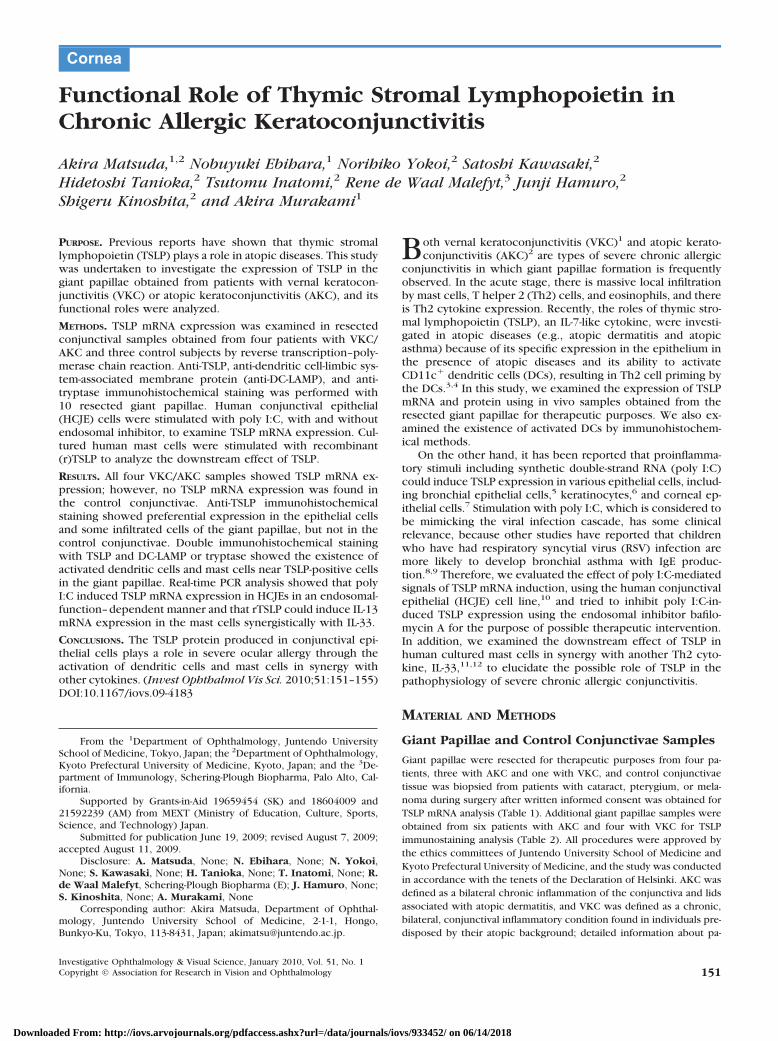
Functional Role of Thymic Stromal Lymphopoietin inChronic Allergic Keratoconjunctivitis
Akira Matsuda,1,2 Nobuyuki Ebihara,1 Norihiko Yokoi,2 Satoshi Kawasaki,2
Hidetoshi Tanioka,2 Tsutomu Inatomi,2 Rene de Waal Malefyt,3 Junji Hamuro,2
Shigeru Kinoshita,2 and Akira Murakami1
PURPOSE. Previous reports have shown that thymic stromallymphopoietin (TSLP) plays a role in atopic diseases. This studywas undertaken to investigate the expression of TSLP in thegiant papillae obtained from patients with vernal keratocon-junctivitis (VKC) or atopic keratoconjunctivitis (AKC), and itsfunctional roles were analyzed.
METHODS. TSLP mRNA expression was examined in resectedconjunctival samples obtained from four patients with VKC/AKC and three control subjects by reverse transcription–poly-merase chain reaction. Anti-TSLP, anti-dendritic cell-limbic sys-tem-associated membrane protein (anti-DC-LAMP), and anti-tryptase immunohistochemical staining was performed with10 resected giant papillae. Human conjunctival epithelial(HCJE) cells were stimulated with poly I:C, with and withoutendosomal inhibitor, to examine TSLP mRNA expression. Cul-tured human mast cells were stimulated with recombinant(r)TSLP to analyze the downstream effect of TSLP.
RESULTS. All four VKC/AKC samples showed TSLP mRNA ex-pression; however, no TSLP mRNA expression was found inthe control conjunctivae. Anti-TSLP immunohistochemicalstaining showed preferential expression in the epithelial cellsand some infiltrated cells of the giant papillae, but not in thecontrol conjunctivae. Double immunohistochemical stainingwith TSLP and DC-LAMP or tryptase showed the existence ofactivated dendritic cells and mast cells near TSLP-positive cellsin the giant papillae. Real-time PCR analysis showed that polyI:C induced TSLP mRNA expression in HCJEs in an endosomal-function–dependent manner and that rTSLP could induce IL-13mRNA expression in the mast cells synergistically with IL-33.
CONCLUSIONS. The TSLP protein produced in conjunctival epi-thelial cells plays a role in severe ocular allergy through theactivation of dendritic cells and mast cells in synergy withother cytokines. (Invest Ophthalmol Vis Sci. 2010;51:151–155)DOI:10.1167/iovs.09-4183
Both vernal keratoconjunctivitis (VKC)1 and atopic kerato-conjunctivitis (AKC)2 are types of severe chronic allergic
conjunctivitis in which giant papillae formation is frequentlyobserved. In the acute stage, there is massive local infiltrationby mast cells, T helper 2 (Th2) cells, and eosinophils, and thereis Th2 cytokine expression. Recently, the roles of thymic stro-mal lymphopoietin (TSLP), an IL-7-like cytokine, were investi-gated in atopic diseases (e.g., atopic dermatitis and atopicasthma) because of its specific expression in the epithelium inthe presence of atopic diseases and its ability to activateCD11c� dendritic cells (DCs), resulting in Th2 cell priming bythe DCs.3,4 In this study, we examined the expression of TSLPmRNA and protein using in vivo samples obtained from theresected giant papillae for therapeutic purposes. We also ex-amined the existence of activated DCs by immunohistochem-ical methods.
On the other hand, it has been reported that proinflamma-tory stimuli including synthetic double-strand RNA (poly I:C)could induce TSLP expression in various epithelial cells, includ-ing bronchial epithelial cells,5 keratinocytes,6 and corneal ep-ithelial cells.7 Stimulation with poly I:C, which is considered tobe mimicking the viral infection cascade, has some clinicalrelevance, because other studies have reported that childrenwho have had respiratory syncytial virus (RSV) infection aremore likely to develop bronchial asthma with IgE produc-tion.8,9 Therefore, we evaluated the effect of poly I:C-mediatedsignals of TSLP mRNA induction, using the human conjunctivalepithelial (HCJE) cell line,10 and tried to inhibit poly I:C-in-duced TSLP expression using the endosomal inhibitor bafilo-mycin A for the purpose of possible therapeutic intervention.In addition, we examined the downstream effect of TSLP inhuman cultured mast cells in synergy with another Th2 cyto-kine, IL-33,11,12 to elucidate the possible role of TSLP in thepathophysiology of severe chronic allergic conjunctivitis.
MATERIAL AND METHODS
Giant Papillae and Control Conjunctivae Samples
Giant papillae were resected for therapeutic purposes from four pa-tients, three with AKC and one with VKC, and control conjunctivaetissue was biopsied from patients with cataract, pterygium, or mela-noma during surgery after written informed consent was obtained forTSLP mRNA analysis (Table 1). Additional giant papillae samples wereobtained from six patients with AKC and four with VKC for TSLPimmunostaining analysis (Table 2). All procedures were approved bythe ethics committees of Juntendo University School of Medicine andKyoto Prefectural University of Medicine, and the study was conductedin accordance with the tenets of the Declaration of Helsinki. AKC wasdefined as a bilateral chronic inflammation of the conjunctiva and lidsassociated with atopic dermatitis, and VKC was defined as a chronic,bilateral, conjunctival inflammatory condition found in individuals pre-disposed by their atopic background; detailed information about pa-
From the 1Department of Ophthalmology, Juntendo UniversitySchool of Medicine, Tokyo, Japan; the 2Department of Ophthalmology,Kyoto Prefectural University of Medicine, Kyoto, Japan; and the 3De-partment of Immunology, Schering-Plough Biopharma, Palo Alto, Cal-ifornia.
Supported by Grants-in-Aid 19659454 (SK) and 18604009 and21592239 (AM) from MEXT (Ministry of Education, Culture, Sports,Science, and Technology) Japan.
Submitted for publication June 19, 2009; revised August 7, 2009;accepted August 11, 2009.
Disclosure: A. Matsuda, None; N. Ebihara, None; N. Yokoi,None; S. Kawasaki, None; H. Tanioka, None; T. Inatomi, None; R.de Waal Malefyt, Schering-Plough Biopharma (E); J. Hamuro, None;S. Kinoshita, None; A. Murakami, None
Corresponding author: Akira Matsuda, Department of Ophthal-mology, Juntendo University School of Medicine, 2-1-1, Hongo,Bunkyo-Ku, Tokyo, 113-8431, Japan; [email protected].
Cornea
Investigative Ophthalmology & Visual Science, January 2010, Vol. 51, No. 1Copyright © Association for Research in Vision and Ophthalmology 151
Downloaded From: http://iovs.arvojournals.org/pdfaccess.ashx?url=/data/journals/iovs/933452/ on 06/14/2018
tient selection was described elsewhere.13 Upper bulbar conjunctivaeresected from six patients with conjunctivochalasis were used ascontrol samples, as previously described,13 after informed consent wasobtained (Table 3).
Antibodies, Reagents, and Cell Lines
We purchased mouse anti-DC-LAMP (CD208) monoclonal antibodyfrom Beckman Coulter Japan (Tokyo, Japan), mouse anti-humantryptase antibody from Dako Japan (Kyoto, Japan), and Alexa-488-conjugated donkey anti-rat IgG and Alexa-594-conjugated donkey anti-mouse IgG antibodies from Invitrogen Japan (Tokyo, Japan). Rat anti-human TSLP monoclonal antibody was prepared as previouslydescribed.4 HCJE was kindly provided by Ilene K. Gipson (SchepensEye Research Institute, Philadelphia, PA) and maintained with definedkeratinocyte serum-free medium (KSFM; Invitrogen Japan). The humanmast cell line LAD2 was kindly provided by Arnold Kirshenbaum(National Institutes of Health, Bethesda, MD), and maintained as pre-viously described.14 Recombinant human (r)TSLP and recombinanthuman (r)IL-33 were obtained from Peprotech (London, UK), poly I:Cwas obtained from InvivoGen (San Diego, CA), and bafilomycin A1 wasobtained from Sigma-Aldrich (St. Louis, MO).
Reverse-Transcription–PolymeraseChain Reaction
Total RNA was extracted from the giant papillae tissue (NucleoSpin IIRNA isolation kit; Macherey-Nagel GmbH & Co., Duren, Germany), andcDNAs were prepared from 1 �g of total RNA by using random primersand reverse transcriptase (Superscript II; Invitrogen) according to themanufacturer’s protocol. PCR primers for TSLP amplification were5�-aacaagtgtcacaattacaag-3� (forward) and 5�-aatgtcccttagaaaagtatg-3�(reverse), which are designed for amplifying the common region ofTSLP transcript variants 1 and 2 (GenBank accession numbers:NM_033035.4 and NM_138551.3, respectively; 849-bp length; http://www.ncbi.nlm.nih.gov/Genbank; provided in the public domain bythe National Center for Biotechnology Information, Bethesda, MD).PCR reaction was performed as follows: initial denaturation at 94°C for5 minutes and at 94°C for 1 minute, annealing at 60°C for 1 minute, and
extension at 72°C for 1 minute (35 cycles). IL-415 and IL-1316 amplifi-cation was performed according to previously published methods,with the following pairs of primers: IL-4: 5�-ctcacagagcagaagactctgtg-caccgag-3�(forward), and 5�-cacaggacaggaattcaagcccgccaggcc-3� (re-verse); and IL-13: 5�-ccacggtcattgctctctcacttgcc-3� (forward), 5�-ccttgt-gcgggcagaatccgctca-3� (reverse).
Immunohistochemistry
Giant papillae were frozen in OCT compound, and cryostat sectionswere then cut, mounted on slides, and fixed in 4% paraformaldehydein PBS. Nonspecific staining was blocked (30 minutes) with blockingbuffer (10% normal donkey serum, 1% bovine serum albumin [BSA] inPBS). Anti-TSLP monoclonal antibody (10 �g/mL) was then applied andreacted overnight at 4°C. After they were washed with PBS, the slideswere incubated for 30 minutes with Alexa 488-conjugated anti-rat IgG.Double immunohistochemical staining was performed with pairs ofanti-TSLP and anti-tryptase antibodies and anti-TSLP and anti-CD208antibodies. The pair of primary antibodies was applied to the samplessimultaneously, and the secondary antibodies (Alexa 488 anti-rat IgGand Alexa and 594 anti-mouse IgG antibodies) were then applied afterthe samples were washed with PBS.
HCJE Stimulation with Poly I:C and the Effect ofBafilomycin A for Poly I:C Stimulation
HCJE cells were grown in 12-well dishes and used in the subconfluentstate. Poly I:C (5 �g/mL) was added to HCJE cells and incubated for 1,3, and 8 hours in a CO2 incubator. Simultaneously, 10 nM bafilomycinA was added to some wells to inhibit endosomal functions in the HCJEcells.
Mast Cell Stimulation with rTSLP/rIL-33 andDownstream Signal Analysis
LAD2 cells (2 � 104 cells per well in a 24-well dish) were stimulatedwith rTSLP (10 ng/mL) for 1, 3, and 16 hours. rIL-33 (10 ng/mL) was
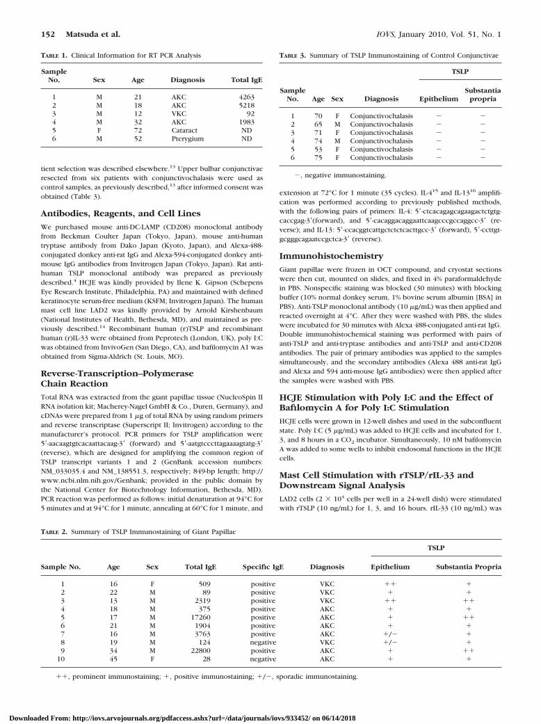
TABLE 1. Clinical Information for RT PCR Analysis
SampleNo. Sex Age Diagnosis Total IgE
1 M 21 AKC 42632 M 18 AKC 52183 M 12 VKC 924 M 32 AKC 19835 F 72 Cataract ND6 M 52 Pterygium ND
TABLE 2. Summary of TSLP Immunostaining of Giant Papillae
Sample No. Age Sex Total IgE Specific IgE Diagnosis
TSLP
Epithelium Substantia Propria
1 16 F 509 positive VKC �� �2 22 M 89 positive VKC � �3 13 M 2319 positive VKC �� ��4 18 M 375 positive AKC � �5 17 M 17260 positive AKC � ��6 21 M 1904 positive AKC � �7 16 M 3763 positive AKC �/� �8 19 M 124 negative VKC �/� �9 34 M 22800 positive AKC � ��
10 45 F 28 negative AKC � �
��, prominent immunostaining; �, positive immunostaining; �/�, sporadic immunostaining.
TABLE 3. Summary of TSLP Immunostaining of Control Conjunctivae
SampleNo. Age Sex Diagnosis
TSLP
EpitheliumSubstantia
propria
1 70 F Conjunctivochalasis � �2 65 M Conjunctivochalasis � �3 71 F Conjunctivochalasis � �4 74 M Conjunctivochalasis � �5 53 F Conjunctivochalasis � �6 75 F Conjunctivochalasis � �
�, negative immunostaining.
152 Matsuda et al. IOVS, January 2010, Vol. 51, No. 1
Downloaded From: http://iovs.arvojournals.org/pdfaccess.ashx?url=/data/journals/iovs/933452/ on 06/14/2018
added alone or simultaneously to LAD2 cells. IL-13 mRNA expressionin LAD2 cells was quantified by real-time PCR.
Real-Time PCR Analysis of TSLP and IL-33mRNA Expression
Total RNA was extracted from HCJE and LAD2 cells and cDNAs wereprepared from 1 �g of total RNA by using random primers as justdescribed. We used real-time PCR probes (TaqMan; Applied Biosys-tems [ABI], Foster City, CA) and primers specific for human TSLP(Hs01572934_g1), human IL-13 (Hs00174379_m1), and 18SrRNA (As-say-on-Demand gene expression products; ABI). Real-time PCR analysiswas performed on a sequence-detection system (Prism 7300; ABI). Theexpression of TSLP in the HCJE cells was quantified by the standardcurve method, by using 18SrRNA expression in the same cDNA as acontrol. We calculated a standard curve with full-length human TSLPcDNA obtained by PCR reaction and subcloned into pCRII dual pro-moter plasmid (Invitrogen). For IL-13 mRNA expression, the compar-ative Ct method was used, which utilizes 18SrRNA expression in thesame cDNA as a control.
RESULTS
RT-PCR Analysis of Giant Papillae Obtained fromPatients with VKC/AKC
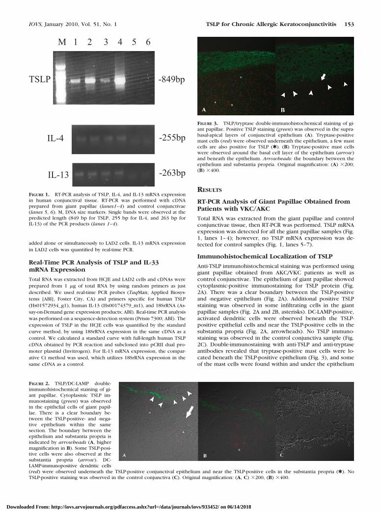
Total RNA was extracted from the giant papillae and controlconjunctivae tissue, then RT-PCR was performed. TSLP mRNAexpression was detected for all the giant papillae samples (Fig.1, lanes 1–4); however, no TSLP mRNA expression was de-tected for control samples (Fig. 1, lanes 5–7).
Immunohistochemical Localization of TSLP
Anti-TSLP immunohistochemical staining was performed usinggiant papillae obtained from AKC/VKC patients as well ascontrol conjunctivae. The epithelium of giant papillae showedcytoplasmic-positive immunostaining for TSLP protein (Fig.2A). There was a clear boundary between the TSLP-positiveand -negative epithelium (Fig. 2A). Additional positive TSLPstaining was observed in some infiltrating cells in the giantpapillae samples (Fig. 2A and 2B, asterisks). DC-LAMP-positive,activated dendritic cells were observed beneath the TSLP-positive epithelial cells and near the TSLP-positive cells in thesubstantia propria (Fig. 2A, arrowheads). No TSLP immuno-staining was observed in the control conjunctiva sample (Fig.2C). Double-immunostaining with anti-TSLP and anti-tryptaseantibodies revealed that tryptase-positive mast cells were lo-cated beneath the TSLP-positive epithelium (Fig. 3), and someof the mast cells were found within and under the epithelium
M 1 2 3 4 5 6
pb948-PLST
IL-4 -255bp
IL-13 -263bp
FIGURE 1. RT-PCR analysis of TSLP, IL-4, and IL-13 mRNA expressionin human conjunctival tissue. RT-PCR was performed with cDNAprepared from giant papillae (lanes1–4) and control conjunctivae(lanes 5, 6). M, DNA size markers. Single bands were observed at thepredicted length (849 bp for TSLP, 255 bp for IL-4, and 263 bp forIL-13) of the PCR products (lanes 1–4).
FIGURE 2. TSLP/DC-LAMP double-immunohistochemical staining of gi-ant papillae. Cytoplasmic TSLP im-munostaining (green) was observedin the epithelial cells of giant papil-lae. There is a clear boundary be-tween the TSLP-positive- and -nega-tive epithelium within the samesection. The boundary between theepithelium and substantia propria isindicated by arrowheads (A, highermagnification in B). Some TSLP-posi-tive cells were also observed at thesubstantia propria (arrow). DC-LAMP-immunopositive dendritic cells(red) were observed underneath the TSLP-positive conjunctival epithelium and near the TSLP-positive cells in the substantia propria (✱). NoTSLP-positive staining was observed in the control conjunctiva (C). Original magnification: (A, C) �200, (B) �400.
FIGURE 3. TSLP/tryptase double-immunohistochemical staining of gi-ant papillae. Positive TSLP staining (green) was observed in the supra-basal-apical layers of conjunctival epithelium (A). Tryptase-positivemast cells (red) were observed underneath the epithelium, a few mastcells are also positive for TSLP (✱). (B) Tryptase-positive mast cellswere observed around the basal cell layer of the epithelium (arrow)and beneath the epithelium. Arrowheads: the boundary between theepithelium and substantia propria. Original magnification: (A) �200;(B) �400.
IOVS, January 2010, Vol. 51, No. 1 TSLP for Chronic Allergic Keratoconjunctivitis 153
Downloaded From: http://iovs.arvojournals.org/pdfaccess.ashx?url=/data/journals/iovs/933452/ on 06/14/2018
(Fig. 3B). A few mast cells were also positive for TSLP immu-nostaining (Fig. 3A, asterisks). The immunostaining results aresummarized in Tables 2 and 3.
Poly I:C-Induced TSLP Expression in HCJE Cells
cDNAs were synthesized from total RNA isolated from polyI:C-stimulated HCJE cells. TSLP mRNA induction was observedin HCJE cells stimulated with poly I:C (5 �g/mL) at 3 hoursafter stimulation (Fig. 4). Simultaneous addition of 10 nMbafilomycin A showed inhibition of poly I:C-induced TSLPmRNA expression (Fig. 4).
TSLP-Induced IL-33 mRNA Expression in Synergywith IL-33
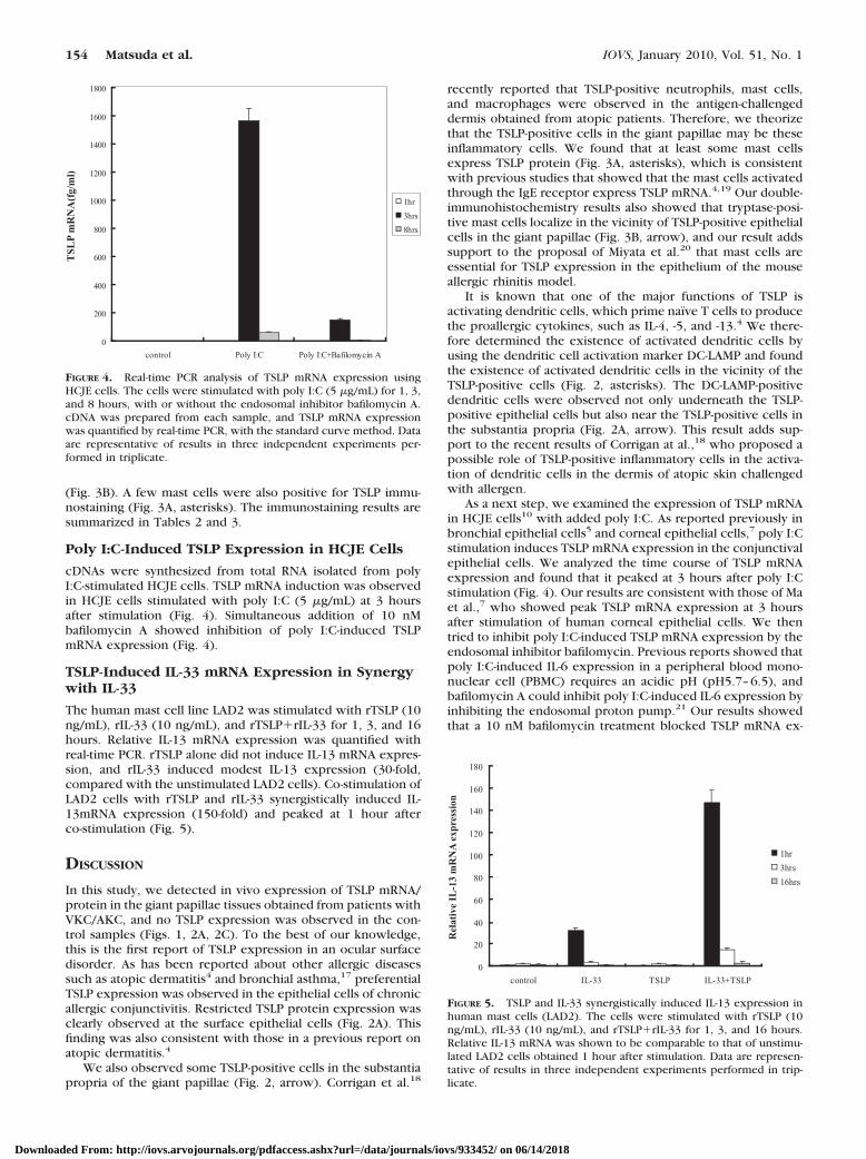
The human mast cell line LAD2 was stimulated with rTSLP (10ng/mL), rIL-33 (10 ng/mL), and rTSLP�rIL-33 for 1, 3, and 16hours. Relative IL-13 mRNA expression was quantified withreal-time PCR. rTSLP alone did not induce IL-13 mRNA expres-sion, and rIL-33 induced modest IL-13 expression (30-fold,compared with the unstimulated LAD2 cells). Co-stimulation ofLAD2 cells with rTSLP and rIL-33 synergistically induced IL-13mRNA expression (150-fold) and peaked at 1 hour afterco-stimulation (Fig. 5).
DISCUSSION
In this study, we detected in vivo expression of TSLP mRNA/protein in the giant papillae tissues obtained from patients withVKC/AKC, and no TSLP expression was observed in the con-trol samples (Figs. 1, 2A, 2C). To the best of our knowledge,this is the first report of TSLP expression in an ocular surfacedisorder. As has been reported about other allergic diseasessuch as atopic dermatitis4 and bronchial asthma,17 preferentialTSLP expression was observed in the epithelial cells of chronicallergic conjunctivitis. Restricted TSLP protein expression wasclearly observed at the surface epithelial cells (Fig. 2A). Thisfinding was also consistent with those in a previous report onatopic dermatitis.4
We also observed some TSLP-positive cells in the substantiapropria of the giant papillae (Fig. 2, arrow). Corrigan et al.18
recently reported that TSLP-positive neutrophils, mast cells,and macrophages were observed in the antigen-challengeddermis obtained from atopic patients. Therefore, we theorizethat the TSLP-positive cells in the giant papillae may be theseinflammatory cells. We found that at least some mast cellsexpress TSLP protein (Fig. 3A, asterisks), which is consistentwith previous studies that showed that the mast cells activatedthrough the IgE receptor express TSLP mRNA.4,19 Our double-immunohistochemistry results also showed that tryptase-posi-tive mast cells localize in the vicinity of TSLP-positive epithelialcells in the giant papillae (Fig. 3B, arrow), and our result addssupport to the proposal of Miyata et al.20 that mast cells areessential for TSLP expression in the epithelium of the mouseallergic rhinitis model.
It is known that one of the major functions of TSLP isactivating dendritic cells, which prime naïve T cells to producethe proallergic cytokines, such as IL-4, -5, and -13.4 We there-fore determined the existence of activated dendritic cells byusing the dendritic cell activation marker DC-LAMP and foundthe existence of activated dendritic cells in the vicinity of theTSLP-positive cells (Fig. 2, asterisks). The DC-LAMP-positivedendritic cells were observed not only underneath the TSLP-positive epithelial cells but also near the TSLP-positive cells inthe substantia propria (Fig. 2A, arrow). This result adds sup-port to the recent results of Corrigan at al.,18 who proposed apossible role of TSLP-positive inflammatory cells in the activa-tion of dendritic cells in the dermis of atopic skin challengedwith allergen.
As a next step, we examined the expression of TSLP mRNAin HCJE cells10 with added poly I:C. As reported previously inbronchial epithelial cells5 and corneal epithelial cells,7 poly I:Cstimulation induces TSLP mRNA expression in the conjunctivalepithelial cells. We analyzed the time course of TSLP mRNAexpression and found that it peaked at 3 hours after poly I:Cstimulation (Fig. 4). Our results are consistent with those of Maet al.,7 who showed peak TSLP mRNA expression at 3 hoursafter stimulation of human corneal epithelial cells. We thentried to inhibit poly I:C-induced TSLP mRNA expression by theendosomal inhibitor bafilomycin. Previous reports showed thatpoly I:C-induced IL-6 expression in a peripheral blood mono-nuclear cell (PBMC) requires an acidic pH (pH5.7–6.5), andbafilomycin A could inhibit poly I:C-induced IL-6 expression byinhibiting the endosomal proton pump.21 Our results showedthat a 10 nM bafilomycin treatment blocked TSLP mRNA ex-
0
20
40
60
80
100
120
140
160
180
control IL-33 TSLP IL-33+TSLP
1hr
3hrs
16hrs
Rel
ativ
e IL
-13
mR
NA
exp
ress
ion
FIGURE 5. TSLP and IL-33 synergistically induced IL-13 expression inhuman mast cells (LAD2). The cells were stimulated with rTSLP (10ng/mL), rIL-33 (10 ng/mL), and rTSLP�rIL-33 for 1, 3, and 16 hours.Relative IL-13 mRNA was shown to be comparable to that of unstimu-lated LAD2 cells obtained 1 hour after stimulation. Data are represen-tative of results in three independent experiments performed in trip-licate.
0
200
400
600
800
1000
1200
1400
1600
1800
control Poly I:C Poly I:C+Bafilomycin A
1hr
3hrs
8hrs
TSL
P m
RN
A(f
g/m
l)
FIGURE 4. Real-time PCR analysis of TSLP mRNA expression usingHCJE cells. The cells were stimulated with poly I:C (5 �g/mL) for 1, 3,and 8 hours, with or without the endosomal inhibitor bafilomycin A.cDNA was prepared from each sample, and TSLP mRNA expressionwas quantified by real-time PCR, with the standard curve method. Dataare representative of results in three independent experiments per-formed in triplicate.
154 Matsuda et al. IOVS, January 2010, Vol. 51, No. 1
Downloaded From: http://iovs.arvojournals.org/pdfaccess.ashx?url=/data/journals/iovs/933452/ on 06/14/2018
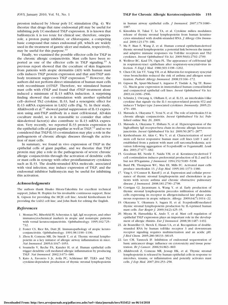
pression induced by 3-hour poly I:C stimulation (Fig. 4). Wetheorize that drugs that raise endosomal pH may be useful forinhibiting poly I:C-mediated TSLP expression. It is known thatbafilomycin A is too toxic for clinical use; therefore, omepra-zole, a proton pump inhibitor, or chloroquine, a competingbasic compound that raises endosomal pH, which are widelyused in the treatment of gastric ulcer and malaria, respectively,may be useful for this purpose.22
Finally, we examined the possible effector cells for TSLP inthe chronic allergic conjunctivitis. Mast cells have been re-ported as one of the effector cells in TSLP signaling.23 Aprevious report showed that the coculture of skin fragmentsfrom patients with atopic dermatitis (AD) with human mastcells induces TSLP protein expression and that anti-TSLP anti-body treatment suppresses TSLP expression.23 However, theauthors did not perform direct stimulation of human mast cellswith recombinant (r)TSLP. Therefore, we stimulated humanmast cells with rTSLP and found that rTSLP treatment aloneinduced a minimum of IL-13 mRNA induction. A surprisingfinding showed that co-stimulation with another epithelialcell–derived Th2 cytokine, IL-33, had a synergistic effect forIL-13 mRNA expression in LAD2 cells (Fig. 5). In their study,Allakhverdi et al.23 showed partial suppression of IL-13 expres-sion using anti-TSLP antibody in a mast cell-lesional, AD-skincoculture model, so it is reasonable to consider that otherskin-derived factor(s) also contribute to IL-13 mRNA expres-sion. Very recently, we reported IL-33 protein expression inthe epithelial cells of giant papillae as well as TSLP,12 and so weconsidered that TSLP-IL-33 co-stimulation may play a role in thepathogenesis of chronic allergic diseases through the activa-tion of mast cells.
In summary, we found in vivo expression of TSLP in theepithelial cells of giant papillae, and we theorize that TSLPprotein may play a role in the pathogenesis of severe chronicallergic conjunctivitis through the activation of dendritic cellsor mast cells in synergy with other proinflammatory cytokinessuch as IL-33. The double-stranded RNA molecule, associatedwith viral infection, may induce expression of TSLP, and theendosomal inhibitor bafilomycin may be useful for inhibitingthis activation.
Acknowledgments
The authors thank Hisako Hitora-Takeshita for excellent technicalsupport; Julian M. Hopkin for his invaluable continuous support; IleneK. Gipson for providing the HCJE cell line; Arnold Kirshenbaum forproviding the LAD2 cell line; and John Bush for editing the English.
References
1. Montan PG, Biberfeld PJ, Scheynius A. IgE, IgE receptors, and otherimmunocytochemical markers in atopic and nonatopic patientswith vernal keratoconjunctivitis. Ophthalmology. 1995;102:725–732.
2. Foster CS, Rice BA, Dutt JE. Immunopathology of atopic kerato-conjunctivitis. Ophthalmology. 1991;98:1190–1196.
3. Zhou B, Comeau MR, De Smedt T, et al. Thymic stromal lympho-poietin as a key initiator of allergic airway inflammation in mice.Nat Immunol. 2005;6:1047–1053.
4. Soumelis V, Reche PA, Kanzler H, et al. Human epithelial cellstrigger dendritic cell mediated allergic inflammation by producingTSLP. Nat Immunol. 2002;3:673–680.
5. Kato A, Favoreto S Jr, Avila PC, Schleimer RP. TLR3- and Th2cytokine-dependent production of thymic stromal lymphopoietin
in human airway epithelial cells. J Immunol. 2007;179:1080–1087.
6. Kinoshita H, Takai T, Le TA, et al. Cytokine milieu modulatesrelease of thymic stromal lymphopoietin from human keratino-cytes stimulated with double-stranded RNA. J Allergy Clin Immu-nol. 2009;123:179–186.
7. Ma P, Bian F, Wang Z, et al. Human corneal epithelium-derivedthymic stromal lymphopoietin: a potential link between the innateand adaptive immune responses via Toll-like receptors and Th2cytokines. Invest Ophthalmol Vis Sci. 2009;50(6):2702–2709.
8. Welliver RC, Kaul TN, Ogra PL. The appearance of cell-bound IgEin respiratory-tract epithelium after respiratory-syncytial-virus in-fection. N Engl J Med. 1980;303:1198–1202.
9. Chen CH, Lin YT, Yang YH, et al. Ribavirin for respiratory syncytialvirus bronchiolitis reduced the risk of asthma and allergen sensi-tization. Pediatr Allergy Immunol. 2008;19:166–172.
10. Gipson IK, Spurr-Michaud S, Argueso P, Tisdale A, Ng TF, RussoCL. Mucin gene expression in immortalized human corneal-limbaland conjunctival epithelial cell lines. Invest Ophthalmol Vis Sci.2003;44:2496–2506.
11. Schmitz J, Owyang A, Oldham E, et al. IL-33, an interleukin-1-likecytokine that signals via the IL-1 receptor-related protein ST2 andinduces T helper type 2-associated cytokines. Immunity. 2005;23:479–490.
12. Matsuda A, Okayama Y, Terai N, et al. The role of interleukin-33 inchronic allergic conjunctivitis. Invest Ophthalmol Vis Sci. Pub-lished online May 20, 2009.
13. Matsuda A, Okayama Y, Ebihara N, et al. Hyperexpression of thehigh-affinity IgE receptor-beta chain in chronic allergic keratocon-junctivitis. Invest Ophthalmol Vis Sci. 2009;50:2871–2877.
14. Kirshenbaum AS, Akin C, Wu Y, et al. Characterization of novelstem cell factor responsive human mast cell lines LAD 1 and 2established from a patient with mast cell sarcoma/leukemia; acti-vation following aggregation of FcepsilonRI or FcgammaRI. LeukRes. 2003;27:677–682.
15. Goodman RE, Nestle F, Naidu YM, et al. Keratinocyte-derived Tcell costimulation induces preferential production of IL-2 and IL-4but not IFN-gamma. J Immunol. 1994;152:5189–5198.
16. Burd PR, Thompson WC, Max EE, Mills FC. Activated mast cellsproduce interleukin 13. J Exp Med. 1995;181:1373–1380.
17. Ying S, O’Connor B, Ratoff J, et al. Expression and cellular prove-nance of thymic stromal lymphopoietin and chemokines in pa-tients with severe asthma and chronic obstructive pulmonarydisease. J Immunol. 2008;181:2790–2798.
18. Corrigan CJ, Jayaratnam A, Wang Y, et al. Early production ofthymic stromal lymphopoietin precedes infiltration of dendriticcells expressing its receptor in allergen-induced late phase cuta-neous responses in atopic subjects. Allergy. 2009;64(7):1014–22.
19. Okayama Y, Okumura S, Sagara H, et al. FcepsilonRI-mediatedthymic stromal lymphopoietin production by IL-4-primed humanmast cells. Eur Respir J. 2009;34(2):425–35.
20. Miyata M, Hatsushika K, Ando T, et al. Mast cell regulation ofepithelial TSLP expression plays an important role in the develop-ment of allergic rhinitis. Eur J Immunol. 2008;38:1487–1492.
21. de Bouteiller O, Merck E, Hasan UA, et al. Recognition of double-stranded RNA by human toll-like receptor 3 and downstreamreceptor signaling requires multimerization and an acidic pH.J Biol Chem. 2005;280:38133–38145.
22. Lee CM, Tannock IF. Inhibition of endosomal sequestration ofbasic anticancer drugs: influence on cytotoxicity and tissue pene-tration. Br J Cancer. 2006;94:863–869.
23. Allakhverdi Z, Comeau MR, Jessup HK, et al. Thymic stromallymphopoietin is released by human epithelial cells in response tomicrobes, trauma, or inflammation and potently activates mastcells. J Exp Med. 2007;204:253–258.
IOVS, January 2010, Vol. 51, No. 1 TSLP for Chronic Allergic Keratoconjunctivitis 155
Downloaded From: http://iovs.arvojournals.org/pdfaccess.ashx?url=/data/journals/iovs/933452/ on 06/14/2018