Embed Size (px)
Citation preview
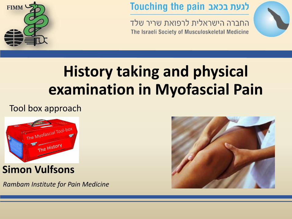
History taking and physical examination in Myofascial Pain
Rambam Institute for Pain Medicine
Simon Vulfsons
Tool box approach
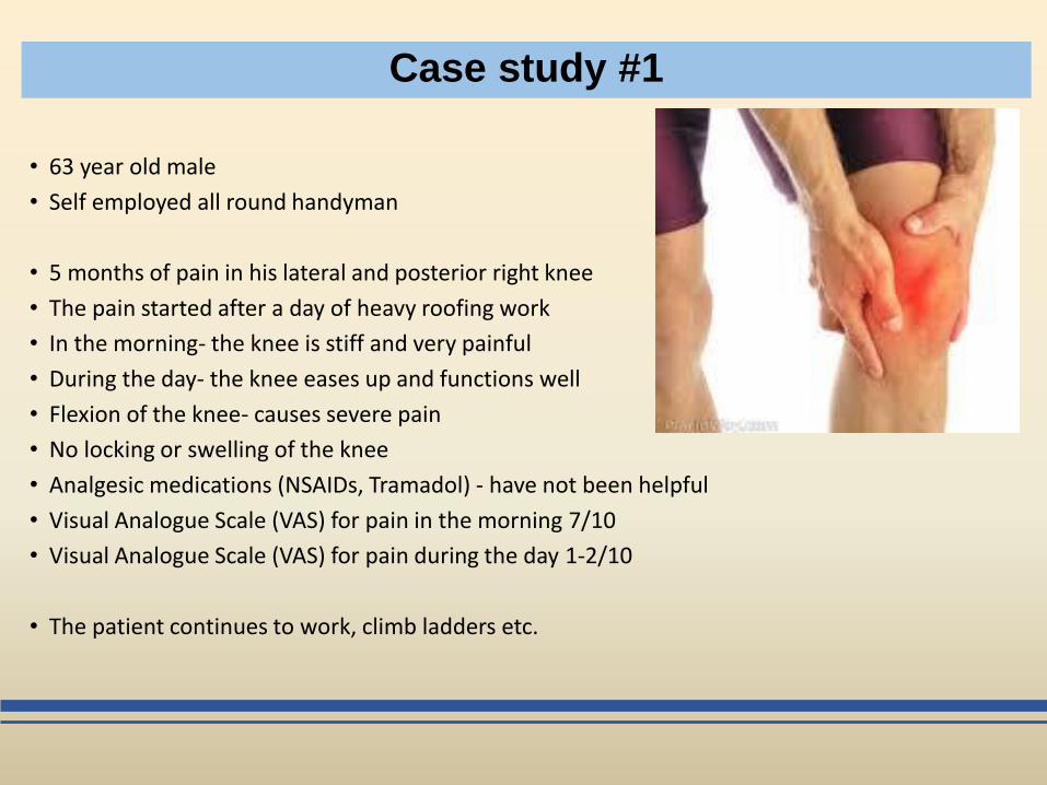
Case study #1
• 63 year old male
• Self employed all round handyman
• 5 months of pain in his lateral and posterior right knee
• The pain started after a day of heavy roofing work
• In the morning- the knee is stiff and very painful
• During the day- the knee eases up and functions well
• Flexion of the knee- causes severe pain
• No locking or swelling of the knee
• Analgesic medications (NSAIDs, Tramadol) - have not been helpful
• Visual Analogue Scale (VAS) for pain in the morning 7/10
• Visual Analogue Scale (VAS) for pain during the day 1-2/10
• The patient continues to work, climb ladders etc.
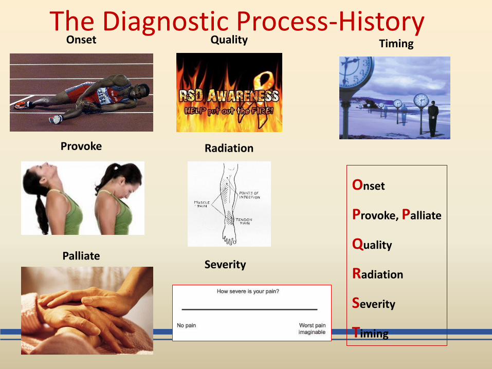
The Diagnostic Process-History Onset
Provoke
Palliate
Quality
Radiation
Severity
Timing
Onset
Provoke, Palliate
Quality
Radiation
Severity
Timing

Case #1 Physical examination
What are we looking for in the physical examination?
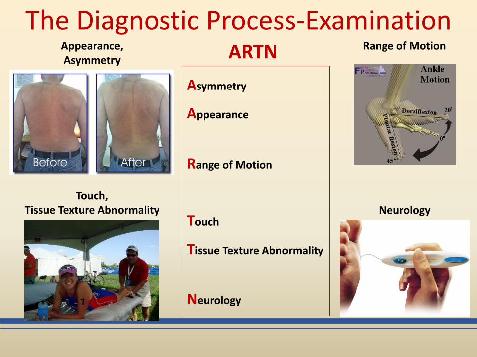
The Diagnostic Process-Examination Appearance, Asymmetry
Range of Motion
Asymmetry
Appearance
Range of Motion
Touch
Tissue Texture Abnormality
Neurology
Touch, Tissue Texture Abnormality Neurology
ARTN
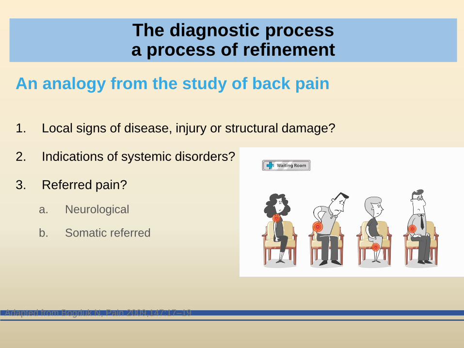
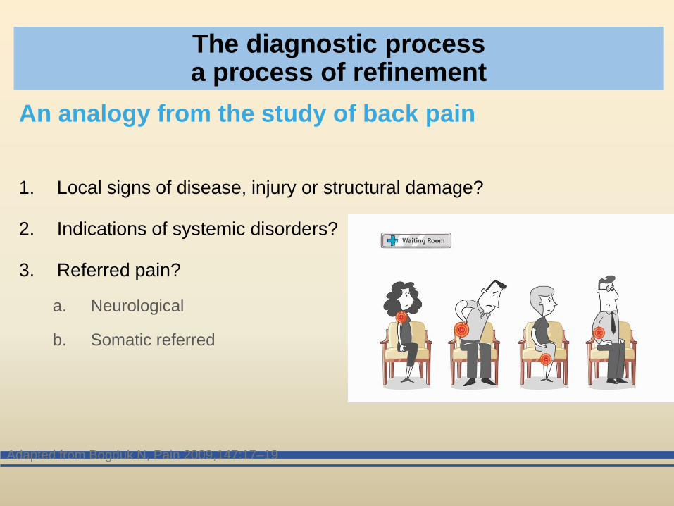
The diagnostic process a process of refinement
1. Local signs of disease, injury or structural damage?
2. Indications of systemic disorders?
3. Referred pain?
a. Neurological
b. Somatic referred
Adapted from Bogduk N, Pain 2009,147:17–19
An analogy from the study of back pain
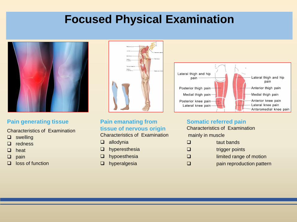
Focused Physical Examination
Pain generating tissue
Characteristics of Examination
swelling
redness
heat
pain
loss of function
Somatic referred pain Characteristics of Examination
mainly in muscle
taut bands
trigger points
limited range of motion
pain reproduction pattern
Pain emanating from
tissue of nervous origin Characteristics of Examination
allodynia
hyperesthesia
hypoesthesia
hyperalgesia
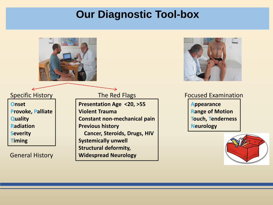
Our Diagnostic Tool-box
Onset Provoke, Palliate Quality Radiation Severity Timing
Presentation Age <20, >55 Violent Trauma Constant non-mechanical pain Previous history Cancer, Steroids, Drugs, HIV Systemically unwell Structural deformity, Widespread Neurology
Specific History The Red Flags
Appearance Range of Motion Touch, Tenderness Neurology
Focused Examination
General History
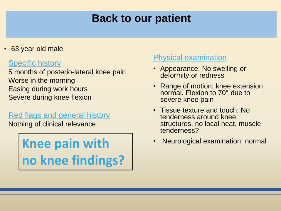
Back to our patient
• 63 year old male
Physical examination
• Appearance: No swelling or deformity or redness
• Range of motion: knee extension normal. Flexion to 70° due to severe knee pain
• Tissue texture and touch: No tenderness around knee structures, no local heat, muscle tenderness?
• Neurological examination: normal Knee pain with no knee findings?
Specific history 5 months of posterio-lateral knee pain
Worse in the morning
Easing during work hours
Severe during knee flexion
Red flags and general history Nothing of clinical relevance
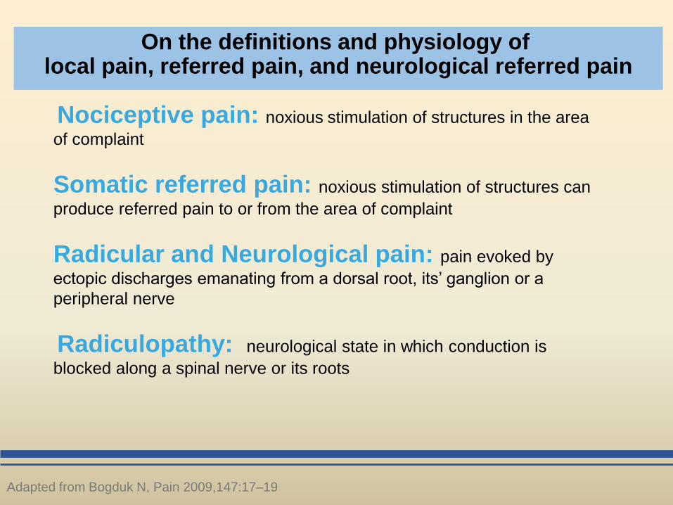
On the definitions and physiology of local pain, referred pain, and neurological referred pain
Adapted from Bogduk N, Pain 2009,147:17–19
Nociceptive pain: noxious stimulation of structures in the area
of complaint
Somatic referred pain: noxious stimulation of structures can
produce referred pain to or from the area of complaint
Radicular and Neurological pain: pain evoked by
ectopic discharges emanating from a dorsal root, its’ ganglion or a
peripheral nerve
Radiculopathy: neurological state in which conduction is
blocked along a spinal nerve or its roots
Citation credit - Ariel 14 – dark gray
This position
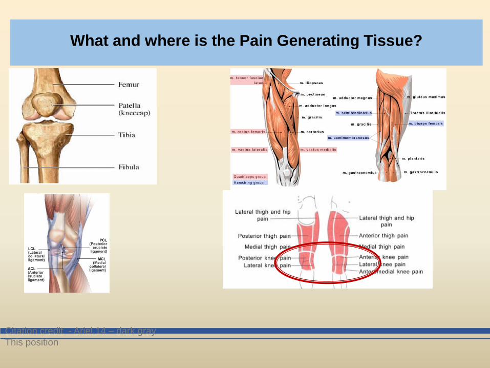
What and where is the Pain Generating Tissue?
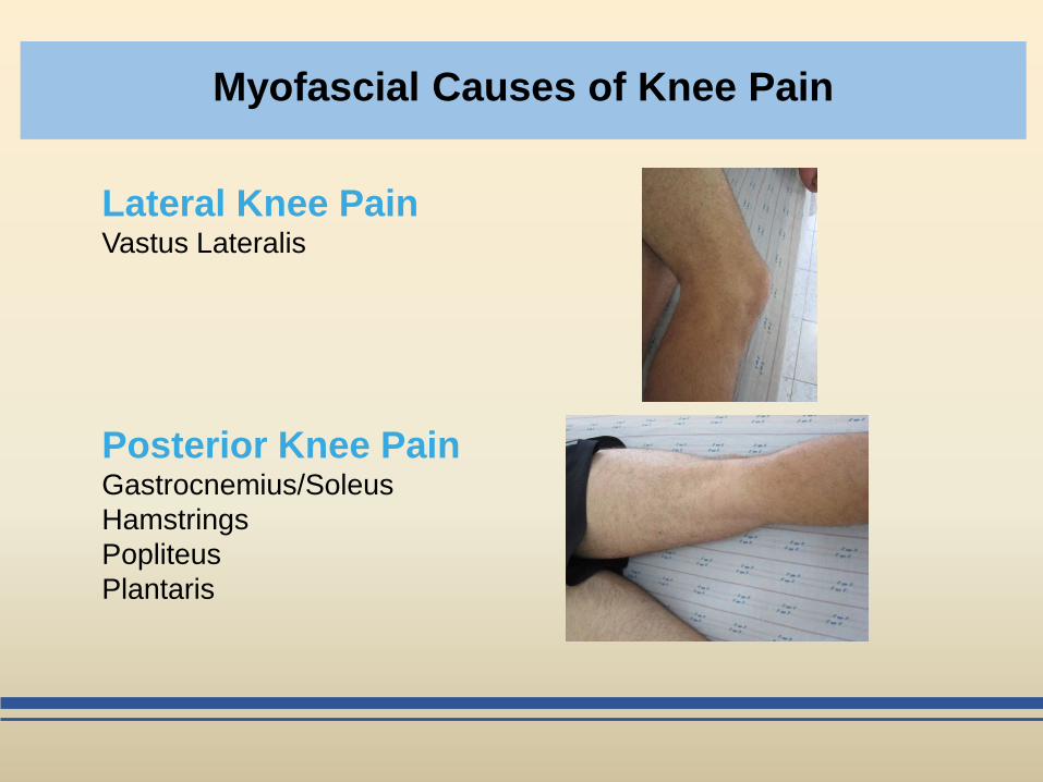
Myofascial Causes of Knee Pain
Lateral Knee Pain Vastus Lateralis
Posterior Knee Pain Gastrocnemius/Soleus
Hamstrings
Popliteus
Plantaris
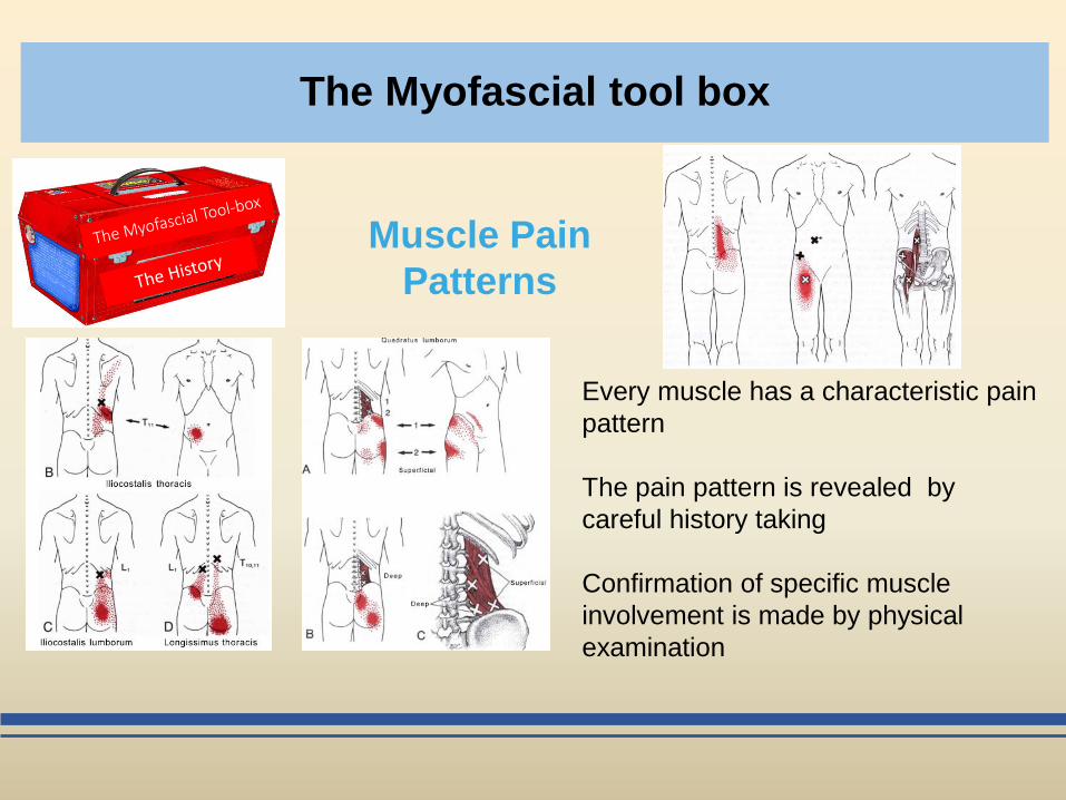
The Myofascial tool box
Muscle Pain
Patterns
Every muscle has a characteristic pain
pattern
The pain pattern is revealed by
careful history taking
Confirmation of specific muscle
involvement is made by physical
examination
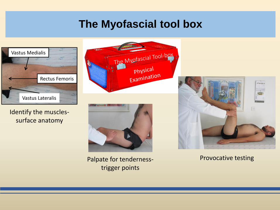
The Myofascial tool box
Identify the muscles- surface anatomy
Palpate for tenderness- trigger points
Provocative testing
Vastus Medialis
Rectus Femoris
Vastus Lateralis
Myofascial Pain Definition
• Regional muscle pain syndrome accompanied by
Trigger Points (TrP)
• TrP- hyperirritable spot within a taut band of skeletal
muscle or muscle fascia
– characteristic referral pain patterns
– painful on compression
– tenderness and autonomic phenomena
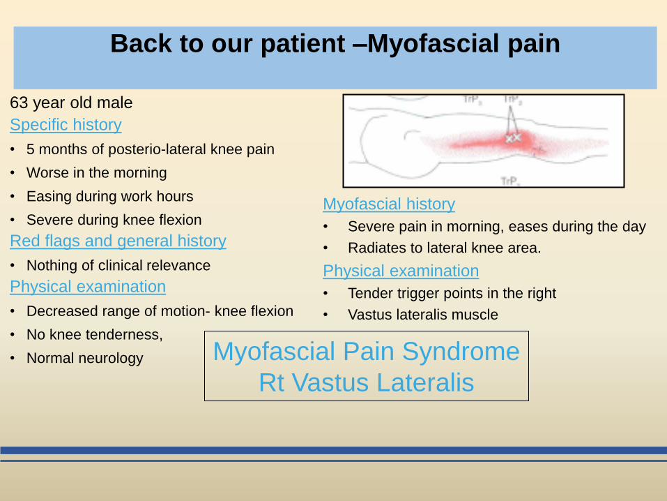
Back to our patient –Myofascial pain
63 year old male
Specific history
• 5 months of posterio-lateral knee pain
• Worse in the morning
• Easing during work hours
• Severe during knee flexion
Red flags and general history
• Nothing of clinical relevance
Physical examination
• Decreased range of motion- knee flexion
• No knee tenderness,
• Normal neurology
Myofascial Pain Syndrome
Rt Vastus Lateralis
Myofascial history
• Severe pain in morning, eases during the day
• Radiates to lateral knee area.
Physical examination
• Tender trigger points in the right
• Vastus lateralis muscle
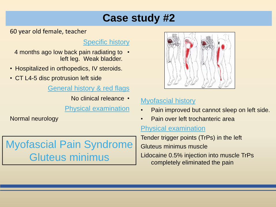
Case study #2 60 year old female, teacher
historySpecific
•4 months ago low back pain radiating to left leg. Weak bladder.
• Hospitalized in orthopedics, IV steroids.
• CT L4-5 disc protrusion left side
history & red flagsGeneral
•No clinical releance
Physical examination
Normal neurology
Myofascial Pain Syndrome
Gluteus minimus
Myofascial history
• Pain improved but cannot sleep on left side.
• Pain over left trochanteric area
Physical examination
Tender trigger points (TrPs) in the left
Gluteus minimus muscle
Lidocaine 0.5% injection into muscle TrPs completely eliminated the pain
The diagnostic process a process of refinement
An analogy from the study of back pain
1. Local signs of disease, injury or structural damage?
2. Indications of systemic disorders?
3. Referred pain?
a. Neurological
b. Somatic referred
Adapted from Bogduk N, Pain 2009,147:17–19
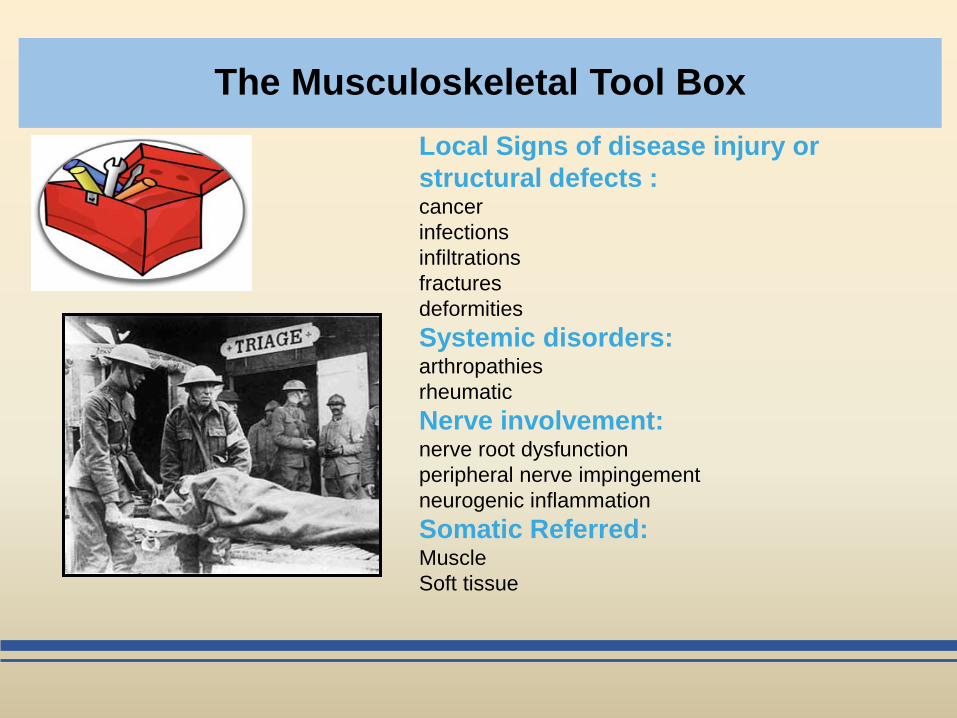
The Musculoskeletal Tool Box
Local Signs of disease injury or
structural defects : cancer
infections
infiltrations
fractures
deformities
Systemic disorders: arthropathies
rheumatic
Nerve involvement: nerve root dysfunction
peripheral nerve impingement
neurogenic inflammation
Somatic Referred: Muscle
Soft tissue
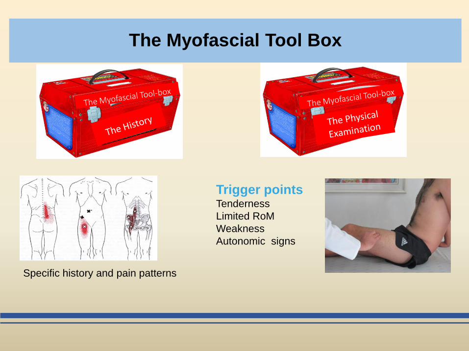
The Myofascial Tool Box
Specific history and pain patterns
Trigger points Tenderness
Limited RoM
Weakness
Autonomic signs
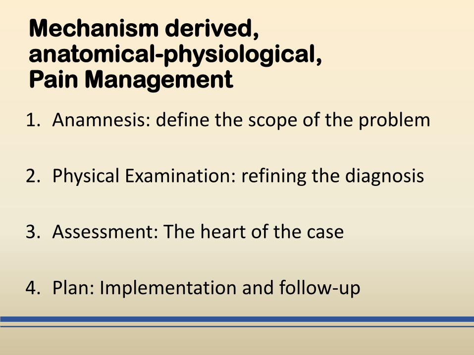
Mechanism derived, anatomical-physiological, Pain Management
1. Anamnesis: define the scope of the problem
2. Physical Examination: refining the diagnosis
3. Assessment: The heart of the case
4. Plan: Implementation and follow-up
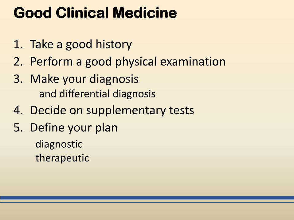
Good Clinical Medicine
1. Take a good history
2. Perform a good physical examination
3. Make your diagnosis and differential diagnosis
4. Decide on supplementary tests
5. Define your plan diagnostic
therapeutic
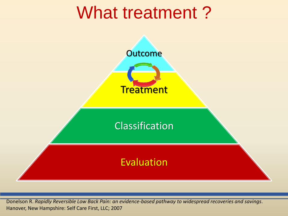
What treatment ?
Outcome
Treatment
Classification
Evaluation
Donelson R. Rapidly Reversible Low Back Pain: an evidence-based pathway to widespread recoveries and savings. Hanover, New Hampshire: Self Care First, LLC; 2007
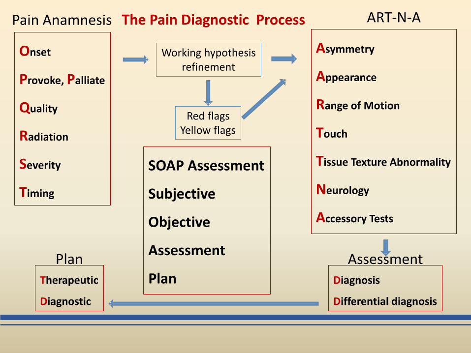
Onset
Provoke, Palliate
Quality
Radiation
Severity
Timing
Pain Anamnesis
Asymmetry
Appearance
Range of Motion
Touch
Tissue Texture Abnormality
Neurology
Accessory Tests
ART-N-A
Working hypothesis refinement
Red flags Yellow flags
Diagnosis
Differential diagnosis
Assessment
Therapeutic
Diagnostic
Plan
SOAP Assessment
Subjective
Objective
Assessment
Plan
The Pain Diagnostic Process
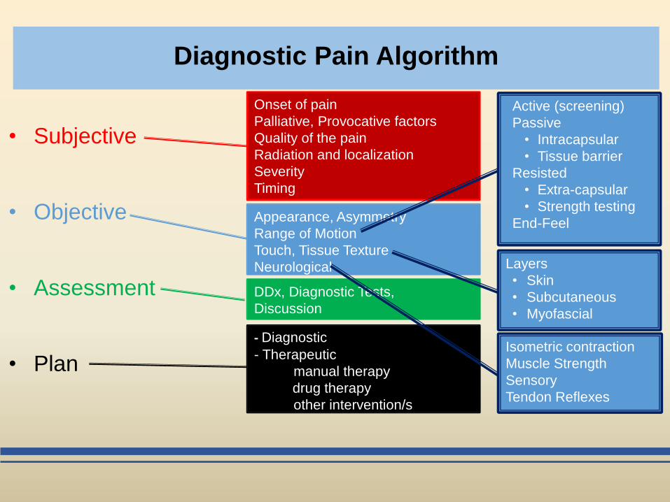
Diagnostic Pain Algorithm
• Subjective
• Objective
• Assessment
• Plan
Onset of pain
Palliative, Provocative factors
Quality of the pain
Radiation and localization
Severity
Timing
Appearance, Asymmetry
Range of Motion
Touch, Tissue Texture
Neurological
DDx, Diagnostic Tests,
Discussion
- Diagnostic
- Therapeutic
manual therapy
drug therapy
other intervention/s
Active (screening)
Passive
• Intracapsular
• Tissue barrier
Resisted
• Extra-capsular
• Strength testing
End-Feel
Layers
• Skin
• Subcutaneous
• Myofascial
Isometric contraction
Muscle Strength
Sensory
Tendon Reflexes