Embed Size (px)
Citation preview
Hypercementosis, daganatok, fejlődési rendellenességek
Dr.Ackermann Gábor
Daganatok • Klinikai vizsgálat
– stomato-onkológiai szűrés!!! • Röntgenvizsgálat
– Diagnózis • Felismerés (intraoralis, panoráma)
– Lokalizálás, tervezés – Terápia
Daganatok – Lokalizálás, tervezés
• CT – csont-/lágyszövet • MRI – lágyszövet • Ultrahang vizsgálat- nyirokcsomók • Nukleáris medicina (PET, SPECT) –
funkciós képalkotás – Terápia (angiográfia)
• Embolizáció • Célzott kemoterápia
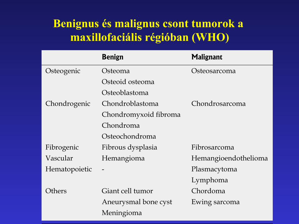
Benignus és malignus csont tumorok a maxillofaciális régióban (WHO)
Abdel Razek AAK. Imaging of maxillofacial bone tumors
BENIGN TUMOR OsteomaOsteoma is the most common osseous tumor in the maxillofacial region. Osteoma is most commonly seen in the 5th to 6th decades of life and the male-to-female ratio is 1.3:1. Osteoma occurs more commonly in the fronto-ethmoidal sinus and is rarely seen in the maxillary and sphenoid sinuses[2]. All osteomas contain three main components: compact bone (ivory), cancellous bone (trabeculae), and fibrous (spongy) tissue. Osteomas are named according to the dominant component. Compact osteomas most often involve the frontal sinus and grow gradually. Cancellous osteomas are located mostly in the maxillary and ethmoid sinuses and grow relatively quickly. Osteomas are slow growing benign tumors. Multiple cra-niofacial osteomas may be a part of Gardner syndrome. It is usually asymptomatic but may be associated with facial swelling, deformity, mucocele, proptosis, ocular disturbances and pneumocephalus[2,7]. Osteoma appears as a characteristic sharp, well delineated sclerotic lesion attached by a broad base or pedicle to the bone. Osteoma composed exclusively of compact bone is radiodense (Figure 1), while those containing cancellous bone show evidence of internal trabecular structure. CT multiplanar reconstructions allow the precise identification of the site of origin of the lesion, to fully depict course and patency of all sinus paths, and to correctly assess the integrity of thin bony walls such as the lamina papyracea or the cribriform plate. Compact osteomas produce a complete signal void on all MR sequences, so they are often indis-tinguishable from the surrounding air in the paranasal sinuses and are thus overlooked. Fibrous osteomas have low to absent signal intensity on all MR sequences[2,4,7,8].
Osteoid osteomaOsteoid osteoma is a rare tumor in maxillofacial regions (with a few case reports in the ethmoid region) that af-fects young males in the 2nd to 3rd decades of life. It is a benign osteoblastic lesion characterized by varying intermixtures of osteoid, newly formed bone, and highly vascular supporting osseous tissue (nidus) surrounded by a distinctive surrounding zone of reactive bone forma-tion. Osteoid osteoma appears on CT scan as a charac-teristic radio-opaque lesion with a nidus (less than 1.5 cm in diameter) which has a radiolucent center surrounded by dense sclerosis. Occasionally, the nidus may have a radio-opaque calcified center with a surrounding radiolu-cent area. The osteoid osteoma may even be completely sclerotic. MR appearance of osteoid osteoma depends upon the amount of calcification within the nidus, the size of the fibrovascular zone and reactive sclerosis; so it may not be diagnostic. The mass demonstrates patchy enhancement[9].
OsteoblastomaOsteoblastoma is typically seen in male patients during
the 2nd decade of life. It may be seen in the maxilla, eth-moid, nasal cavity and orbit. It shows a marked amount of osteoid tissue produced by osteoblasts. The osteo-clasts are numerous and the background is highly vascu-lar. Histologically, osteoblastoma show some similarity to osteoid osteoma, but they are larger without nidus or zonal architecture and show a stronger, more progres-sive, occasionally even destructive growth; thus, they are sometimes called aggressive osteoblastoma. The patient presents with pain, facial swelling and asymmetry of the face[4,10]. It is commonly seen as an expansile lytic lesion with cortical shell (Figure 2A), or it may show as mixed lytic and sclerotic or predominately sclerotic bone form-ing a lesion. Ossification foci with ground glass appear-ance, cloudy confluent mineralization in the central part of the lesion (75%) may be seen. It exhibits intermediate to low signal intensity on T1-weighted images and high to low signal intensity on T2-weighted images depending upon the amount of ossification. Areas of mineralization appear as zones of low signal intensity on T2-weighted images. It shows variable patterns of contrast enhance-ment[4,10,11] (Figure 2B and C).
126 May 28, 2011|Volume 3|Issue 5|WJR|www.wjgnet.com
�������� ��������������� ����������������������������� ��������� ��������������������������������������� ���[1]
Benign Malignant
Osteogenic Osteoma OsteosarcomaOsteoid osteomaOsteoblastoma
Chondrogenic Chondroblastoma ChondrosarcomaChondromyxoid fibromaChondromaOsteochondroma
Fibrogenic Fibrous dysplasia FibrosarcomaVascular Hemangioma HemangioendotheliomaHematopoietic - Plasmacytoma
LymphomaOthers Giant cell tumor Chordoma
Aneurysmal bone cyst Ewing sarcomaMeningioma
Figure 1 Compact Osteoma. Axial computed tomography scan of the para-nasal sinus shows a pathognomonic dense sclerotic mass (arrow) in the frontal sinus.
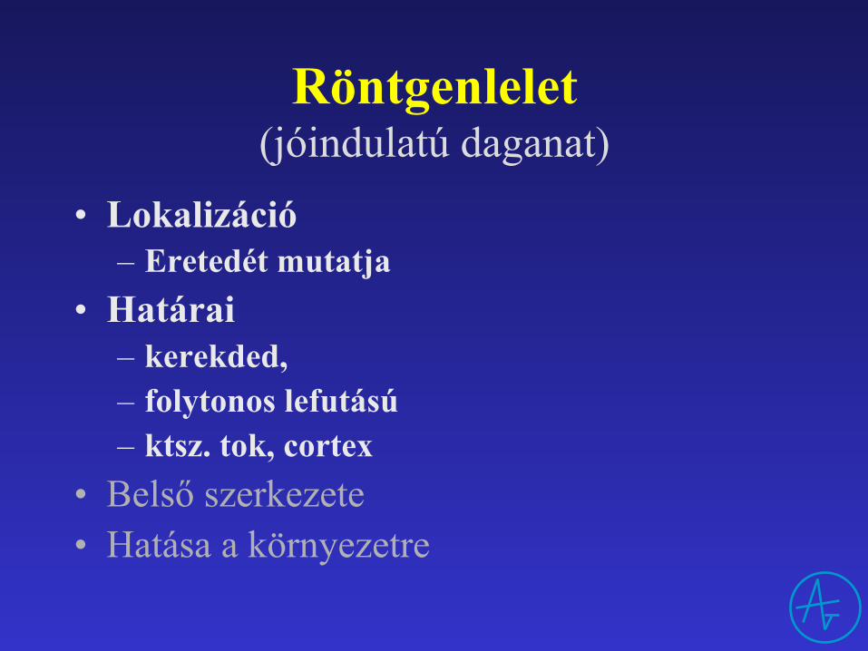
Röntgenlelet (jóindulatú daganat)
• Lokalizáció – Eretedét mutatja
• Határai – kerekded, – folytonos lefutású – ktsz. tok, cortex
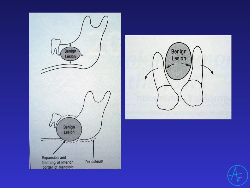
• Belső szerkezete • Hatása a környezetre
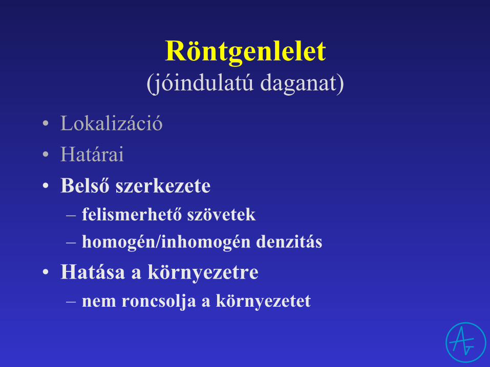
Röntgenlelet (jóindulatú daganat)
• Lokalizáció • Határai • Belső szerkezete
– felismerhető szövetek – homogén/inhomogén denzitás
• Hatása a környezetre – nem roncsolja a környezetet
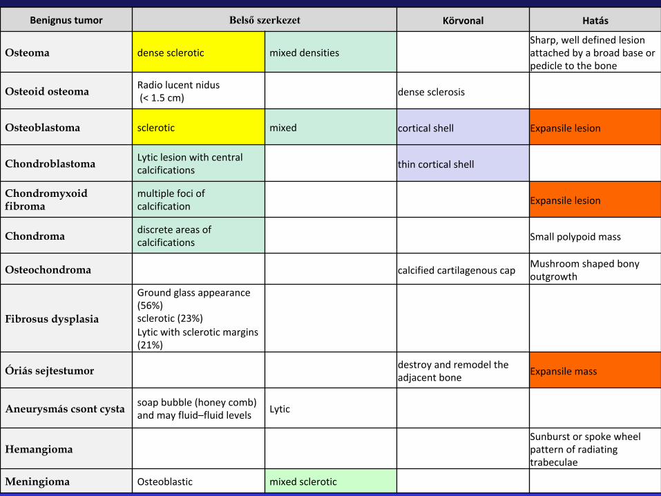
Benignustumor Belső szerkezet Körvonal Hatás
Osteoma densesclerotic mixeddensitiesSharp,welldefinedlesionattachedbyabroadbaseorpedicletothebone
Osteoid osteoma Radiolucentnidus(<1.5cm) densesclerosis
Osteoblastoma sclerotic mixed corticalshell Expansilelesion
Chondroblastoma Lyticlesionwithcentralcalcifications thincorticalshell
Chondromyxoid fibroma
multiplefociofcalcification Expansilelesion
Chondroma discreteareasofcalcifications Smallpolypoidmass
Osteochondroma calcifiedcartilagenouscap Mushroomshapedbonyoutgrowth
Fibrosus dysplasia
Groundglassappearance(56%)sclerotic(23%)Lyticwithscleroticmargins(21%)
Óriás sejtestumor destroyandremodeltheadjacentbone Expansilemass
Aneurysmás csont cysta soapbubble(honeycomb)andmayfluid–fluidlevels Lytic
Hemangioma Sunburstorspokewheelpatternofradiatingtrabeculae
Meningioma Osteoblastic mixedsclerotic
Benignus daganatok
• Valódi neoplasma (pl.ameloblastoma) • Hamartoma (pl. odontoma) • Hyerplasia
– enostosis, exostosis, torus palatinalis, torus mandibularis, hypercementosis
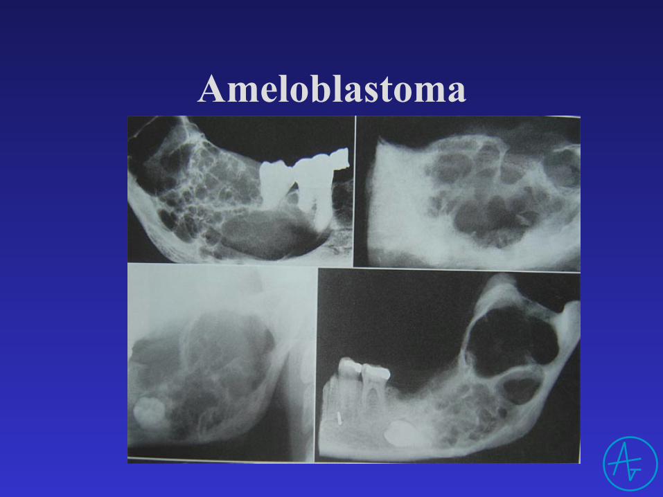
Ameloblastoma
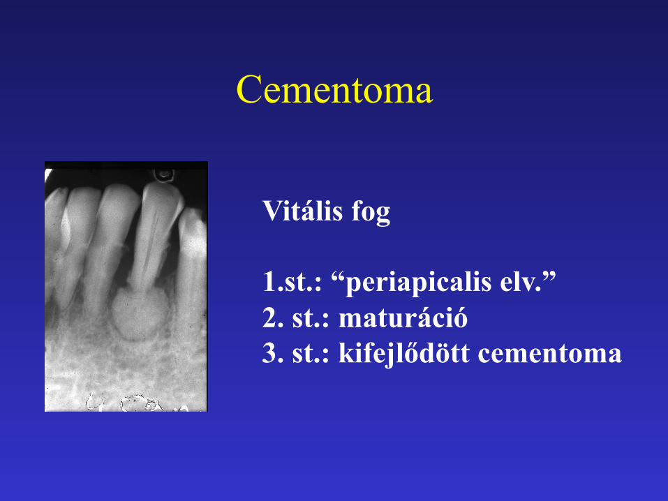
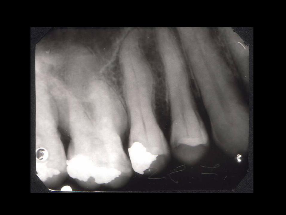
Cementoma
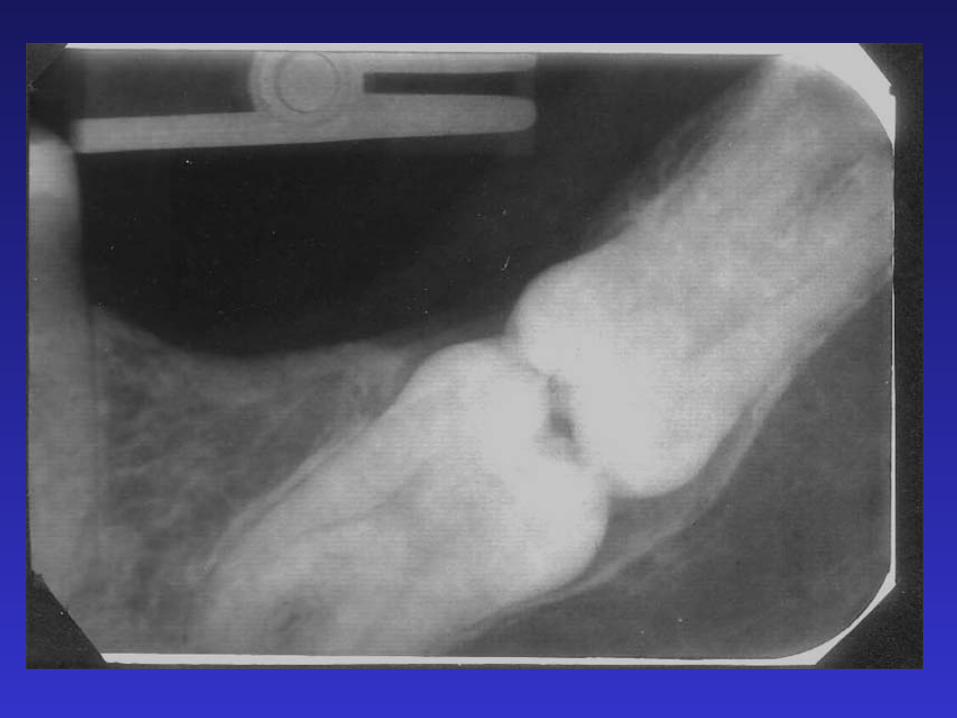
Vitális fog 1.st.: “periapicalis elv.” 2. st.: maturáció 3. st.: kifejlődött cementoma
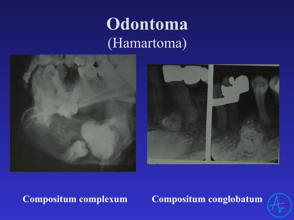
Odontoma (Hamartoma)
Compositum complexum Compositum conglobatum
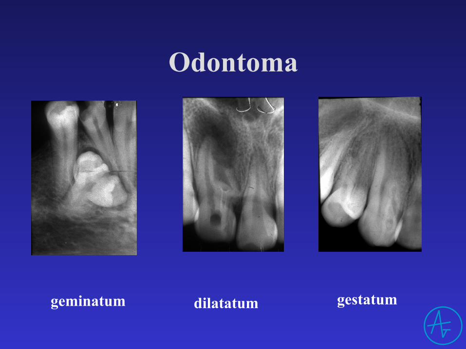
Odontoma
geminatum dilatatum gestatum
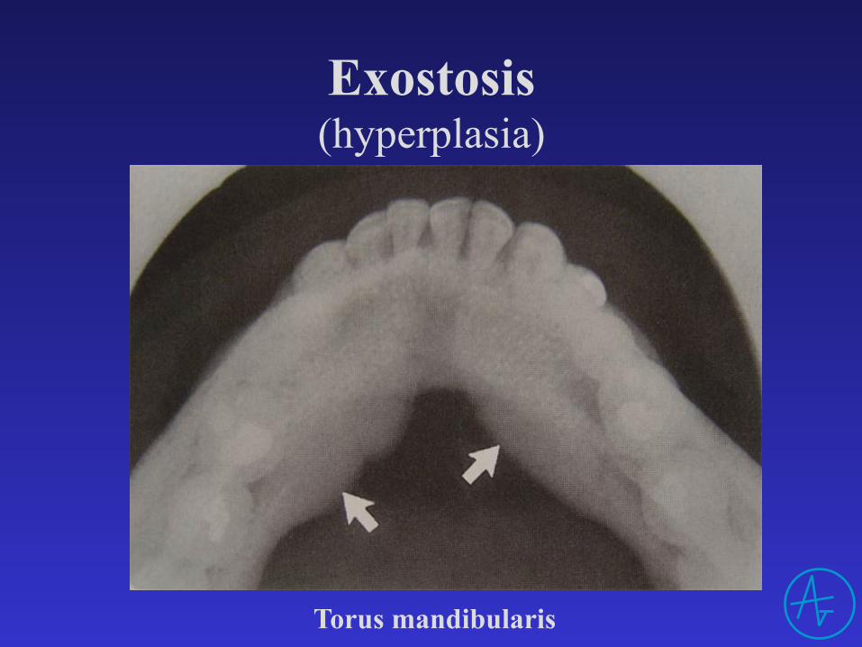
Exostosis (hyperplasia)
Torus mandibularis
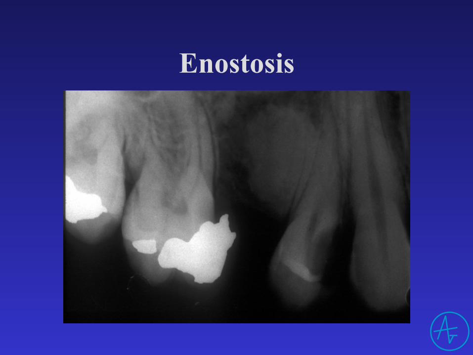
Enostosis
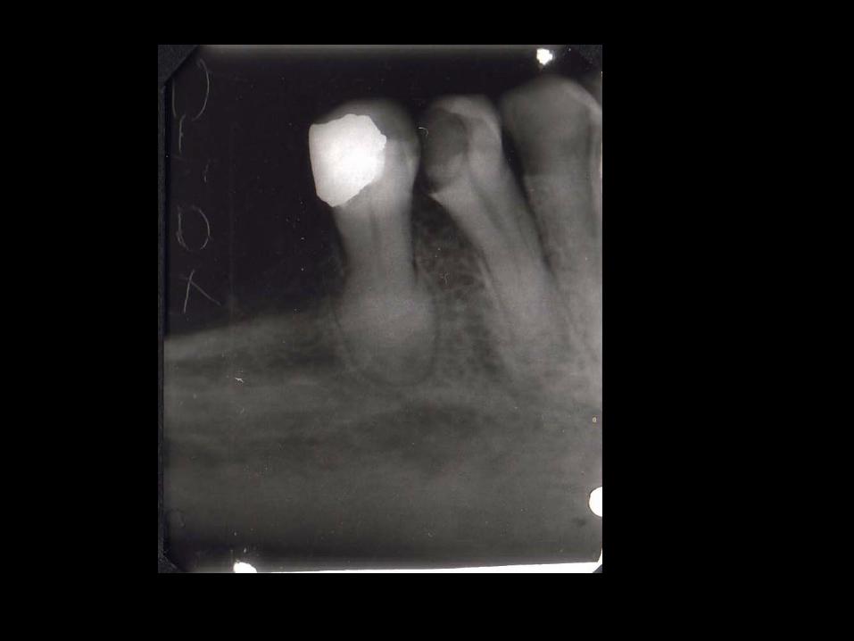
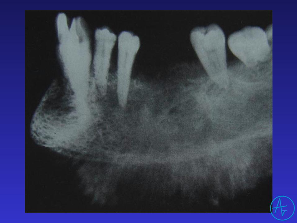
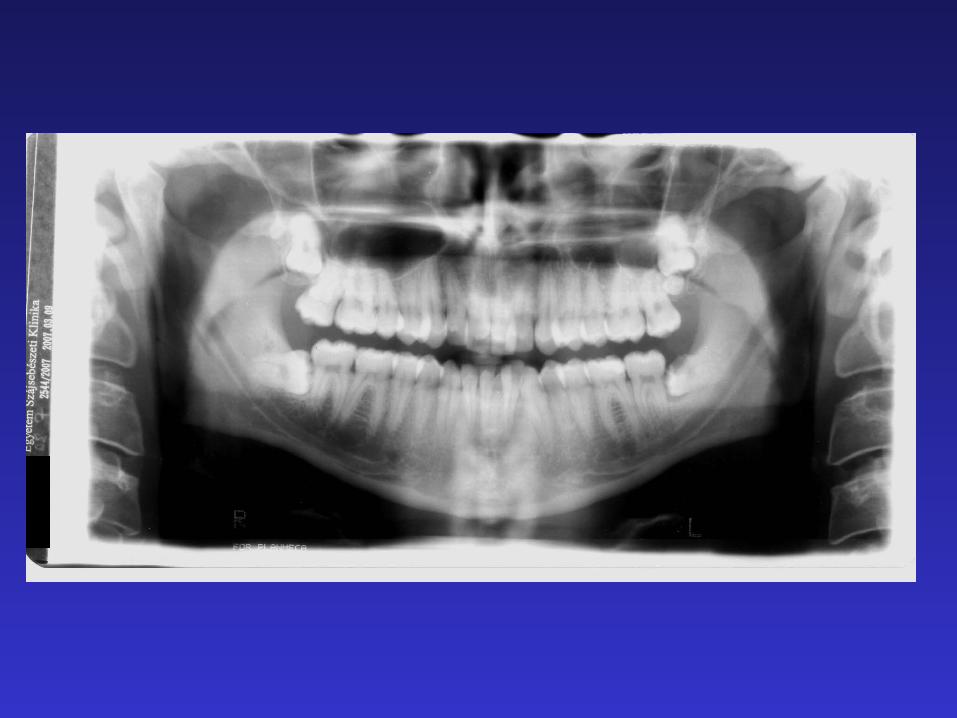
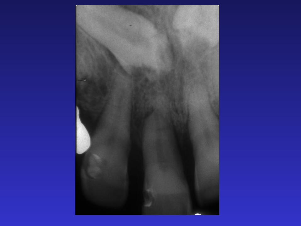
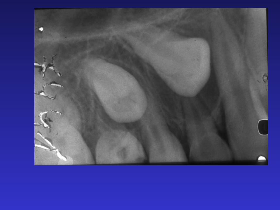
Hypercememtosis
• gyökércsúcson: – nem élő fogakon, chr.gyulladás
• diffúz oldalsó: – túlterhelés
• multiplex: – idült izületi gyull., ált. betegségek
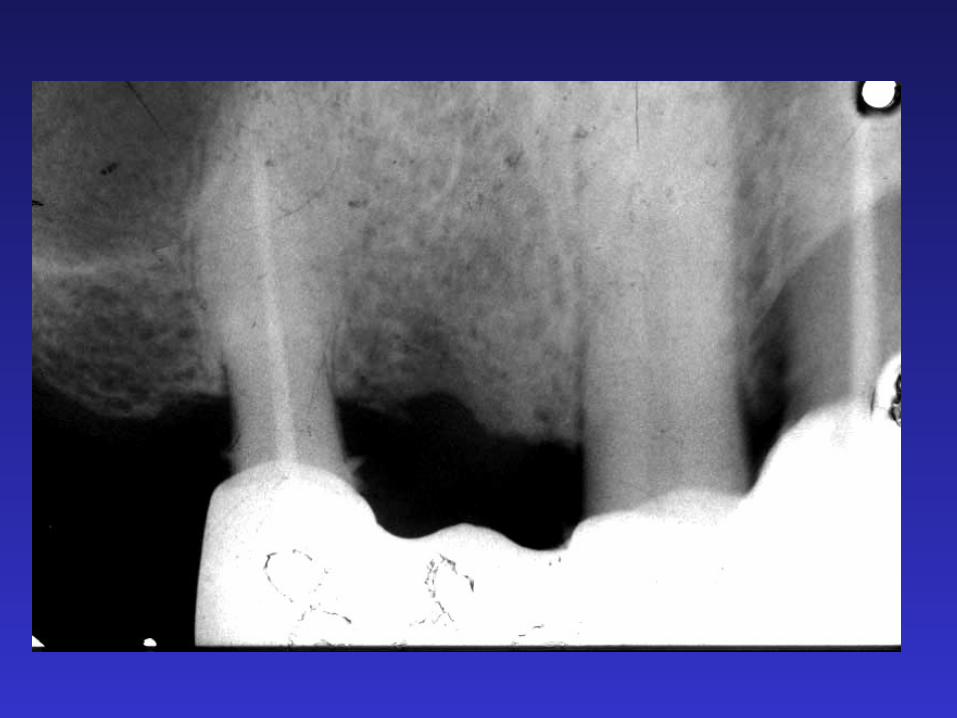
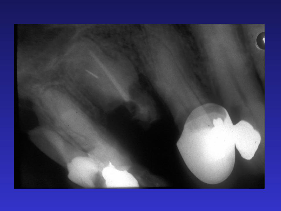
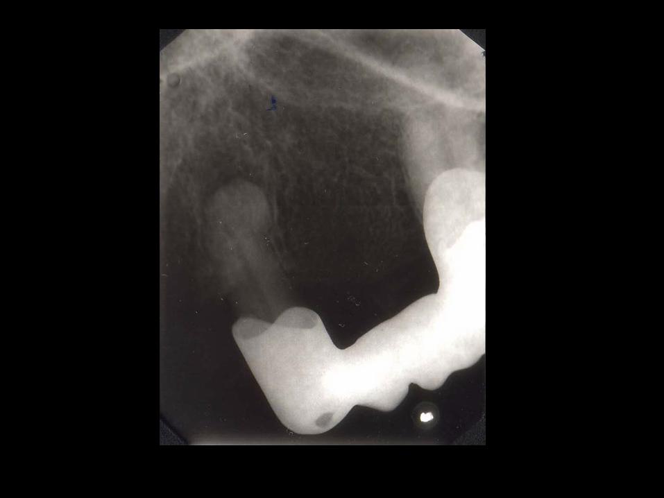
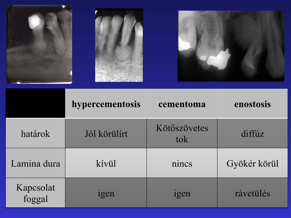
hypercementosis cementoma enostosis
határok Jól körülírt Kötőszövetes tok diffúz
Lamina dura kívül nincs Gyökér körül
Kapcsolat foggal igen igen rávetülés
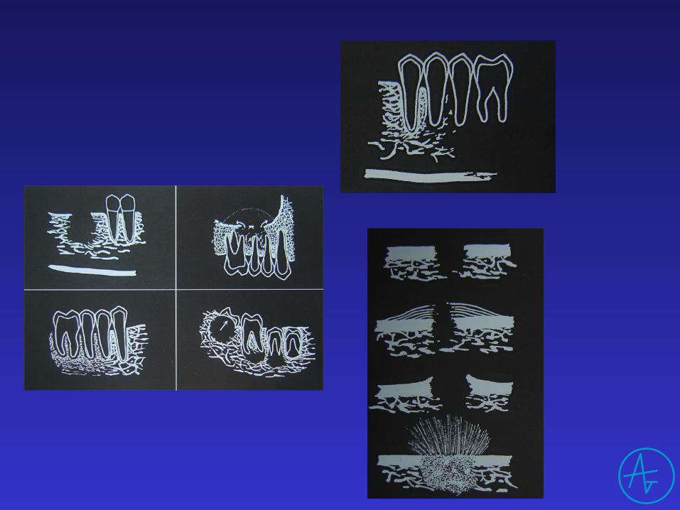
Röntgenlelet (rosszindulatú daganat)
• Lokalizáció • Határai, alakja
– invasiv rajzolat – nincs cortex, ktsz. tok
• Belső szerkezete • Hatása a környezetre
Röntgenlelet (rosszindulatú daganat)
• Lokalizáció • Határai • Belső szerkezete
– általában radiolucens – csontszigetek
• Hatása a környezetre – gyors, destruktív hatás
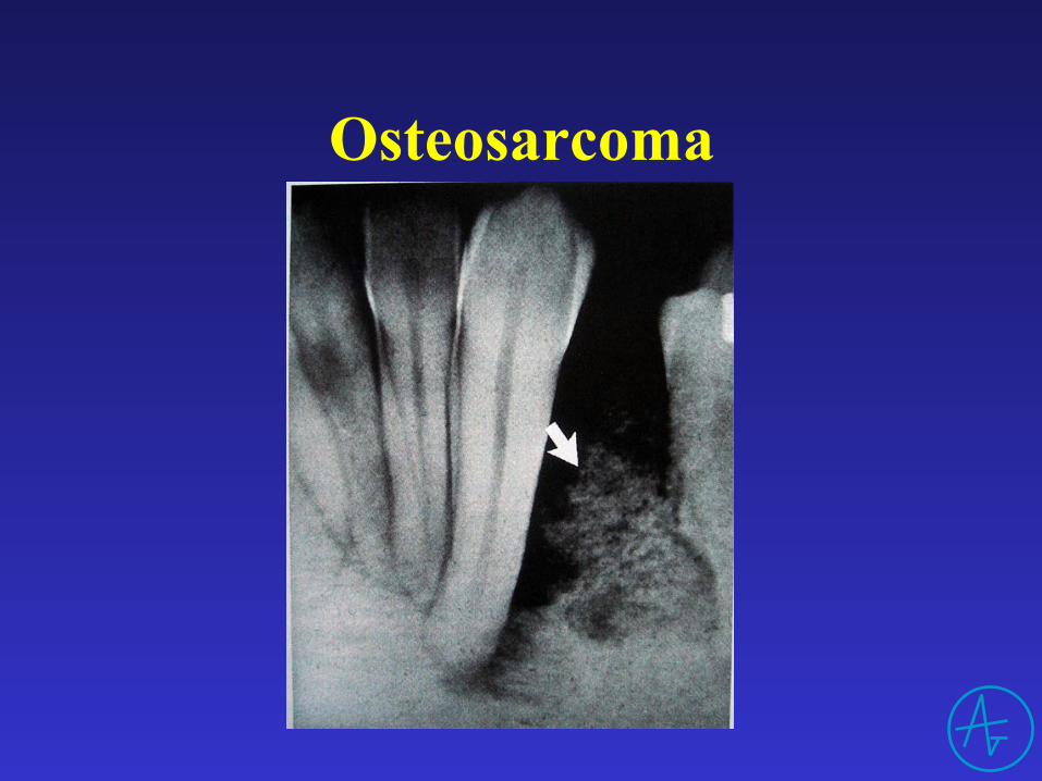
Osteosarcoma
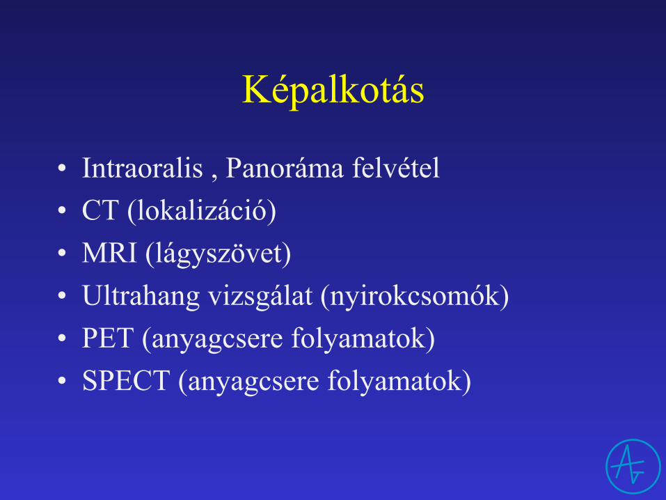
Képalkotás
• Intraoralis , Panoráma felvétel • CT (lokalizáció) • MRI (lágyszövet) • Ultrahang vizsgálat (nyirokcsomók) • PET (anyagcsere folyamatok) • SPECT (anyagcsere folyamatok)
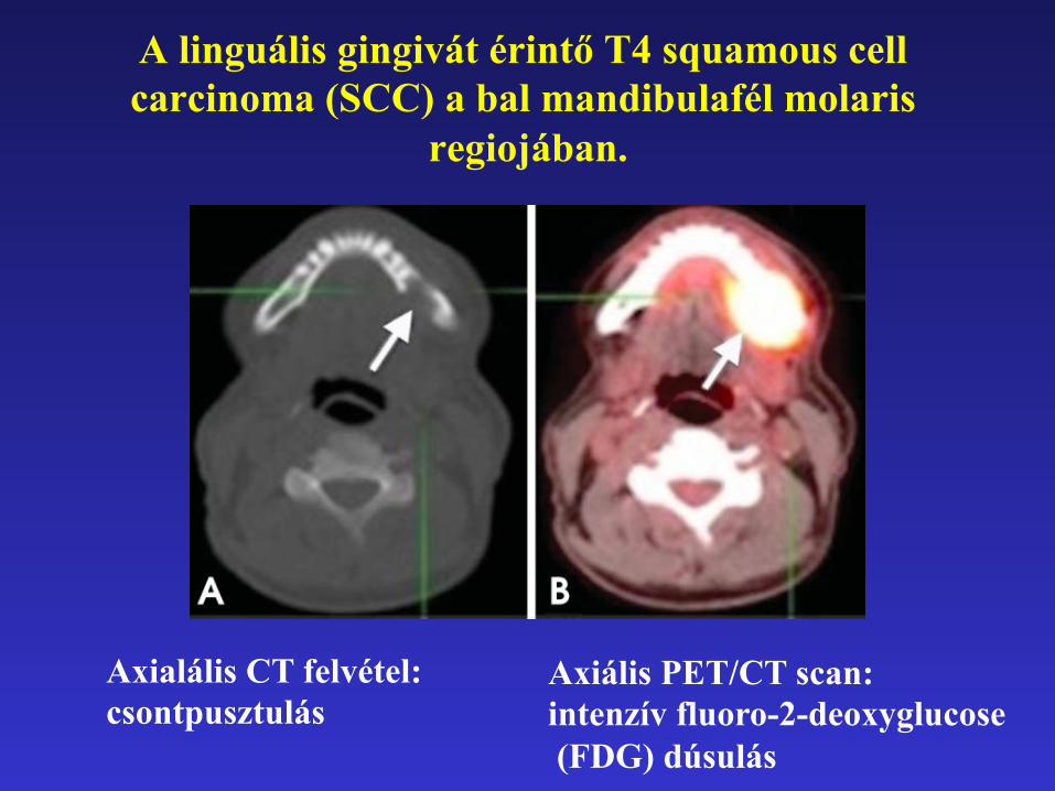
A linguális gingivát érintő T4 squamous cell carcinoma (SCC) a bal mandibulafél molaris
regiojában.
Axialális CT felvétel: csontpusztulás
Axiális PET/CT scan: intenzív fluoro-2-deoxyglucose (FDG) dúsulás
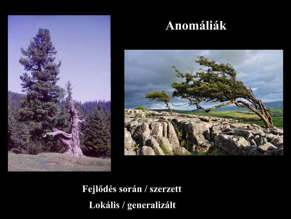
Anomáliák
Lokális / generalizált
Fejlődés során / szerzett
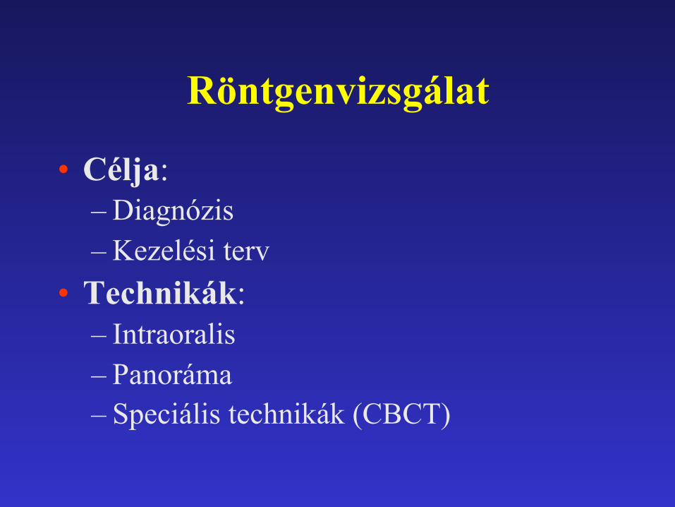
Röntgenvizsgálat
• Célja: – Diagnózis – Kezelési terv
• Technikák: – Intraoralis – Panoráma – Speciális technikák (CBCT)
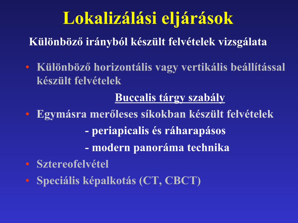
Lokalizálási eljárások Különböző irányból készült felvételek vizsgálata
• Különböző horizontális vagy vertikális beállítással készült felvételek
Buccalis tárgy szabály • Egymásra merőleses síkokban készült felvételek
- periapicalis és ráharapásos - modern panoráma technika
• Sztereofelvétel • Speciális képalkotás (CT, CBCT)
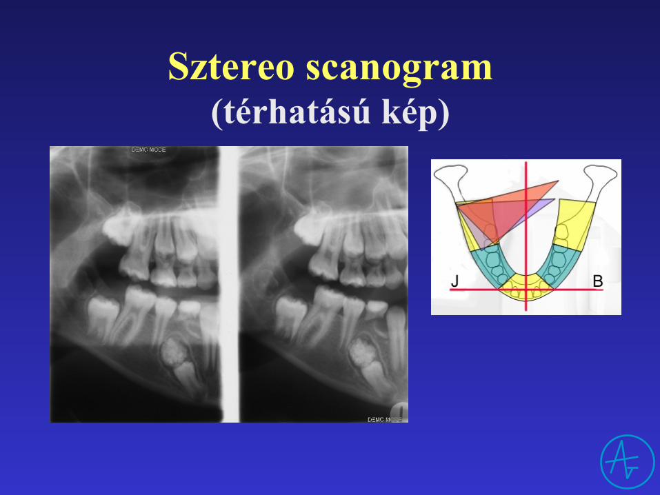
Sztereo scanogram (térhatású kép)
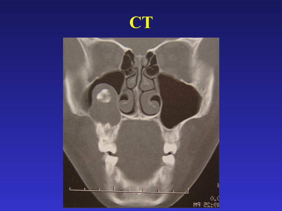
CT
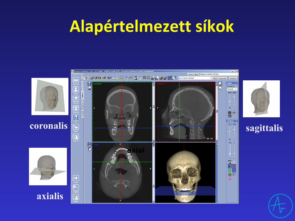
axial
coronalis sagittalis
axialis
Alapértelmezettsíkok
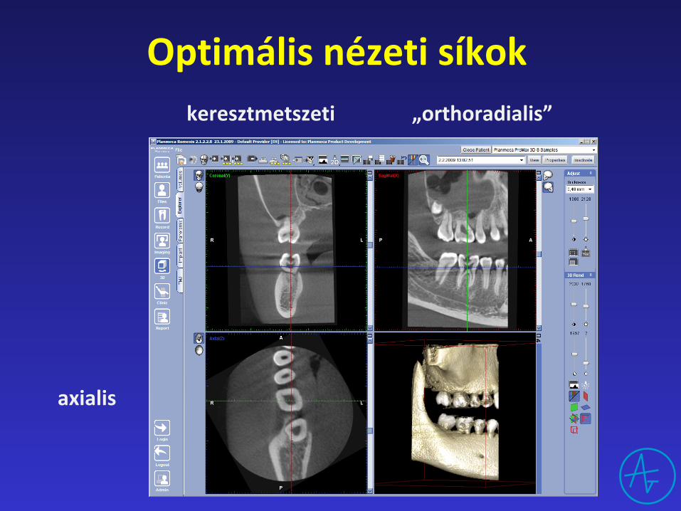
keresztmetszeti „orthoradialis”
axialis
Optimálisnézetisíkok
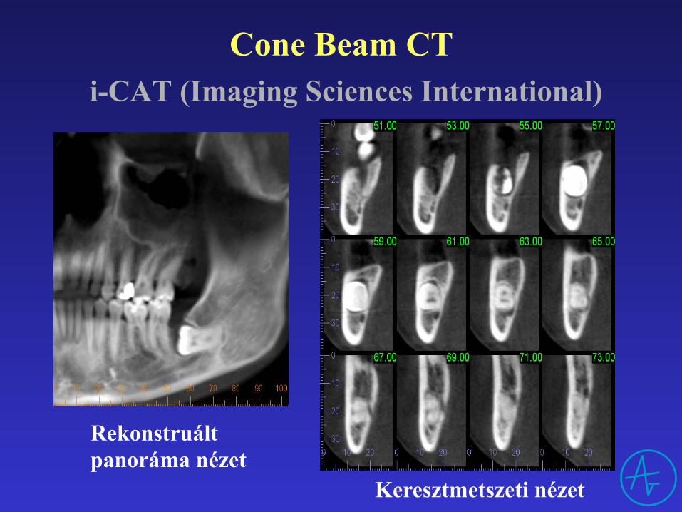
Cone Beam CT i-CAT (Imaging Sciences International)
Rekonstruált panoráma nézet
Keresztmetszeti nézet
Anomáliák
• Nagyságbeli • Alaki • Számbeli • Helyzeti • Előtörési • Szerkezeti rendellenességek • Kettőzött fogak • Hasadékok
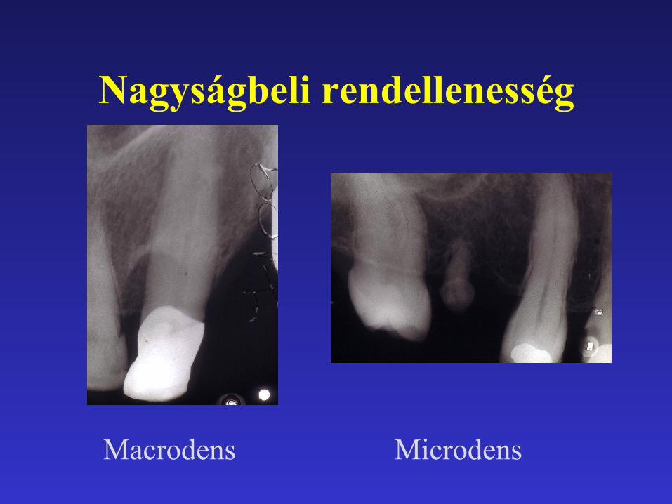
Macrodens Microdens
Nagyságbeli rendellenesség
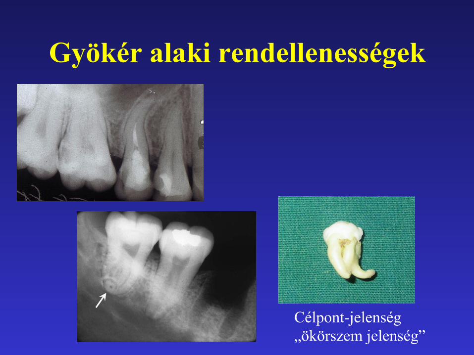
Gyökér alaki rendellenességek
Célpont-jelenség „ökörszem jelenség”
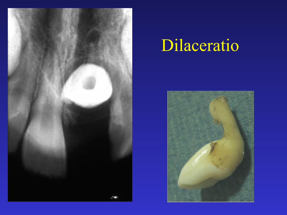
Dilaceratio
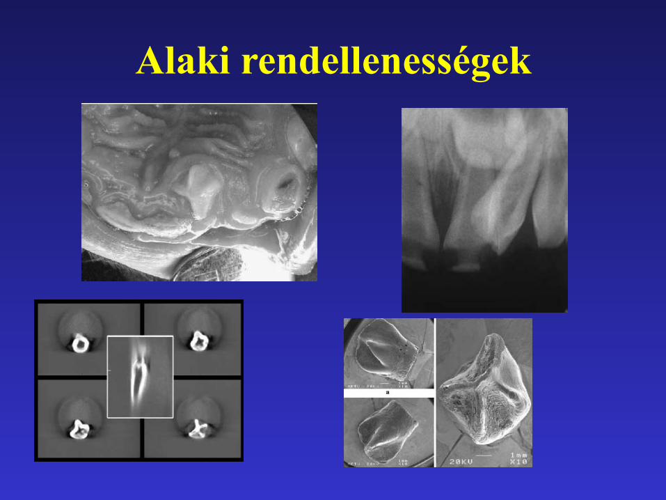
Alaki rendellenességek
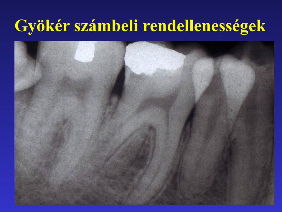
Gyökér számbeli rendellenességek
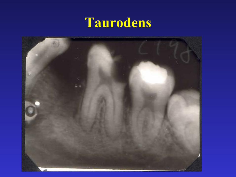
Taurodens
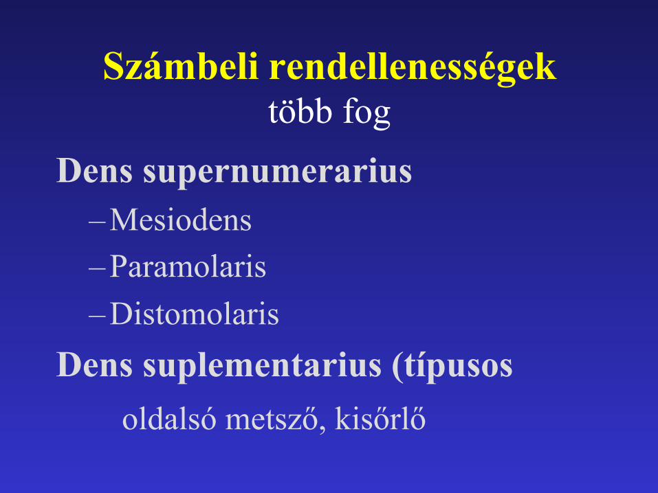
Számbeli rendellenességek több fog
Dens supernumerarius – Mesiodens – Paramolaris – Distomolaris
Dens suplementarius (típusos oldalsó metsző, kisőrlő
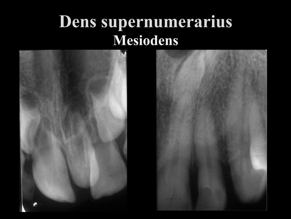
Dens supernumerarius Mesiodens
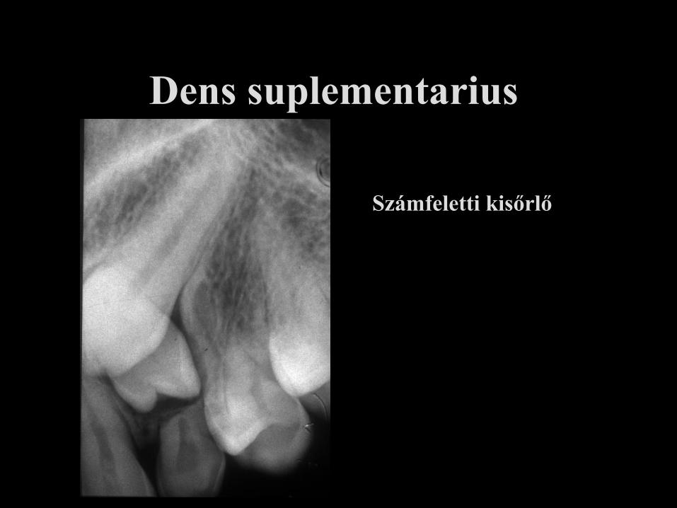
Dens suplementarius
Számfeletti kisőrlő
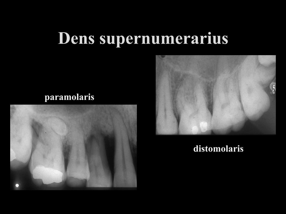
Dens supernumerarius
paramolaris
distomolaris
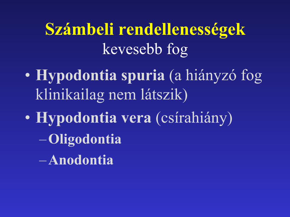
Számbeli rendellenességek kevesebb fog
• Hypodontia spuria (a hiányzó fog klinikailag nem látszik)
• Hypodontia vera (csírahiány) – Oligodontia – Anodontia
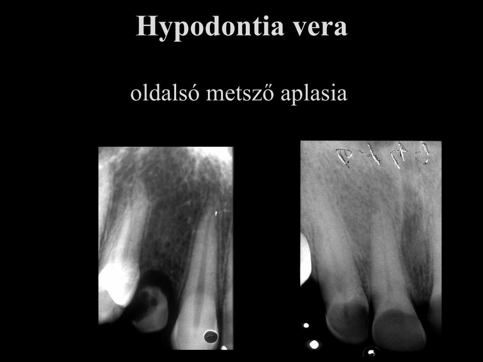
Hypodontia vera
oldalsó metsző aplasia
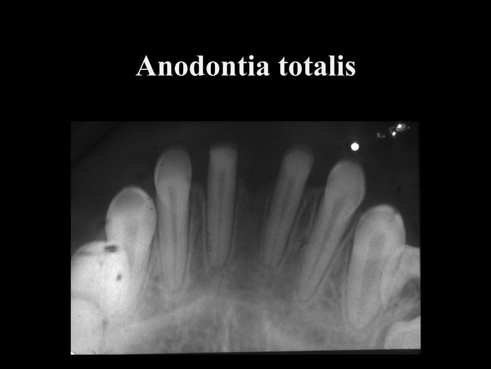
Anodontia totalis
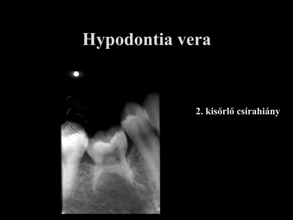
Hypodontia vera
2. kisőrlő csírahiány
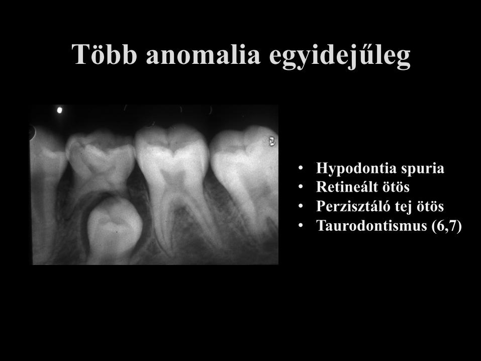
Több anomalia egyidejűleg
• Hypodontia spuria • Retineált ötös • Perzisztáló tej ötös • Taurodontismus (6,7)
Retentio rtg.vizsgálat szerepe
• Retentio ténye, foka • Retineált fogak száma, helyzete • Retentio oka, következménye
• Műtéti terv – Lokalizálás – Anatómiai képletek védelme
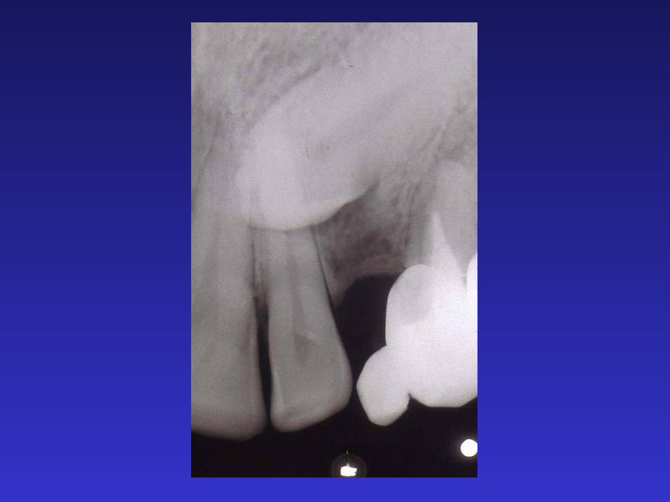
Helyzeti rendellenességek
• Distalisatio • Rotatio, torsio • Intra-, extraversio • Supra-, infraocclusio • Transpositio • Heterotopia
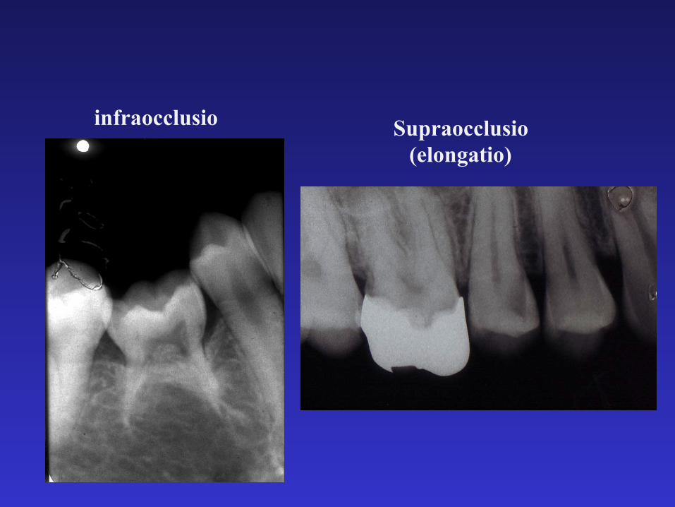
infraocclusio Supraocclusio (elongatio)
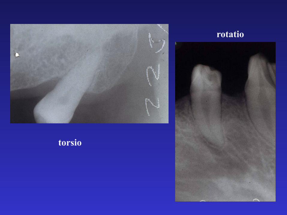
rotatio
torsio
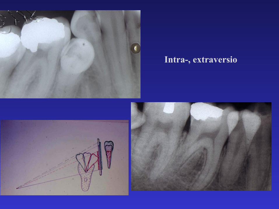
Intra-, extraversio
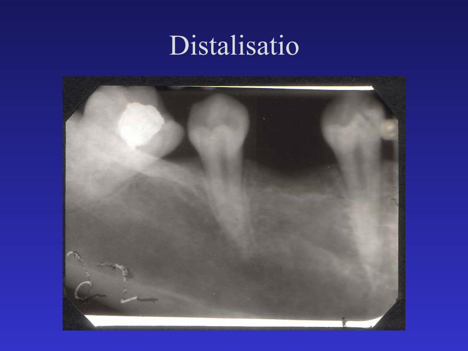
Distalisatio
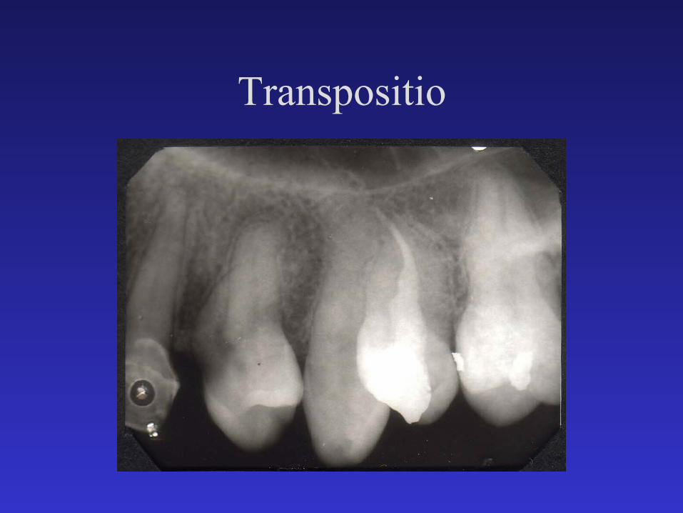
Transpositio
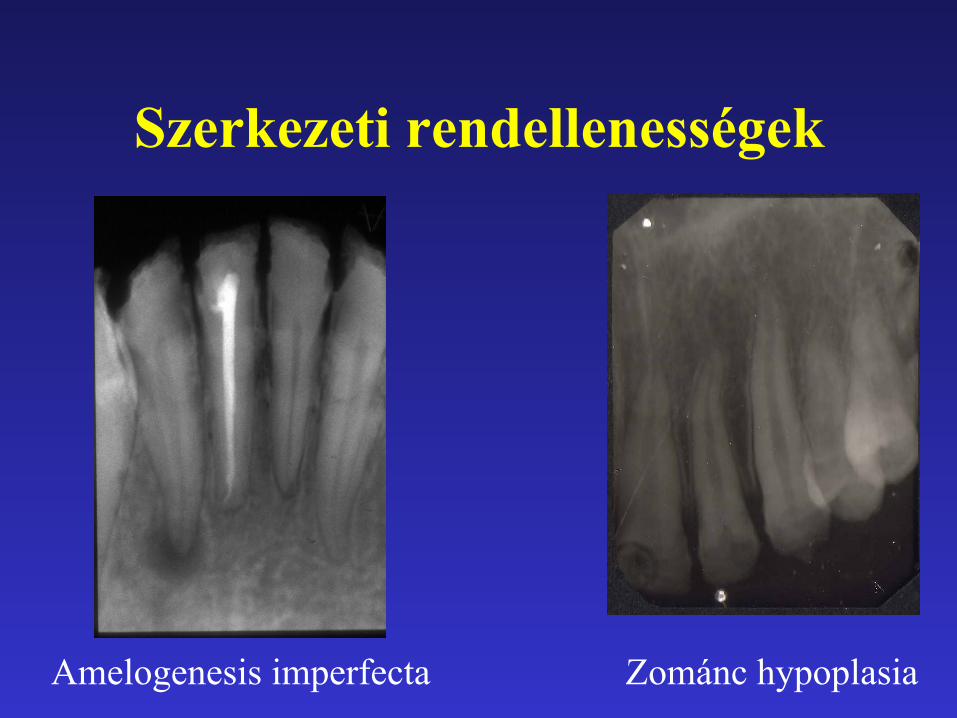
Amelogenesis imperfecta Zománc hypoplasia
Szerkezeti rendellenességek
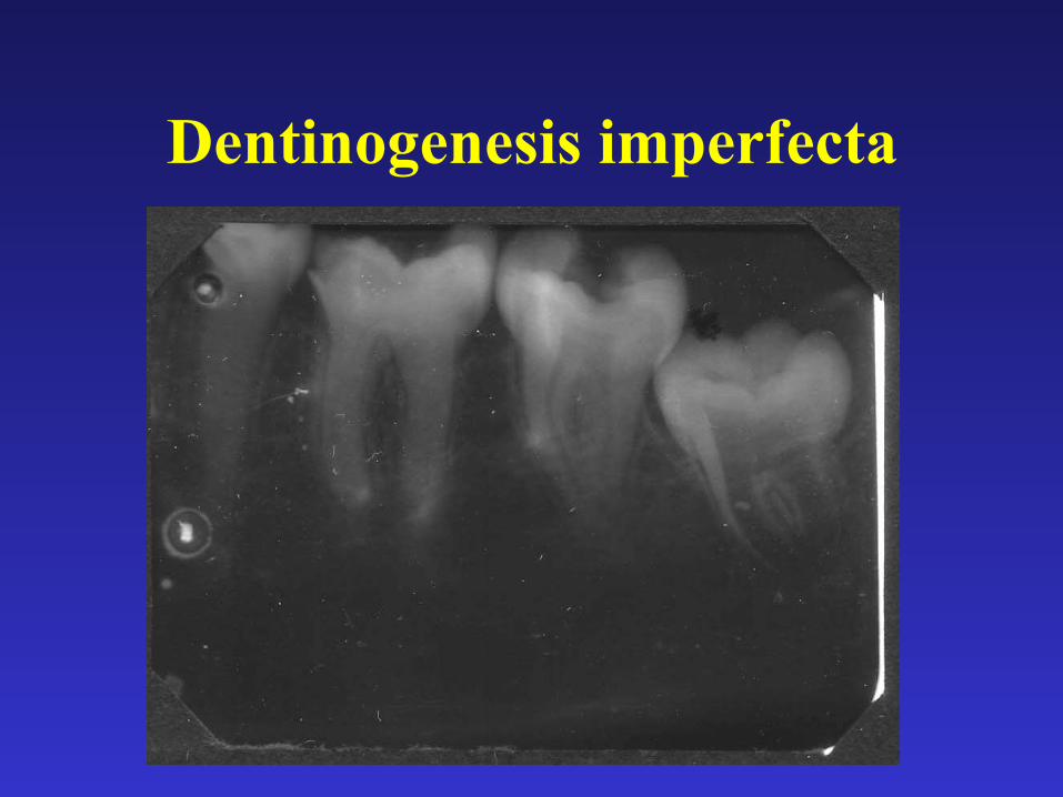
Dentinogenesis imperfecta
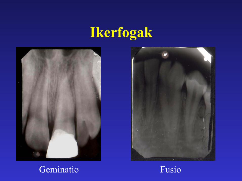
Ikerfogak
Geminatio Fusio