Embed Size (px)
Citation preview

UNIVERSITI SAINS MALAYSIA PENYELIDlKAN & PEMBANGUNAN • RESEARCH & DEVELOPMENT
11800. PULAU PINANG • MALAYSIA
Rujukan Kami: FPP 98/1 18
Tarikh: 28 DISEMBER 200 I
Dr. Ahmad Sukari Halim Pusat Pengajian Sains Perubatan Kampus Kesihatan USM I 5990 Kubang Kerian KELANT AN DARUl NAIM
r:~~~:,: ' /'
"i
l j .. ~ .~~ 2001 f
Tuan, I : J",f ',ito ,l:,I, .. :",W,fl f laporan Akhir Projek Penyelidikan Geran USM Jangka-r-endek :"'".---------",' "A Pilot Study of AnkLe InstabiLity Following Long Segment FibuLar Graft Harvesting" .
Saya ingin merujuk surat tuan bertarikh 10 Oktober 200 I berhubung tajuk projek tersebut di atas. T erlebih dahulu suka saya ucapkan terima kasih di atas satu salinan laporan akhir untuk projek penyelidikan USM jangka pendek "A Pilot Study of AnkLe Instability Following L~ng Segment FibuLar Graft Harvesting"
SeLerusnya . walaupun projek ini telah selesai, Jabatan Bendahari telah dinasihatkan untuk menangguhkan penutupan akaun projek kepada 31 JANUARI 2001. T empoh ini diberi untuk membolehkan penjelasan semua urusan tuntutan dan bayaran yang telah dikomitkan di dalam tempoh projek.
Walau bagaimanapun, tuan dinasihatkan supaya tidak mengeluarkan borang-borang pesanan baru di dalam tempoh
Sekian, terima kasih.
njalankan tugas,

Universiti Sains lVIalaysia ill ltm! ill !
Pusat Pengajian Sains Perubatan School of Medical Sciences
Ruj. Kami U SMIPP SP( Sc.P)/01(34)
10 Oktober 2001
PUGn Mazula Sabudin . Pem. Ketua Penolong Pendaftar Bah. Penyelidikan & Pembangunan Universiti Sains Malaysia 11800 Minden Pulau Pi nang
Puan
Per: Laporen Akhir Penyelidikan USM J angka Pendek "A Pilot Study of Ankle Instability Following Long Segment Fibular Graft HarVesting"
Merujuk kepada surat puan bertarikh 3 Oktober 2001, rujukan fail FPP 2000/108, soya telah mengemukakon loporan tersebut kepada Dekan, PPSP seperti yang dipinta.
Bersama-sama ini soya sertakan salinan iaperan tersebut.
Sekian, terima kasih.
"BERKHIDMAT UNTUK NEGARA" "CINTAILA ASA KITA"
s.k 1.
2.
Prof. Madya Zabidi Azhar Mohd. Hussin Dekan, PPSP En. Halim Othman, Bah. Penyefidikan, PPSP

Semua laporan kemajuan dan laporan akhir yang dikemukakan kepada Bahagian Penyelidikan dan Pembangunan perlu terlebih dahulu disampaikan untuk penelitian dan perakuan Iawatankuasa Penyelidikan di Pusat Pengajian.
USMJP-06
1)
2)
3)
BAHAGIAN PENYELIDlKAN UNIVERSITI SAINS MALAYSIA
Laporan Akhir Projek Penyelidikan Jangka Pendek
Nama Penyelidik: _____ ~A~HMA~D~S~U~K~AR~I=_=B~._H~A;.!.;:L:..:;:I;;!..:M~ ______ _
Nama Penyelidik-Penyelidik Lain: (Jika berkaitan) DR. ZULMI WAN
DR. MAHAYIDDIN MUHAMAD
DR. MOHD YUSOF IBRAHIM
Pusat P~ngajianJPusat/Unit: __ ---:!PwU:!!=S~A..;.T~P....:E:!!o.lN~G~AJ~I=.!.A~N~SA~I=.!.N!.!OS~PE~R~U'_=B~A"""TAu..lN.!././ __ _
UNIT SAINS REKONSTRUKTIF
Tajuk Projek: ______ AU-.lP....,jI'-.IoL~O""'T.....JSol...Tu.I.u.JD ....... Y_O"""F"___'_A ... NwK .... L:...E_I .... N ..... S .... T .... A ...... B..,IL ..... I ..... T .... y.-...FO>o<.I .... ,L""'O""'W.,.I ...... NG""'-.
LONG SEGMENT FIBULAR GRAFT HARVESTING

(b) Senaraikan Kata Kunci yang digunakan di dalam abstrak:
Bahasa Malaysia Bahasa Inggeris
KESTABILAN SENDI ANKLE INSTABILITY
KAWASAN PEND ERMA DONOR SITE
MORBIDITI SENDI :-:'- . ANKLE MORBIDITY
PERGELANGAN KAKI
GRAF FIBULA FIBULAR GRAFT
5. Output Dan Faedah Projek (a) Penerbitan (termasuk Zaporan/kertas seminar)
(Sila nyatakan jenis, tajuk, pengarang, tahun terbitan dan di mana telah diterbitkanldibentangkan)
1) PEMBENTANGAN KERTAS DAN ABSTRAK
" CLINICAL AND RADIOGRAPHIC ASSESSMENT OF ANKLE MORBIDITY
FOLLOWING A LONG SEGMENT FIB'ULAR RESECTION "
2) PEMBENTANGAN KERTAS DAN ABSTRAK
" CLINICAL AND RADIOGRAPHIC EVALUATION OF ANKLE TNSTABILITY
FOLLOWING LONG SEGMENT FIBULA HARVESTING "
3) PEMBENTANGAN KERTAS SERTA PROCEEDING
" CRITICAL CLINICO-RADIOGRAPHICAL ASSESSMENT OF ANKLE AFTER
FREE FIBULA TRANFER "
( RUJUK LAMPIRAN - 1,2 DAN 3 )

6. Peralatan Yang Telah Dibeli:
1) PERALATAN PAKAI HABIS
- DISKET & PERMANENT MARKERS
- CANON BC 20 CARTRIDGE
- BATERI
- CD
2) ASET - TIADA
3) PERKHIDMATAN
- CT SCAN & X- RAY
- HONORARIUM PESAKIT
- FAX, COURIER
- PROCEEDING KONFEREN
UNTUK KEGUNAAN JA WATANKUASA PENYELIDIKAN UNlVERSITI
TANDATANGAN PENGERUSI JAWATANKUASA PENYELIDIKAN PUSAT PENGAJIAN fn:borangladlinaimclnak

ABSTRAK
KAJ1AN P1LOT KETAKSTABILAN SENDI PERGELANGAN KAKI SElEPAS
PENGAMBILAN GRAF SEGMEN PANJANG TUlANG FIBULA
Graffibula adalah satu teknik; untuk mengembalikan keutuhan tulang rangka
akibat kecacatan tulang. Walaupun teknik ini amat berguna, namun banyak
fapuran berkaitan dengan tSU pengambilan tulang fibula ini terufamanya
berkaitan ketidakstabilan sendi pergelangan kaki.
Ini adaIah kajian kohort kemas pesakit-pesakit yang tatah menjalani
pengambilan graf segmen panjang tulang fibula sekurang-kurangnya 15 sm ..
Seramai sepuJuh pesakit berumur 12 hlnggaB4tahun tertibat daJam kajian inL
Sepufuh pasang kaki telah diperiksa dengan kaki sebelah berlawanan
(kontralateral) yang normal sebagai kontrol (20 kakisemuanya). Kajian objektif
berkaitan morbiditi sendi pergelangan kaki adalah berdasarkan sistem skore
kaki Maryland (Maryland Foot Score). Kajian radiofogi termasukaJah
pengambilan X-ray secara pandangan sidesmosis sendi pergelangan kaki
untuk kedua-dua keadaan, semasa berdiri dan semasa berbarlng. Imej CT scan
secara melintang di kedudukkan 9 mm diatas plafond tulang tibia drkaji dan
pengukuran jarak sindesmosis depan dan belakang dijaJankan. Analisis keatas
simptoms-simptom subjektif menghasilkan markah diantara 78 hingga 99%
berdasarkan analisrs menggunakan system permarkahan kaki Maryland
(Maryland Foot score scoring system). Perubahan osteoporosis keatas baki
tu/angfibula distal sebelah kaki kawasan penderma terjadi sebanyak ·89 %
daripada keseluruhan subjek kajian.

ABSTRACT
A PILOT STUDY OF ANKLE INSTABILITY FOLLOWING LONG SEGMENT FIBULAR GRAFT HARVESTING
Fibufar graft is a useful technique to restore skeletal integrity of bony defects.
Despite the benefits of this procedure, there are reported problems associated
with donor site, particularly with regards to ankle stability.
A cohort study was performed on patients who had undergone tong segment
fibular graft resection of minimum 15 cm in length. A total of ten patients ranging
from 1.2 to 64 years old were included in the study .The assessment performed
after a minimum of 4 months following the operation. Ten pairs of legs were
evaluated with contralateral normal legs as control (a total of ten legs). The
objective assessment of ankle morbidity was done based on Maryland Foot
scoring system. Radiological assessments included pfain radiograph in
syndesmotic views, on both non-weight bearing supine position and weight
bearing standing position. The axial CT scan slice done at the level of 9 mm
above the tibial plafond was assessed to measure the anterior and posterior
syndesmotic interval. Subjective assessments revealed score ranging from 78
to 99% according to Maryland Foot scoring system. OsteoporosiS of distal fibula
of the donors' side was present in 89 %.
Despite the significant radiological changes of the residual distal fibula and the
ankle, the subjective assessment of the ankle morbidity yielded good or
excellent results. The residual distal fibula of 5.S cm or less was associated with

No. 5.(a) Output dan Feadah Projek (Rujuk Lampiran -1, 2 dan 3)
" Clinkal :and Radiographi~ Assessm'ents of Ankle Morbidity Following a Long Segment Fibular Resection"
M. Yusof, A.S Halim, ~I. Mabayuddin & ZuImi Wan
1st Asean Conference on Medical Sciences 18 - 21 May 2001 Ballroom, Renaissance Kota Bharu Hotel
" Clinical and Radiographic Evaluation of Ankle Instability Following Long Segment Fibula Harvesting"
AS Halirn,M Yusof, M j\tIabayuddin , ZuImi "Van
Association & College 'Of Surgeons, Academy of ~Iedicine of Malaysia 24 - 27 l\lay 2001 Santubong }(uchiug Resort, Sarawak.
," Critical Clinico-radiographicaJ Assessment of Ankle after Free Fibula Transfer"
NaUm AS, Ibrahim l\1Y, Mohamad M, Zulmi Wan
Inaugural Congress the \Vorld Society for Reconstructive Microsurgery October 29 - November 3, .2001 Taipei, Taiwan.

1st ASEAN Conference on Medical Sciences .18 -21 May 2001 .Universiti Sains Malaysia
Abstract No. 0-26 THE VALUE OF CONTRAST MEDIUMIN CRANIAL COMPUTED TOMOGRAPHY IN PATIENTS WITHOUT FOCAL NEUROLOGICAL FEATURES (PRELIMINARY REPORT)
Win Mar & Mahayiddin Mohame~ Department of Radiology, School of Medical Sciences, Universiti Sains Malaysia, Kubang Kerian, Kelantan, Malaysia.
The value of contrast enhancement during cranial computed tomography (CT) is well known. Contrast enhancement has been regarded as unhelpful in patients in whom the uncontrasted scan is entirely normal. It is only helpful in limited numbers of patients with symptoms and signs suggesting focal intracranial pathology. This study was done to assess the value of contrast medium administration in patients presenting with generalised features without focal neuorological signs. The study was done both retrospectively and prospectively. The patients with above features who had boh non-contrasted CT scan (NCeT) and contrastenhanced CT scan (CECT) were recruited. Only the NCCTs were shown to the radiologist and phase II radiology residents. The target sample is 212 cases and abcut 100 cases had been analysed. There were five abnormal cases out of 100. Intravenous contrast enhancement only contributes to the diagnosis if suspicious abnormality is seen on NCCT (5%). In remaining patients (95%) there is no diagnostic contribution. Both sensitivity and specificity for the radiologist was 100% and 96.8% respectively, and for the medical officers were 100% and 93.7-97.9%. Intravenous contrast enhancement is unlikely to be of value in those patients without focal neurological features and who have a norma! uncontrasted scan. However, a reduction in the use of contrast medium in patients with focal treatabl!l lesions being missed and therefore it still has an important but limited role.
Abstract No. 0-28 CLINICAL AND RADIOGRAPHIC ASSESSMENTS OF ANKLE MORBIDITY FOLLOWING A LONG SEGMENT FIBULAR
/' RESECTION
M. YusoL AS. Haliin,' M. Mahayuddin & Zulmi Wan Hospital Universiti Sains Malaysia, Kubang Kerian, Kelantan, ~alaysia
Fibula graft is a useful technique to restore skeletal integrity of bOny defectS. D~spite the benefits of this procedure, there are problems associated with donor site, particularly with regards to ankle stability.
Objective: To determine the clinical signs and symptoms, as well as radiographic fmdings and their correlation with ankle stability.
A cohort study was carried out on patients who had undergone long segment fibula graft harvesting. A total of nine .patients were included in the study. Nine pairs oflegs were evaluated with contralateral normal legs as control. Evaluation was based on interviews and physical examination. Radiological assessments included plain radiograph and CT scan.
Subjective ~essments were based on Maryland foot score which revealed score ranging from 78 to 99%. The range of ankle motion was decreased on loaded dorsiflexion in 2 patients but range in loaded plantar flexion were all normal. Based on the X-rays fmdings, the distal residual length of fibula were ranged from 5.0 em to 11:0 em. The abnormalities detected on plain radiograph were proximal migration of the fibula and medial tilt of the distal residual fibula proximal to the ankle (tilting angle offibula). The CT scan study was for assessment of distal tibio-fibula syndesmosis for evidence of widening and rotation of the fibula at the syndesmotic level. The correlation between the Maryland foot score and radiological fmdings with the residual length of distal fibula was assessed.
There is minimal ankle morbidity following a long segment fibula graft harvested despite the radiological findings of increased tilting angle of fibula and proximal migration of the fibula.
Abstract No. 0-27 ENHANCING FAT SATURATION TECHNIQUE IN MRI
WA Kamil A Sabri & Khalid Osman Department of Radiology, School of Medical Sciences, Universiti Sains Malaysia, Kubang Kerian, Kelantan, Malaysia.
This p~esentation will highlight the rinciples of fat saturation method, some new adjustinent parameters that had been determined and the resulti;g enchanced images obtained using MRI facilities at Universiti Sains Ma~sia Hospital.
The technique of at saturation in MRI examination IS an excellent choice to exclude fatty tissues from the vicinity of suspected soft tumor. Routine exmaination uses a set of pre-determined scan parameters available on the instrument console, assuming that the magnetic field homogeneity for large field-of-view (FOV) is shimmed properly. In practical case where the external magnetic field at the firnge ofFOV is not quite homogeneous, adjustments have been made and tailored to the new field situation due to the presence of a subject undergoing MRI examinttions.
Two sets of MRI images were obtained by selecting firstly, the automatic fat suppression format and secondly, using the manual method. In the manual !p.ode, several values ofRF pulse were within an acceptable range"Comparison was made between the two sets of results.
. Images obtamed by inserting manually the value of an RF pulse to suppress the fat were found to be significantly better than the images acqurred automatically. Fine-tuning the RF pulse value is necessary because of small disturbance in the magnetic field homogeneity due to the presence of a patient in the MRI chamber.

• .......---~_-L_._.'''''.-'''''."._'''''._'''''_'''''_:::!'~==''''' .• '''''~-~~'''''.""""'''''''''-'-'---'---~-----!'-~----------'---'~.!.-'--'\ FP 3.8 !
CLINICAL AND RADIOGRAPHIC EVALUATION OF ANKLE INSTABILITY FOLLOWING LONG SEGMENT FIBUIA HARVESTING AS flAUM, M YUSOF, M MAHYUDDIN, ZULMI WAN Department of Orthopaedic Surgery, Universiti Sains Malaysia, Kubang Kenan, Kelantan
Introduction: Fibula graft is a useful technique to restore skeletal integrity of bony defects. Despite the benefits of this procedure, there are problem associated with donor site particularly with regard to ankle. instab iIi ty.
Objective : To determine the clinical signs and symptoms as well as radiographic findings and their correlation with the ankle instability
Methodology : A cohort study on patient whom had undergone long segment fibula graft harvesting. Evaluation was based on interview and physical examination, radiological assessment include plain radiograph andCf scan .'
Results : A total of nine patients were iitcluded in the study. ·Nine pairs of legs were evaluated with contralateral normal legs as controL Subjective assessment were based on Maryland Foot score which revealed of score ranging from 78 to 99%. The range of anklemotion'was decreased on loaded dorsiflexion in 2 patients but loaded plantar fleXion were all normaL Based on the X-rays findings, the distal residual length of fibula were ranges from 5.0cm to II-Oem. The abnormality detected on plain radiograph are proximal migration of the fibula and medially tilt of the -distal residual fibula proximal to the ankle (tilting angle of fibula). The cr scan study were foi: assessment of distal tibia-fibula syndesmotic for evidence of widening and rotation of the fibula at the syndesmotic leveL 'The correlation between the Maryland foot score and radiological findings'with the residual length of distal fibula is assessed.
Conclusion: There is' minimal ankle morbidity following a long segment fibula graft harvested despite the radiological findings such as increased tilting angle of,fibula and proximal migration of the fibula.
I I i I I i i-
I II
I
I
t~h<;;:-l',~r4.l li'n,rr.' ...... v .. ~l! D"".,fp ... 1J"i.-'" ........ - -. -""~. ....~ I .J. \,;.;, ........ .:. Videu ',theck file' categol.J of Tuur paper:
I 0 1 • J TT..1 , XI ' I 11K '11 r' • n . J /11 r . . nCOL0gtCfu .neaa. ana. l"'i eCi£ lv.l.a...XlliO-\~f'Cli1iO-L'aC1al/ lvlicfosurgerj I Upper E:lI.i::refi1ity
! Low~ Extremity I Nerve ! Flaps ! Aesthetic Surgery/.Nficrosurgery ! Breast
! Paediatrics 11 .. ficros1.u:~"Crv ! Ali ftT ' . . .n..tlograr rrulspLantatlOll
! Replantation ! Tissue En~-inecring and Prefabricated flap
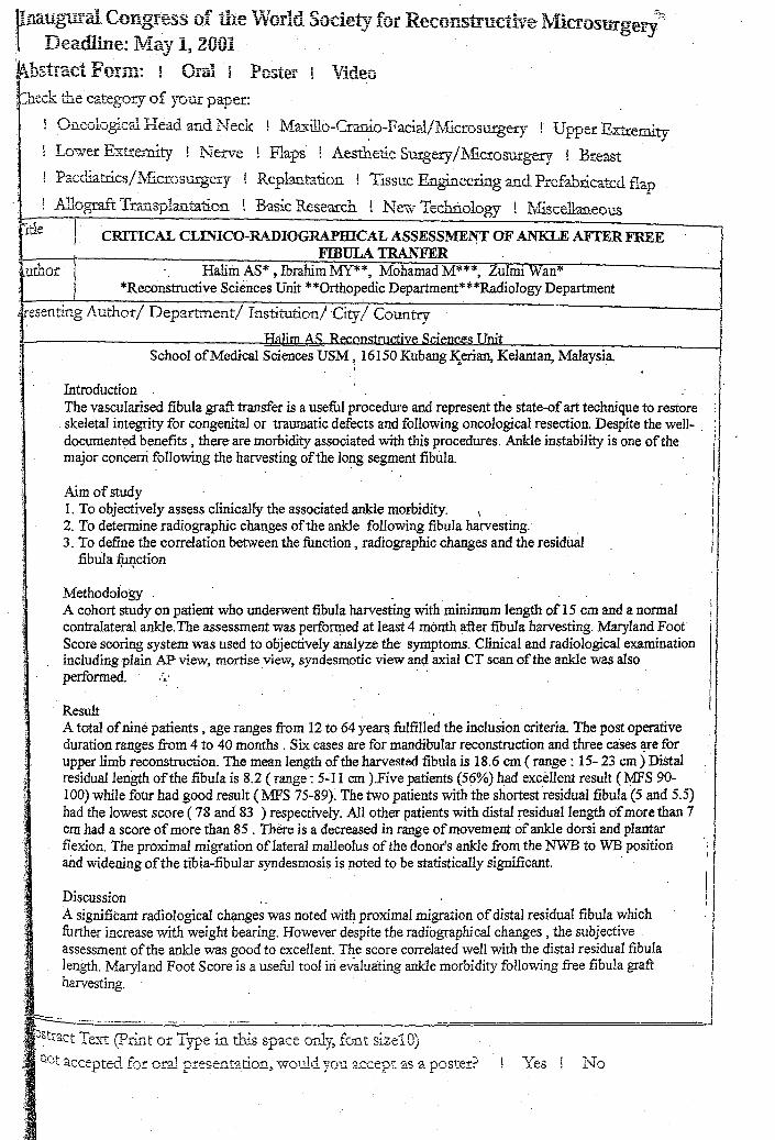
! Basic Reseru:ch ! New Technology ! ~Y1iscellaueous I itle I CRITICAL CLINICO-RADIOGRAPIDCAL ASSESSMENT OF ANKLE AFTER FREE
FIBULA TRANFER ~uthor Halim AS* , Ibrahim MY**, Mohamad M***, Zulmi Wan* ) *Reconstructive Sciences Unit **Orthopedic Department***Radiology Department
iresenting Author/ Department/ Institution/City/ Country----------1
H~lim A~ Rf'!r.nnstructive Sciences Unit School of Medical Sciences USM ~ 16150 Kubang I\erian, Kelantan, Malaysia
I
Introduction The vascularised fibula graft transfer is a useful procedure and represent the state-of art technique to restore skeletal integrity for congenital or traumatic defects and following oncological resection. Despite the well-. ! I documented benefits, there are morbidity associated with this procedures. Ankle instability is one of the ! I major concern following the harvesting of the long segment fibula. /1
~~~y I 1. To objectively assess clinically the associated ankle morbidity. \ I
2. To determine radiographic changes of the ankle following fibula harvesting. Ii
3. To define the correlation between the function, radiographic changes and the residual fibula function . ,
Methodology A cohort study on patient who underwent fibula harvesting with minimum length of 15 cm and a normal
, I contralateral ankle. The assessment was performed at least 4 month after fibula harvesting. Maryland Foot I Score scoring system was used to objectively analyze the symptoms_ Clinical and radiological examination including plain AP-view, mortise view, syndesmotic view and axial CT scan of the ankle was also performed. '.
Result A total of nine patients, age ranges from 12 to 64 yearS fulfilled the inclusion criteria. The post operative duration ranges from 4 to 40 months. Six cases are for mandibular reconstruction and three cases !!Ie for upper limb reconstruction. The mean length of the harvestt'.d fibula is 18.6 em (range: 15- 23 em) Distal residuallen~h of the fibula is 8.2 (range: 5-1 I cm ).Five patients (56%) ~ad excellent result (:MFS 90-100) while four had good result (1fFS 75-89). The two patients with the shortest 'residual fibula (5 and 5.5) had the lowest score (78 and 83 ) respectively. All other patients with distal residual length of more than 7 cm had a score of more than 85 . There is a decreased in range of movement of ankle dorsi and plantar flexion. The proximal migration of lateral malleolus of the donor's ankle from the NWB to WB position and widening of the tibia-fibular syndesmosis is poted to be statistically significant. .
Discussion .. A significant radiological chf,mges was noted with proximal migration of distal residual fibula which further increase with weight bearing. However despite the radiographical changes, the subjective assessment of the ankle was good to excellent. The score correlated well with the distal residual fibula length. Maryland Foot Score is a useful tool in evitluating ankle morbidity following free fibula graft harvesting. .
·1
I ; ~--_ .. ~~.~ .. ~-~.-~-===--~.~.~.~-~-~~~.-~--~ __ ~ __ ~ ______________ ~._~======================-======~i ; • \('11- .... ,..,,.... ~ (p. 'T:! • ~i_~ 1...· rl\
',: .<.J~~l.a.'-t ..te1..rt .1 tmt Of' .1. ype 111 trrts sp-ace Ofl...y, !G11t size10) .Q~'~ " -l t=: 1 • • -l ~
i \,.JL a.cceptet.L .Lor orat presentation, \tTOll.h .. :.. you accept as a poster!"' f Yes! N, Tr. .!. V

distance of the tip of lateral malleolus in relation to the tip of medial malleolus was assessed. The tilting angle of the residual distal fibular was measured. The axial CT scan slice done at a level of 9 mm above the tibial plafond was assessed to measure the anterior and postellor syndesmotic interval. The average of these two intervals was used as the syndesmotic interval of the ankle , whereas the difference was used to determine the rotation of the fibula. A comparison was made with the contralateral nonnal ankle. Statistica1analysis using student-t test was performed to assess the results of the donor's ankle as compared to the normal ankle.
Results
Subjective assessments revealed a score ranging from 78 to 99% according to the Maryland Foot Scoring System. The average plantar flexion of the donors' ankles was 46.33° , compared to the normal ankles of 48.11°. The average range of loaded dorsiflexion of the donors~ankles was 27.78° compared to 30.89j) of the normal ankles. Osteoporosis of distal fibula ofthe donors' side was present in 89%, In normal ankles, the tip of lateral malleolus was distal than the tip of medial malleolus with an average of 922mm during non-weight bearing, increasing to 1 L33m:m with weight bearing. In donors' ankles, the tip of the lateral malleolus was also distal than the tip of medial malleolus but with an average of 7.33mm during non-weight bearing and further decreasing to 6.56mm on weight bearing. The mean tilting angle of distal fi.buI:a of normal legs on syndesmotic view of plain radiograph was 90.33° with nonweight bearing increasing to 91.67° with weight bearing. However, the mean tilting angle for distal fibula of donors' legs on syndesmotic view of plain radiograph was 90.llowith non-weight bearing but decreased ftrrther to 88.22° with weight bearing. Both normal and donors' ankles had posterior syndesmotic interval greater than anterior interval. The average width of tibiofibular interval at 9mm above the tibial plafond of nonnal ankles was 2.939mm, whereas the donors' ankle was 3.50Omm. The average difference of anterior and posterior interval in the normal ankles was L 700mm and 2.022 mm for the donors·' ankles.
Conclusion
There were significant radiological changes of the ankle following a long segment fibular graft resection. The proximal migration of the residual distal fibula occurred and f'urther ,during weight bearing. The tilting angle of fibula decreused whilc weight bearing. The syndesmotic interval also widened due to lateral displacement of the :fibula following a long segment fibular graft resection but no rotation of fibula was noted.
Despite the above-mentioned radiological changes, the scoring marks for the subjective assessment of the ankle morbidity were either good or excellent with the minimum residual distal fibula of 5.0cm.However, leaving a minimum 7 cm length of the residual distal fibula is suggested to minimize the symptoms of ankle instability as itscorre1ates with the Mmyland Foot score above 85%.
References


![BAB II TINJAUAN PUSTAKA A. Anatomi Fisiologi Tulang Kakieprints.umm.ac.id/61987/3/BAB_II[1].pdf · a. Struktur Tulang Regio Ankle Bagian distal dari tulang tibia dan fibula berartikulasi](https://img.pdfslide.tips/doc/110x75/6074b98c00be752dfa1c0ce9/bab-ii-tinjauan-pustaka-a-anatomi-fisiologi-tulang-1pdf-a-struktur-tulang.jpg)