Embed Size (px)
Citation preview
INSTITUTO OSWALDO CRUZ
Pós-Graduação em Biologia Celular e Molecular
VÍTOR ENNES VIDAL
Identificação de homólogos das calpaínas em
Trypanosoma cruzi e avaliação do efeito do MDL28170, um
inibidor de calpaínas, sobre o parasito
Dissertação apresentada ao Instituto Oswaldo Cruz
como parte dos requisitos para obtenção do título
de Mestre
Orientadora: Prof. Dra. Claudia Mansini d‟Avila Levy
RIO DE JANEIRO
2010
ii
INSTITUTO OSWALDO CRUZ
Pós-Graduação em Biologia Celular e Molecular
VÍTOR ENNES VIDAL
Identificação de homólogos das calpaínas em
Trypanosoma cruzi e avaliação do efeito do MDL28170, um
inibidor de calpaínas, sobre o parasito
ORIENTADOR: Prof. Dra. Claudia Masini d’Avila Levy
Aprovada em: _____/_____/_____
EXAMINADORES:
Prof. Dra. Solange Lisboa de Castro – Presidente
Prof. Dra. Patricia Cuervo Escobar
Prof. Dr. Herbert Leonel de Matos Guedes
Prof. Dr. Eduardo Caio Torres dos Santos
Prof. Dra. Patricia Maria Lourenço Dutra
Rio de Janeiro, 15 de julho de 2010

iii
DEDICATÓRIA
Aos meus pais Fernando e Márcia e a
minha esposa Isabela por todo apoio,
amor e compreensão durante esses dois
maravilhosos anos.
iv
AGRADECIMENTOS
Primeiramente gostaria de agradecer a minha orientadora, Dra. Claudia Masini
d‟Avila Levy, por todo apoio e confiança que teve por mim; além de ótima orientadora, foi
também uma excelente amiga desde que entrei no laboratório.
À Dra. Constança Britto por ser a maior responsável pela minha vinda ao Laboratório
de Biologia Molecular e Doenças Endêmicas, que me acolheu muito bem e permitiu a
realização de meus objetivos profissionais e pessoais.
Ao Dr. Carlos Roberto Alves e ao Dr. Otacílio Moreira da Cruz pelas dicas e
conselhos nas prévias de entrevista de mestrado, seminário discente e defesa da tese.
Às tecnologistas do laboratório, Luiza e Angélica, por terem me ajudado muito a me
adaptar ao novo ambiente de trabalho e às novas metodologias empregadas no meu estudo.
Ao Dr. André Luís Souza dos Santos e à Dra. Marta Helena Branquinha pela
elaboração do projeto que abracei com grande dedicação e carinho, e pelas críticas e sugestões
nos artigos antes da submissão.
Ao Dr. Rubem Figueiredo Sadok Menna Barreto por toda a colaboração prestada
durante boa parte do meu trabalho, além de ser um amigo.
A todos os demais alunos e pesquisadores pelo companheirismo e apoio durante o
desenvolvimento deste trabalho, além da compreensão por terem que dividir o técnico do
laboratório com o mestrado.
À plataforma de citometria de fluxo que muitas vezes precisei usar para leitura dos
meus experimentos.
Aos meus colegas do Laboratório de Pesquisas em Malária que sempre me apoiaram,
em especial à Dra. Maria de Fátima Ferreira da Cruz que me abriu as portas da Fiocruz.
Aos meus amigos da época da faculdade, Rodrigo, Melissa e Larissa, por sempre
acreditarem que eu chegaria mais longe na minha vida acadêmica.
Ao meu irmão de sangue, Igor, e aos meus outros seis irmãos de coração (André,
Bruno, Fábio, Júlio, Léo, Vinicius) que sempre me apoiaram e compreenderam minha
ausência durante os períodos mais críticos de dedicação ao presente trabalho.

v
A todos os meus familiares e demais amigos pelo incentivo e carinho.
A todos não mencionados anteriormente que de alguma forma contribuíram para a
realização deste trabalho.
Por fim, ao nosso Mestre amado que nos permite viver, aprender e triunfar, sempre em
busca de sua melhor compreensão. Obrigado, Senhor!
vi
ÍNDICE
RESUMO viii
ABSTRACT ix
LISTA DE ABREIATURAS x
I. INTRODUÇÃO 1-25
1. A família Trypanosomatidae 1
2. A doença de Chagas
3. O Trypanosoma cruzi
3
7
4. O tratamento da doença de Chagas 11
5. Peptidases 12
6. Peptidases no Trypanosoma cruzi 14
7. Calpaínas 17
8. Calpaínas nos tripanossomatídeos 20
9. Inibidores proteolíticos 23
II. OBJETIVOS 27
III. RESULTADOS 28-63
Artigo 1: “Arrested growth of Trypanosoma cruzi by the calpain inhibitor
MDL28170 and detection of calpain homologues in epimastigote forms”
29
Artigo 2: “Effects of the calpain inhibitor MDL28170 on the clinically relevant
forms of Trypanosoma cruzi in vitro”
39
Artigo 3: “MDL28170, a potent calpain inhibitor, affects Trypanosoma cruzi
metacyclogenesis, ultrastructure and attachment to the luminal midgut surface of
Rhodnius prolixus”
44
IV. DISCUSSÃO 71-79
V. CONCLUSÕES 80
VI. BIBLIOGRAFIA 81-98
viii
RESUMO
CALPAÍNAS EM TRYPANOSOMA CRUZI: PARTICIPAÇÃO NO CICLO DE VIDA,
DIFERENCIAÇÃO E INFECTIVIDADE
Vítor Ennes Vidal
DISSERTAÇÃO DE MESTRADO
As calpaínas constituem uma família de cisteína-peptidases neutras dependentes de
cálcio presentes numa ampla variedade de organismos. Em virtude da relevância fisiológica
dessas proteases, inibidores de calpaínas já vêm sendo desenvolvidos para o tratamento de
doenças humanas e microbianas. Estudos recentes vêm relatando a presença de diversas
proteínas relacionadas às calpaínas em tripanossomatídeos, mas pouco se sabe a respeito das
funções específicas dessas proteínas nesses micro-organismos. Nesse contexto, uma vez que
os fármacos atualmente disponíveis para o tratamento da doença de Chagas apresentam sérios
efeitos colaterais e podem ser ineficazes, inibidores proteolíticos poderiam ser uma alternativa
no tratamento desta doença. Portanto, o presente trabalho investiga a presença de moléculas
similares às calpaínas no Trypanosoma cruzi e o efeito inibidor do inibidor III de calpaínas
(MDL28170) sobre a proliferação, viabilidade, diferenciação, ultraestrutura e interação das
diferentes formas do parasito com células hospedeiras em ensaios in vitro. Nossos resultados
revelam a reatividade cruzada de anticorpos produzidos contra calpaínas já bem
caracterizadas de Drosophila melanogaster, Homarus americanus e Trypanosoma brucei
contra moléculas de superfície do T. cruzi, conforme demonstrado por imunofluorescência e
citometria de fluxo. Em ensaios de Western Blotting foi possível observar que o anticorpo
anti-Dm-calpaína foi capaz de reagir contra uma proteína de 80 kDa. Pesquisas realizadas no
GenBank demonstraram a presença de 4 seqüências homólogas à calpaína de D. melanogaster
no genoma do T. cruzi. Essas 4 seqüências foram identificadas como cisteína-peptidases de
massa molecular predita em torno de 80 kDa. Nos ensaios de inibição com o MDL28170 foi
possível observar a redução da proliferação das formas epimastigotas ao longo de 5 dias de
cultura; e a redução significativa da viabilidade das formas tripomastigotas sanguíneas dos
parasitos tratados com 25 μM do composto. O inibidor, adicionado nas concentrações de 6,25
à 25 μM, também foi capaz de diminuir de forma dose- e tempo-dependente o número de
macrófagos parasitados e o número de parasitas interiorizados nos ensaios de interação com
macrófagos peritoneais murinos. O MDL28170 em concentrações a partir de 12,5 μM teve
ainda um efeito inibitório significativo sobre a adesão de formas epimastigotas ao epitélio
intestinal de Rhodnius prolixus; assim como os anticorpos anti-calpaínas foram capazes de
inibir significativamente a interação com o inseto vetor. Por fim, foi possível observar uma
redução significativa no processo de diferenciação por metaciclogênese em meio TAU e
alterações ultraestuturais em reservossomos, Golgi e membrana plasmática quando formas
epimastigotas do T. cruzi foram tratadas com MDL28170. Embora mais estudos sejam
necessários para melhor caracterizar moléculas similares às calpaínas no T. cruzi, o nosso
trabalho acrescenta novos conhecimentos sobre as possíveis funções dessas moléculas e sobre
a possibilidade de utilização de inibidores de calpaínas como uma alternativa promissora para
o desenvolvimento de compostos mais potentes e seletivos para o tratamento da doença de
Chagas.
ix
ABSTRACT
CALPAÍNAS EM TRYPANOSOMA CRUZI: PARTICIPAÇÃO NO CICLO DE VIDA,
DIFERENCIAÇÃO E INFECTIVIDADE
Vítor Ennes Vidal
DISSERTAÇÃO DE MESTRADO
Calpains constitute a large family of calcium-regulated cytosolic cysteine peptidases
present in a broad range of organisms. Since these enzymes may participate in a variety of
physiological processes, calpain inhibitors are under development for the treatment of human
and infectious diseases. Previous works have shown the presence of a large and diverse family
of calpains in trypanosomatids, but less is known about the specific functions of these
molecules. In this context, since the current chemotherapy to treat Chagas‟ disease presents
several side effects and can be ineffective, proteolytic inhibitors could be an alternative to the
treatment of this disease. In the present work, we aimed to detect calpain-like molecules in
Trypanosoma cruzi and to explore the effects of the calpain inhibitor III (MDL28170) on the
growth, viability, differentiation process, ultrastructure and interaction of the T. cruzi with
host cells in vitro. Our results show the cross-reactivity of molecules present on the cell
surface of T. cruzi epimastigotes and anti-calpain antibodies raised against well characterized
calpains from Drosophila melanogaster, Homarus americanus and Trypanosoma brucei.
Furthermore, an 80 kDa reactive molecule was detected by Western blotting assays using the
anti-Dm-calpain. Based on sequence analysis in GenBank, we identified 4 T. cruzi sequences
that share the same conserved domain with the fragment of the D. melanogaster calpain that
was employed to generate the anti-Dm-calpain antibody. All these 4 sequences were cysteine
peptidases of predicted molecular mass around 80 kDa. Our results on the growth pattern of
treated parasites showed that MDL28170 arrested the growth of epimastigote forms
maintained for 5 days in culture; and significantly decreased the bloodstream trypomastigotes
viability treated with 25 μM. MDL28170 at concentrations ranging from 6,25 to 25 μM was
also capable of reducing the infection of murine macrophages and the number of internalized
parasites in a dose- and time-dependent manner in the T. cruzi-macrophage interaction assay
in vitro. In addition, MDL28170 at concentrations from 12 M on was also capable of
reducing the number of bound T. cruzi epimastigotes on the luminal surface midgut of the
insect vector Rhodnius prolixus; anti-calpain antibodies also significantly impaired this
interaction. Finally, we observed a significant decrease in the metacyclogenesis process on
TAU medium and ultrastructural disorganizations on reservosomes, Golgi and plasma
membrane from T. cruzi epimastigote forms treated with MDL28170. Although more
investigations may be necessary for better understanding the presence of calpain-like
molecules in T. cruzi, our work add new insights into the possible functions of these
molecules and into the possibility of exploring calpain inhibitors as a promising alternative to
the development of more powerful an selective drugs to treat Chagas‟ disease.
x
LISTA DE ABREVIATURAS
μl- Microlitro
μM- Micromolar
BSA- Soro Albumina Bovina
Ca+2
- Íons cálcio
CDPs- “Calcium Dependent Peptidases” (Peptidases Dependentes de Cálcio)
DMEM- “Dubelco´s Modified Eagle Medium” (Meio Eagle Modificado por Dubelco)
DMSO- Dimetilsulfóxido
DNA- “Deoxyribonucleic acid” (Ácido Desoxirribonucléico)
EC- Comitê de Nomenclatura Enzimática
EDTA- Ácido Etilenodiaminotetracético
EGTA- Ácido Etileno Glicol Tetracético
FACS- “Fluorescence Activated Cell Sorting” (Separador de Células Ativado por
Fluorescência)
FITC- “Fluorescein Isothiocyanate” (Isotiocianato de Fluoresceína)
gp63- Glicoproteína 63
gp57/51- Cruzipaína
GPI- Glicosilfosfatidilinositol
h- Hora
HEPES- Etanosulfônico 4-2 Hidroxietil Piperazina-1
HIV- Vírus da Imunodeficiência Humana
IC50- Referente à concentração da droga que causa uma redução em 50% da sobrevivência
dos parasitos em comparação à curva de crescimento controle
IOC- Instituto Oswaldo Cruz
IgG- Imunoglobulina G
kDa- Kilodalton
k-DNA- DNA do cinetoplasto
M- Molar
MET- Microscopia Eletrônica de Transmissão
min- Minuto
MSP- “Major Surface Peptidase” (Principal Peptidase de Superfície)
MTT- Bromidrato de 3-(4,5-dimetiltiazol-2-il)-2,5-difeniltetrazolio
mM- Milemolar
NO- Óxido Nítrico
PBS- Tampão Fosfato Salina
pH- Potencial Hidrogeniônico
PKDL- Leishmaniose Dérmica Pós-Calazar
PSP- “Promastigote Surface Peptidase” (Peptidase de Superfície de Promastigotas)
RFLP- “Restriction Fragment Lenght Polymorfism” (Polimorfismo do Tamanho do
Fragmento de Restrição)

xi
RNA- Ácido ribonucleico
SDS- “Sodium Dodecyl Sulfate” (Lauril Sulfato de Sódio)
SFB- Soro Fetal Bovino
sp.- Espécie
spp.- Várias espécies
v/v- Volume por Volume
1
I. INTRODUÇÃO
1. A Família Trypanosomatidae
A família Trypanosomatidae pertence ao sub-reino Protozoa da ordem Kinetoplastida
que compreende um grande número de parasitos eucariotos monoflagelados. Os
microrganismos desta família compartilham características singulares, como a presença de
uma mitocôndria única e ramificada que percorre todo o corpo celular e cujo DNA possui um
arranjo único na natureza, formando o cinetoplasto. O DNA desta estrutura corresponde a 20-
30 % do DNA total, é denominado kDNA e organizado em redes de cadeias circulares,
concatenadas e compactas. Em muitos tripanossomatídeos, a posição relativa do cinetoplasto
em relação ao núcleo varia de acordo com a forma evolutiva do parasito que muda de acordo
com o ciclo de vida (MASLOV & SIMPSON, 1995). O glicossoma também é uma organela
singular dessa família e está relacionado com o peroxissoma de organismos eucariotos
superiores. Além de tornar a transformação de glucose em piruvato mais eficiente em
tripanossomatídeos que em outros organismos eucariotos, já que compartimentaliza a via
glicolítica (revisto por VICKERMANN, 1994), o glicossoma também possui outras funções
relacionadas à biossíntese de pirimidinas, recuperação de purinas, síntese de éter-lipídios e β-
oxidação de ácidos graxos (revisto por MICHELS et al., 2000).
Outro componente marcante dos tripanossomatídeos é o fato de apresentarem um
único flagelo locomotor, que pode estar livre ou próximo ao corpo celular (VICKERMAN &
PRESTON, 1976). Neste caso, com o movimento flagelar esta área da membrana é puxada
junto com o flagelo, dando a impressão da formação de uma membrana ondulante
(SCHMIDT & ROBERTS, 1989). Este flagelo é composto basicamente por um axonema e
um corpo paraxial, que emerge da bolsa flagelar e se encontra ancorado à célula pelos corpos
basais associados à mitocôndria (revisto por LANDFEAR & IGNATUSHCHENKO, 2001).
Em algumas espécies, o flagelo pode participar na adesão do microrganismo aos tecidos
hospedeiros (VICKERMAN, 1994; VICKERMAN & TETLEY, 1990).
Nesta família, o citoesqueleto é caracterizado por microtúbulos subpeliculares que
formam ligações cruzadas entre si e com a membrana plasmática garantindo a sustentação da
célula (revisto por GULL, 1999). O citoesqueleto representa barreira importante ao transporte
vesicular, logo o principal sítio de endocitose e exocitose de macromoléculas é a bolsa
flagelar, uma vez que é uma das poucas regiões da membrana que não possui microtúbulos
subpeliculares ligados (revisto por OVERATH et al., 1997). No entanto, existe uma estrutura
2
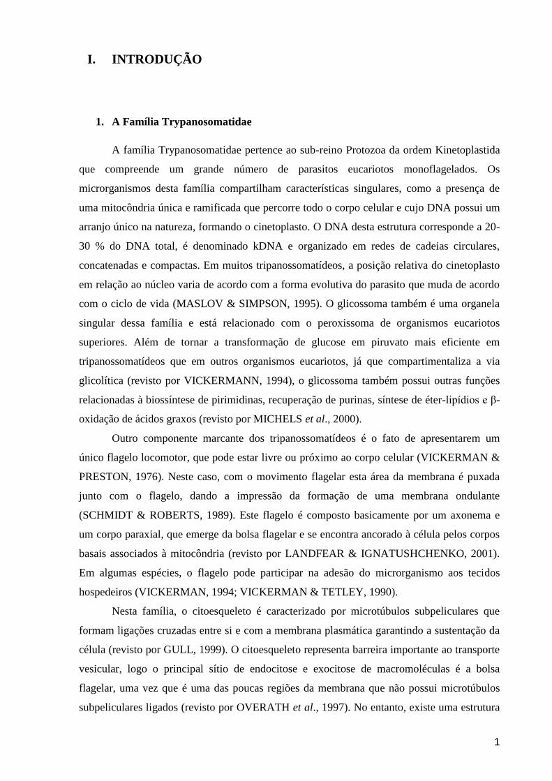
responsável por processos de endocitose/exocitose, o citóstomo. Esta estrutura se apresenta
como uma invaginação da membrana plasmática que pode atingir até a região do núcleo
celular (DE SOUZA, 2008). A arquitetura celular de um tripanossomatídeo pode ser
observada no esquema representativo da figura 1.
Figura 1: Representação esquemática da morfologia dos tripanossomatídeos
(adaptado de DOCAMPO et al., 2005).
Atualmente, a família Trypanosomatidae é dividida em nove gêneros de acordo com
as características morfológicas e a especificidade do hospedeiro: Blastocrithidia, Crithidia,
Endotrypanum, Herpetomonas, Leptomonas, Leishmania, Phytomonas, Rhynchoidomonas e
Trypanosoma. De acordo com o primeiro critério, a distinção dos gêneros é baseada nas
formas evolutivas observadas para cada gênero, que incluem características como a
morfologia do corpo celular, a presença ou ausência de “membrana ondulante” e de flagelo
extracelular aparente, local de emersão do flagelo e posição do complexo formado pelo
flagelo, bolsa flagelar, e cinetoplasto em relação ao núcleo (HOARE & WALLACE, 1966;
JANOVY et al., 1974; VICKERMAN & PRESTON, 1976; WALLACE, 1977; YOSHIDA et
al., 1978; VICKERMAN, 1994; TEIXEIRA et al., 1997). Quanto ao hospedeiro, os
tripanossomatídeos podem ser divididos em monoxênicos quando desenvolvem todo o seu
ciclo de vida em apenas um hospedeiro (geralmente um invertebrado), ou heteroxênicos
quando desenvolvem o seu ciclo de vida em hospedeiros diferentes (invertebrado e planta ou
invertebrado e vertebrado). Porém, alguns estudos sugerem a criação de três novos gêneros
3
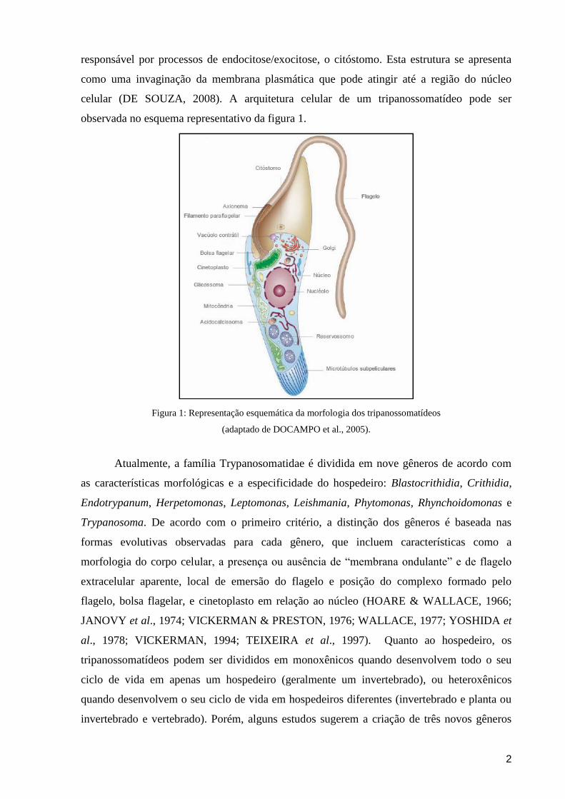
(Wallaceina, Strigomonas e Angomonas) para reagrupar os tripanossomatídeos de acordo com
a sua filogenia (PODLIPAEV & ROKITSKAYA, 1999; BRANDÃO et al., 2000; D‟AVILA-
LEVY et al., 2004), reforçando a necessidade de uma revisão taxonômica na família
Trypanosomatidae (Figura 2).
Figura 2: Relação espécie-hospedeiro dos tripanossomatídeos. Tripanossomatídeos heteroxênicos alternam entre
hospedeiros invertebrados e vertebrados (setas vermelhas) ou plantas (setas verdes). O desenvolvimento dos
parasitos monoxênicos ocorre em um único hospedeiro invertebrado (seta azul escura), embora
tripanossomatídeos de insetos já tenham sido isolados de plantas e vertebrados (setas azuis claras). Os parasitos
com nome em destaque (preto) representam os novos gêneros propostos (adaptado de SANTOS et al., 2007).
2. A doença de Chagas
A história da descoberta da doença de Chagas se inicia no interior de Minas Gerais em
abril de 1909, quando Carlos Chagas comunicou ao mundo a descoberta de uma nova doença
humana, seu agente etiológico (o protozoário Trypanosoma cruzi) e o inseto que o transmitia
(triatomíneo conhecido como “barbeiro”). As descobertas de Carlos Chagas, consideradas
únicas na história da medicina, constituíram um marco na história da ciência e da saúde
brasileira. Além da contribuição inovadora à medicina tropical, Carlos Chagas trouxe a
público a realidade sanitária e social do interior do país, chamando a atenção sobre a
necessidade do combate as endemias do interior do país intimamente associadas à pobreza
que estão presentes até os dias atuais (KROPF, 2010).
4
Epidemiologicamente, a doença de Chagas é prevalente em populações rurais, onde se
encontram milhares de insetos vetores nas moradias de adobe, e estima-se que existam cerca
de 12 a 14 milhões de pessoas infectadas em toda América Latina (DIAS, 2007). Notável
esforço para se eliminar a transmissão domiciliar por Triatoma infestans foi realizado no
Brasil durante as últimas décadas, passando o país a ser considerado livre desta modalidade de
infecção pela Comissão Intergovernamental do Cone Sul (DIAS et al., 2000). Entretanto, tal
situação não considera importante os aspectos regionais. No Nordeste brasileiro, por exemplo,
há espécies de triatomíneos silvestres que se adaptam muito bem às moradias e são atraídos
pelas luzes das casas. A espécie predominante é Triatoma brasiliensis, e também existem
outros vetores importantes do T. cruzi (DIAS et al., 2000; BORGES-PEREIRA et al., 2002).
A transmissão do T. cruzi faz-se principalmente por intermédio do vetor, insetos
triatomíneos em cujas fezes encontram-se as formas infectantes do parasito. Segue-se em
importância a transmissão transfusional e a congênita, que vêm se tornando cada vez mais
importante. O fenômeno da migração rural em direção aos centros urbanos fez surgir a
transmissão por transfusão sanguínea no Brasil, sendo que cerca de 70% dos indivíduos
infectados estão vivendo nas cidades, o que torna o risco de transmissão por transfusão
sanguínea muito alto, caso não haja rigoroso controle nos bancos de sangue. Embora menos
comuns, podem ocorrer ainda a transmissão por acidente de laboratório e por leite materno
(DIAS, 2007).
As principais formas de controle para a transmissão são: o combate aos vetores com
produtos químicos aplicados diretamente nas moradias e proximidades, e os programas de
melhoria de moradias rurais, que dificultam a domicialização dos insetos vetores (DIAS,
1986). Porém, recentemente ocorreram surtos de doença de Chagas com forma aguda e morte
por ingestão de formas tripomastigotas presentes em bebidas, como suco de cana e açaí, em
que os insetos vetores foram triturados durante o preparo ou suas fezes contaminaram o
alimento (NÓBREGA et al., 2009).
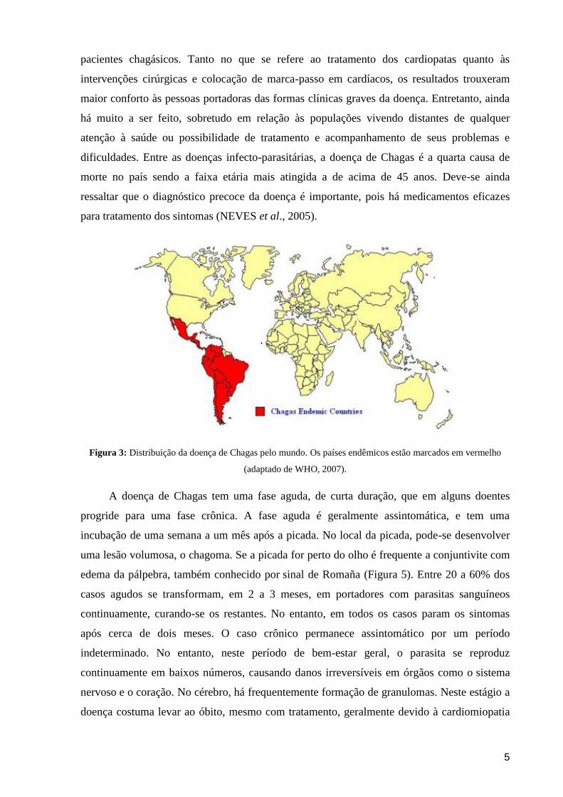
A doença de Chagas segue como problema de saúde pública por todos os países da
América Latina, sendo que sua distribuição cobre quase toda América do Sul e Central
(Figura 3). A iniciativa dos países do cone Sul em promoverem ações para controle do vetor
foi bem sucedida, com a participação da Argentina, Bolívia, Brasil, Chile, Paraguai e Uruguai,
como revisto por Schofield & Dias (1999). No entanto a iniciativa, como sua persistência,
deve-se muito à participação ativa de cientistas, ao manterem junto aos governos a atenção
voltada para o controle e prevenção da doença. No Brasil, com a progressiva melhora do
Sistema Único de Saúde (SUS), houve avanços consideráveis em relação à atenção aos
5
pacientes chagásicos. Tanto no que se refere ao tratamento dos cardiopatas quanto às
intervenções cirúrgicas e colocação de marca-passo em cardíacos, os resultados trouxeram
maior conforto às pessoas portadoras das formas clínicas graves da doença. Entretanto, ainda
há muito a ser feito, sobretudo em relação às populações vivendo distantes de qualquer
atenção à saúde ou possibilidade de tratamento e acompanhamento de seus problemas e
dificuldades. Entre as doenças infecto-parasitárias, a doença de Chagas é a quarta causa de
morte no país sendo a faixa etária mais atingida a de acima de 45 anos. Deve-se ainda
ressaltar que o diagnóstico precoce da doença é importante, pois há medicamentos eficazes
para tratamento dos sintomas (NEVES et al., 2005).
Figura 3: Distribuição da doença de Chagas pelo mundo. Os países endêmicos estão marcados em vermelho
(adaptado de WHO, 2007).
A doença de Chagas tem uma fase aguda, de curta duração, que em alguns doentes
progride para uma fase crônica. A fase aguda é geralmente assintomática, e tem uma
incubação de uma semana a um mês após a picada. No local da picada, pode-se desenvolver
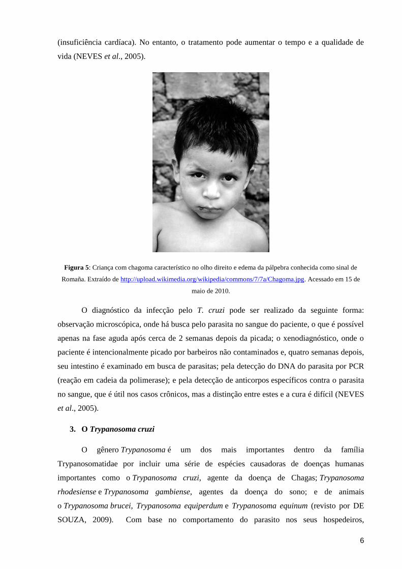
uma lesão volumosa, o chagoma. Se a picada for perto do olho é frequente a conjuntivite com
edema da pálpebra, também conhecido por sinal de Romaña (Figura 5). Entre 20 a 60% dos
casos agudos se transformam, em 2 a 3 meses, em portadores com parasitas sanguíneos
continuamente, curando-se os restantes. No entanto, em todos os casos param os sintomas
após cerca de dois meses. O caso crônico permanece assintomático por um período
indeterminado. No entanto, neste período de bem-estar geral, o parasita se reproduz
continuamente em baixos números, causando danos irreversíveis em órgãos como o sistema
nervoso e o coração. No cérebro, há frequentemente formação de granulomas. Neste estágio a
doença costuma levar ao óbito, mesmo com tratamento, geralmente devido à cardiomiopatia
6
(insuficiência cardíaca). No entanto, o tratamento pode aumentar o tempo e a qualidade de
vida (NEVES et al., 2005).
Figura 5: Criança com chagoma característico no olho direito e edema da pálpebra conhecida como sinal de
Romaña. Extraído de http://upload.wikimedia.org/wikipedia/commons/7/7a/Chagoma.jpg. Acessado em 15 de
maio de 2010.
O diagnóstico da infecção pelo T. cruzi pode ser realizado da seguinte forma:
observação microscópica, onde há busca pelo parasita no sangue do paciente, o que é possível
apenas na fase aguda após cerca de 2 semanas depois da picada; o xenodiagnóstico, onde o
paciente é intencionalmente picado por barbeiros não contaminados e, quatro semanas depois,
seu intestino é examinado em busca de parasitas; pela detecção do DNA do parasita por PCR
(reação em cadeia da polimerase); e pela detecção de anticorpos específicos contra o parasita
no sangue, que é útil nos casos crônicos, mas a distinção entre estes e a cura é difícil (NEVES
et al., 2005).
3. O Trypanosoma cruzi
O gênero Trypanosoma é um dos mais importantes dentro da família
Trypanosomatidae por incluir uma série de espécies causadoras de doenças humanas
importantes como o Trypanosoma cruzi, agente da doença de Chagas; Trypanosoma
rhodesiense e Trypanosoma gambiense, agentes da doença do sono; e de animais
o Trypanosoma brucei, Trypanosoma equiperdum e Trypanosoma equinum (revisto por DE
SOUZA, 2009). Com base no comportamento do parasito nos seus hospedeiros,
7
principalmente no vetor, o gênero Trypanosoma foi dividido em dois grupos. O primeiro,
chamado de Stercoraria, inclui tripanossomos que se desenvolvem no tubo digestivo do vetor,
progredindo no sentido da porção intestinal com liberação de formas infectantes pelas fezes,
onde temos o T. cruzi e o T. lewisi. O segundo, chamado de Salivaria, inclui tripanossomos
que se desenvolvem inicialmente no tubo digestivo e que posteriormente atravessam o epitélio
digestivo e atingem as glândulas salivares onde podemos encontrar as formas infectantes que
são inoculadas mecanicamente. Neste grupo encontramos o T. brucei, T. congolense e T.
rangeli. A grande complexidade no comportamento biológico dos tripanossomatídeos do
gênero Trypanosoma levou à criação de alguns subgêneros. No caso do T. cruzi cabe
mencionar que ele pertence ao subgênero Schizotrypanum, onde também estão incluídos
outros tripanossomos encontrados principalmente em morcegos do velho e do novo mundo,
como é o caso do T. dionisii, T. vespertilionis, T. myoti. Todos estes apresentam um ciclo
evolutivo em culturas axênicas e de células muito semelhante ao do T. cruzi (revisto por
HUGHES & PIONTKIVSKA, 2003).
Epidemiologicamente podem ser considerados três ciclos de transmissão vetorial do T.
cruzi. O ciclo de maior importância é o doméstico, já que perpetua a infecção nos seres
humanos. No ciclo silvestre, participam triatomíneos que, uma vez contaminados, infectam
roedores, marsupiais e outros animais silvestres. O terceiro ciclo é o peridoméstico, do qual
participam roedores, marsupiais, gatos e cães que entram e saem das residências, e os insetos
silvestres atraídos às casas pela disponibilidade de alimento. Esse ciclo serve de ligação entre
os ciclos doméstico e silvestre (REY, 2008). Mais de 130 espécies de triatomíneos
representam potenciais vetores de T. cruzi, mas somente 5 espécies possuem particular
importância na transmissão da doença de Chagas no Brasil (COURA & DIAS, 2009).
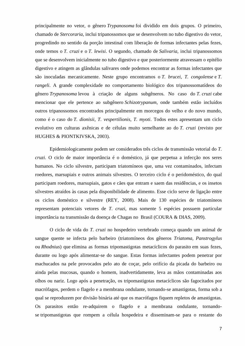
O ciclo de vida do T. cruzi no hospedeiro vertebrado começa quando um animal de
sangue quente se infecta pelo barbeiro (triatomíneos dos gêneros Triatoma, Panstrogylus
ou Rhodnius) que elimina as formas tripomastigotas metacíclicos do parasito em suas fezes,
durante ou logo após alimentar-se do sangue. Estas formas infectantes podem penetrar por
machucados na pele provocados pelo ato de coçar, pelo orifício da picada do barbeiro ou
ainda pelas mucosas, quando o homem, inadvertidamente, leva as mãos contaminadas aos
olhos ou nariz. Logo após a penetração, os tripomastigotas metacíclicos são fagocitados por
macrófagos, perdem o flagelo e a membrana ondulante, tornando-se amastigotas, forma sob a
qual se reproduzem por divisão binária até que os macrófagos fiquem repletos de amastigotas.
Os parasitos então re-adquirem o flagelo e a membrana ondulante, tornando-
se tripomastigotas que rompem a célula hospedeira e disseminam-se para o restante do
8
organismo pela circulação sanguínea. Quando chegam ao coração, esôfago ou cólon,
principais órgãos atingidos, penetram ativamente nas fibrilas musculares desses órgãos e
repetem o ciclo intracelular. Os tripomastigotas podem ainda ser fagocitados por macrófagos
nos órgãos do sistema fagocitário mononuclear (fígado, baço, linfonodos, medula óssea), onde
se reproduzem novamente como amastigotas (revisto por TYLER & ENGMAN, 2001).
Em dado momento da infecção, os tripomastigotas sanguíneos são ingeridos por um
novo repasto sanguíneo de outro triatomíneo que se alimenta de sangue humano, dando assim
início a parte do ciclo no hospedeiro invertebrado. Portanto, uma vez ingeridas pelo inseto as
formas tripomastigotas aderem à parede epitelial do intestino médio e se diferenciam em
epimastigotas, que são formas proliferativas flageladas de corpo mais largo e rígido. Após
intensa proliferação, as formas epimastigotas se diferenciam novamente em tripomastigotas
metacíclicos conforme migram em direção ao intestino posterior do triatomíneo estando
prontas para iniciar uma nova infecção ao hospedeiro vertebrado (revisto por TYLER &
ENGMAN, 2001). O ciclo de vida do T. cruzi pode ser visualizado na figura 4.
Figura 4: Representação esquemática do ciclo evolutivo do Trypanosoma cruzi nos hospedeiros vertebrados e
invertebrados. Extraído de http://upload.wikipedia.org/Chagas_ciclo_de _doenças.jpg/300. Acessado em 15 de
maio de 2010.
Diversas moléculas pertencentes ao T. cruzi e às suas células hospedeiras vêm sendo
implicadas como tendo participação no processo de adesão, como as glicoproteínas, os
9
glicolipídeos e as proteínas tipo lectina. Uma das moléculas mais estudadas, presente na
membrana plasmática de formas tripomastigotas (mas também em amastigotas, embora em
menor quantidade), é uma proteína com atividade neuraminidásica e transialidásica. Esta
última enzima remove resíduos de ácido siálico de glicoproteínas, glicolipídeos e
oligosacarídeos presentes no meio e os transfere para moléculas aceptoras presentes na
membrana plasmática das formas tripomastigotas. Dependendo da forma evolutiva do T.
cruzi estudada, diferentes moléculas foram identificadas como participantes do processo de
infecção da célula hospedeira. Nos tripomastigotas metacíclicos, por exemplo, a gp82, as
moléculas semelhantes a mucinas e a gp90, todas ancoradas na membrana plasmática do
parasita via âncora de glicosil fosfatidil inositol (GPI), reconhecem receptores nas células
hospedeiras ainda não identificados e disparam a maquinaria de sinalização tanto no parasito
quanto na célula hospedeira. Em seguida, após a entrada de formas tripomastigotas, a célula
hospedeira ativa um processo de sinalização que leva a um aumento transiente dos níveis
citoplasmáticos de cálcio, tanto no parasito quanto na célula hospedeira. Em células
fagocíticas profissionais, como os macrófagos, ocorre ativação de tirosinas cinases,
recrutamento de PI-3 cinase e actina para o local de entrada do parasito, o que demonstra que
o principal mecanismo de entrada é por fagocitose e que é crucial a participação de moléculas
das células hospedeiras. (revisto por CARVALHO, 2009). Outras enzimas, capazes de
quebrar ligações peptídicas e conhecidas como peptidases, também desempenham uma série
de funções críticas e essencias para o estabelecimento da infecção e persistência do parasito,
entre outras funções (VERMELHO et al., 2007)
O T. cruzi é representado por um conjunto de populações que circulam em hospedeiros
mamíferos e insetos vetores denominados isolados ou cepas que apresentam grande
heterogeneidade de comportamento biológico como, por exemplo, diferentes graus de
virulência para animais experimentais e humanos, variações na sensibilidade a drogas e
tropismo tissular. A explicação para esta diversidade fenotípica reside no fato do T. cruzi
ser um organismo diplóide com alta variabilidade genética entre seus diferentes isolados e
que se multiplica predominantemente por divisão binária. Desta forma, o genoma de cada
isolado do parasito evolui de forma independente. É interessante notar que a doença de
Chagas também apresenta uma diversidade de apresentações clínicas (formas indeterminada,
cardíaca e digestiva). Assim, um grande desafio para a comunidade científica vem sendo
identificar marcadores genéticos dos isolados capazes de classificá-los em grupos discretos,
visando sua caracterização do ponto de vista epidemiológico e de patogenia (revisto por
CAMPBELL et al., 2004). Ao longo dos anos, várias abordagens têm sido usadas para
caracterizar a estrutura populacional do T. cruzi, visando à definição do número de subgrupos
10
relevantes. Assim, estes subgrupos receberam denominações diferentes, incluindo:
zimodemas (MILES et al., 1977, 1978, 1981; ROMANHA et al., 1979), baseados no perfil
eletroforético de isoenzimas utilizadas como marcadores; schizodemas (MOREL et al., 1980);
biodemas (ANDRADE & MAGALHÃES, 1996); clones (TIBAYRENC & AYALA, 1991);
linhagens (SOUTO et al., 1996); clados (KAWASHITA et al., 2001); e, mais recentemente,
unidades discretas de tipagem (DTUs) (TIBAYRENC, 2003) e haplótipos (HERRERA et al.,
2007). Portanto, em 23 de agosto de 2009 foi realizada uma reunião em Búzios (RJ-Brasil),
que precedeu o XIII Congresso Internacional de Protistology, para padronização da
nomenclatura deste parasito. Por consenso, a comissão de peritos reconheceu que a
nomenclatura para cepas do T. cruzi deve ser classificada em seis DTUs, T. cruzi I-VI, e
emitiu recomendações com justificações detalhadas e suas implicações (ZINGALES et al.,
2009).
4. Tratamento da doença de Chagas
O tratamento de escolha para a doença de Chagas é frequentemente através da
administração de nitroimidazóis (benzonidazol) e de 5-nitrofuranos (nifurtimox). Entretanto, o
nifurtimox não se encontra mais disponível comercialmente no Brasil e nenhum destes
compostos é ideal para o combate da doença uma vez que: não são ativos durante a fase
crônica da doença; apresentam sérios efeitos colaterais; requerem administração por longos
períodos de tempo sob supervisão médica; há grande variação na susceptibilidade de isolados
do parasito a ação destas drogas; e apresentam alto custo (FAIRLAMB, 2003; CAVALLI &
BOLOGNESI, 2009; WILKINSON & KELLY, 2009; MCKERROW et al., 2009; URBINA,
2010). O benzonidazol tem sido principalmente utilizado no tratamento de pacientes agudos e
crônicos recentes, nos quais se observam resultados positivos médio de cura em torno de
80%, inclusive em crianças.
Drogas para o tratamento da doença de Chagas não são do interesse de indústrias
farmacêuticas, sendo os principais problemas o alto custo dos investimentos e a falta de um
mercado potencial e seguro nos países em desenvolvimento. Desta forma, apesar da redução
na incidência da transmissão, ainda há desafios com dois problemas críticos: o tratamento de
pacientes na fase crônica, e a ocorrência de novos casos agudos em algumas regiões da
América Latina. De um modo geral, o desenvolvimento de uma nova quimioterapia
antiparasitária ocorre pela investigação de drogas já aprovadas para o tratamento de outras
doenças, uma vez que elas já foram submetidas a ensaios clínicos muito dispendiosos, ou
através da determinação de um ou mais alvos específicos identificados em vias metabólicas
importantes para o parasito (revisto por SOEIRO & DE CASTRO, 2009; URBINA, 2009).
11
Nas últimas décadas, estudos têm demonstrado que o T. cruzi, como a maioria dos
fungos e leveduras, requer esteróis específicos, conhecidos como ergosteróis, para sua a
viabilidade e proliferação celular em todas as fases do ciclo de vida. Assim, neste parasito,
etapas da biossíntese de esteróis que são divergentes em relação à síntese realizada por células
de mamíferos têm sido quimicamente validadas como o mais promissor alvo para o
tratamento da doença de Chagas (URBINA, 2002; URBINA & DOCAMPO, 2003). Nesse
contexto, derivados triazólicos, como o D0870 e o posaconazol, têm-se mostrado capazes de
induzir cura em modelos murinos de fase aguda e crônica da doença de Chagas (URBINA et
al., 1996, 1998, 2002; URBINA E DOCAMPO, 2003). Um estudo mais recente com
posaconazol demonstrou que este composto pode erradicar formas amastigotas intracelulares
do T. cruzi de cultura de cardiomiócitos e, ao mesmo tempo, permitir a reconstituição
completa do citoesqueleto das células hospedeiras (SILVA et al., 2006). No entanto, o
elevado custo de produção do posaconazol e sua necessidade de administração oral
juntamente com uma refeição rica em gorduras podem limitar seu uso no tratamento de
infecções crônicas por T. cruzi (MORRIS, 2009). Entre os demais alvos quimioterápicos com
compostos em desenvolvimento para o tratamento da doença de Chagas se destacam: as
diamidinas aromáticas que se associam à fenda menor do DNA em sítios ricos em AT, que
são capazes de interferir na função do cinetoplasto em tripanossomatídeos (CATERINA et al.,
2008); o sistema glutationa/glutationa redutase, que reduz os níveis de radicais livres
contribuindo para a manutenção de um ambiente intracelular redutor (HEBY et al., 2007); a
hipoxantina-guanina fosforibosiltransferase, enzima responsável pela conversão de bases de
purina a ribonucleotídeos, essencial para a síntese de material genético nos
tripanossomatídeos (WENCK et al., 2004); e as peptidases do parasito, em especial a
cruzipaína, cujos inibidores irreversíveis peptídicos tais como diazometilcetonas,
fluorometilcetonas, alilsulfonas, vinilsulfonas e vinilsulfonamidas, vêm apresentando bons
resultados no tratamento de infecções experimentais (revisto por MCKERROW et al., 2009).
Com o conhecimento acumulado sobre a biologia e a bioquímica do T. cruzi, os
esforços dirigidos à compreensão do mecanismo de ação de potenciais compostos contra este
parasito se fazem necessários. O desenvolvimento de novas drogas pode vir a requerer a
interação interdisciplinar dos diferentes campos da ciência como a biologia molecular e
celular, química, bioquímica, farmacologia e toxicologia. Os avanços da genômica, da
bioinformática, da química combinatória e das triagens automatizadas fortalecem a interação
entre grupos de diferentes especialidades de modo a permitir a identificação de compostos
eficazes com baixa toxicidade e a baixo custo de produção (revisto por SOEIRO & DE
CASTRO, 2009).
12
5. As peptidases
As peptidases, proteases ou peptídeo-hidrolases (E.C. 3.4) são enzimas que
quebram ligações peptídicas entre os aminoácidos das proteínas, clivando assim proteínas ou
fragmentos protéicos (BARRET, 1994; BARRET et al., 2001). A clivagem proteolítica é um
mecanismo comum de ativação ou inativação de enzimas que está envolvida principalmente
na digestão e na coagulação sanguínea. Como uma molécula de água é utilizada no processo,
as proteases pertencem ao grupo das hidrolases. Muitos microrganismos podem secretar
peptidases para o meio externo com a finalidade de degradar proteínas, cujos produtos de
hidrólise são fontes de carbono e nitrogênio para o seu crescimento (VERMELHO et al.,
2007).
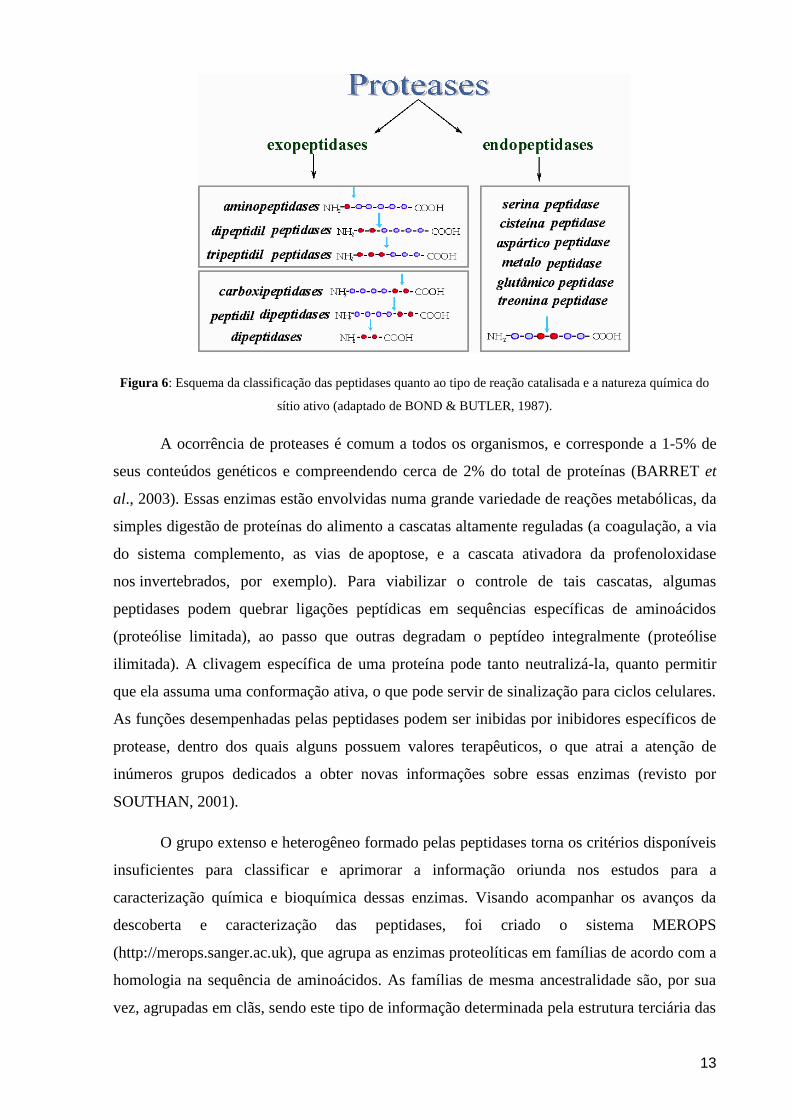
As peptidases constituem uma grande família, podendo ser divididas em
endopeptidases ou proteinases e exopetidases, de acordo com a posição da ligação peptídica a
ser clivada na cadeia peptídica. As endopeptidases atuam preferencialmente nas regiões
internas da cadeia polipeptídica; enquanto as exopeptidases atuam somente nos finais das
cadeias polipeptídicas na região N- ou C-terminal.. As exopeptidases que atuam na região
amino-terminal livre liberam um único resíduo de aminoácido (aminopeptidases), um
dipeptídeo (dipeptidil-peptidases) ou um tripeptídeo (tripeptidil-peptidases); já aquelas que
atuam na região carboxi-terminal livre liberam um único aminoácido (carboxipeptidases) ou
um dipeptídeo (peptidil-dipeptidases) (BOND & BUTLER, 1987). As endopeptidases podem
também ser classificadas pela natureza química do seu sítio catalítico, podendo ser divididas
em subclasses do tipo serina-, cisteína-, aspártico-, metalo-, treonina-, e glutâmico-peptidases.
Algumas peptidases não se encaixam nestas subclasses e formam a subclasse 3.4.99, de
mecanismo catalítico desconhecido (BARRET et al., 2001). O esquema da classificação das
peptidases pode ser observado na figura 6.
13
Figura 6: Esquema da classificação das peptidases quanto ao tipo de reação catalisada e a natureza química do
sítio ativo (adaptado de BOND & BUTLER, 1987).
A ocorrência de proteases é comum a todos os organismos, e corresponde a 1-5% de
seus conteúdos genéticos e compreendendo cerca de 2% do total de proteínas (BARRET et
al., 2003). Essas enzimas estão envolvidas numa grande variedade de reações metabólicas, da
simples digestão de proteínas do alimento a cascatas altamente reguladas (a coagulação, a via
do sistema complemento, as vias de apoptose, e a cascata ativadora da profenoloxidase
nos invertebrados, por exemplo). Para viabilizar o controle de tais cascatas, algumas
peptidases podem quebrar ligações peptídicas em sequências específicas de aminoácidos
(proteólise limitada), ao passo que outras degradam o peptídeo integralmente (proteólise
ilimitada). A clivagem específica de uma proteína pode tanto neutralizá-la, quanto permitir
que ela assuma uma conformação ativa, o que pode servir de sinalização para ciclos celulares.
As funções desempenhadas pelas peptidases podem ser inibidas por inibidores específicos de
protease, dentro dos quais alguns possuem valores terapêuticos, o que atrai a atenção de
inúmeros grupos dedicados a obter novas informações sobre essas enzimas (revisto por
SOUTHAN, 2001).
O grupo extenso e heterogêneo formado pelas peptidases torna os critérios disponíveis
insuficientes para classificar e aprimorar a informação oriunda nos estudos para a
caracterização química e bioquímica dessas enzimas. Visando acompanhar os avanços da
descoberta e caracterização das peptidases, foi criado o sistema MEROPS
(http://merops.sanger.ac.uk), que agrupa as enzimas proteolíticas em famílias de acordo com a
homologia na sequência de aminoácidos. As famílias de mesma ancestralidade são, por sua
vez, agrupadas em clãs, sendo este tipo de informação determinada pela estrutura terciária das
14
peptidases (BARRET et al., 2003). Assim, as peptidases são agrupadas em famílias de acordo
com a semelhança da sequência de aminoácidos do seu sítio catalítico em relação à enzima
representativa da família, que por sua vez são agrupadas em seguida por clãs específicos, de
acordo com as semelhanças na estrutura secundária, terciária e na ordem dos resíduos
catalíticos ou sequências motivo em torno dos resíduos. As famílias são representadas por
uma letra, que indica o mecanismo catalítico e um número. O clã é representado por duas
letras: a primeira indica o tipo de mecanismo catalítico e a segunda é adicionada
sequencialmente. As letras usadas são „A‟ (aspártico), „C‟ (cisteína), „M‟ (metalo), „S‟
(serina), „T‟ (treonina), „G‟ (glutâmico) ou „U‟ (tipo desconhecido, do inglês, “unknown”)
(BARRET et al., 2001).
6. Peptidases no Trypanosoma cruzi
As peptidases desempenham uma série de funções básicas e essenciais no
metabolismo e no processo de patogênese de diversos microrganismos, cujas doenças
parasitárias representam grave problema de saúde global (revisto por MCKERROW et al.,
2006). Nos parasitos membros da família Trypanosomatidae, essas enzimas estão envolvidas
em eventos cruciais do ciclo de vida destes parasitos, tais como a diferenciação e remodelação
do parasito nas fases de seu ciclo de vida, ativação ou degradação de enzimas ou proteínas
regulatórias e processamento nutricional de proteínas. Uma vez que os tripanossomatídeos são
parasitos obrigatórios, as peptidases não poderiam deixar de estar associadas à invasão celular
através da ruptura de células hospedeiras e degradação do citoesqueleto, participando assim
do processo de interação parasito-hospedeiro. As peptidases também são de extrema
relevância na prevenção da resposta imune do hospedeiro, uma vez que podem promover o
escape e a modulação do sistema imunológico por degradação ou ativação de moléculas do
sistema imune, como o sistema complemento e a coagulação sanguínea, além de serem
responsáveis pela variabilidade antigênica do parasito (revisto por McKerrow et al., 1993;
Sajid & McKerrow, 2002).
Entre as seis classes principais de peptidases, as mais comumente detectadas nos
tripanossomatídeos são as metalo- e as cisteína-peptidases (BRANQUINHA et al., 1996;
SANTOS et al., 2005; VILLALTA et al., 2008). A cruzipaína é uma das principais proteases
do T. cruzi. Esta enzima consiste de uma cisteína-peptidase também conhecida como gp57/51,
que consiste em uma glicoproteína monomérica de 60 kDa altamente imunogênica e cuja
atividade máxima pode ser observada em intervalos de pH 5 a 7,5 (MURTA et al., 1990).
Esta enzima é expressa em todas as formas evolutivas de todas as cepas e clones de T.
cruzi até hoje testados, com os maiores níveis de atividade sendo detectados na forma
15
epimastigota (TOMAS & KELLY, 1996), e pode também ser detectada no meio extracelular,
onde se acredita que sua forma secretada possa estar envolvida no processo de invasão de
células hospedeiras (APARICIO et al., 2004). A participação desta peptidase na invasão da
célula hospedeira e no desenvolvimento intracelular foi primeiramente descrita através do uso
de derivados de diazometano-peptidil, uma classe de inibidores irreversíveis da cisteína-
peptidases (MEIRELLES et al., 1992).
A cruzipaína possui homologia com a catepsina L de mamíferos, estando associada
principalmente ao sistema endocítico-lisossomal e reservossomas em epimastigotas (MURTA
et al., 1990; SOUTO-PADRÓN et al., 1990; SOARES et al., 1992). Esta enzima também está
presente na superfície de amastigotas e epimastigotas e na bolsa flagelar das três formas
evolutivas (SOUTO-PADRÓN et al., 1990). Várias funções já foram atribuídas à cruzipaína
como: degradação protéica, aumento da metaciclogênese in vitro (BONALDO et al., 1991;
TOMAS et al., 1997), invasão celular (SOUTO-PADRÓN et al., 1990; MEIRELLES et al.,
1992; HARTH et al., 1993, SCHARFSTEIN et al., 2000; APARICIO et al., 2004),
multiplicação do parasito nas células do hospedeiro (MEIRELLES et al., 1992), no escape do
sistema imune (GRUPPI et al., 1997; BENÍTEZ-HERNÁNDEZ et al., 2010) e em processos
inflamatórios e ativação do sistema de morte (CAZZULO et al., 2001).
Em T. cruzi, já foram identificadas glicoproteínas da família das cisteína-peptidases
com massa molecular de 30 kDa que podem estar envolvidas em eventos celulares como a
diferenciação nas células hospedeiras e transmissão intracelular de tripomastigotas
(CAZZULO et al., 1990; GARCIA et al., 1998). Embora apresente maior atividade
enzimática em epimastigotas, essas glicoproteínas estão presentes nas três formas evolutivas
do parasito, apresentando similaridade da sua sequência N-terminal com a catepsina B de
humanos, Leishmania mexicana e Caenorhabditis elegans (GARCIA et al., 1998).
As proteases da classe das metalopeptidases também são amplamente encontradas nos
tripanossomatídeos, inclusive no T. cruzi que possui uma metalopeptidase homóloga à gp63
da Leishmania (Tcgp63) (CUEVAS et al., 2003). Esta glicoproteína de 60-65 kDa é
abundantemente expressa em promastigotas de Leishmania, sendo de extrema importância no
processo de patogênese da leishmaniose (revisto por YAO et al., 2003). O T. cruzi tem 10 ou
mais genes de gp63 ou moléculas homólogas à gp63 e sua expressão é aumentada na fase
amastigota do parasito, indicando que esta enzima possa ter um papel na sobrevivência e
desenvolvimento do T. cruzi no citoplasma da célula hospedeira (GRANDGENETT et al.,
2000). A Tcgp63 provavelmente está envolvida na infecção de células do hospedeiro
vertebrado, uma vez que modificações pós-traducionais que ocorrem exclusivamente em
16
isoformas de tripomastigotas metacíclicos participam do processo de infecção experimental
de mioblastos (KULKARNI et al., 2009).
Outras peptidases como as pertencentes aos grupos serina e aspártico também têm sido
identificadas em T. cruzi (BURLEIGH et al., 1997; PAUGAM et al., 2003; ALVES et al.,
2005; PINHO et al., 2009). Porém, as aspártico-peptidases ainda não estão bem caracterizadas
na família Trypanosomatidae. Este grupo de peptidases está dividido em várias famílias e
geralmente funcionam em condições ácidas, o que limita a função das aspártico peptidases a
alguns locais específicos nos diferentes organismos e as tornam menos abundante do que a de
outros grupos de peptidases (DASH et al., 2003). Apesar disso, estas enzimas têm sido
isoladas e estudadas em uma ampla variedade de organismos, em vertebrados, fungos,
plantas, vírus e até em bactérias (HILL & PHYLIP, 1997; JAMES, 1998; DASH et al., 2003;
TAKAHASHI et al., 2006; ALVES et al., 2005; VALDIVIESO et al., 2007; PINTI et al.,
2007). Recentemente, duas aspártico-peptidases foram identificadas e isoladas de formas
epimastigotas do T. cruzi: a cruzipsina-I (CZP-I) e a cruzipsina-II (CZP-II), ambas com massa
molecular em torno de 120 kDa. Estas enzimas tiveram uma alta atividade enzimática
específica detectada, hidrolisando substrato de catepsina B, e sendo inibidas por inibidores
específicos dessa classe de peptidases, como a pepstatina A. Contudo, as funções fisiológicas
dessas enzimas ainda são desconhecidas e há a necessidade de novos estudos (PINHO et al.,
2009).
Outro grupo de peptidases encontradas no T. cruzi que necessita de mais investigações
são as serina-peptidases. Uma dessas enzimas está intimamente relacionada com os membros
da família prolil-oligopeptidase de serina-endopeptidases de mamíferos e, assim, é
denominada oligopeptidase B (Tc-OP) (BURLEIGH et al., 1997). Formas tripomastigotas do
parasito mutantes de Tc-OP apresentam deficiência na mobilização de cálcio nas células de
mamíferos, o que prejudica o estabelecimento da infecção in vitro e in vivo (CALER et al.,
1998). Outra serina-peptidase amplamente estudada no T. cruzi é uma prolil-oligopeptidase
não-lisossomal secretada de 80 kDa conhecida como POP Tc80 (SANTANA et al., 1997).
Este enzima é encontrada em todas as formas evolutivas do ciclo evolutivo do parasito e tem
alta especificidade para colágenos humanos tipo I e IV, o que sugere a participação da POP
Tc80 na disseminação do parasito no hospedeiro (JOYEAU et al., 2000), além de participar
no processo de invasão celular (GRELLIER et al., 2001; BASTOS et al., 2005).
Recentemente, uma nova serina-peptidase também extracelular foi isolada de epimastigotas.
A nova enzima apresenta massa molecular de 75 kDa e sua localização intracelular sugere
uma participação em eventos de proteólise nos reservossomas, embora possivelmente esta
17
seja secretada pela bolsa flagelar (SILVA-LOPEZ et al., 2008). Portanto, peptidases
secretadas para o meio extracelular podem funcionar como um mecanismo adaptativo
importante durante o ciclo de vida dos microrganismos (MCKERROW et al., 1993; MONOD
et al., 2002; SANTOS et al., 2007).
7. As Calpaínas
A família das calpaínas consiste de um grupo heterogêneo de cisteína-peptidases com
um amplo padrão de expressão, que inclui múltiplas isoformas ubíquas no organismo e de
tecido específicos. Também denominadas CDPs (“Calcium Dependent Peptidases”), as
calpaínas são proteases neutras dependentes de cálcio cujas suas duas principais isoformas,
denominadas μ- e m-calpaínas, têm sido distinguidas com base nas diferenças de seus
requerimentos micro- e milimolar para ativação in vitro por cálcio. Estas proteínas são
formadas por duas subunidades, uma catalítica de 80 kDa de massa molecular e uma
subunidade regulatória de 30 kDa. Ambas as subunidades contêm um domínio de ligação para
cálcio do tipo calmodulina, sendo que nas células de animais, a subunidade menor nas
isoenzimas é idêntica, enquanto que a subunidade maior apresenta identidade de sequência
limitada (revisto por JOHNSON & GUTTMAN, 1997). A estrutura da m-calpaína é
demonstrada na figura 7.
18
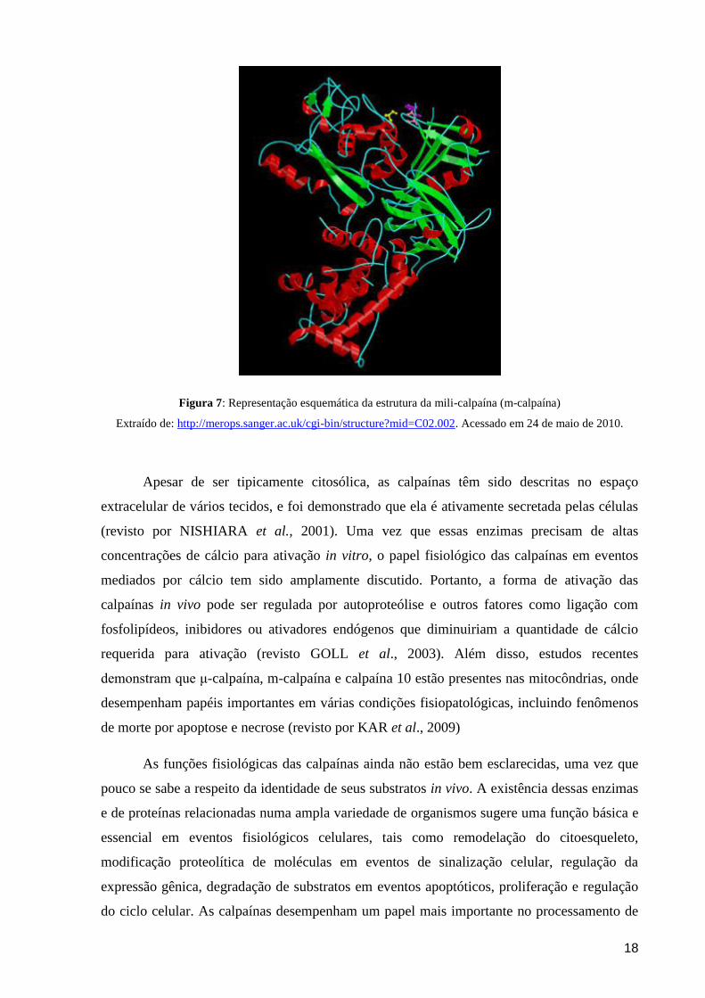
Figura 7: Representação esquemática da estrutura da mili-calpaína (m-calpaína)
Extraído de: http://merops.sanger.ac.uk/cgi-bin/structure?mid=C02.002. Acessado em 24 de maio de 2010.
Apesar de ser tipicamente citosólica, as calpaínas têm sido descritas no espaço
extracelular de vários tecidos, e foi demonstrado que ela é ativamente secretada pelas células
(revisto por NISHIARA et al., 2001). Uma vez que essas enzimas precisam de altas
concentrações de cálcio para ativação in vitro, o papel fisiológico das calpaínas em eventos
mediados por cálcio tem sido amplamente discutido. Portanto, a forma de ativação das
calpaínas in vivo pode ser regulada por autoproteólise e outros fatores como ligação com
fosfolipídeos, inibidores ou ativadores endógenos que diminuiriam a quantidade de cálcio
requerida para ativação (revisto GOLL et al., 2003). Além disso, estudos recentes
demonstram que μ-calpaína, m-calpaína e calpaína 10 estão presentes nas mitocôndrias, onde
desempenham papéis importantes em várias condições fisiopatológicas, incluindo fenômenos
de morte por apoptose e necrose (revisto por KAR et al., 2009)
As funções fisiológicas das calpaínas ainda não estão bem esclarecidas, uma vez que
pouco se sabe a respeito da identidade de seus substratos in vivo. A existência dessas enzimas
e de proteínas relacionadas numa ampla variedade de organismos sugere uma função básica e
essencial em eventos fisiológicos celulares, tais como remodelação do citoesqueleto,
modificação proteolítica de moléculas em eventos de sinalização celular, regulação da
expressão gênica, degradação de substratos em eventos apoptóticos, proliferação e regulação
do ciclo celular. As calpaínas desempenham um papel mais importante no processamento de
19
substratos, ativando-os através da remoção de domínios auto-inibitórios, do que na
degradação completa das proteínas (revisto por CARAFOLI & MOLINARI, 1998; GOLL et
al., 2003; MARSHALL et al., 2005; KAR et al., 2009).
A grande variedade de funções fisiológicas das calpaínas está relacionada a diversos
processos patológicos intimamente relacionados com a ação destas enzimas. A desregulação
de sua atividade tem sido envolvida, principalmente, em diversas desordens neurológicas
como o mal de Alzheimer, a doença de Huntington, doença de Parkinson e esclerose múltipla
(revisto por SAEZ et al., 2006). Em pacientes com Alzheimer, as calpaínas estão envolvidas
em eventos moleculares que conduzem a hiperfosforilação de Tau, a principal proteína
encontrada nos arranjos fibrilares (HIGUCHI et al., 2005). Além disso, a atividade
proteolítica das calpaínas sobre Tau e outras proteínas de neurofilamentos está relacionada
com a morte celular por necrose observada nesta doença (PARK & FERREIRA, 2005). Na
doença de Huntington, foi identificada a ação de calpaínas, além da participação de
proteassomas e caspases. Neste caso, a ação das calpaínas gera fragmentos protéicos tóxicos
que levam a danos neuronais (GAFNI & ELLERBY, 2002). O aumento no nível de
marcadores da atividade de calpaínas in vivo, como os gerados pela clivagem da αII-
espectrina, abundantes em neurônios, tem sido observado em condições de injúrias hipóxicas
e isquêmicas do cérebro, coração e pulmões (VANDERKLISH & BAHR, 2000). Em outros
processos patológicos como no câncer (ATENCIO et al., 2000), na catarata (BISWAS, 2004)
e nas infecções virais (UPLA et al., 2008), a atividade desregulada das calpaínas também está
associada (revisto por SAEZ et al., 2006). Sendo assim, a participação das calpaínas em
diferentes eventos celulares, o envolvimento em diversas doenças humanas, as características
moleculares e o mecanismo de ação destas enzimas fazem com que estas se tornem
interessantes alvos para o desenvolvimento de drogas.
Um avanço significativo no estudo de homólogos das calpaínas veio através do avanço
das técnicas de manipulações genéticas que permitiram a descoberta destas moléculas em
outros organismos e uma maior facilidade de determinação de suas funções. Através dessas
análises, foi demonstrado, por exemplo, que o gene tra-3 está envolvido na cascata de
determinação do sexo em Caenorhabditis elegans (DEAR et al., 1997). Enquanto que em
Drosophila melanogaster, foi descoberto que a proteína Sol está envolvida com o
desenvolvimento do sistema nervoso, uma vez que o gene sol defectivo leva a uma
degeneração específica nos lobos óticos, resultando numa redução do tamanho dos lobos
óticos e na ausência de certas classes de neurônios (KAMEI et al., 1998). Também foi
possível demonstrar que a adaptação a ambientes alcalinos no fungo Aspergillus nidulans
20
requer atividade da proteína PalB, que é homóloga à superfamília das calpaínas (revisto por
SORIMACHI et al., 1997; FRANZ et al., 1999; SAEZ et al., 2006). Nos protozoários
Plasmodium falciparum e Toxoplasma gondii, agentes causadores da malária e toxoplasmose,
respectivamente, foi observada a utilização de calpaínas das células hospedeiras para facilitar
o escape do vacúolo parasitóforo e/ou membrana plasmática hospedeira. A imunodepleção ou
a inibição da calpaína-1 da célula hospedeira impediu o escape de P. falciparum, e um efeito
similar foi observado em T. gondii pela supressão ou deleção dos genes das calpaínas de
fibroblastos (CHANDRAMOHANADAS et al., 2009).
8. Calpaínas nos tripanossomatídeos
Estudos voltados para a descoberta de moléculas homólogas às calpaínas em
microrganismos vêm descrevendo a presença de calpaínas na família Trypanosomatidae. O
primeiro relato de atividade proteolítica relacionada com calpaínas em tripanossomatídeos
ocorreu no gênero Leishmania, onde uma cisteína-peptidase dependente de cálcio foi
detectada nas formas promastigotas de L. donovani. Esta enzima foi denominada
caldonopaína devido à sua similaridade bioquímica com a família das calpaínas, embora a
reação cruzada ou homologia com genes de outras calpaínas não tenha sido reportada
(BHATTACHARYA et al., 1993). Apesar da identidade da proteína não ter sido confirmada,
posteriormente foi demonstrado que a atividade da caldonopaína facilitaria a invasão de
macrófagos por L. donovani (DEY et al., 2006).
O tripanossomatídeo cuja caracterização de homólogos de calpaínas está mais
avançada é o Trypanosoma brucei. Uma proteína associada ao citoesqueleto (CAP5.5) com
similaridade com a região catalítica das calpaínas foi identificada neste protozoário,
entretanto, não foi verificado se a proteína apresentava atividade enzimática (HERTZ-
FOWLER, et al., 2001). A CAP5.5 é detectada exclusivamente nas formas procíclicas do
parasito e foi, de fato, o primeiro homólogo de calpaína caracterizado em tripanossomatídeos.
Posteriormente, o mesmo grupo descreveu em tripomastigotas metacíclicos a expressão
aumentada de uma variante análoga desta proteína, denominada CAP5.5V. Neste trabalho foi
demonstrado que ambos homólogos de calpaínas, CAP5.5 e CAP5.5V, estão relacionados à
correta morfogênese do T. brucei, conforme foi observado em estudos ultraestruturais com
parasitos silenciados para essas duas proteínas (OLEGO-FERNANDEZ et al., 2009). Mais
recentemente, um estudo sobre a expressão e localização celular dos homólogos de calpaínas
descritos no genoma do T. brucei demonstrou mais uma vez haver uma expressão
diferenciada destas moléculas nas diferentes fases do ciclo de vida do parasito. Também foi
21
relatada a relação das modificações pós-traducionais com a localização celular dos homólogos
de calpaínas expressos nas diferentes formas evolutivas (LIU et al., 2010).
Após a caracterização da proteína CAP5.5 nas formas procíclicas do T. brucei (Hertz-
Fowler et al., 2001), Ersfeld e colaboradores (2005) mostraram a presença de uma grande e
diversa família de proteínas relacionadas às calpaínas nos tripanossomatídeos T. brucei, T.
cruzi e L. major. Essas proteínas foram classificadas em cinco grupos com base em suas
características estruturais. Os membros dos grupos 1 e 2 apresentam quatro domínios e são
denominadas proteínas calpaína-like (CALPs), destacando-se por seus domínios N-terminal:
enquanto o grupo 1 contém o domínio IK, que é altamente conservado em tripanossomatídeos,
o grupo 2 contém os domínios não-conservados IH. A caracterização dessas sequências
revelou que o domínio responsável pela atividade catalítica, o domínio II, está bem
conservado, embora os resíduos de aminoácidos críticos para a atividade catalítica estejam
alterados. Embora os sítios de ligação do cálcio também estejam ausentes no domínio IV, em
ambos os grupos, os resíduos de aminoácidos que são essenciais para a ligação do cálcio
dentro do domínio II em calpains de mamíferos estão conservados em algumas sequências
desses protozoários. Os membros do grupo 3 são conhecidos como proteína de
kinetoplastídeos relacionadas às calpaínas (SKCRPs – samll kinetoplastid calpain related
protein), consistindo apenas do domínio IK. Seis SKRCPs foram encontrados em T. brucei,
nove em T. cruzi e dez em L. major, com um comprimento médio de cerca de 200
aminoácidos. Este domínio não mostrou semelhanças com outras proteínas bem
caracterizadas e não há nenhuma indicação quanto à sua função. Os grupos 4 e 5 contêm
sequências de calpaínas altamente divergentes ainda não estudadas: no grupo 4, as proteínas
são caracterizadas pela presença de três repetições de domínios II e III, e no grupo 5 por
repetições N-terminais dos domínios II e III (ERSFELD et al., 2005).
Boa parte dos avanços no estudo das calpaínas nos tripanossomatídeos se deve ao
sequenciamento do genoma desses microrganismos, que permitiu a identificação de diversas
sequências com homologia às calpaínas (ERSFELD et al., 2005). Em cepas de L. donovani,
por exemplo, a análise da sequência de seis clones genômicos demonstrou a existência de
diferenças na expressão de uma proteína tipo calpaína em isoladas de pacientes que haviam
desenvolvido leishmaniose visceral e leishmaniose cutânea pós-calazar (PKDL) quando
comparada com pacientes que apresentavam o quadro original de leishmaniose visceral. O
homólogo de calpaína identificado neste estudo é um polipeptídeo pequeno, de
aproximadamente 14 kDa, similar ao encontrado em T. brucei (CAB95480) (SALOTRA et
al., 2006). Em seguida, foi reportado por análises proteômicas a presença de uma proteína
22
relacionada à calpaína (SKCRP-14.1) intimamente envolvida com o programa de morte
celular induzido por drogas, onde esta proteína promove a apoptose induzida por antimoniais
e previne a apoptose induzida pela miltefosina (VERGNES et al., 2007).
No T. cruzi, até o momento, pouco foi descrito a respeito de moléculas homólogas às
calpaínas. Através de análises proteômicas, um estudo recente identificou a presença de
proteínas tipo calpaínas em cepas de T. cruzi resistentes ao benzonidazol. A expressão da
proteína similar à calpaína identificada foi 21 vezes maior nas amostras de cepas resistentes
selecionadas in vivo, enquanto que as cepas resistentes in vitro não apresentaram diferença na
expressão desta proteína (ANDRADE et al., 2008). Posteriormente, GIESE e colaboradores
(2008) clonaram um gene pertencente à família das calpaínas em T. cruzi, denominado
TcCALPx11, que corresponde a uma proteína hipotética de 80 kDa (XP_816697.1) específica
das formas epimastigotas, submetidas ao estresse nutricional que precede a metaciclogênese.
Nenhuma atividade proteolítica foi verificada para este homólogo de calpaína do T. cruzi, e a
sua expressão diferenciada sugere que a proteína poderia estar exercendo um papel na
resposta ao estresse e/ou na transdução de sinal durante o processo de diferenciação do
parasito (GIESE et al., 2008).
Nesse contexto, nosso grupo se envolveu no estudo de homólogos das calpaínas em
tripanossomatídeos. D‟AVILA-LEVY e colaboradores (2003) ao investigarem as proteínas
secretadas para o meio extracelular de uma cepa apossimbiótica Crithidia deanei se
depararam com uma cisteína-peptidase que migrava na faixa de 80 kDa. Este protease foi
purificada e caracterizada como uma enzima neutra dependente de cálcio que poderia ser
completamente inibida por E-64 e EGTA, apresentando assim características semelhantes à
família das calpaínas. Outra evidência da relação desta enzima com a família das calpaínas foi
obtida através da reação cruzada contra um anticorpo específico para uma calpaína de
Drosophila melanogaster (anti-Dm-calpaína). Em um estudo voltado para a busca de
calpaínas em Leishmania amazonensis, nosso grupo identificou uma molécula homóloga à
calpaína na superfície celular de formas promastigotas reativa contra o anti-Dm-calpaína. No
mesmo estudo pode-se observar o efeito do potente inibidor de calpaína III (MDL28170)
sobre a taxa de multiplicação do parasito. Este inibidor foi capaz de promover notáveis
alterações celulares, culminando com a morte do parasito (D'AVILA-LEVY et al., 2006).
Recentemente, PEREIRA e colaboradores (2009) também demonstraram a presença de uma
proteína similar à calpaína de D. melanogaster em promastigotas de Herpetomonas
samuelpessoai, a qual era modulada ao longo do processo de diferenciação celular neste
tripanossomatídeo monoxênico. Sendo assim, uma melhor caracterização das calpaínas em
23
tripanossomatídeos monoxênicos e heteroxênicos poderá ajudar a determinar as funções
destas moléculas na família Trypanosomatidae colaborando para um maior entendimento das
funções desempenhadas por estas moléculas.
9. Inibidores Proteolíticos
Essenciais participantes de uma sequência extremamente regulada e orquestrada de
eventos denominados de cascata proteolítica, as peptidases estão envolvidas na regulação de
uma grande variedade de processos metabólicos e fisiológicos essencias em todos os seres
vivos. Estas enzimas estão envolvidas, por exemplo, em eventos como a coagulação
sanguínea, a apoptose mediada por caspases, a cascata de reação das metalopeptidases de
matriz (MMP) e a cascata do sistema complemento. Entretanto, a ativação desregulada das
peptidases nestes eventos pode levar a graves patologias, como artrite, câncer, esclerose
múltipla, osteoporose, doenças cardiovasculares, entre outras. Portanto, em vista da
importância das peptidases, as indústrias farmacêuticas e a comunidade científica têm
dedicado grandes esforços no estudo destas enzimas visando o desenvolvimento de drogas
efetivas para o tratamento destas patologias (revisto por AMOUR et al., 2004).
Além disso, as peptidases são fatores de virulência cruciais em um grande número de
microrganismos, portanto, os inibidores proteolíticos têm sido explorados no controle e
prevenção de infecções (MCKERROW et al., 1993; ABAD-ZAPATERO et al., 1996;
PORTER & SCULLY, 1998; RAO et al., 1998; MUNRO & HUBE, 2002; VERMELHO et
al., 2007). O caso de aplicação destes inibidores no tratamento de doenças infecciosas que
mais atraiu a atenção dos pesquisadores consiste no uso dos inibidores de aspártico-peptidases
do vírus da imudeficiência humana (HIV) para o tratamento da síndrome da imunodeficiência
adquirida (AIDS) (HO et al., 1995). Estes inibidores não só demonstraram eficiência contra a
AIDS, como também foram capazes de reduzir drasticamente o número de infecções
oportunistas nos pacientes (PALELA et al., 1998). Portanto, outros inibidores de aspártico-
peptidases vêm sendo desenvolvidos como possíveis agentes terapêuticos, como os inibidores
da plasmepsina para o tratamento da malária, e os inibidores das SAPs (secreted aspartic
peptidases) para o tratamento da candidíase (revisto por DASH et al., 2003).
Nos tripanossomatídeos, a classe de peptidases que recebe maior atenção como
possível alvo para o desenvolvimento de novos quimioterápicos é a da cisteína-peptidase.
Estas enzimas foram escolhidas porque possuem atividade proteolítica proeminente nos
parasitos e atividade estágio-específica. Estudos com mutantes ou com inibidores de cisteína-
peptidases revelaram que estas enzimas estão envolvidas com a virulência dos parasitos,
24
através da modulação da resposta imune do hospedeiro, diferenciação do parasito, invasão das
células hospedeiras e sobrevivência (revisto por MOTTRAM et al., 1998; VERMELHO et
al., 2007). Nesse contexto, inibidores irreversíveis da cruzipaína, que é a principal cisteína-
peptidase do T. cruzi, tais como diazometilcetonas, fluorometilcetonas, alilsulfonas,
vinilsulfonas e vinilsulfonamidas vêm sendo investigados como potenciais agentes
quimioterápicos para o tratamento da doença de Chagas (revisto por SOEIRO & DE
CASTRO, 2009). O inibidor que apresenta resultados mais promissores é o K777 (N-
piperazinil-F-Ala-homoF-Ala-vinilsulfona-fenila), que tem sido utilizado no tratamento da
infecção causada por T. cruzi em culturas de células e em modelos de camundongos que
apresentavam doença de Chagas. Além disso, este inibidor pode ser administrado por via oral,
e estudos toxicológicos mostraram que é significantemente seguro para uma terapia (ENGEL
et al, 1998a). O tratamento de camundongos infectados com o K777 levou à redução da carga
parasitária e das lesões cardíacas, enquanto que em cães da raça Beagle foi observado uma
regressão do dano do miocárdio induzido pela infecção por T. cruzi (BARR et al., 2005).
Os inibidores proteolíticos podem ter efeito terapêutico seletivo em doenças de origem
microbiana (SELZER et al., 1999), apesar de existir uma relativa homologia entre as enzimas
proteolíticas encontradas em tripanossomatídeos e em mamíferos (BARRETT et al., 2001).
Porém, as células do hospedeiro apresentam redundância de genes codificando atividade
proteolítica, o que não ocorre nos parasitos, que aparentemente concentram o inibidor no seu
citoplasma (SELZER et al., 1999). Além disso, algumas das propriedades das peptidases dos
tripanossomatídeos diferem significativamente das peptidases homólogas de mamíferos, o que
facilita a busca por inibidores seletivos (revisto por SAJID & MCKERROW, 2002). Além
disso, compostos eficientes devem ter uma boa afinidade de ligação, com alta seletividade
entre as numerosas peptidases presentes nos sistemas biológicos para evitar efeitos colaterais
graves (revisto por CAZZULO et al., 2001). Portanto, inibidores de atividade proteolítica
podem ser uma alternativa à terapia convencional em doenças causadas por protozoários.
Particularmente, as calpaínas chamam a atenção pelos extensos estudos que implicam
o aumento desregulado da atividade destas enzimas num amplo espectro de importantes
doenças e processos biológicos, o que justificam a existência de numerosos grupos de
pesquisa dedicados ao estudo dessas enzimas. Nesse contexto, um grande esforço tem sido
feito no campo de pesquisa para desenvolver um meio de identificar inibidores seletivos de
calpaínas, uma vez que muitos destes também inibem outras cisteína-peptidases, serina-
peptidases ou até mesmo o proteossomo. Os inibidores de calpaínas sintéticos podem ser
subdivididos em peptídicos e não-peptídicos. Estes primeiros podem ser também subdivididos
25
em reversíveis (peptídeo-aldeídos e peptídeo acetamidas) e irreversíveis (peptídeo epóxidos),
mas ambos compartilham o mesmo mecanismo de ação, que consiste na formação da ligação
covalente entre o grupo tiol (-SH) da cisteína do sítio ativo e o centro eletrofílico do inibidor.
Acredita-se que os inibidores reversíveis são mais apropriados, em virtude da distribuição
ubíqua das diferentes isoformas das calpaínas e da sua inibição não-seletiva (revisto por
SAEZ et al., 2006). Um dos maiores desafios encontrados no desenvolvimento de inibidores
de calpaína é a permeabilidade celular, que vem sendo resolvido, por exemplo, através da
esterificação do grupo carboxil com os grupos lipofílicos durante a síntese química da
molécula (DONKOR, 2000; CARRAGHER, 2006). Quanto à seletividade, os peptídeos
epóxidos e aldeídos são relativamente mais seletivos para as cisteína-peptidases em
comparação com as serina-peptidases, mas apresentam baixa seletividade para calpaínas sobre
outras peptidases (Barret, 1994). Os peptídeos cetoamidas, como os derivados de morfolina
(ieAK275, AK295), benzodioxothianzines (derivado do SJA-6017) e cromonas, são altamente
específicos para calpaínas quando comparados com outras cisteína-peptidases, e
correspondem a terceira geração de inibidores de calpaínas desenvolvidos para melhorar a
potência, a permeabilidade celular e a seletividade (NEFFE & ABELL, 2005). A quarta
geração de inibidores de calpaínas é constituída pelos inibidores não-peptídicos, que possuem
uma maior estabilidade, seletividade e perfis farmacológicos que os inibidores peptídicos.
Esta classe de compostos são inibidores reversíveis não-competitivos que não são dirigidos
contra o sítio ativo da enzima, mas que interagem com domínios calpaínas relevantes para a
sua ativação (CARRAGHER, 2006).
Apesar de todo o esforço voltado para o desenvolvimento de diversos inibidores das
quatro gerações nos últimos anos, nenhum inibidor de calpaínas foi aprovado em todas as
fases de triagem clínica, embora venham apresentando resultados promissores na
experimentação animal (revisto por WANG et al., 2006). Os quelantes de cálcio aprovados
pela FDA, como o EDTA, já vem sendo utilizados para remoção de metais pesados em casos
de envenenamento e em tratamentos de autismo e poderiam ser uma alternativa como
inibidores de calpaínas, entretanto os recentes relatos de mortes associadas ao tratamento com
esses compostos vem tornando sua utilização controversa (BROWN et al., 2006; BAXTER
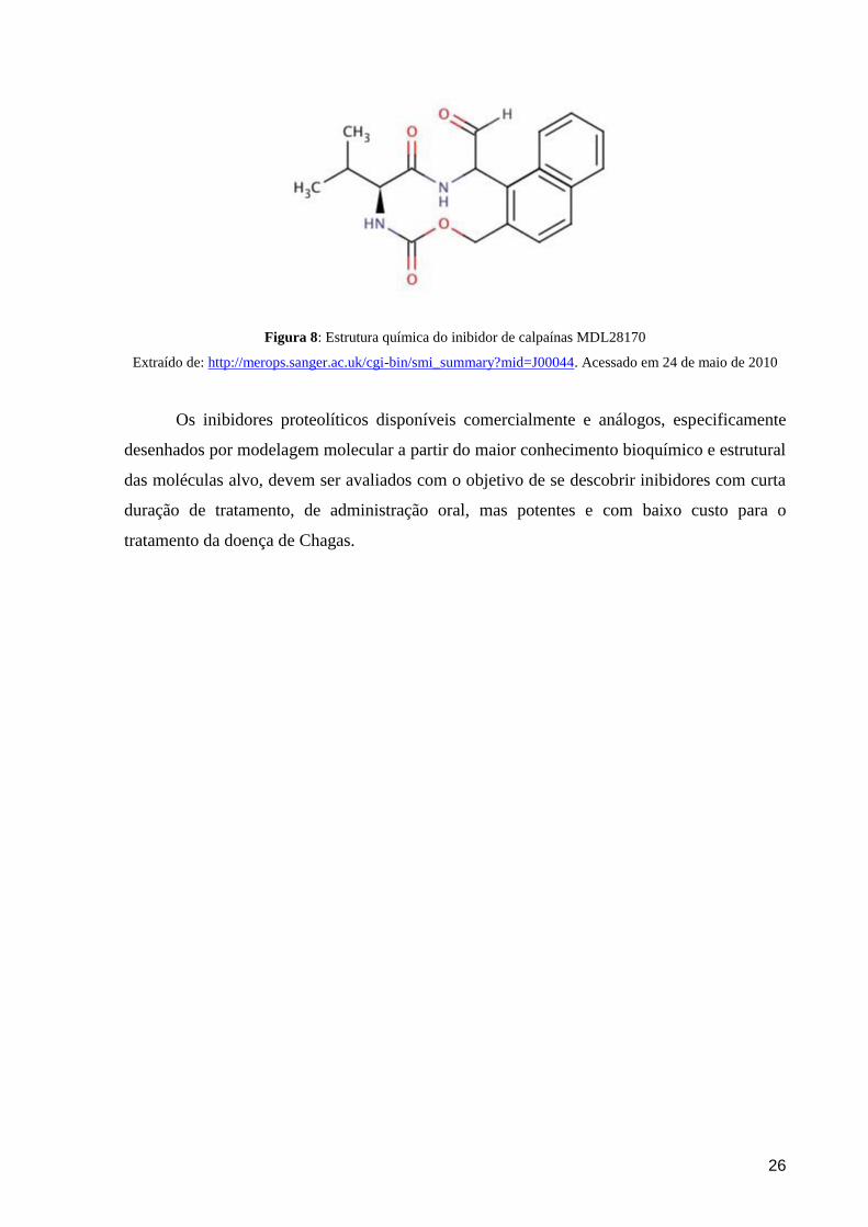
& KRENZELOK, 2008). O inibidor de calpaínas utilizado neste estudo é MDL28170 (Z-Val-
Phe-CHO), também conhecido como inibidor III de calpaínas. Este inibidor apresenta alta
permeabilidade celular, um mecanismo de ação reversível e atua de forma competitiva se
ligando ao sítio catalítico das calpaínas. A estrutura química do MDL28170 está representada
na figura 8.
26
Figura 8: Estrutura química do inibidor de calpaínas MDL28170
Extraído de: http://merops.sanger.ac.uk/cgi-bin/smi_summary?mid=J00044. Acessado em 24 de maio de 2010
Os inibidores proteolíticos disponíveis comercialmente e análogos, especificamente
desenhados por modelagem molecular a partir do maior conhecimento bioquímico e estrutural
das moléculas alvo, devem ser avaliados com o objetivo de se descobrir inibidores com curta
duração de tratamento, de administração oral, mas potentes e com baixo custo para o
tratamento da doença de Chagas.

27
2. OBJETIVOS
O presente trabalho tem como objetivo geral identificar moléculas similares às
calpaínas em Trypanosoma cruzi e avaliar a influência do inibidor III de calpaínas sobre o
ciclo de vida, diferenciação e infectividade destes flagelados.
28
3. RESULTADOS
Os resultados obtidos nesta dissertação deram origem aos seguintes artigos:
1. Sangenito LS#, Ennes-Vidal V#, Marinho FA, Da Mota FF, Santos ALS, d‟Avila-Levy
CM, Branquinha MH (2009) Arrested growth of Trypanosoma cruzi by the calpain inhibitor
MDL28170 and detection of calpain homologues in epimastigote forms. Parasitology
136(4):433-411. # Os autores contribuíram igualmente para este trabalho.
2. Ennes-Vidal V, Menna-Barreto RF, Santos ALS, Branquinha MH, d'Avila-Levy CM
(2010) Effects of the calpain inhibitor MDL28170 on the clinically relevant forms of
Trypanosoma cruzi in vitro. Journal of Antimicrobial Chemotherapy: in press.
3. Ennes-Vidal V, Menna-Barreto RF, Santos ALS, Branquinha MH, d'Avila-Levy CM
(2010) MDL28170, a potent calpain inhibitor, affects T. cruzi metacyclogenesis, ultrastructure
and attachment to the luminal midgut surface of Rhodnius prolixus. Submetido para
publicação na revista eletrônica Plos Neglected Tropical Diseases.
29
Artigo 1: Arrested growth of Trypanosoma cruzi by the calpain inhibitor MDL28170 and
detection of calpain homologues in epimastigote forms
Sangenito LS#, Ennes-Vidal V#, Marinho FA, Da Mota FF, Santos ALS, d‟Avila-Levy
CM, Branquinha MH.
# Esses autores contribuíram igualmente para este trabalho.
Parasitology 136(4):433-411, 2009
Este primeiro artigo trata dos primeiros 4 objetivos da dissertação, onde pudemos
identificar moléculas homólogas às calpaínas nas formas epimastigotas de diferentes
linhagens do Trypanosoma cruzi e verificamos o efeito do inibidor III (MDL28170) de
calpaínas sobre esses parasitos. A identificação das moléculas similares às calpaínas foi
verificada através da reatividade cruzada dos anticorpos direcionados contra as calpaínas de
Drosophila melanogaster e Trypanosoma brucei, observados por citometria de fluxo. O anti-
Dm-calpaína também foi capaz de reagir contra uma proteína com massa molecular ao redor
de 80 kDa no ensaio de Western Blotting. Quando analisamos a homologia existente entre
proteínas do T. cruzi e a proteína utilizada para gerar o anticorpo anti-calpaína de D.
melanogaster, nós pudemos observar que o parasita apresenta 4 seqüências com similaridade
à calpaína do inseto em seu genoma, e massa molecular predita em torno de 80 kDa, o que
reforça a possibilidade do anticorpo estar reconhecendo calpaínas no T. cruzi. O efeito do
inibidor III de calpaínas (MDL28170) foi verificado em epimastigotas de três linhagens
evolutivas diferentes do T. cruzi, com pequenas variações entre as mesmas. O inibidor teve
um efeito tripanostático, uma vez que epimastigotas lavados e cultivados em novo meio livre
de drogas retomam seu crescimento. Por fim, verificamos a queda na expressão da cruzipaína
pelo ensaio dec citometria de fluxo nos parasitos tratados com a concentração do IC50 do
MDL28170. Os resultados desse trabalho motivaram nosso grupo a continuar estudando a
presença desses homólogos de calpaínas ainda pouco caracterizados no T. cruzi e os efeitos do
inibidor de calpaínas sobre as diferentes formas evolutivas do parasito. A primeira autoria do
artigo ficou dividida com um aluno colaborador do Laboratório de Bioquímica de Proteases
do Instituto de Microbiologia da UFRJ, que desenvolveu um trabalho similar durante sua
monografia, uma vez que ambos contribuíram igualmente nos resultados e na preparação do
artigo.
Arrested growth of Trypanosoma cruzi by the calpain
inhibitor MDL28170 and detection of calpain
homologues in epimastigote forms
L. S. SANGENITO1#, V. ENNES-VIDAL2#, F. A. MARINHO1, F. F. DA MOTA3,
A. L. S. SANTOS1, C. M. D’AVILA-LEVY2 and M. H. BRANQUINHA1*
1Departamento de Microbiologia Geral, Instituto de Microbiologia Prof. Paulo de Goes, Universidade Federal do Rio deJaneiro, Rio de Janeiro, Brazil2Laboratorio de Biologia Molecular e Doencas Endemicas, Instituto Oswaldo Cruz, Fundacao Oswaldo Cruz, Rio de Janeiro,Brazil3Laboratorio de Genomica Funcional e Bioinformatica e Laboratorio de Biologia Computacional e Sistemas, Instituto OswaldoCruz, Fundacao Oswaldo Cruz, Rio de Janeiro, Brazil
(Received 14 November 2008; revised 26 December 2008 and 8 January 2009; accepted 9 January 2009)
SUMMARY
In this paper, we aimed to explore the effects of the calpain inhibitor III (MDL28170) and to detect calpain-like molecules
(CALPs) in epimastigote forms ofTrypanosoma cruzi isolate Dm28c. MDL28170 at 70 mM promoted a powerful reduction
in the growth rate after 48 h. The IC50 value was calculated to be 31.7 mM. This inhibitor promoted an increase in the
cellular volume, but not cell lysis, resulting in a trypanostatic effect. T. cruzi CALPs presented a strong cross-reactivity
with anti-Drosophila melanogaster calpain and anti-cytoskeleton-associated protein from Trypanosoma brucei antibodies,
and labelling was found mainly intracellularly. Furthermore, an 80 kDa reactive protein was detected by Western blotting
assays. No significant cross-reactivity was found with anti-human brain calpain antibody. The expression of CALPs was
decreased in cells kept for long periods in axenic cultures in comparison to a strain recently isolated frommice, as well as in
MDL28170-treated cells, the latter being paralleled by an increased expression of cruzipain. Different levels of CALPs
expression were also detected in distinct phylogenetic lineages, like Y strain (lineage TCI), Dm28c (TCII) and INPA6147
strain (Z3 zymodeme). These results may contribute for the investigation of the functions of CALPs in trypanosomatids.
Key words: Trypanosoma cruzi, calpain, peptidase, inhibitor.
INTRODUCTION
Trypanosoma cruzi is the aetiological agent of Chagas
disease, also known as American trypanosomiasis.
Chagas disease affects from 16 to 18million people in
the Americas, causing 21000 deaths yearly and 40
million are at risk of infection throughout Central
and South America (WHO, 2005). The life cycle of
the parasite involves several stages of differentiation;
in the mammalian host, the parasite has an obligate
intracellular replicative form, the amastigote, and a
non-replicative form, the bloodstream trypomasti-
gote. The major forms present in the insect vector
are also a replicative stage, the epimastigote, and a
non-replicative stage, the infective metacyclic trypo-
mastigote. The epimastigote form can be readily
obtained in axenic culture, and has been used for this
reason in most biochemical studies performed on the
parasite (Cazzulo et al. 2001).
Peptidases of microbial pathogens, including
T. cruzi, have attracted the attention of many lab-
oratories because of their roles in pathogenesis
(Cazzulo et al. 2001; Vermelho et al. 2007), but less is
known about the presence of a group of peptidases,
the calpains. These enzymes constitute a large family
of calcium-dependent cytosolic cysteine peptidases
that have been characterized mainly in humans
and that exist in 2 major isoforms, m-calpain and
m-calpain, which require millimolar and micromolar
concentrations of Ca2+, respectively, for their acti-
vation. The role of calpain remains poorly under-
stood, but it may participate in a variety of cellular
processes, including the rearrangement of cytoskel-
etal proteins, different signal transduction pathways
and apoptosis (Goll et al. 2003). Calpain activation in
humans seems to be increased during normal ageing
and in muscular dystrophy, cataract, arthritis and
Alzheimer’s disease, and in many acute traumas.
A variety of calpain inhibitors are under develop-
ment and the potential clinical utility of these
* Corresponding author: Departamento de MicrobiologiaGeral, Instituto de Microbiologia Prof. Paulo de Goes,Universidade Federal doRio de Janeiro (UFRJ), Centro deCiencias da Saude (CCS), Av. Carlos Chagas Filho, 373,Bloco I, Cidade Universitaria, Rio de Janeiro, RJ, 21941-902, Brazil. Tel : +55 21 2562 6743. Fax: +55 21 25608344. E-mail : [email protected]# These authors contributed equally to the work.
1
Parasitology, Page 1 of 9. f 2009 Cambridge University Press
doi:10.1017/S0031182009005629 Printed in the United Kingdom
inhibitors has been shown to treat Alzheimer’s dis-
ease (Battaglia et al. 2003) and to minimize neuronal
death after ischaemia (Hayes et al. 1998).
The presence of large numbers of calpain-related
proteins (CALPs) in trypanosomatids has been re-
vealed by some groups, although there are no data as
yet on the specific functions of the calpain-like pro-
teins in these microorganisms (Hertz-Fowler et al.
2001; Ramos et al. 2004; Ersfeld et al. 2005; Salotra
et al. 2006; Vergnes et al. 2007; Andrade et al. 2008).
Our group became involved in the study of these
proteins though the purification of a proteolytically
active CALP in the culture supernatant of the insect
trypanosomatid Crithidia deanei, yet its function was
not addressed (d’Avila-Levy et al. 2003). More re-
cently, our group investigated the effect of calpain
inhibitor III (MDL28170), a potent calpain inhibi-
tor, on the growth of Leishmania amazonensis. This
inhibitor promoted cellular alterations and arrested
the cellular growth of the parasite, and a CALP was
identified and detected on the cell surface of the
flagellate (d’Avila-Levy et al. 2006a).
In this paper, we extended our studies on the
presence of CALPs in trypanosomatids by in-
vestigating the inhibitory capability of MDL28170
against T. cruzi, aiming to show its effects on the
growth and morphology of epimastigote forms. In
addition, we have demonstrated the presence of
calpain homologues in 3 distinct T. cruzi strains.
MATERIALS AND METHODS
Parasites and cultivation
Epimastigotes of Trypanosoma cruziDm28c (lineage
TCII), Y (lineage TCI) and INPA 4167 (Z3 zymo-
deme) strains were grown in 3.7% brain heart
infusion medium supplemented with 10% heat-
inactivated fetal bovine serum at 28 xC for 4 days to
reach late-log phase growth. Alternatively, epimasti-
gote forms of Dm28c were obtained from the dif-
ferentiation of trypomastigotes after a blood passage
in mice; in this case, epimastigotes were kept for no
more than 3 passages in the axenic culture.
Effects of the calpain inhibitor on growth rate
and cell morphology
The effects of calpain inhibitor III (MDL28170;
Z-Val-Phe-CHO; purchased from Calbiochem,
San Diego, CA, USA) on epimastigotes of T. cruzi
were assessed by a method similar to that pre-
viously described elsewhere (d’Avila-Levy et al.
2006a). Briefly, epimastigotes were counted using a
Neubauer chamber and resuspended in fresh me-
dium to a final concentration of 5r106 viable epi-
mastigotes per ml. The viability was assessed by
mobility and lack of staining after challenging with
trypan blue. MDL28170 was added to the culture at
final concentrations in the 10–70 mM range (starting
from a 5 mM solution in dimethylsulfoxide (DMSO)
that was serially diluted in culture medium). A di-
lution of DMSO corresponding to that used to pre-
pare the highest drug concentration was assessed in
parallel. After incubation for 24, 48, 72 and 96 h at
28 xC, the number of viable, motile epimastigotes
was quantified by counting the flagellates in a
Neubauer chamber. Alternatively, parasites grown
for 72 h in the absence and in the presence of the
calpain inhibitor were washed 5 times in cold phos-
phate-buffered saline (PBS; 150 mM NaCl, 20 mM
phosphate buffer, pH 7.2) prior to resuspension in a
drug-free fresh medium and allowed to grow for
another 72 h, in order to evaluate the trypanocidal or
trypanostatic effect. The number of live epimasti-
gotes was evaluated as well as cell morphology (after
Giemsa staining) under optical microscopy at 24-h
intervals (Mendonca-Filho et al. 2004). The 50%
inhibitory concentration (IC50) was evaluated after
72 h. This value was determined by linear regression
analysis, by plotting the number of viable epimasti-
gotes versus log drug concentration by use of Origin
Pro 7.5 computer software.
Identification of CALPs by Western blotting
T. cruzi Dm28c cells were collected by centri-
fugation (500 g/5 min/4 xC) and washed 3 times with
cold PBS. Trypanosomes (108 cells) were resus-
pended in 100 ml of PBS and lysed by the addition of
1% SDS (Santos et al. 2006). TheDrosophila extracts
(kindly provided by Dr Alexandre Afranio Peixoto,
FIOCRUZ, RJ, Brazil) were prepared in the ex-
traction buffer (25 mM Hepes pH 7.2, 5 mM EDTA,
5 mMEGTA, 10 mM b-mercaptoethanol and 250 mM
sucrose). About 10 frozen flies were crushed, sus-
pended with 200 ml of cold extraction buffer and
homogenized in a Teflon/glass homogenizer with
10–12 strokes at 4 xC. The homogenate was carefully
mixed and centrifuged at 100000 g for 1 h at 4 xC.
The supernatant was stored at x80 xC. Protein
concentration was determined by the method de-
scribed by Lowry et al. (1951), using BSA as stan-
dard.
Immunoblot analysis was performed with total
cellular extracts equivalent to 100 mg of protein, as
previously described (d’Avila-Levy et al. 2005).
The primary antibody used was a rabbit antiserum
raised against Drosophila melanogaster calpain (anti-
Dm-calpain; Emori and Saigo, 1994), at a dilution
of 1 : 500. The secondary antibody used was peroxi-
dase-conjugated goat anti-rabbit IgG at a dilution
of 1 : 25 000 followed by chemiluminescence im-
munodetection after reaction with ECL reagents
(d’Avila-Levy et al. 2005). The relative molecular
mass of the reactive polypeptide was calculated by
comparison with the mobility of SDS–PAGE stan-
dards.
L. S. Sangenito and others 2
Flow cytometry and immunofluorescence microscopy
for calpain detection
Epimastigotes (1r107 cells) used for these exper-
iments were fixed at 4 xC in 0.4% paraformaldehyde
in PBS (pH 7.2) for 30 min, followed by extensive
washing in the same buffer. Alternatively, the fixed
cells were permeabilized by 0.01% Triton X-100
in PBS for 15 min at room temperature and then
washed twice in PBS. The fixed and permeabilized
cells maintained their morphological integrity, as
verified by optical microscopic observation. After
this step, the cells were incubated for 1 h at room
temperature with a 1 : 500 dilution of the following
polyclonal antibodies: rabbit anti-Dm-calpain poly-
clonal antibody (Emori and Saigo, 1994) ; anti-C21,
raised against the whole molecule of human brain
m-calpain (Grynspan et al. 1997) ; and anti-CAP5.5,
raised against the cytoskeleton-associated protein
from Trypanosoma brucei (Hertz-Fowler et al. 2001).
Cells were then incubated for an additional hour
with a 1 : 200 dilution of fluorescein isothiocyanate
(FITC)-labelled goat anti-rabbit IgG (d’Avila-Levy
et al. 2006b). The cells were then washed 3 times
in PBS and observed in a Zeiss epifluorescence
microscope (Axioplan 2). Alternatively, the parasite-
associated fluorescence was excited at 488 nm and
quantified on a flow cytometer (FACSCalibur, BD
Bioscience, USA) equipped with a 15 mW argon
laser emitting at 488 nm. Non-treated cells and those
treated with the secondary antibody alone were run
in parallel as controls. Each experimental population
was then mapped by using a two-parameter histo-
gram of forward-angle light scatter versus side scat-
ter. The mapped population (n=10 000) was then
analysed for log green fluorescence by using a single-
parameter histogram.
Effects of the calpain inhibitor on peptidase
expression
Epimastigotes (5r106 cells) of Dm28c were incu-
bated with MDL28170 at the IC50 concentration
for 24 h at 28 xC. Thereafter, cells were fixed and
processed for flow cytometry analysis as pre-
viously described, employing a 1 : 500 dilution of the
polyclonal antibodies anti-Dm-calpain and anti-
cruzipain, the latter raised against the major cysteine
peptidase of T. cruzi epimastigote forms (Cazzulo
et al. 2001).
Statistical analysis
All experiments were performed in triplicate, in 3
independent experimental sets. The data were ana-
lysed statistically by means of Student’s t-test using
EPI-INFO 6.04 (Database and Statistics Program
for Public Health) computer software. P values of
0.05 or less were considered statistically significant.
Sequence data analysis
A search for Dm-calpain homologous proteins in
T. cruzi was performed using the BlastP algorithm
and the nr database at NCBI (GenBank). The
theoretical molecular mass of homologous proteins
was calculated using the ExPASy Server facilities
(http://expasy.org). Identification of conserved do-
mains was performed using the CD-Search Tool and
Conserved Domain Database (CDD 27036 PSSMs)
at NCBI.
RESULTS
The calpain inhibitor III (MDL28170) was added to
T. cruzi clone Dm28c epimastigote forms in con-
centrations ranging from 20 to 70 mM, and the cellular
growth was monitored for 4 days in vitro. Our results
showed that MDL28170 arrested the growth in a
dose-dependent manner (Fig. 1A). The lowest con-
centration of the drug (20 mM) presented a significant
inhibitory effect only after 72 h of growth, but the
remaining concentrations promoted reduced levels
of growth that were statistically significant after 48 h
(Fig. 1A). The calpain inhibitor at 50 mM decreased
the growth rate by approximately 70–75% in the
48–96 h interval, and at 70 mM promoted a reduction
in the cellular growth rate of around 91% after 48 h,
and this ratio was also maintained up to 96 h of
growth (Fig. 1A). Conversely, DMSO did not sig-
nificantly affect the parasite growth behaviour
(Fig. 1A). The same conditions performed for
Dm28c were employed for the analysis of the effects
of MDL28170 upon Y and INPA4167 strains, and
similar profiles of growth inhibition were observed
(data not shown). These strains were chosen to rep-
resent members of distinct T. cruzi lineages: TCI
(Y strain), TCII (Dm28c) and Z3 zymodeme
(INPA4167 strain). The IC50 was calculated for each
strain, and similar values were detected: 31.7 mM(Dm28c), 34.3 mM (Y) and 37.4 mM (INPA4167).
The anti-trypanosomal activity of this inhibitor
was reversible, since T. cruzi cells pre-treated for
72 h with the calpain inhibitor at 70 mM resumed
growth when subcultured in a drug-free fresh
medium (data not shown). Optical microscopy ob-
servations of T. cruzi Dm28c cells treated with the
calpain inhibitor in the 20–70 mM range revealed,
irrespective to the drug concentration, an increase in
the cell volume, with the flagellates becoming round
and some of them presenting no detectable flagellum
(Fig. 1C, b–i), but no cell lysis was observed, corrob-
orating the trypanostatic effect on the epimastigote
growth. Cells cultured in the presence of DMSO at a
dose used to dissolve the highest concentration of
MDL28170 did not present any detectable mor-
phological alterations (data not shown).
As demonstrated by flow cytometry and fluor-
escence microscopy analyses, the anti-Dm-calpain
Effects of the calpain inhibitor MDL28170 on T. cruzi 3
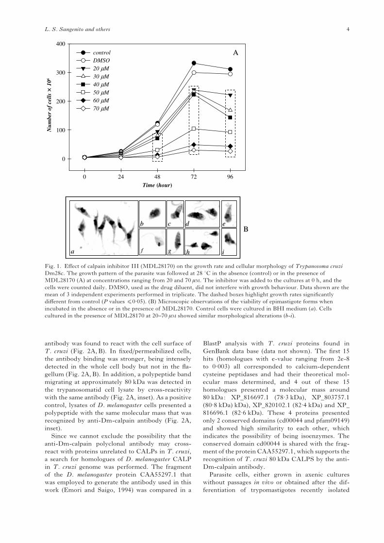
antibody was found to react with the cell surface of
T. cruzi (Fig. 2A,B). In fixed/permeabilized cells,
the antibody binding was stronger, being intensely
detected in the whole cell body but not in the fla-
gellum (Fig. 2A,B). In addition, a polypeptide band
migrating at approximately 80 kDa was detected in
the trypanosomatid cell lysate by cross-reactivity
with the same antibody (Fig. 2A, inset). As a positive
control, lysates of D. melanogaster cells presented a
polypeptide with the same molecular mass that was
recognized by anti-Dm-calpain antibody (Fig. 2A,
inset).
Since we cannot exclude the possibility that the
anti-Dm-calpain polyclonal antibody may cross-
react with proteins unrelated to CALPs in T. cruzi,
a search for homologues of D. melanogaster CALP
in T. cruzi genome was performed. The fragment
of the D. melanogaster protein CAA55297.1 that
was employed to generate the antibody used in this
work (Emori and Saigo, 1994) was compared in a
BlastP analysis with T. cruzi proteins found in
GenBank data base (data not shown). The first 15
hits (homologues with e-value ranging from 2e-8
to 0.003) all corresponded to calcium-dependent
cysteine peptidases and had their theoretical mol-
ecular mass determined, and 4 out of these 15
homologues presented a molecular mass around
80 kDa: XP_816697.1 (78.3 kDa), XP_803757.1
(80.8 kDa) kDa), XP_820102.1 (82.4 kDa) and XP_
816696.1 (82.6 kDa). These 4 proteins presented
only 2 conserved domains (cd00044 and pfam09149)
and showed high similarity to each other, which
indicates the possibility of being isoenzymes. The
conserved domain cd00044 is shared with the frag-
ment of the protein CAA55297.1, which supports the
recognition of T. cruzi 80 kDa CALPS by the anti-
Dm-calpain antibody.
Parasite cells, either grown in axenic cultures
without passages in vivo or obtained after the dif-
ferentiation of trypomastigotes recently isolated
Time (hour)0 24 48 72 96
Num
ber
of c
ells
× 1
06
0
100
200
300
400
controlDMSO20 µM30 µM40 µM50 µM60 µM70 µM
AA
aa
bb cc dd ee
ff gg hh ii
BB
Fig. 1. Effect of calpain inhibitor III (MDL28170) on the growth rate and cellular morphology of Trypanosoma cruzi
Dm28c. The growth pattern of the parasite was followed at 28 xC in the absence (control) or in the presence of
MDL28170 (A) at concentrations ranging from 20 and 70 mM. The inhibitor was added to the cultures at 0 h, and the
cells were counted daily. DMSO, used as the drug diluent, did not interfere with growth behaviour. Data shown are the
mean of 3 independent experiments performed in triplicate. The dashed boxes highlight growth rates significantly
different from control (P values f0.05). (B) Microscopic observations of the viability of epimastigote forms when
incubated in the absence or in the presence of MDL28170. Control cells were cultured in BHI medium (a). Cells
cultured in the presence of MDL28170 at 20–70 mM showed similar morphological alterations (b-i).
L. S. Sangenito and others 4
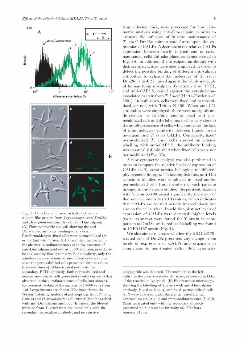
from infected mice, were processed for flow cyto-
metric analysis using anti-Dm-calpain in order to
estimate the influence of in vitro maintenance of
T. cruzi Dm28c epimastigote forms upon the ex-
pression of CALPs. A decrease in the relative CALPs
expression between newly isolated and in vitro-
maintained cells did take place, as demonstrated in
Fig. 3A. In addition, 2 anti-calpain antibodies with
distinct specificities were also employed in order to
detect the possible binding of different anti-calpain
antibodies to calpain-like molecules of T. cruzi
Dm28c: anti-C21, raised against the whole molecule
of human brain m-calpain (Grynspan et al. 1997) ;
and anti-CAP5.5, raised against the cytoskeleton-
associated protein fromT. brucei (Hertz-Fowler et al.
2001). In both cases, cells were fixed and permeabi-
lized, or not, with Triton X-100. When anti-C21
antibodies were employed, there were no significant
differences in labelling among fixed and per-
meabilized cells and the labelling itself is very close to
the autofluorescence of cells, which indicates the lack
of immunological similarity between human brain
m-calpain and T. cruzi CALPs. Conversely, fixed/
permeabilized T. cruzi cells showed an intense
labelling with anti-CAP5.5; the antibody binding
was drastically diminished when fixed cells were not
permeabilized (Fig. 3B).
A flow cytometric analysis was also performed in
order to compare the relative levels of expression of
CALPs in T. cruzi strains belonging to different
phylogenetic lineages. To accomplish this, anti-Dm
calpain antibodies were employed in fixed and/or
permeabilized cells from members of each parasite
lineage. In the 3 strains studied, the permeabilization
with Triton X-100 raised significantly the mean of
fluorescence intensity (MFI) values, which indicates
that CALPs are located mainly intracellularly but
also on the cell surface. In addition, distinct levels of
expression of CALPs were detected: higher levels
(twice as many) were found for Y strain in com-
parison to Dm28c, and a reduced labelling was found
in INPA4167 strain (Fig. 4).
We also aimed to assess whether the MDL28170-
treated cells of Dm28c presented any change in the
levels of expression of CALPs and cruzipain in
comparison to non-treated cells. Flow cytometry
BB
a b
c d
e f
101033
128128
001010 00 101011 1010 22
FluorescenceFluorescence intensityintensity
Eve
nts
Eve
nts
autofluorescenceautofluorescencenpnp
pp
8080
aa bb cc
AA
Fig. 2. Detection of cross-reactivity between a
calpain-like protein from Trypanosoma cruzi Dm28c
and Drosophila melanogaster calpain (Dm-calpain).
(A) Flow cytometric analysis showing the anti-
Dm-calpain antibody binding to T. cruzi.
Paraformaldehyde-fixed cells were permeabilized (p)
or not (np) with Triton X-100 and then incubated in
the absence (autofluorescence) or in the presence of
anti-Dm-calpain antibody at 1 : 500 dilution, in order to
be analysed by flow cytometry. For simplicity, only the
autofluorescence of non-permeabilized cells is shown,
since the permeabilized cells presented similar values
(data not shown). When treated only with the
secondary-FITC antibody, both permeabilized and
non-permeabilized cells generated similar curves to that
observed in the autofluorescence of cells (not shown).
Representative data of the analysis of 10 000 cells from
1 of 3 experiments are shown. The inset shows the
Western blotting analysis of polypeptides from T. cruzi
(lane a) and D. melanogaster cell extract (lane b) probed
with anti-Dm-calpain antibody. In lane c, the blotted
proteins from T. cruzi were incubated only with the
secondary-peroxidase antibody, and no reactive
polypeptide was detected. The number on the left
indicates the apparent molecular mass, expressed in kDa,
of the reactive polypeptide. (B) Fluorescence microscopy
showing the labelling of T. cruzi with anti-Dm-calpain
antibody. Fixed cells (a–d) and fixed-permeabilized cells
(e, f) were analysed under differential interferential
contrast images (a, c, e) and immunofluorescence (b, d, f).
Parasites treated only with the secondary antibody
presented no fluorescence intensity (b). The bars
represent 1 mm.
Effects of the calpain inhibitor MDL28170 on T. cruzi 5
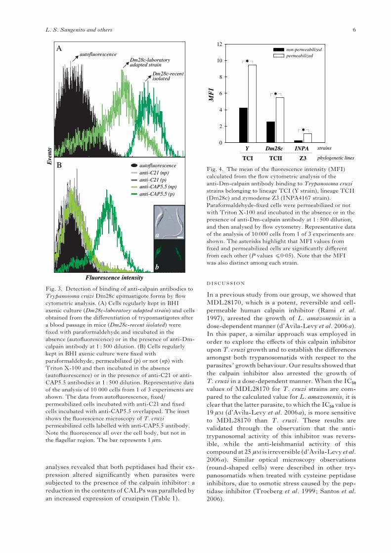
analyses revealed that both peptidases had their ex-
pression altered significantly when parasites were
subjected to the presence of the calpain inhibitor: a
reduction in the contents of CALPswas paralleled by
an increased expression of cruzipain (Table 1).
DISCUSSION
In a previous study from our group, we showed that
MDL28170, which is a potent, reversible and cell-
permeable human calpain inhibitor (Rami et al.
1997), arrested the growth of L. amazonensis in a
dose-dependent manner (d’Avila-Levy et al. 2006a).
In this paper, a similar approach was employed in
order to explore the effects of this calpain inhibitor
upon T. cruzi growth and to establish the differences
amongst both trypanosomatids with respect to the
parasites’ growth behaviour. Our results showed that
the calpain inhibitor also arrested the growth of
T. cruzi in a dose-dependent manner. When the IC50
values of MDL28170 for T. cruzi strains are com-
pared to the calculated value for L. amazonensis, it is
clear that the latter parasite, to which the IC50 value is
19 mM (d’Avila-Levy et al. 2006a), is more sensitive
to MDL28170 than T. cruzi. These results are
validated through the observation that the anti-
trypanosomal activity of this inhibitor was revers-
ible, while the anti-leishmanial activity of this
compoundat 25 mMis irreversible (d’Avila-Levy et al.
2006a). Similar optical microscopy observations
(round-shaped cells) were described in other try-
panosomatids when treated with cysteine peptidase
inhibitors, due to osmotic stress caused by the pep-
tidase inhibitor (Troeberg et al. 1999; Santos et al.
2006).
MF
I
0
2
4
6
8
10
12non-permeabilizedpermeabilized
TCITCI TCIITCII Z3Z3
Y Y Dm28c Dm28c INPA INPA strainsstrains
phylogeneticphylogenetic lineslines
Fig. 4. The mean of the fluorescence intensity (MFI)
calculated from the flow cytometric analysis of the
anti-Dm-calpain antibody binding to Trypanosoma cruzi
strains belonging to lineage TCI (Y strain), lineage TCII
(Dm28c) and zymodeme Z3 (INPA4167 strain).
Paraformaldehyde-fixed cells were permeabilized or not
with Triton X-100 and incubated in the absence or in the
presence of anti-Dm-calpain antibody at 1 : 500 dilution,
and then analysed by flow cytometry. Representative data
of the analysis of 10 000 cells from 1 of 3 experiments are
shown. The asterisks highlight that MFI values from
fixed and permeabilized cells are significantly different
from each other (P values f0.05). Note that the MFI
was also distinct among each strain.
autofluorescenceautofluorescenceDm28cDm28c--laboratorylaboratoryadapted strainadapted strain
Dm28cDm28c--recent recent isolatedisolated
autofluorescenceautofluorescence
antianti--C21 (C21 (npnp))antianti--C21 (p)C21 (p)antianti--CAP5.5 (CAP5.5 (npnp))
antianti--CAP5.5 (p)CAP5.5 (p)
FluorescenceFluorescence intensityintensity
Eve
nts
Eve
nts
AA
BB
a
b
Fig. 3. Detection of binding of anti-calpain antibodies to
Trypanosoma cruzi Dm28c epimastigote forms by flow
cytometric analysis. (A) Cells regularly kept in BHI
axenic culture (Dm28c-laboratory adapted strain) and cells
obtained from the differentiation of trypomastigotes after
a blood passage in mice (Dm28c-recent isolated) were
fixed with paraformaldehyde and incubated in the
absence (autofluorescence) or in the presence of anti-Dm-
calpain antibody at 1 : 500 dilution. (B) Cells regularly
kept in BHI axenic culture were fixed with
paraformaldehyde, permeabilized (p) or not (np) with
Triton X-100 and then incubated in the absence
(autofluorescence) or in the presence of anti-C21 or anti-
CAP5.5 antibodies at 1 : 500 dilution. Representative data
of the analysis of 10 000 cells from 1 of 3 experiments are
shown. The data from autofluorescence, fixed/
permeabilized cells incubated with anti-C21 and fixed
cells incubated with anti-CAP5.5 overlapped. The inset
shows the fluorescence microscopy of T. cruzi
permeabilized cells labelled with anti-CAP5.5 antibody.
Note the fluorescence all over the cell body, but not in
the flagellar region. The bar represents 1 mm.
L. S. Sangenito and others 6
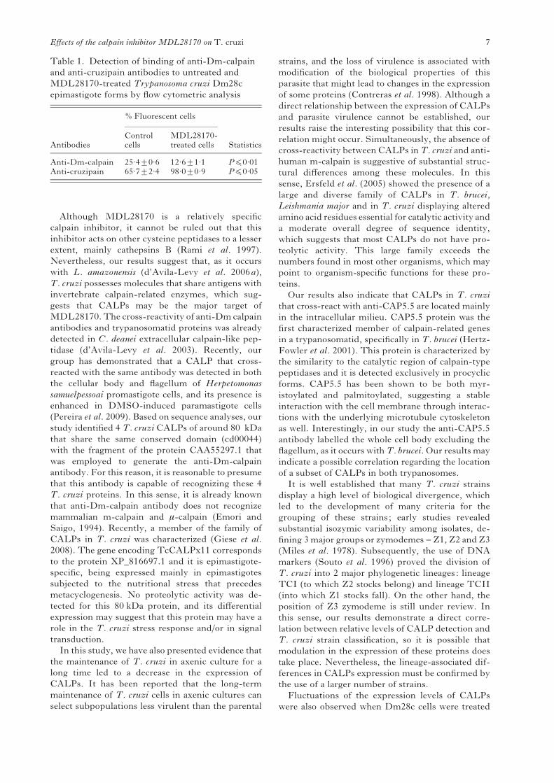
Although MDL28170 is a relatively specific
calpain inhibitor, it cannot be ruled out that this
inhibitor acts on other cysteine peptidases to a lesser
extent, mainly cathepsins B (Rami et al. 1997).
Nevertheless, our results suggest that, as it occurs
with L. amazonensis (d’Avila-Levy et al. 2006a),
T. cruzi possesses molecules that share antigens with
invertebrate calpain-related enzymes, which sug-
gests that CALPs may be the major target of
MDL28170. The cross-reactivity of anti-Dm calpain
antibodies and trypanosomatid proteins was already
detected in C. deanei extracellular calpain-like pep-
tidase (d’Avila-Levy et al. 2003). Recently, our
group has demonstrated that a CALP that cross-
reacted with the same antibody was detected in both
the cellular body and flagellum of Herpetomonas
samuelpessoai promastigote cells, and its presence is
enhanced in DMSO-induced paramastigote cells
(Pereira et al. 2009). Based on sequence analyses, our
study identified 4 T. cruzi CALPs of around 80 kDa
that share the same conserved domain (cd00044)
with the fragment of the protein CAA55297.1 that
was employed to generate the anti-Dm-calpain
antibody. For this reason, it is reasonable to presume
that this antibody is capable of recognizing these 4
T. cruzi proteins. In this sense, it is already known
that anti-Dm-calpain antibody does not recognize
mammalian m-calpain and m-calpain (Emori and
Saigo, 1994). Recently, a member of the family of
CALPs in T. cruzi was characterized (Giese et al.
2008). The gene encoding TcCALPx11 corresponds
to the protein XP_816697.1 and it is epimastigote-
specific, being expressed mainly in epimastigotes
subjected to the nutritional stress that precedes
metacyclogenesis. No proteolytic activity was de-
tected for this 80 kDa protein, and its differential
expression may suggest that this protein may have a
role in the T. cruzi stress response and/or in signal
transduction.
In this study, we have also presented evidence that
the maintenance of T. cruzi in axenic culture for a
long time led to a decrease in the expression of
CALPs. It has been reported that the long-term
maintenance of T. cruzi cells in axenic cultures can
select subpopulations less virulent than the parental
strains, and the loss of virulence is associated with
modification of the biological properties of this
parasite that might lead to changes in the expression
of some proteins (Contreras et al. 1998). Although a
direct relationship between the expression of CALPs
and parasite virulence cannot be established, our
results raise the interesting possibility that this cor-
relation might occur. Simultaneously, the absence of
cross-reactivity between CALPs inT. cruzi and anti-
human m-calpain is suggestive of substantial struc-
tural differences among these molecules. In this
sense, Ersfeld et al. (2005) showed the presence of a
large and diverse family of CALPs in T. brucei,
Leishmania major and in T. cruzi displaying altered
amino acid residues essential for catalytic activity and
a moderate overall degree of sequence identity,
which suggests that most CALPs do not have pro-
teolytic activity. This large family exceeds the
numbers found in most other organisms, which may
point to organism-specific functions for these pro-
teins.
Our results also indicate that CALPs in T. cruzi
that cross-react with anti-CAP5.5 are located mainly
in the intracellular milieu. CAP5.5 protein was the
first characterized member of calpain-related genes
in a trypanosomatid, specifically in T. brucei (Hertz-
Fowler et al. 2001). This protein is characterized by
the similarity to the catalytic region of calpain-type
peptidases and it is detected exclusively in procyclic
forms. CAP5.5 has been shown to be both myr-
istoylated and palmitoylated, suggesting a stable
interaction with the cell membrane through interac-
tions with the underlying microtubule cytoskeleton
as well. Interestingly, in our study the anti-CAP5.5
antibody labelled the whole cell body excluding the
flagellum, as it occurs withT. brucei. Our results may
indicate a possible correlation regarding the location
of a subset of CALPs in both trypanosomes.
It is well established that many T. cruzi strains
display a high level of biological divergence, which
led to the development of many criteria for the
grouping of these strains; early studies revealed
substantial isozymic variability among isolates, de-
fining 3major groups or zymodemes – Z1, Z2 and Z3
(Miles et al. 1978). Subsequently, the use of DNA
markers (Souto et al. 1996) proved the division of
T. cruzi into 2 major phylogenetic lineages: lineage
TCI (to which Z2 stocks belong) and lineage TCII
(into which Z1 stocks fall). On the other hand, the
position of Z3 zymodeme is still under review. In
this sense, our results demonstrate a direct corre-
lation between relative levels of CALP detection and
T. cruzi strain classification, so it is possible that
modulation in the expression of these proteins does
take place. Nevertheless, the lineage-associated dif-
ferences in CALPs expression must be confirmed by
the use of a larger number of strains.
Fluctuations of the expression levels of CALPs
were also observed when Dm28c cells were treated
Table 1. Detection of binding of anti-Dm-calpain
and anti-cruzipain antibodies to untreated and
MDL28170-treated Trypanosoma cruzi Dm28c
epimastigote forms by flow cytometric analysis
Antibodies
% Fluorescent cells
StatisticsControlcells
MDL28170-treated cells
Anti-Dm-calpain 25.4¡0.6 12.6¡1.1 Pf0.01Anti-cruzipain 65.7¡2.4 98.0¡0.9 Pf0.05
Effects of the calpain inhibitor MDL28170 on T. cruzi 7
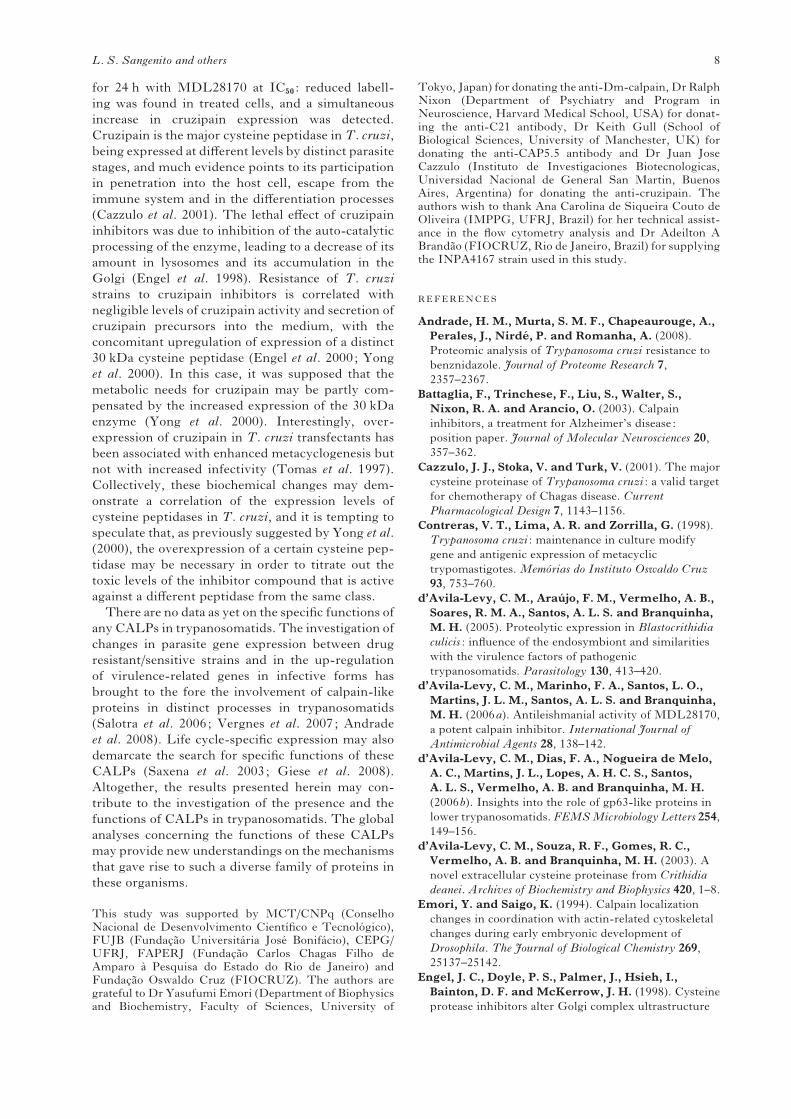
for 24 h with MDL28170 at IC50 : reduced labell-
ing was found in treated cells, and a simultaneous
increase in cruzipain expression was detected.
Cruzipain is the major cysteine peptidase inT. cruzi,
being expressed at different levels by distinct parasite
stages, and much evidence points to its participation
in penetration into the host cell, escape from the
immune system and in the differentiation processes
(Cazzulo et al. 2001). The lethal effect of cruzipain
inhibitors was due to inhibition of the auto-catalytic
processing of the enzyme, leading to a decrease of its
amount in lysosomes and its accumulation in the
Golgi (Engel et al. 1998). Resistance of T. cruzi
strains to cruzipain inhibitors is correlated with
negligible levels of cruzipain activity and secretion of
cruzipain precursors into the medium, with the
concomitant upregulation of expression of a distinct
30 kDa cysteine peptidase (Engel et al. 2000; Yong
et al. 2000). In this case, it was supposed that the
metabolic needs for cruzipain may be partly com-
pensated by the increased expression of the 30 kDa
enzyme (Yong et al. 2000). Interestingly, over-
expression of cruzipain in T. cruzi transfectants has
been associated with enhanced metacyclogenesis but
not with increased infectivity (Tomas et al. 1997).
Collectively, these biochemical changes may dem-
onstrate a correlation of the expression levels of
cysteine peptidases in T. cruzi, and it is tempting to
speculate that, as previously suggested by Yong et al.
(2000), the overexpression of a certain cysteine pep-
tidase may be necessary in order to titrate out the
toxic levels of the inhibitor compound that is active
against a different peptidase from the same class.
There are no data as yet on the specific functions of
any CALPs in trypanosomatids. The investigation of
changes in parasite gene expression between drug
resistant/sensitive strains and in the up-regulation
of virulence-related genes in infective forms has
brought to the fore the involvement of calpain-like
proteins in distinct processes in trypanosomatids
(Salotra et al. 2006; Vergnes et al. 2007; Andrade
et al. 2008). Life cycle-specific expression may also
demarcate the search for specific functions of these
CALPs (Saxena et al. 2003; Giese et al. 2008).
Altogether, the results presented herein may con-
tribute to the investigation of the presence and the
functions of CALPs in trypanosomatids. The global
analyses concerning the functions of these CALPs
may provide new understandings on the mechanisms
that gave rise to such a diverse family of proteins in
these organisms.
This study was supported by MCT/CNPq (ConselhoNacional de Desenvolvimento Cientıfico e Tecnologico),FUJB (Fundacao Universitaria Jose Bonifacio), CEPG/UFRJ, FAPERJ (Fundacao Carlos Chagas Filho deAmparo a Pesquisa do Estado do Rio de Janeiro) andFundacao Oswaldo Cruz (FIOCRUZ). The authors aregrateful to Dr Yasufumi Emori (Department of Biophysicsand Biochemistry, Faculty of Sciences, University of
Tokyo, Japan) for donating the anti-Dm-calpain, DrRalphNixon (Department of Psychiatry and Program inNeuroscience, Harvard Medical School, USA) for donat-ing the anti-C21 antibody, Dr Keith Gull (School ofBiological Sciences, University of Manchester, UK) fordonating the anti-CAP5.5 antibody and Dr Juan JoseCazzulo (Instituto de Investigaciones Biotecnologicas,Universidad Nacional de General San Martin, BuenosAires, Argentina) for donating the anti-cruzipain. Theauthors wish to thank Ana Carolina de Siqueira Couto deOliveira (IMPPG, UFRJ, Brazil) for her technical assist-ance in the flow cytometry analysis and Dr Adeilton ABrandao (FIOCRUZ, Rio de Janeiro, Brazil) for supplyingthe INPA4167 strain used in this study.
REFERENCES
Andrade, H. M., Murta, S. M. F., Chapeaurouge, A.,
Perales, J., Nirde, P. and Romanha, A. (2008).
Proteomic analysis of Trypanosoma cruzi resistance to
benznidazole. Journal of Proteome Research 7,
2357–2367.
Battaglia, F., Trinchese, F., Liu, S., Walter, S.,
Nixon, R. A. and Arancio, O. (2003). Calpain
inhibitors, a treatment for Alzheimer’s disease:
position paper. Journal of Molecular Neurosciences 20,
357–362.
Cazzulo, J. J., Stoka, V. and Turk, V. (2001). The major
cysteine proteinase of Trypanosoma cruzi : a valid target
for chemotherapy of Chagas disease. Current
Pharmacological Design 7, 1143–1156.
Contreras, V. T., Lima, A. R. and Zorrilla, G. (1998).
Trypanosoma cruzi : maintenance in culture modify
gene and antigenic expression of metacyclic
trypomastigotes. Memorias do Instituto Oswaldo Cruz
93, 753–760.
d’Avila-Levy, C. M., Araujo, F. M., Vermelho, A. B.,
Soares, R. M. A., Santos, A. L. S. and Branquinha,
M. H. (2005). Proteolytic expression in Blastocrithidia
culicis : influence of the endosymbiont and similarities
with the virulence factors of pathogenic
trypanosomatids. Parasitology 130, 413–420.
d’Avila-Levy, C. M., Marinho, F. A., Santos, L. O.,
Martins, J. L. M., Santos, A. L. S. and Branquinha,
M. H. (2006a). Antileishmanial activity of MDL28170,
a potent calpain inhibitor. International Journal of
Antimicrobial Agents 28, 138–142.
d’Avila-Levy, C. M., Dias, F. A., Nogueira de Melo,
A. C., Martins, J. L., Lopes, A. H. C. S., Santos,
A. L. S., Vermelho, A. B. and Branquinha, M. H.
(2006b). Insights into the role of gp63-like proteins in
lower trypanosomatids.FEMSMicrobiology Letters 254,
149–156.
d’Avila-Levy, C. M., Souza, R. F., Gomes, R. C.,
Vermelho, A. B. and Branquinha, M. H. (2003). A
novel extracellular cysteine proteinase from Crithidia
deanei. Archives of Biochemistry and Biophysics 420, 1–8.
Emori, Y. and Saigo, K. (1994). Calpain localization
changes in coordination with actin-related cytoskeletal
changes during early embryonic development of
Drosophila. The Journal of Biological Chemistry 269,
25137–25142.
Engel, J. C., Doyle, P. S., Palmer, J., Hsieh, I.,
Bainton, D. F. and McKerrow, J. H. (1998). Cysteine
protease inhibitors alter Golgi complex ultrastructure
L. S. Sangenito and others 8
and function in Trypanosoma cruzi. The Journal of Cell
Science 111, 597–606.
Engel, J. C., Torres, C., Hsieh, I., Doyle, P. S. and
McKerrow, J. H. (2000). Upregulation of the secretory
pathway in cysteine protease inhibitor-resistant
Trypanosoma cruzi. The Journal of Cell Science 113,
1345–1354.
Ersfeld, K., Barraclough, H. and Gull, K. (2005).
Evolutionary relationships and protein domain
architecture in an expanded calpain superfamily in
kinetoplastid parasites. The Journal of Molecular
Evolution 61, 742–757.
Giese, V., Dallagiovanna, B., Marchini, F. K.,
Pavoni, D. P., Krieger, M. A. and Goldenberg, S.
(2008). Trypanosoma cruzi : a stage-specific calpain-like
protein is induced after various kinds of stress.Memorias
do Instituto Oswaldo Cruz 103, 598–601.
Goll, D. E., Thompson, V. F., Li, H., Wei, W. and
Cong, J. (2003). The calpain system. Physiological
Reviews 83, 731–801.
Grynspan,F.,Griffin,W. R., Cataldo,A.,Katayama, S.
and Nixon, R. A. (1997). Active site-directed
antibodies identify calpain II as an early-appearing
and pervasive component of neurofibrillary
pathology in Alzheimer’s disease. Brain Research 763,
145–158.
Hayes, R. L., Wang, K. K., Kampfl, A.,
Posmantur, R. M., Newcomb, J. K. and
Clifton, G. L. (1998). Potential contribution of
proteases to neuronal damage. Drug News Perspectives
11, 215–222.
Hertz-Fowler, C., Ersfeld, K. and Gull, K. (2001).
CAP5.5, a life-cycle-regulated, cytoskeleton-associated
protein is a member of a novel family of calpain-related
proteins in Trypanosoma brucei. Molecular and
Biochemical Parasitology 116, 25–34.
Lowry, O. H., Roseborough, N. J., Farr, A. L. and
Randall, R. J. (1951). Protein measurement with the
Folin phenol reagent. The Journal of Biological
Chemistry 193, 264–275.
Mendonca-Filho, R. R., Rodrigues, I. A.,
Alviano, D. S., Santos, A. L. S., Soares, R. M. A.,
Alviano, C. S., Lopes, A. H. C. S. and Rosa, M. S.
(2004). Leishmanicidal activity of polyphenolic-rich
extract from husk fiber of Cocos nucifera Linn. (Palmae).
Research in Microbiology 155, 136–143.
Miles, M. A., Souza, A., Povoa, M., Shaw, J. J.,
Lainson, R. and Toye, P. J. (1978). Isozymic
heterogeneity of Trypanosoma cruzi in the first
autochtonous patients with Chagas’ disease in
Amazonian Brazil. Nature, London 272, 819–821.
Pereira, F. M., Elias, C. G. R., d’Avila-Levy, C. M.,
Branquinha, M. H. and Santos, A. L. S. (2009).
Cysteine peptidases in Herpetomonas samuelpessoai are
modulated by temperature and dimethylsulfoxide-
triggered differentiation. Parasitology 136, 45–54.
Rami, A., Ferger, D. and Krieglstein, J. (1997).
Blockade of calpain proteolytic activity rescues neurons
from glutamate excitotoxicity. Neuroscience Research 27,
93–97.
Ramos, C. S., Franco, F. A. L., Smith, D. F. and
Uliana, S. R. B. (2004). Characterisation of a new
Leishmania META gene and genomic analysis of the
META cluster. FEMS Microbiology Letters 238,
213–219.
Salotra, P., Duncan, R. C., Singh, R., Raju, B. V. S.,
Sreenivas, G. and Nakhasi, H. L. (2006).
Upregulation of surface proteins in Leishmania donovani
isolated from patients of post kala-azar dermal
leishmaniasis. Microbes and Infection 8, 637–644.
Santos, A. L. S., d’Avila-Levy, C. M., Dias, F. A.,
Ribeiro, R. O., Pereira, F. M., Elias, C. G.,
Souto-Padron, T., Lopes, A. H., Alviano, C. S.,
Branquinha, M. H. and Soares, R. M. A. (2006).
Phytomonas serpens : cysteine peptidase inhibitors
interfere with growth, ultrastructure and host adhesion.
International Journal for Parasitology 36, 47–56.
Saxena, A., Worthey, E. A., Yan, S., Leland, A.,
Stuart, K. D. and Myler, P. J. (2003). Evaluation of
differential gene expression inLeishmania majorFriedlin
procyclics and metacyclics using DNA microarray
analysis. Molecular and Biochemical Parasitology 129,
103–114.
Souto, R. P., Fernandes, O., Macedo, A. M.,
Campbell, D. A. and Zingales, B. (1996). DNA
markers define two major phylogenetic lineages of
Trypanosoma cruzi. Molecular and Biochemical
Parasitology 83, 141–152.
Tomas, A. M., Miles, M. A. and Kelly, J. M. (1997).
Overexpression of cruzipain, the major cysteine
proteinase of Trypanosoma cruzi, is associated with
enhanced metacyclogenesis. European Journal of
Biochemistry 244, 596–603.
Troeberg, L., Morty, R. E., Pike, R. N., Lonsdale-
Eccles, J. D., Palmer, J. T., McKerrow, J. H. and
Coetzer, T. H. (1999). Cysteine proteinase inhibitors
kill cultured bloodstream forms of Trypanosoma brucei
brucei. Experimental Parasitology 91, 349–355.
Vergnes, B., Gourbal, B., Gorard, I., Sundar, S.,
Drummelsmith, J. and Ouellette, M. (2007).
A proteomics screen implicates HSP83 and a small
kinetoplastid calpain-related protein in drug resistance
in Leishmania donovani clinical field isolates by
modulating drug-induced programmed cell death.
Molecular Cell Proteomics 6, 88–101.
Vermelho, A. B., Giovanni-de-Simone, S.,
d’Avila-Levy, C. M., Santos, A. L. S., Melo, A. C. N.,
Silva Jr, F. P., Bom, E. P. S. and Branquinha, M. H.
(2007). Trypanosomatidae peptidases: a target for drugs
development. Current Enzyme Inhibition 3, 19–48.
World Health Organization (2005). Infectious Diseases
Home Burdens and Trends. Available from: <http://
www.who.int/ctd/chagas/burdens.htm>.
Yong, V., Schmitz, V., Vannier-Santos, M. A.,
Lima, A. P. C. A., Lalmanach, G., Juliano, L.,
Gauthier, F. and Scharfstein, J. (2000). Altered
expression of cruzipain and a cathepsin B-like target in a
Trypanosoma cruzi cell line displaying resistance to
synthetic inhibitors of cysteine-proteinases. Molecular
and Biochemical Parasitology 109, 47–59.
Effects of the calpain inhibitor MDL28170 on T. cruzi 9
39
Artigo 2: Effects of the calpain inhibitor MDL28170 on the clinically relevant forms of
Trypanosoma cruzi in vitro
Ennes-Vidal V, Menna-Barreto RF, Santos ALS, Branquinha MH, d'Avila-Levy CM.
Journal of Antimicrobial Chemotherapy: in press, 2010
Nosso segundo trabalho aborda os efeitos do inibidor III de calpaínas (MDL28170)
sobre as formas clínicas relevantes do Trypanosoma cruzi, os amastigotas e os tripomastigotas
sanguíneos, através de ensaios de interação com macrófagos peritoneais murinos in vitro.
Primeiramente, demonstramos que o MDL 28170 é capaz de diminuir significativamente a
viabilidade das formas tripomastigotas sanguíneos mantidas em cultura por 24 horas, e seu
IC50 calculado foi de 20,4 µM. Em seguida, pudemos verificar que o pré-tratamento dos
tripomastigotas sanguíneos por 1 hora com concentrações crescentes de MDL28170 (6,25 –
50 µM) é capaz de inibir significativamente a infecção de macrófagos murinos em ensaios in
vitro. Para verificar a toxicidade do inibidor de calpaínas aos macrófagos foi realizado um
ensaio colorimétrico com MTT, que revelou que concentrações até 25 µM de MDL28170 não
alteram a viabilidade dessas células hospedeiras. Portanto, passamos a verificar o efeito do
inibidor de calpaínas sobre os macrófagos murinos previamente infectados in vitro e mantidos
em cultura nas concentrações de 6,25 à 25 µM de MDL28170 por 24, 48 e 72 horas. Foi
possível verificar uma redução significativa tanto do número de macrófagos infectados quanto
do número de parasitos internalizados nas células infectadas de uma forma dose- e tempo-
dependente em todas as concentrações testadas no experimento. Os resultados obtidos nesse
trabalho sugerem que os inibidores de calpaínas em desenvolvimento possam ser uma
alternativa interessante para o tratamento da doença de Chagas, uma que sua eficácia seja
confirmada. Toda abordagem experimental deste artigo foi desenvolvida por mim, com a
colaboração do Dr. Rubem Menna-Barreto do Laboratório de Biologia Celular do Instituto
Oswaldo Cruz e orientação dos demais autores.
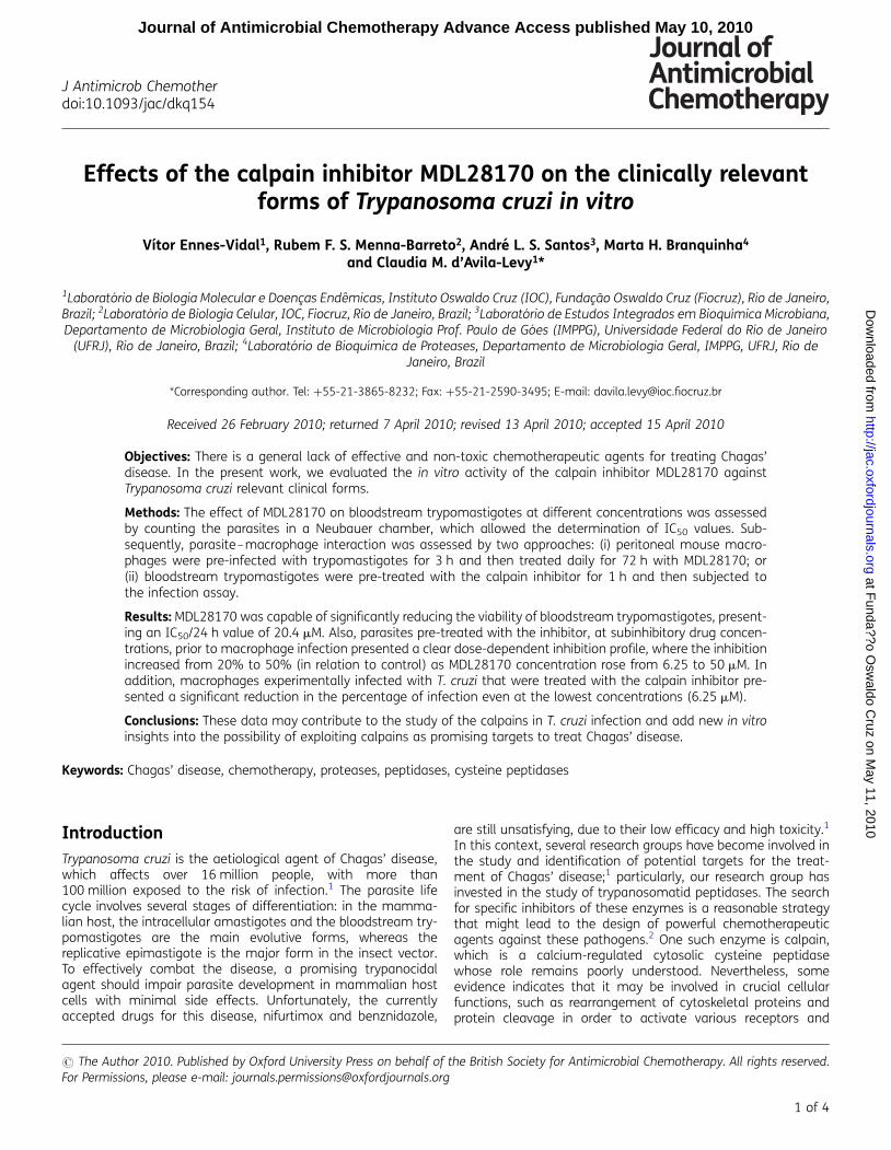
Effects of the calpain inhibitor MDL28170 on the clinically relevantforms of Trypanosoma cruzi in vitro
Vıtor Ennes-Vidal1, Rubem F. S. Menna-Barreto2, Andre L. S. Santos3, Marta H. Branquinha4
and Claudia M. d’Avila-Levy1*
1Laboratorio de Biologia Molecular e Doencas Endemicas, Instituto Oswaldo Cruz (IOC), Fundacao Oswaldo Cruz (Fiocruz), Rio de Janeiro,Brazil; 2Laboratorio de Biologia Celular, IOC, Fiocruz, Rio de Janeiro, Brazil; 3Laboratorio de Estudos Integrados em Bioquımica Microbiana,Departamento de Microbiologia Geral, Instituto de Microbiologia Prof. Paulo de Goes (IMPPG), Universidade Federal do Rio de Janeiro
(UFRJ), Rio de Janeiro, Brazil; 4Laboratorio de Bioquımica de Proteases, Departamento de Microbiologia Geral, IMPPG, UFRJ, Rio deJaneiro, Brazil
*Corresponding author. Tel: +55-21-3865-8232; Fax: +55-21-2590-3495; E-mail: [email protected]
Received 26 February 2010; returned 7 April 2010; revised 13 April 2010; accepted 15 April 2010
Objectives: There is a general lack of effective and non-toxic chemotherapeutic agents for treating Chagas’disease. In the present work, we evaluated the in vitro activity of the calpain inhibitor MDL28170 againstTrypanosoma cruzi relevant clinical forms.
Methods: The effect of MDL28170 on bloodstream trypomastigotes at different concentrations was assessedby counting the parasites in a Neubauer chamber, which allowed the determination of IC50 values. Sub-sequently, parasite–macrophage interaction was assessed by two approaches: (i) peritoneal mouse macro-phages were pre-infected with trypomastigotes for 3 h and then treated daily for 72 h with MDL28170; or(ii) bloodstream trypomastigotes were pre-treated with the calpain inhibitor for 1 h and then subjected tothe infection assay.
Results: MDL28170 was capable of significantly reducing the viability of bloodstream trypomastigotes, present-ing an IC50/24 h value of 20.4 mM. Also, parasites pre-treated with the inhibitor, at subinhibitory drug concen-trations, prior to macrophage infection presented a clear dose-dependent inhibition profile, where the inhibitionincreased from 20% to 50% (in relation to control) as MDL28170 concentration rose from 6.25 to 50 mM. Inaddition, macrophages experimentally infected with T. cruzi that were treated with the calpain inhibitor pre-sented a significant reduction in the percentage of infection even at the lowest concentrations (6.25 mM).
Conclusions: These data may contribute to the study of the calpains in T. cruzi infection and add new in vitroinsights into the possibility of exploiting calpains as promising targets to treat Chagas’ disease.
Keywords: Chagas’ disease, chemotherapy, proteases, peptidases, cysteine peptidases
IntroductionTrypanosoma cruzi is the aetiological agent of Chagas’ disease,which affects over 16 million people, with more than100 million exposed to the risk of infection.1 The parasite lifecycle involves several stages of differentiation: in the mamma-lian host, the intracellular amastigotes and the bloodstream try-pomastigotes are the main evolutive forms, whereas thereplicative epimastigote is the major form in the insect vector.To effectively combat the disease, a promising trypanocidalagent should impair parasite development in mammalian hostcells with minimal side effects. Unfortunately, the currentlyaccepted drugs for this disease, nifurtimox and benznidazole,
are still unsatisfying, due to their low efficacy and high toxicity.1
In this context, several research groups have become involved inthe study and identification of potential targets for the treat-ment of Chagas’ disease;1 particularly, our research group hasinvested in the study of trypanosomatid peptidases. The searchfor specific inhibitors of these enzymes is a reasonable strategythat might lead to the design of powerful chemotherapeuticagents against these pathogens.2 One such enzyme is calpain,which is a calcium-regulated cytosolic cysteine peptidasewhose role remains poorly understood. Nevertheless, someevidence indicates that it may be involved in crucial cellularfunctions, such as rearrangement of cytoskeletal proteins andprotein cleavage in order to activate various receptors and
# The Author 2010. Published by Oxford University Press on behalf of the British Society for Antimicrobial Chemotherapy. All rights reserved.For Permissions, please e-mail: [email protected]
J Antimicrob Chemotherdoi:10.1093/jac/dkq154
1 of 4
Journal of Antimicrobial Chemotherapy Advance Access published May 10, 2010 at F
unda??o Osw
aldo Cruz on M
ay 11, 2010 http://jac.oxfordjournals.org
Dow
nloaded from
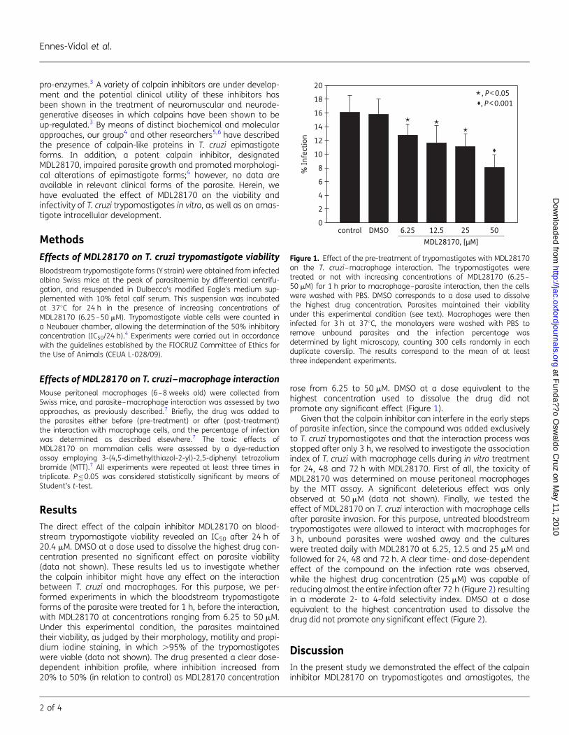
pro-enzymes.3 A variety of calpain inhibitors are under develop-ment and the potential clinical utility of these inhibitors hasbeen shown in the treatment of neuromuscular and neurode-generative diseases in which calpains have been shown to beup-regulated.3 By means of distinct biochemical and molecularapproaches, our group4 and other researchers5,6 have describedthe presence of calpain-like proteins in T. cruzi epimastigoteforms. In addition, a potent calpain inhibitor, designatedMDL28170, impaired parasite growth and promoted morphologi-cal alterations of epimastigote forms;4 however, no data areavailable in relevant clinical forms of the parasite. Herein, wehave evaluated the effect of MDL28170 on the viability andinfectivity of T. cruzi trypomastigotes in vitro, as well as on amas-tigote intracellular development.
Methods
Effects of MDL28170 on T. cruzi trypomastigote viabilityBloodstream trypomastigote forms (Y strain) were obtained from infectedalbino Swiss mice at the peak of parasitaemia by differential centrifu-gation, and resuspended in Dulbecco’s modified Eagle’s medium sup-plemented with 10% fetal calf serum. This suspension was incubatedat 378C for 24 h in the presence of increasing concentrations ofMDL28170 (6.25–50 mM). Trypomastigote viable cells were counted ina Neubauer chamber, allowing the determination of the 50% inhibitoryconcentration (IC50/24 h).4 Experiments were carried out in accordancewith the guidelines established by the FIOCRUZ Committee of Ethics forthe Use of Animals (CEUA L-028/09).
Effects of MDL28170 on T. cruzi–macrophage interactionMouse peritoneal macrophages (6–8 weeks old) were collected fromSwiss mice, and parasite–macrophage interaction was assessed by twoapproaches, as previously described.7 Briefly, the drug was added tothe parasites either before (pre-treatment) or after (post-treatment)the interaction with macrophage cells, and the percentage of infectionwas determined as described elsewhere.7 The toxic effects ofMDL28170 on mammalian cells were assessed by a dye-reductionassay employing 3-(4,5-dimethylthiazol-2-yl)-2,5-diphenyl tetrazoliumbromide (MTT).7 All experiments were repeated at least three times intriplicate. P≤0.05 was considered statistically significant by means ofStudent’s t-test.
ResultsThe direct effect of the calpain inhibitor MDL28170 on blood-stream trypomastigote viability revealed an IC50 after 24 h of20.4 mM. DMSO at a dose used to dissolve the highest drug con-centration presented no significant effect on parasite viability(data not shown). These results led us to investigate whetherthe calpain inhibitor might have any effect on the interactionbetween T. cruzi and macrophages. For this purpose, we per-formed experiments in which the bloodstream trypomastigoteforms of the parasite were treated for 1 h, before the interaction,with MDL28170 at concentrations ranging from 6.25 to 50 mM.Under this experimental condition, the parasites maintainedtheir viability, as judged by their morphology, motility and propi-dium iodine staining, in which .95% of the trypomastigoteswere viable (data not shown). The drug presented a clear dose-dependent inhibition profile, where inhibition increased from20% to 50% (in relation to control) as MDL28170 concentration
rose from 6.25 to 50 mM. DMSO at a dose equivalent to thehighest concentration used to dissolve the drug did notpromote any significant effect (Figure 1).
Given that the calpain inhibitor can interfere in the early stepsof parasite infection, since the compound was added exclusivelyto T. cruzi trypomastigotes and that the interaction process wasstopped after only 3 h, we resolved to investigate the associationindex of T. cruzi with macrophage cells during in vitro treatmentfor 24, 48 and 72 h with MDL28170. First of all, the toxicity ofMDL28170 was determined on mouse peritoneal macrophagesby the MTT assay. A significant deleterious effect was onlyobserved at 50 mM (data not shown). Finally, we tested theeffect of MDL28170 on T. cruzi interaction with macrophage cellsafter parasite invasion. For this purpose, untreated bloodstreamtrypomastigotes were allowed to interact with macrophages for3 h, unbound parasites were washed away and the cultureswere treated daily with MDL28170 at 6.25, 12.5 and 25 mM andfollowed for 24, 48 and 72 h. A clear time- and dose-dependenteffect of the compound on the infection rate was observed,while the highest drug concentration (25 mM) was capable ofreducing almost the entire infection after 72 h (Figure 2) resultingin a moderate 2- to 4-fold selectivity index. DMSO at a doseequivalent to the highest concentration used to dissolve thedrug did not promote any significant effect (Figure 2).
DiscussionIn the present study we demonstrated the effect of the calpaininhibitor MDL28170 on trypomastigotes and amastigotes, the
control DMSO 6.25 12.5 25MDL28170, [µM]
50
% In
fect
ion
0
2
4
6
8
10
12
14
16
18
20
�
�
��
�, P < 0.05�, P < 0.001
Figure 1. Effect of the pre-treatment of trypomastigotes with MDL28170on the T. cruzi–macrophage interaction. The trypomastigotes weretreated or not with increasing concentrations of MDL28170 (6.25–50 mM) for 1 h prior to macrophage–parasite interaction, then the cellswere washed with PBS. DMSO corresponds to a dose used to dissolvethe highest drug concentration. Parasites maintained their viabilityunder this experimental condition (see text). Macrophages were theninfected for 3 h at 378C, the monolayers were washed with PBS toremove unbound parasites and the infection percentage wasdetermined by light microscopy, counting 300 cells randomly in eachduplicate coverslip. The results correspond to the mean of at leastthree independent experiments.
Ennes-Vidal et al.
2 of 4
at Funda??o O
swaldo C
ruz on May 11, 2010
http://jac.oxfordjournals.orgD
ownloaded from
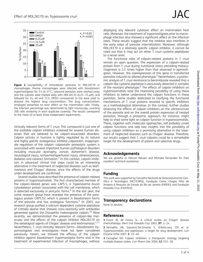
clinically relevant forms of T. cruzi. This compound is just one ofthe available calpain inhibitors screened for several human dis-eases that are believed to be calpain-associated disorders.Calpain activity in humans is tightly regulated by its naturaland highly specific endogenous inhibitor calpastatin. Inappropri-ate regulation of the calpain–calpastatin proteolytic system isassociated with several important human pathological disordersincluding muscular dystrophy, cancer, Alzheimer’s disease,neurological injury, ischaemia/reperfusion injury, atherosclerosis,diabetes and cataract formation.3 In this context, calpain inhibi-tors in advanced clinical trial steps could be an interestingalternative in the treatment of neglected diseases such as leish-maniasis and Chagas’ disease, once the effects of the drugsunder development are confirmed.
Several studies have described the presence of calpain-relatedproteins in trypanosomatids. The first characterized member ofthe calpain-related genes was CAP5.5, a Trypanosoma bruceicytoskeleton protein associated with the cell membrane, whichis detected exclusively in procyclic forms.8 In the last year, thesame research group have revealed the presence of the para-logous protein CAP5.5V, which is present in bloodstream formsof the parasite and has analogous functions.9 In 2003, ourresearch group purified a calcium-dependent cysteine peptidaseof Crithidia deanei that showed cross-reactivity with antibodiesgenerated against the Drosophila melanogaster calpain.10 Morerecently, we demonstrated the presence of calpain-like mol-ecules and the effects of the calpain inhibitor MDL28170 onthe aetiological agents of Chagas’ disease and leishmaniasis.4,11
Nevertheless, T. cruzi clinically relevant forms—bloodstream try-pomastigotes and amastigotes—have not been consideredpreviously. Herein, we showed the efficacy of the calpaininhibitor against bloodstream trypomastigote forms and in thetreatment of experimental infection of macrophages, without
displaying any relevant cytotoxic effect on mammalian hostcells. Moreover, the treatment of trypomastigotes prior to macro-phage infection also showed a significant effect on the infectionrates. These results suggest that the inhibitor also interferes inthe early steps of parasite internalization. Moreover, althoughMDL28170 is a relatively specific calpain inhibitor, it cannot beruled out that it may act on other T. cruzi cysteine peptidasesto a lesser level.
The functional roles of calpain-related proteins in T. cruziremain an open question. The expression of a calpain-relatedprotein from T. cruzi during nutritional stress preceding metacy-clogenesis is 2.5 times higher than that observed in epimasti-gotes. However, the overexpression of this gene in transfectedparasites induced no altered phenotype.5 Nevertheless, a proteo-mic analysis of T. cruzi resistance to benznidazole revealed that acalpain-like cysteine peptidase is exclusively detected in samplesof the resistant phenotype.6 The effects of calpain inhibitors ontrypanosomatids raise the interesting possibility of using theseinhibitors to better understand the calpain functions in theseparasites. Some studies dedicated to clarifying the functionalmechanisms of T. cruzi proteins resorted to specific inhibitorsas a methodological alternative. In this context, further studiesassessing the effects of calpain inhibitors on the ultrastructureof the parasite and on the overall protein expression of treatedparasites, through a proteomic approach, for instance, mighthelp to shed some light on calpain function in trypanosomatids.These, together with molecular approaches, will probably unveilcalpain functions and add new insights into the possibility ofusing calpain inhibitors as a promising alternative in the treat-ment of neglected diseases such as Chagas’ disease. Therefore,our results suggest that T. cruzi calpains could be a remarkabletarget for the development of potent and selective drugs.
AcknowledgementsWe are grateful to Marcos Meuser and Michelle Fernandes for theirexcellent technical assistance.
FundingThis work was supported by Conselho Nacional de Desenvolvimento Cien-tıfico e Tecnologico (MCT/CNPQ), Fundacao Carlos Chagas Filho deAmparo a Pesquisa do Estado do Rio de Janeiro (FAPERJ) and FundacaoOswaldo Cruz (FIOCRUZ).
Transparency declarationsNone to declare.
References1 Coura JR, de Castro SL. A critical review on Chagas’ diseasechemotherapy. Mem Inst Oswaldo Cruz 2002; 97: 3–24.
2 Vermelho AB, Giovanni-De-Simone S, d’Avila-Levy CM et al.Trypanosomatids and peptidases: a target for drug development. CurrEnzyme Inhib 2007; 3: 19–48.
3 Carragher NO. Calpain inhibition: a therapeutic strategy targetingmultiple disease states. Curr Pharm Des 2006; 12: 615–38.
024 h 48 h 72 h
5
10
15
20
% In
fect
ion
25
30
35ControlDMSO6.25 µM12.5 µM25 µM
�, P < 0.05
���
�
�
�
�
�
�
�, P < 0.001
Figure 2. Susceptibility of intracellular parasites to MDL28170 inmacrophages. Murine macrophages were infected with bloodstreamtrypomastigotes for 3 h at 378C, unbound parasites were washed awayand the cultures were treated daily with MDL28170 (6.25–25 mM), andfollowed for 24, 48 and 72 h. DMSO corresponds to the dose used todissolve the highest drug concentration. The drug concentrationsemployed presented no toxic effect on the mammalian cells. Finally,the infection percentage was determined by light microscopy, counting300 cells randomly in each duplicate coverslip. The results correspondto the mean of at least three independent experiments.
Effect of MDL28170 on Trypanosoma cruzi
3 of 4
JAC at F
unda??o Osw
aldo Cruz on M
ay 11, 2010 http://jac.oxfordjournals.org
Dow
nloaded from
4 Sangenito LS, Ennes-Vidal V, Marinho FA et al. Arrested growth ofTrypanosoma cruzi by the calpain inhibitor MDL28170 and detection ofcalpain homologues in epimastigote forms. Parasitology 2009; 136:433–41.
5 Giese V, Dallagiovanna B, Marchini FK et al. Trypanosoma cruzi: astage-specific calpain-like protein is induced after various kinds ofstress. Mem Inst Oswaldo Cruz 2008; 103: 598–601.
6 Andrade HM, Murta SMF, Chapeaurouge A et al. Proteomic analysis ofTrypanosoma cruzi resistance to benznidazole. J Proteome Res 2008; 7:2357–67.
7 Santos LO, Marinho FA, Altoe EF et al. HIV aspartyl peptidase inhibitorsinterfere with cellular proliferation, ultrastructure and macrophageinfection of Leishmania amazonensis. PLoS ONE 2009; 4: e4918.
8 Hertz-Fowler C, Ersfeld K, Gull K. CAP5.5, a life-cycle-regulated,cytoskeleton-associated protein is a member of a novel family ofcalpain-related proteins in Trypanosoma brucei. Mol Biochem Parasitol2001; 116: 25–34.
9 Olego-Fernandez S, Vaughan S, Shaw MK et al. Cell morphogenesis ofTrypanosoma brucei requires the paralogous, differentially expressedcalpain-related proteins CAP5.5 and CAP5.5V. Protist 2009; 160: 576–90.
10 d’Avila-Levy CM, Souza RF, Gomes RC et al. A novel extracellularcysteine proteinase from Crithidia deanei. Arch Biochem Biophys 2003;420: 1–8.
11 d’Avila-Levy CM, Marinho FA, Santos LO et al. Antileishmanial activityof MDL28170, a potent calpain inhibitor. Int J Antimicrob Agents 2006;28: 138–42.
Ennes-Vidal et al.
4 of 4
at Funda??o O
swaldo C
ruz on May 11, 2010
http://jac.oxfordjournals.orgD
ownloaded from
44
Artigo 3: MDL28170, a potent calpain inhibitor, affects T. cruzi metacyclogenesis,
ultrastructure and attachment to the luminal midgut surface of Rhodnius prolixus
Ennes-Vidal V, Menna-Barreto RF, Santos ALS, Branquinha MH, d'Avila-Levy CM.
Submetido para publicação na revista eletrônica Plos Neglected Tropical Diseases, 2010
Nosso terceiro e último artigo desenvolvido durante esse dois anos de mestrado é
focado nos 3 últimos objetivos desta dissertação: avaliar o efeito do inibidor III de calpaínas
MDL28170 no processo de interação de epimastigotas do Trypanosoma cruzi com o epitélio
intestinal de Rhodnius prolixus em ensaios in vitro, na metaciclogênese induzida
artificialmente e sobre a ultraestrutura. Contudo, não nos restringimos a responder apenas a
essas perguntas, também avaliamos a reatividade cruzada de outros anticorpos anti-calpaínas,
assim como o efeito desses anticorpos na interação de formas epimastigotas do T. cruzi com o
epitélio intestinal de R. prolixus. Nossos resultados demonstraram que o MDL28170 é capaz
de: reduzir significativamente a adesão dos epimastigotas ao epitélio do inseto vetor; diminuir
de forma dose-dependente o processo de diferenciação de epimastigotas à tripomastigotas
metacíclicos em meio TAU (triatomine artificial urine); e provocar desorganizações
ultraestruturais nos reservossomos, no Golgi e na membrana plasmáticas das formas
epimastigotas do T. cruzi tratadas por 72 horas com a concentração correspondente ao IC50
calculado para este inibidor. Além disso, observamos também que os anticorpos anti-
calpaínas capazes de reagir contra moléculas expressas na superfície de formas epimastigotas
do T. cruzi são capazes de reduzir significativamente a adesão do parasito ao epitélio
intestinal de R. prolixus, e que MDL28170 é capaz de diminuir de forma significante a
viabilidade de tripomastigotas sanguíneos mantidos em cultura por 24 horas. Dessa forma,
nosso conjunto de dados sugere que as moléculas similares às calpaínas presentes no T. cruzi
possam estar participando de eventos essenciais do ciclo de vida desse parasito, o que torna
essas moléculas alvos interessantes para o desenvolvimento de inibidores menos tóxicos e
mais seletivos para o tratamento da doença de Chagas. Os experimentos realizados nesse
trabalho foram realizados por mim e pelo Dr. Rubem Menna-Barreto sobre a orientação dos
demais autores.
1
MDL28170, a potent calpain inhibitor, affects Trypanosoma cruzi 1
metacyclogenesis, ultrastructure and attachment to the luminal midgut 2
surface of Rhodnius prolixus 3
4
Vítor Ennes-Vidal1, Rubem F. S. Menna-Barreto2, André L. S. Santos3, Marta H. 5
Branquinha4, Claudia M. d’Avila-Levy1*. 6
7 1Laboratório de Biologia Molecular e Doenças Endêmicas, Instituto Oswaldo Cruz 8
(IOC), Fundação Oswaldo Cruz (Fiocruz), Rio de Janeiro, Brazil; 9
2Laboratório de Biologia Celular, IOC, Fiocruz, Rio de Janeiro, Brazil; 10 3Laboratório de Estudos Integrados em Bioquímica Microbiana, Departamento de 11
Microbiologia Geral, Instituto de Microbiologia Prof. Paulo de Góes (IMPPG), 12
Universidade Federal do Rio de Janeiro (UFRJ), Rio de Janeiro, Brazil; 13 4Laboratório de Bioquímica de Proteases, Departamento de Microbiologia Geral, 14
IMPPG, UFRJ, Rio de Janeiro, Brazil. 15
16
17
18 * Corresponding author. Tel.: +5521 3865-8232; Fax: +5521 2590-3495 19
E-mail address: [email protected], [email protected] (Claudia M. 20
d’Avila-Levy) 21
22
23
Running title: MDL28170 effects on T. cruzi 24
25
26
27
28
29
2
Abstract 1
2
Background: Trypanosoma cruzi is the etiological agent of Chagas’ disease. During the 3
parasite life cycle, several molecules are involved in the differentiation process and 4
infectivity, but less is known about which of them are key molecules. Peptidases are 5
relevant for crucial steps of T. cruzi life cycle, as such, it is conceivable that they may 6
participate in the metacyclogenesis and interaction with the invertebrate host. 7
Methodology/Principal Findings: In this paper, we have investigated the effect of the 8
calpain inhibitor MDL28170 on the attachment of T. cruzi epimastigotes to the luminal 9
midgut surface of Rhodnius prolixus, as well as on the metacyclogenesis process and 10
ultrastructure. In addition, we have analyzed the effect of anti-calpain antibodies on the 11
interaction of epimastigotes to the midgut surface of the insect. MDL28170 treatment 12
was capable of significantly reducing the number of bound epimastigotes to the luminal 13
surface midgut of the insect. Once the cross-reactivity of the anti-Dm-calpain was 14
assessed, it was possible to block calpain molecules by the antibody, leading to a 15
significant reduction in the capacity of adhesion to the insect guts by T. cruzi. 16
Moreover, the in vitro metacyclogenesis process was impaired by the calpain inhibitor 17
presenting a significant reduction in the number of metacyclic trypomastigotes. The 18
calpain inhibitor also demonstrated a direct effect against bloodstream trypomastigotes. 19
Ultrastructural analysis of epimastigotes treated with the calpain inhibitor revealed 20
disorganization in the reservosomes, Golgi and plasma membrane disruption. 21
Conclusions/Significance: The presence of calpain and calpain-like molecules in a 22
wide range of organisms suggests that these proteins could be necessary for basic 23
cellular functions. Herein, we demonstrated the effects of MDL28170 in crucial steps of 24
the T. cruzi life cycle, such as attachment to the insect midgut and metacyclogenesis, as 25
well as in parasite viability and morphology. Together with our previous findings, these 26
results help to shed some light on the functions of T. cruzi calpains. Considering the 27
potential roles of these molecules on the interaction with both invertebrate and 28
vertebrate hosts, it is conceivable to explore them as an alternative target to treat 29
Chagas’ disease. 30
31
Keywords: Chagas’ disease, Trypanosoma cruzi, invertebrate host, metacyclogenesis, 32
peptidases, calpain. 33
3
Author’s Summary 1
2
Chagas’ disease is one of the most important neglected diseases of Latin 3
America, caused by the kinetoplastid protozoa Trypanosoma cruzi. Calpains are 4
cysteine peptidases, described in a broad range of organisms, which are believed to be 5
crucial in several basic cellular functions. In this sense, it is conceivable that calpain like 6
molecules present in T. cruzi may participate in metacyclogenesis and interaction with 7
the invertebrate host. Herein, we described the effects of the calpain inhibitor 8
MDL28170: on the adhesion of T. cruzi epimastigotes on the luminal midgut surface of 9
the insect vector Rhodnius prolixus; on the differentiation process of epimastigotes to 10
metacyclic trypomastigotes; on the viability of bloodstream trypomastigotes; and on the 11
ultrastructure of epimastigotes. Additionally, we demonstrated that anti-calpain 12
antibodies were also capable of reducing the attachment of the epimastigotes to the 13
insect gut. These data may contribute for the study of the functions of T. cruzi calpains 14
and add new insights into the possibility of using calpain inhibitors in treating parasitic 15
infections, since they interfere in key parts of the parasite life cycle. 16
17
Introduction 18
19
Chagas’ disease is a neglected tropical disease, which remains a major health 20
problem in Latin America. Over eight million people are infected with this disease and 21
an estimated of 14,000 people die as a consequence of the infection every year [1]. The 22
etiological agent Trypanosoma cruzi undergoes profound morphological changes during 23
its development in a complex life cycle involving mammalian and invertebrate hosts. 24
The protozoa life cycle comprises three major morphological stages: epimastigotes, 25
trypomastigotes, and amastigotes [2]. During the infection of the invertebrate host, a 26
hemipteran insect of the Reduviidae order, non-infectious epimastigotes adhere to the 27
insect host midgut, begin to proliferate and differentiate (metacyclogenesis process) into 28
metacyclic trypomastigotes, which are non-proliferative forms that are able to infect a 29
mammalian host [3]. The adhesion to the luminal midgut surface of the insect appears to 30
be necessary for the metacyclogenesis, but there is a general lack of information about 31
which molecules are implicated in this process [3,4]. In this context, peptidases, a class 32
of hydrolytic enzymes responsible for breaking the peptides bonds - has attracted the 33
4
attention of our research group because of their role in several crucial steps of the life 1
cycle of the trypanosomatid parasites [5]. Among T. cruzi different peptidases that we 2
considered, the calpains have been presenting interesting finds and seem to be a 3
remarkable target for the development of an alternative target to treat Chagas’ disease 4
and leishmaniasis [6,7,8]. 5
Calpains constitute a large family of calcium-regulated cytosolic cysteine 6
peptidases that have been characterized mainly in humans and whose role still remains 7
poorly understood [9]. Some evidence indicates that these enzymes may participate in a 8
variety of cellular processes, including the rearrangement of cytoskeletal proteins, 9
different signal transduction pathways and apoptosis. In this context, a variety of 10
calpain inhibitors are under development and the potential clinical utility of these 11
compounds have been shown mainly in the treatment of neurodegenerative disorders 12
[10,11,12,13]. In this sense, a classical study employing whole genome analyses 13
showed the presence of a large and diverse family of calpains in Trypanosoma brucei, 14
Leishmania major and T. cruzi [14]. Some years before, the same group had already 15
characterized the first trypanosomatid calpain-like protein in procyclic forms of T. 16
brucei [15]. In addition, our group described the presence of calpain-related proteins in 17
T. cruzi epimastigote forms and the effects of the calpain inhibitor III (MDL28170) on 18
growth, viability and infectivity [7,8]. However, more studies are necessary to better 19
understand the involvement of the calpain homologues in the life cycle of T. cruzi. 20
Here, we have conducted a study to investigate the effect of the calpain inhibitor 21
MDL28170 on the attachment of T. cruzi epimastigotes to the luminal midgut surface of 22
Rhodnius prolixus, as well as on the metacyclogenesis process and ultrastructure. In 23
addition, we have analyzed the effect of anti-calpain antibodies on the interaction of 24
epimastigote forms to the midgut surface of the insect. 25
26
Methods 27
28
Ethics Statement 29
The experiments were carried out in accordance with the guidelines established 30
by the FIOCRUZ Committee of Ethics for the Use of Animals (CEUA L-028/09). 31
32
Chemicals 33
5
The calpain inhibitor III, MDL28170 (carbobenzoxy-valylphenylalanial; Z-Val-1
Phe-CHO), was purchased from Calbiochem (San Diego, CA, USA). Stock solutions of 2
the drug (5 mM) were prepared in dimethylsulfoxide (DMSO). 3
4
5
Parasite culture 6
Epimastigote forms of T. cruzi were grown in 3.7% brain heart infusion medium 7
(BHI), containing hemin and folic acid and supplemented with 10% heat-inactivated 8
fetal bovine serum, at 28 ºC for 4 days to reach late-log phase growth. For the following 9
experiments, epimastigotes were collected, washed three times in 0.15 M NaCl, 0.01 M 10
phosphate-buffer pH 7.2 (PBS) and immediately used. The Y strain of T. cruzi was used 11
in all experiments except for the metacyclogenesis assay, in which the Dm28c strain is 12
the best characterized model for in vitro differentiation [16]. 13
14
Insects 15
Rhodnius prolixus were reared and maintained as previously described [17]. 16
Briefly, fifth-instars larvae were starved for 30 days after the last ecdysis and then 17
allowed to feed on rabbit blood through a membrane feeder. Ten days after the feeding, 18
insects were dissected; the posterior midguts were then removed, longitudinally 19
sectioned and washed three times in PBS to expose their luminal surfaces. After the 20
washing, the tissue fragments were processed as described below. The insects were 21
obtained from the insectary of the Laboratório Nacional e Internacional de Referência 22
em Taxonomia de Triatomíneos, Instituto Oswaldo Cruz, FIOCRUZ. 23
24
Identification of calpain homologues by flow cytometry and fluorescence microscopy 25
Epimastigotes (1 × 10⁷ cells) from the Y strain used for these experiments were 26
fixed at 4 oC in 0.4% paraformaldehyde in PBS (pH 7.2) for 30 min, followed by 27
extensive washing in the same buffer. The fixed cells maintained their morphological 28
integrity, as verified by optical microscopic observation. After this step, the cells were 29
incubated for 1 h at room temperature with a 1:100 dilution of the anti-calpain 30
antibodies. Cells were then incubated for an additional hour with a 1:200 dilution of 31
fluorescein isothiocyanate (FITC)-labelled goat anti-rabbit IgG [18]. The cells were 32
then washed 3 times in PBS and observed in a Zeiss epifluorescence microscope 33
(Axioplan 2). Alternatively, the parasite associated fluorescence was excited at 488 nm 34
6
and quantified on a flow cytometer (FACSCalibur, BD Bioscience, USA) equipped with 1
a 15 mW argon laser emitting at 488 nm. Non-treated cells and those treated with the 2
secondary antibody alone were run in parallel as controls. Each experimental population 3
was then mapped by using a two-parameter histogram of forward-angle light scatter 4
versus side scatter. The mapped population (n=10,000) was then analyzed for log green 5
fluorescence by using a single parameter histogram. The calpain antibodies tested were: 6
a rabbit antiserum raised against Drosophila melanogaster calpain (anti-Dm-calpain) 7
[19], anti-C21, anti-C23 or anti-C24 raised against the whole molecule, the cysteine 8
active site and the histidine active site, respectively, of human brain m-calpain [20], 9
anti-CAP5.5, raised against the cytoskeleton-associated protein from Trypanosoma 10
brucei [15], and anti-CDPIIb and anti-Ha-CalpM raised against Homarus americanus 11
calpains [21,22]. 12
13
In vitro inhibition of T. cruzi-Rhodnius interaction 14
Live epimastigote forms (Y strain) were resuspended in 200 µl of fresh BHI to a 15
density of 1 × 10⁷ cells and treated with the calpain inhibitor for 1 h with sub-inhibitory 16
concentrations (6.25–50 μM) and washed in PBS. Under this experimental condition, 17
the parasite maintains their viability, as previously described [7]. Dilutions of DMSO 18
corresponding to those used to prepare the drug solution were assessed in parallel for 19
control. Alternatively, the parasites were incubated for 1 h with anti-calpain antibodies, 20
or rabbit pre-immune sera, at 1:1000 dilution. After that, binding of protozoa to insect 21
gut was performed as described elsewhere. Briefly, the parasites were incubated for 20 22
min at 28°C with R. prolixus dissected posterior midguts that were sliced open 23
longitudinally. Subsequently, the explanted midguts were spread onto glass slides to 24
count the number of attached parasites per epithelial cells. The experiment was 25
performed three times independently, for each experimental group 4 insect midguts 26
were used and 100 epithelial cells were counted randomly. 27
28
In vitro inhibition of T. cruzi metacyclogenesis 29
For in vitro differentiation, epimastigotes (clone Dm28c) in the stationary phase 30
of growth were incubated for 2 h at 28°C in triatomine artificial urine (TAU) medium 31
(190 mM NaCl, 17 mM KCl, 2 mM MgCl2, 2 mM CaCl2, 8 mM phosphate buffer pH 32
6.0) at a density of 5 × 10⁸ cells/ml. The parasites at a dilution of 1:100 were further 33
incubated for 96 h in TAU3AAG medium (TAU supplemented with 10 mM L-proline, 34
7
50 mM L-sodium glutamate, 2 mM L-sodium aspartate, and 10 mM D-glucose) in 1
culture flasks [16]. For inhibition assays, epimastigotes were incubated in the presence, 2
or absence (control), of increasing concentrations of MDL28170 (6.25–50 µM) in 3
TAU3AAG medium. Culture supernatants were collected after 24, 48, 72, and 96 h of 4
incubation in TAU3AAG medium and the number of epimastigotes and metacyclic 5
trypomastigotes was determined by cell counting in a Neubauer chamber. Under this 6
experimental condition, the parasite maintains their viability, as previously described 7
[7]. These morphological stages can be easily differentiated on morphological grounds 8
by light microscopy, since epimastigotes are broader and have a quite rigid body, while 9
trypomastigotes are slimmer and present a vigorous wavy movement of the whole body. 10
Three independent experiments were performed in triplicate and a DMSO dilution 11
corresponding to highest drug concentration was assessed in parallel. 12
13
Effects of MDL28170 on T. cruzi trypomastigotes viability 14
15
Bloodstream trypomastigote forms, obtained from infected albino Swiss mice at the 16
peak of parasitemia by differential centrifugation, were resuspended in Dulbecco's 17
modified Eagle medium supplemented with 10% fetal calf serum (DMEM). This 18
suspension was incubated at 37°C for 24 h in the presence of increasing concentrations 19
of MDL28170 (6.25–50 μM). Viability was assessed by mobility and lack of staining 20
after challenge with trypan blue. Dilutions of DMSO corresponding to those used to 21
prepare the drug solutions were assessed in parallel. Thereafter, the number of viable 22
motile trypomastigotes was quantified by counting the flagellates in a Neubauer 23
chamber. 24
25
Transmission electron microscopy 26
Briefly, epimastigotes from the Y strain (5 × 10⁶ cells/ml) were treated with the 27
50% inhibitory concentration (IC50) of MDL28170, as previously determined [7] for 72 28
h in BHI medium at 28°C. Afterwards, the parasites were fixed with 2.5% 29
glutaraldehyde in 0.1 M Na-cacodylate buffer (pH 7.2) at room temperature for 40 min 30
at 25°C and post-fixed with a solution of 1% OsO4, 0.8% potassium ferricyanide and 31
2.5 mM CaCl2 in the same buffer for 20 min at 25°C [23]. The cells were dehydrated in 32
an ascending acetone series and embedded in PolyBed 812 resin. Ultrathin sections 33
8
were stained with uranyl acetate and lead citrate and examined in Jeol JEM1011 1
transmission electron microscope. 2
3
Results 4
5
The presence of calpain-like molecules in epimastigote forms of T. cruzi Y strain 6
was accessed by flow cytometry assay and fluorescence microscopy analyses using a 7
panel of antibodies raised against different calpains. The antibodies anti-Dm-Calpain, 8
anti-CDPIIb, anti-Ha-CalpM and anti-CAP5.5 were capable of strongly binding to T. 9
cruzi cells (Fig. 1A), while anti-C21, anti-C23 and anti-C24 did not recognize epitopes 10
on the parasite cell surface (data not shown). However, no cross-reactive of the 11
antibodies with trypomastigote forms were observed in the same analyses (data not 12
shown). Fluorescence microscopy analyzes corroborated the surface distribution of 13
calpain-related molecules in epimastigote forms of T. cruzi (Fig. 1B). 14
In order to assess a potential function for calpain-like molecules in T. cruzi, we 15
have performed binding assays with parasites previously treated with the anti-Dm-16
calpain antibody, at a concentration that did not promote cell agglutination. The 17
blockage of calpain molecules by the antibody led to a significant reduction (P < 0.01) 18
in the capacity of adhesion to the insect guts by T. cruzi by approximately 50% (Fig. 2), 19
similar results were obtained with the other antibodies (data not shown). On the other 20
hand, parasites treated with the pre-immune serum adhered to the guts at a rate similar 21
to that of the control (P < 0.05) (Fig. 2). Some studies dedicated to clarify the functional 22
mechanisms of T. cruzi proteins resorted to specific inhibitors as a methodological 23
alternative [6,7,8,24,25]. In this sense, we performed binding assays with parasites 24
previously treated with MDL28170, a potent calpain inhibitor. The compound reduced 25
significantly the number of parasites adhered to the insect luminal midgut surface in all 26
drug concentrations tested (6.25–50 µM). At the highest doses 25 and 50 µM, the 27
adhesion rate was approximately 0.6 epimastigotes per midgut cell while the control 28
was about 1.4 (Fig. 2), which represents a reduction of 65%. DMSO at a dose 29
equivalent to the highest concentration used to dissolve the drug did not promote any 30
significant effect on the parasite adhesion (Fig. 2). 31
These results led us to investigate whether the T. cruzi MDL28170 might have 32
any effect on the metacyclogenesis process. For this purpose, we performed 33
9
experiments in which late-log phase epimastigotes form the Dm28c clone were 1
submitted to the differentiation assay, and treated or not for 4 days with MDL28170 at 2
concentrations ranging from 6.25 to 50 μM. Under this experimental condition the 3
parasites maintained their viability, as judged by their morphology and motility in 4
which the density of live parasites was approximately the same as the control (data not 5
shown). It should also be pointed out that MDL28170 has a trypanostatic effect on T. 6
cruzi, since cells pre-treated for 72 h with the calpain inhibitor even at 70 μM resumed 7
growth when subcultured in a drug-free fresh medium [7]. Finally, the metacyclogenesis 8
assay revealed that the drug present a time and dose-dependent inhibition profile, since 9
in cultures without MDL28170 the number of metacyclic trypomastigotes increased 10
over time, whereas MDL28170 treatment inhibited the differentiation process almost to 11
50% in the highest drug concentration. DMSO at a dose equivalent to the highest 12
concentration used to dissolve the drug did not interfere in the metacyclogenesis (Fig. 13
3). Furthermore, we decided to evaluate the direct effect of the MDL28170 on 14
bloodstream trypomastigotes viability in vitro after 24 h of treatment at 37ºC. The 15
inhibitor at 25 μM powerfully reduced parasite viability at about 63% in comparison to 16
control (Fig. 4). Bloodstream trypomastigotes cultured in the presence of DMSO at a 17
dose used to dissolve the highest drug concentration presented no significant effect (Fig. 18
4). 19
Given that the calpain inhibitor can reduce the growth of epimastigote forms 20
with a trypanostatic effect [7], interfere in the parasite adhesion to the insect midgut and 21
in the differentiation process of epimastigotes to metacyclic trypomastigotes, we 22
decided to investigate the ultrastructural effects of MDL28170 against the epimastigote 23
forms, by transmission electron microscopy. For this purpose, the morphology of non-24
treated cells (Fig. 5a-b) was compared with the ultrastructure of parasites treated for 72 25
h with 34 µM of MDL28170 (Fig. 5c-g). Results showed an important injury to the 26
reservosomes (Fig. 5c-e), plasma membrane (Fig. 5d,g) and Golgi (Fig. 5f,g). 27
Reservosomes and Golgi were severely affected, showing a washed-out appearance 28
with loss of organelles’ electrondensity and complete disruption of their membranes. 29
30
31
Discussion 32
33
10
Over the past few years some studies have described the presence of calpain-1
related proteins in trypanosomatids. At first, a classical study employing whole genome 2
analyses showed the presence of a large and diverse family of calpains in T. brucei, L. 3
major and T. cruzi [14]. In this sense, our group became involved in the study of these 4
enzymes though the report of the expression of calpain-like molecules and the effects of 5
the calpain inhibitor MDL28170 against the etiological agents of Chagas’ disease and 6
leishmaniasis [6,7,8]. In the present study, we demonstrated that MDL28170 also 7
inhibited T. cruzi adhesion to the insect gut, arrested the metacyclogenesis process, 8
reduced the bloodstream trypomastigote viability and promoted significant 9
ultrastructural effects on reservosomes and Golgi of epimastigote forms. In addition, it 10
was shown the cross-reactivity of the anti-calpain antibodies (anti-Dm-calpain, anti-11
CDPIIb, anti-Ha-CalpM and anti-CAP5.5) with epimastigote cells of the parasite, and 12
their ability to impair parasite adhesion to the midgut surface of R. prolixus in vitro. 13
As peptidases are known to be responsible for several physiological processes, 14
they became a remarkable target for the development of therapeutic agents to treat 15
infectious disease. In this context, T. cruzi peptidases have attracted the attention of 16
many laboratories because of their roles in pathogenesis of Chagas’ disease [5]. The 17
calpain inhibitor MDL28170 impaired significantly the adhesion rate of epimastigotes 18
to the midgut of R. prolixus in vitro suggesting that calpain-like molecules could play an 19
important role in this part of the parasite life cycle. Although MDL28170 is relatively a 20
specific calpain inhibitor, it cannot be ruled out that it may act on other T. cruzi cysteine 21
peptidases to a lesser [26]. Nevertheless, the anti-calpain antibodies were also capable 22
of significantly reducing the number of bound parasites to the luminal midgut surface of 23
the insect host. 24
Little is known about which molecules are relevant for the interaction of T. cruzi 25
with the invertebrate host. Our group has been studying some peptidases believed to be 26
essential in this part of the cycle of trypanosomatids, like gp63 and cruzipain [17,27]. 27
The gp63 homologues found in lower trypanosomatids seem to be indispensable for the 28
nutrition of these parasites as well as in the binding to the insect epithelial cells. 29
Interestingly, gp63 is predominantly expressed in the insect stage of Leishmania, and in 30
all insect and plant trypanosomatids examined so far [17]. Furthermore, we 31
demonstrated that when Phytomonas serpens cells were pre-treated with either cysteine 32
peptidase inhibitors or anti-cruzipain antibody, a significant reduction of the interaction 33
with explanted salivary glands of the phytophagous insect Oncopeltus fasciatus was 34
11
observed [27]. In T. cruzi, it has been shown that glycoinositolphospholipids (GIPLs) 1
are one of the components involved in the adhesion of epimastigote cells to the luminal 2
insect midgut surface and may be one of the determinants of parasite infection in the 3
insect vector [28]. Moreover, it was also shown that a gp72 knockout of T. cruzi 4
displayed a reduced number (less than 1% compared to control) of parasites released in 5
feces of the insect vector Triatoma infestans [29]. The dramatically reduced survival of 6
the mutant may be due to either the lack of GP72 itself, or to the altered properties of 7
the flagellum in this mutant that may be critical for establishment in the insect vector 8
[30]. 9
The metacyclogenesis process consists in the differentiation of non-infectious T. 10
cruzi epimastigotes into pathogenic metacyclic trypomastigotes. During this process, 11
epimastigotes adhere to the epithelium of the insect midgut before transforming into 12
metacyclic trypomastigotes [2]. The adhesion is thought to be a pre-requisite for 13
differentiation to the infective form, but less is known about how epimastigote adhesion 14
triggers the differentiation process after the nutritional stress [31]. Our findings from T. 15
cruzi in vitro metacyclogenesis triggered by an artificial nutritional stress in TAU and 16
TAU 3AAG medium showed that the calpain inhibitor impaired the differentiation 17
process and then decreased the number of metacyclic trypomastigotes without a 18
significantly decrease in the total number of parasites. This result was expected, since 19
previous studies have shown that cysteine peptidases are required during the 20
metacyclogenesis process [31,32]. Despite the toxic effects of MDL28170 against the 21
epimastigote forms previously described [7], it was verified that this inhibitor has 22
reversible effects and just arrested the parasite growth but not exterminate all the 23
epimasgitotes. However, the calpain inhibitor may also act directly inhibiting 24
epimastigote binding to the substrate, since the in vitro binding to the insect midgut was 25
substantially inhibited by MDL28170. The inhibitor could also act directly in the 26
viability of the differentiate cells in the presence of the inhibitor, since MDL28170 is 27
toxic against T. cruzi bloodstream trypomastigote cells [8]. Nevertheless, no 28
trypanocidal effect has already been demonstrated against metacyclic trypomastigotes to 29
date. 30
Finally, our ultrastructural data demonstrated severe effects of MDL28170 in the 31
reservosomes and Golgi, which showed a washed-out appearance with loss of 32
organelles electrondensity and complete disruption of their membranes. These results 33
indicate that the calpain inhibitor strongly affect T. cruzi epimastigote forms, and since 34
12
the reservosomes are essential for the differentiation process, it may suggest that 1
MDL28170 interfered in the metacyclogenesis, [32,33]. Moreover, a recent proteomic 2
analysis showed the presence of calpain-like molecules in a purified fraction of 3
reservosomes [34]. Similar ultrastructural alterations were observed in epimastigotes 4
treated with inhibitors from the major cysteine peptidase of T. cruzi, the cruzipain [35]. 5
The cysteine peptidase inhibitor was capable of modifying the intracellular localization 6
of cruzipain and induced its accumulation in peripheral dilations of Golgi cistern, these 7
abnormalities were followed by distention of endoplasmatic reticulum and nuclear 8
membranes [35]. Taken together, the present results indicate that T. cruzi calpains may 9
be essential proteins involved in the differentiation and infectivity of epimastigote forms 10
of the parasite. 11
It has been almost ten years since the first calpain-like protein CAP5.5 was 12
characterized in T. brucei [15]. Since then, our group has been studying calpains 13
homologues from lower trypanosomatids, like Crithidia deanei [36] and Herpetomonas 14
samuelpessoai [37], and form L. amazonensis [6] and T. cruzi [7,8]. In the latter, it was 15
shown, by a distinct research group, that a calpain-related mRNA is expressed 2.5-times 16
more in epimastigotes under nutritional stress, which precedes the metacyclogenesis, 17
than in epimastigotes in complete medium [38]. Also, a proteomic analysis of T. cruzi 18
cells resistant to benznidazole revealed that a calpain-like cysteine peptidase is 19
exclusively detected in samples of the resistant phenotype [39]. Our first report in T. 20
cruzi demonstrated that epimastigote forms have molecules that share antigens with 21
invertebrate calpain-related enzymes, such as D. melanogaster calpain. These molecules 22
are distributed in distinct phylogenetic lineages of the parasite, and also, the calpain 23
inhibitor MDL28170 arrested the growth of T. cruzi in a dose-dependent manner [7]. 24
Simultaneously, the absence of cross-reactivity between calpain-like molecules in T. 25
cruzi and anti-human m-calpain is suggestive of substantial structural differences 26
among these molecules, as previously described [14]. Here, we extended our findings 27
showing that the cross-reactivity between T. cruzi epitopes and antibodies raised against 28
T. brucei CAP5.5, and two calpains from American lobster Homarus americanus. 29
More recently, we investigated the trypanocidal effect of MDL28170 in the 30
trypomastigote and amastigote forms of T. cruzi by parasite-macrophage interaction 31
assays. The parasites pre-treated with the inhibitor presented a clear dose-dependent 32
inhibition of the interaction process. In addition, macrophages experimentally infected 33
with T. cruzi that were treated with the calpain inhibitor presented a significant 34
13
reduction of the percentage of the infection and in the number of internalized parasites 1
[8]. Herein, we reported the cross-reactivity of distinct anti-calpain antibodies to T. 2
cruzi epimastigote cells and that these antibodies are capable of impairing the parasite 3
attachment to the insect midgut in vitro. It was also reported the effects of MDL28170 4
on T. cruzi-R. prlolixus in vitro interaction assay, on bloodstream trypomastigote 5
viability, on the metacyclogenesis process and on the ultrastructure of epimastigote 6
forms. Although more studies are necessary for better understanding the functional role 7
of calpain-related proteins in T. cruzi, our work adds new insights into the possible 8
functions of these calpain homologues, and into the possibility of using calpain 9
inhibitors as a promising alternative in the treatment of Chagas’ disease. 10
11
Acknowledgments 12
13
This study was supported by MCT/CNPq (Conselho Nacional de Desenvolvimento 14
Científico e Tecnológico), FUJB (Fundação Universitária José Bonifácio), CEPG/UFRJ, 15
FAPERJ (Fundação Carlos Chagas Filho de Amparo à Pesquisa do Estado do Rio de 16
Janeiro) and Fundação Oswaldo Cruz (FIOCRUZ). The funders had no role in study 17
design, data collection and analysis, decision to publish, or preparation of the manuscript. 18
The authors are grateful to Dr. Yasufumi Emori (Department of Biophysics and 19
Biochemistry, Faculty of Sciences, University of Tokyo, Japan) for donating the anti-Dm-20
calpain, Dr. Ralph Nixon (Department of Psychiatry and Program in Neuroscience, 21
Harvard Medical School, USA) for donating the anti-C21, anti-C23 and anti-C24 22
antibodies, Dr. Keith Gull (School of Biological Sciences, University of Manchester, UK) 23
for donating the anti-CAP5.5 antibody and Dr. Donald M. Mykles (Department of 24
Biology, Cell and Molecular Biology Program and Program in Molecular, Cellular and 25
Integrative Neurobiology, Colorado State University, USA) for donating the anti-CDPIIb 26
and anti-Ha-CalpM. 27
28
Author Contributions 29
30
Conceived and designed the experiments: VEV RFSMB ALS MHB CMdL. 31
Performed the experiments: VEV RFSMB. Analyzed the data: VEV RFSMB ALSS 32
14
MHB CMdL. Contributed reagents/materials/analysis tools: RFSMB ALSS MHB 1
CMdL. Wrote the paper: VEV CMdL. 2
3
References 4
5
1. Drugs for Neglected Diseases initiative (2010). New agreement signed to join forces 6
to takle Chagas disease. DNDi. Available from: <http://www.dndi.org/press-7
releases/673-new-agreement-to-tackle-chagas-disease.html> Accessed 2010 Apr 15. 8
9
2. De Souza W (1984) Cell biology of Trypanosoma cruzi. Int Rev Cytol 86: 197-283 10
11
3. Azambuja P, Ratcliffe NA, Garcia ES (2005) Towards an understanding of the 12
interactions of Trypanosoma cruzi and Trypanosoma rangeli within the reduviid insect 13
host Rhodnius prolixus. An Acad Bras Cienc 77: 397-404. 14
15
4. Bonaldo MC, Souto-Padrón T, De Souza W, Goldenberg S (1988) Cell-substrate 16
adhesion during Trypanosoma cruzi differentiation. J Cell Biol 106: 349-1358. 17
18
5. Vermelho AB, Giovanni-de-Simone S, d’Avila-Levy CM, Santos ALS, Melo ACN, 19
et al. (2007) Trypanosomatidae peptidases: a target for drugs development. Curr Enz 20
Inhib 3: 19-48. 21
22
6. d’Avila-Levy CM, Marinho FA, Santos LO, Martins JLM, Santos ALS, et al. (2006) 23
Antileishmanial activity of MDL28170, a potent calpain inhibitor. Int J Antimicrob 24
Agents 28: 138-142. 25
26
7. Sangenito LS, Ennes-Vidal V, Marinho FA, Da Mota FF, Santos ALS, et al. (2009) 27
Arrested growth of Trypanosoma cruzi by the calpain inhibitor MDL28170 and 28
detection of calpain homologues in epimastigote forms. Parasitology 136: 433-441. 29
30
8. Ennes-Vidal V, Menna-Barreto RF, Santos ALS, Branquinha MH, d`Avila-Levy CM 31
(2010) Effects of the calpain inhibitor MDL28170 on the clinically relevant forms of 32
Trypanosoma cruzi in vitro. J Antimicrob Chemother [Epub ahead of print]. 33
15
1
9. Goll DE, Thompson VF, Li H, Wei W, Cong J (2003) The calpain system. Physiol 2
Rev 83: 731-801. 3
4
10. Ray SK, Hogan EL, Banik HL (2003) Calpain in the pathophysiology of spinal cord 5
injury: neuroprotection with calpain inhibitors. Brain Res Rev 42: 169-185. 6
7
11. Lescop C, Herzner H, Siendt H et al. (2005) Novel cell-penetrating alpha-keto-8
amide calpain inhibitors as potential treatment for muscular dystrophy. Bioorg Med 9
Chem Lett 15: 5176-5181. 10
11
12. Araújo-Couto L, Sampaio MN, Hokoc JN et al. (2004) Calpain inhibitor 2 prevents 12
axonal degeneration of opossum optic nerve fibers. J Neurosci Res 77: 410-419. 13
14
13. Saatman KE, Murai H, Bartus RT et al. (1996) Calpain inhibitor AK295 attenuates 15
motor and cognitive deficits following experimental brain injury in the rat. Proc Natl 16
Acad Sci 93: 3428-3433. 17
18
14. Ersfeld K, Barraclough H, Gull K (2005) Evolutionary relationships and protein 19
domain architecture in an expanded calpain superfamily in kinetoplastid parasites. J 20
Mol Evol 61: 742-757. 21
22
15. Hertz-Fowler C, Ersfeld K, Gull K (2001) CAP5.5, a life-cycle-regulated, 23
cytoskeleton-associated protein is a member of a novel family of calpain-related 24
proteins in Trypanosoma brucei. Mol Biochem Parasitol 116: 25–34. 25
26
16. Contreras VT, Salles JM, Thomas N, Morel CM, Goldenberg S (1985) In vitro 27
differentiation of cruzi under chemical1y defined conditions. Mol Biochem Parasitol 16: 28
315-327. 29
30
17. Azambuja P & Garcia ES (1997) Care and maintenance of triatomine colonies. In: 31
Crampton JM, Beard CB, Louid C. (Eds.) Molecular Biology of Insect Disease Vectors: 32
a Methods Manual. Chapman and Hall, London, pp. 56-64. 33
34
16
18. Santos ALS, Branquinha MH, D'Avila-Levy CM (2006) The ubiquitous gp63-like 1
metalloprotease from lower trypanosomatids: in the search for a function. An Acad Bras 2
Cienc 78: 687-714. 3
4
19. Emori Y & Saigo K (1994) Calpain localization changes in coordination with actin-5
related cytoskeletal changes during early embryonic development of Drosophila. J Biol 6
Chem 269: 25137-25142. 7
8
20. Grynspan, F, Griffin WR, Cataldo A, Katayama S, Nixon RA (1997) Active site-9
directed antibodies identify calpain II as an early-appearing and pervasive component of 10
neurofibrillary pathology in Alzheimer’s disease. Brain Res 763: 145-158. 11
12
21. Beyette JR, Emori Y, Mykles DL (1997) Immunological analysis of two calpain-13
like Ca2+-dependent proteinases from lobster striated muscles: relationship to 14
mammalian and Drosophila calpains. Arch Biochem Biophys 337: 232-8. 15
16
22. Yu X & Mykles DL (2003) Cloning of a muscle-specific calpain from the American 17
lobster Homarus americanus: expression associated with muscle atrophy and restoration 18
during moulting. J Exp Biol 206: 561-75. 19
20
23. Meirelles RMS & Soares MJ (2001) Quick fixation of eukaryotic cell suspensions 21
for routine transmission electron microscopy. Acta Microscopica 10:19-22. 22
23
24. Cazzulo JJ, Stoka V, Turk V (2001) The major cysteine proteinase of Trypanosoma 24
cruzi: a valid target for chemotherapy of Chagas disease. Current Pharm Des 7: 1143-25
1156. 26
27
25. Meirelles MN, Juliano L, Carmona E, Silva SG, Costa EM et al. (1992) Inhibitors of 28
the major cysteinyl proteinase (GP57/51) impair host cell invasion and arrest the 29
intracellular development of Trypanosoma cruzi in vitro. J Mol Biochem Parasitol 52: 30
175-184. 31
32
26. Rami A, Ferger D, Krieglstein J (1997) Blockade of calpain proteolytic activity 33
rescues neurons from glutamate excitotoxicity. Neurosci Res 27: 93-97. 34
17
1
27. Santos ALS, d'Avila-Levy CM, Dias FA, Ribeiro RO, Pereira FM et al. (2006) 2
Phytomonas serpens: cysteine peptidase inhibitors interfere with growth, ultrastructure 3
and host adhesion. Int J Parasitol 36: 47-56. 4
5
28. Nogueira NF, Gonzalez MS, Gomes JE, de Souza W, Garcia ES et al. (2007) 6
Trypanosoma cruzi: involvement of glycoinositolphospholipids in the attachment to the 7
luminal midgut surface of Rhodnius prolixus. Exp Parasitol 116: 120-8. 8
9
29. Basombrío MA, Gómez L, Padilla AM, Ciaccio M, Nozaki T et al. (2002) Targeted 10
Deletiono f the gp72 gene decreases the infectivity of Trypanosoma cruzi from mice 11
and insect vectors. J Parasito 88: 489-493. 12
13
30. De Jesus AR, Cooper R, Espinosa M, Gomes JE, Garcia ES et al. (1993) Gene 14
deletion suggests a role for Trypanosoma cruzi surface glycoprotein gp72 in the insect 15
and mammalian stages of the life cycle. J Cell Sci 106: 1023-33. 16
17
31. Figueiredo RCBQ, Rosa DS, Soares MJ (2000) Differentiation of Trypanosoma 18
cruzi epimastigotes: metacyclogenesis and adhesion to substrate are triggered by 19
nutritional stress. J Parasitol 80: 1213-1218. 20
21
32. Figueiredo RCBQ, Steindel M, Soares MJ (1994) The reservosomes of epimastigote 22
forms of Trypanosoma cruzi: occurrence during in vitro cultivation. Parasitology 80: 23
517-522. 24
25
33. Duschak VG, Barboza M, García GA, Lammel EM, Couto AS, et al. (2006) Novel 26
cysteine proteinase in Trypanosoma cruzi metacyclogenesis. Parasitology 132: 345-355. 27
28
34. Sant'Anna C, Nakayasu ES, Pereira MG, Lourenço D, de Souza W, et al. 29
(2009) Subcellular proteomics of Trypanosoma cruzi reservosomes. Proteomics 7: 30
1782-94. 31
32
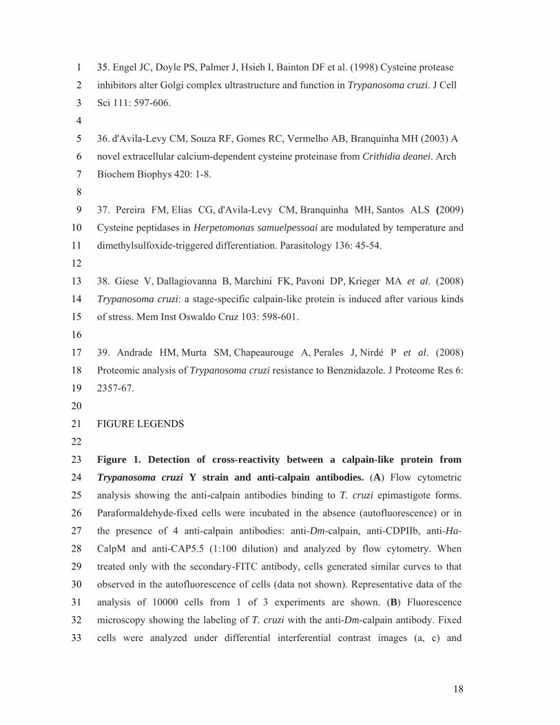
18
35. Engel JC, Doyle PS, Palmer J, Hsieh I, Bainton DF et al. (1998) Cysteine protease 1
inhibitors alter Golgi complex ultrastructure and function in Trypanosoma cruzi. J Cell 2
Sci 111: 597-606. 3
4
36. d'Avila-Levy CM, Souza RF, Gomes RC, Vermelho AB, Branquinha MH (2003) A 5
novel extracellular calcium-dependent cysteine proteinase from Crithidia deanei. Arch 6
Biochem Biophys 420: 1-8. 7
8
37. Pereira FM, Elias CG, d'Avila-Levy CM, Branquinha MH, Santos ALS (2009) 9
Cysteine peptidases in Herpetomonas samuelpessoai are modulated by temperature and 10
dimethylsulfoxide-triggered differentiation. Parasitology 136: 45-54. 11
12
38. Giese V, Dallagiovanna B, Marchini FK, Pavoni DP, Krieger MA et al. (2008) 13
Trypanosoma cruzi: a stage-specific calpain-like protein is induced after various kinds 14
of stress. Mem Inst Oswaldo Cruz 103: 598-601. 15
16
39. Andrade HM, Murta SM, Chapeaurouge A, Perales J, Nirdé P et al. (2008) 17
Proteomic analysis of Trypanosoma cruzi resistance to Benznidazole. J Proteome Res 6: 18
2357-67. 19
20
FIGURE LEGENDS 21
22
Figure 1. Detection of cross-reactivity between a calpain-like protein from 23
Trypanosoma cruzi Y strain and anti-calpain antibodies. (A) Flow cytometric 24
analysis showing the anti-calpain antibodies binding to T. cruzi epimastigote forms. 25
Paraformaldehyde-fixed cells were incubated in the absence (autofluorescence) or in 26
the presence of 4 anti-calpain antibodies: anti-Dm-calpain, anti-CDPIIb, anti-Ha-27
CalpM and anti-CAP5.5 (1:100 dilution) and analyzed by flow cytometry. When 28
treated only with the secondary-FITC antibody, cells generated similar curves to that 29
observed in the autofluorescence of cells (data not shown). Representative data of the 30
analysis of 10000 cells from 1 of 3 experiments are shown. (B) Fluorescence 31
microscopy showing the labeling of T. cruzi with the anti-Dm-calpain antibody. Fixed 32
cells were analyzed under differential interferential contrast images (a, c) and 33
19
immunofluorescence (b, d). Parasites treated only with the secondary antibody 1
presented no fluorescence intensity (b). The bars represent 1 µm. 2
3
Figure 2. Effect of MDL28170 and anti-calpain antibody in the interaction process 4
between T. cruzi and explanted guts of Rhodnius prolixus. Epimastigotes (1.0 × 10⁷ 5
cells) were treated for 30 min at 28 °C with increasing concentrations of MDL28170 6
(6.25 to 50 μM), or incubated for 1 h with the anti-calpain (1:100) or the pre-immune 7
serum (1:100) in 200 μl of BHI. The data from DMSO represents the concentration 8
present in the highest dose of the drug. The viability of the parasites was not affected by 9
the treatments used in this set of experiments. Then, parasites were washed, and 10
incubated for 20 min at 28°C with R. prolixus dissected posterior midguts that were 11
sliced open longitudinally. Subsequently, the explanted midguts were spread onto glass 12
slides to count the number of attached parasites per epithelial cells. For each 13
experimental group 4 insect midguts were used, and 100 epithelial cells were counted 14
randomly. The results are shown as the mean ± standard error of the mean of three 15
independent experiments. 16
17
Figure 3. Effect of MDL28170 on T. cruzi metacyclogenesis in vitro. Epimastigotes 18
from the stationary phase of growth (5.0 × 10⁸ cells) were incubated in TAU3AAG 19
medium for 96 h to induce cellular differentiation to metacyclic trypomastigotes. 20
Parasites were treated with increasing concentrations of MDL28170 (6.25 to 50 μM). 21
The data from DMSO represents the concentration present in the highest dose of the 22
drug. At each time point, culture supernatants were collected and the number of 23
epimastigotes and metacyclic trypomastigotes was determined by cell counting in a 24
Neubauer chamber. The results correspond to the mean of three independent 25
experiments performed in triplicate. 26
27
Figure 4. In vitro efficacy of MDL28170 on T. cruzi bloodstream trypomastigotes 28
viability. Bloodstream trypomastigote forms obtained from Swiss mice (5.0 × 10⁶ cells) 29
were treated with increasing concentrations of MDL28170 (6.25 to 50 μM) for 24 h. 30
The data from DMSO represents the concentration present in the highest dose of the 31
drug. Thereafter, viable parasites were counted by trypan blue exclusion and mobility. 32
20
The results are expressed in viability percentage in relation to control. The results 1
correspond to the mean of three independent experiments performed in triplicate. 2
3
Figure 5. Ultrastructural effects of MDL28170 in T. cruzi epimastigotes. (a,b) 4
Control parasites showing typical elongated morphology with normal kinetoplast (K), 5
mitochondrion (M), nucleus (N) and Golgi (G). (c-g) The treatment of epimastigotes 6
with 34 μM of the inhibitor for 72 h led to an extensive disorganization in the 7
reservosomes (asterisks), plasma membrane alterations (arrows) as well as to the 8
disruption of Golgi cisternae (stars). Bars = 0.5 μm. 9
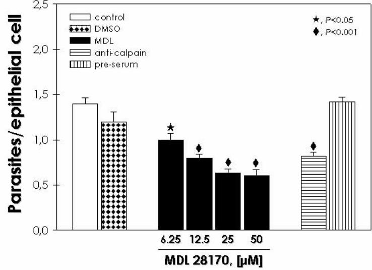
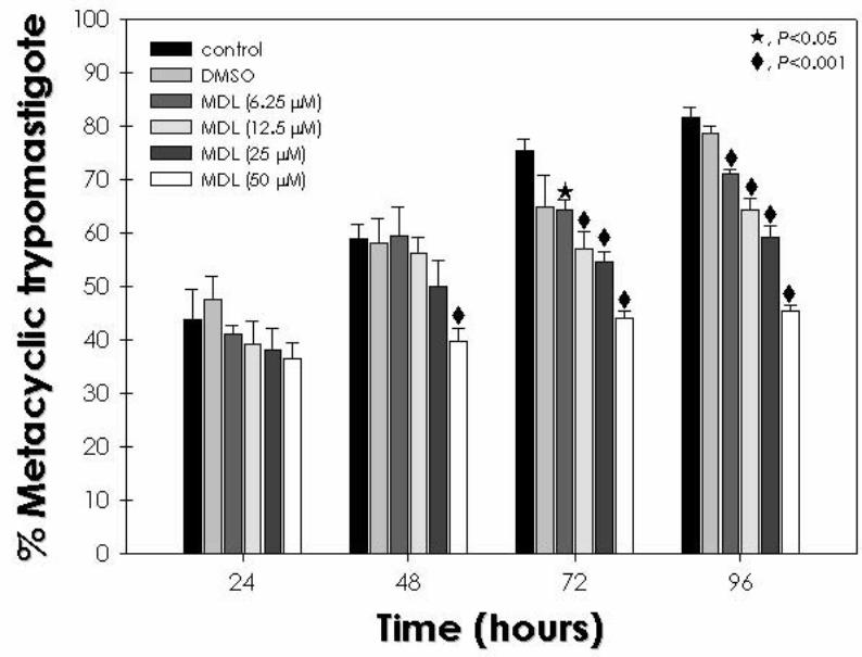
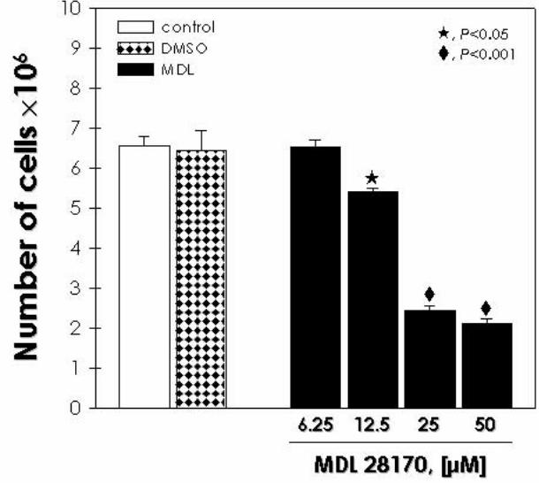
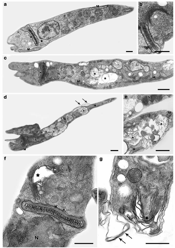
71
IV. DISCUSSÃO
Entre os protozoários, os tripanossomatídeos constituem um grupo de parasitos uni-
flagelados caracterizados pela presença do cinetoplasto, DNA mitocondrial único em sua
estrutura, função e modo de replicação (VICKERMAN, 1994; DE SOUZA et al., 2009). Uma
fração desses parasitos se especializou de forma a parasitar o homem, como o Trypanosoma
brucei, agente etiológico da doença do sono Africano; o Trypanosoma cruzi, agente causador
da doença de Chagas ou tripanossomíase americana, e as diferentes espécies do
gênero Leishmania, responsáveis por um amplo espectro de manifestações clínicas que
variam de cutâneas e mucocutânea à leishmaniose visceral (HANDMAN, 1999;
MATTHEWS, 2005; DE SOUZA, 2002). Apesar dos grandes avanços no combate às doenças
infecciosas no século passado, esses parasitas continuam a constituir um fardo social e
econômico sobre as sociedades humanas, particularmente nos países em
desenvolvimento. Nesse sentido, há uma ausência de agentes quimioterápicos eficazes para o
tratamento de doenças causadas por esses protozoários. A atual terapia é limitada a algumas
drogas de toxicidade inaceitável, de difícil administração e com falhas no tratamento, cujo
aumento na resistência tem se tornado um grave problema (FAIRLAMB, 2003; CAVALLI &
BOLOGNESI, 2009, WILKINSON & KELLY, 2009; MCKERROW et al., 2009; URBINA,
2010). Novos medicamentos devem ser desenvolvidos e, diante desta necessidade, foram
realizados diversos testes baseados na utilização de compostos escolhidos através de estudos
que identifiquem alvos metabólicos do parasito, ou escolhidos empiricamente. Além disso, a
expressão variável das proteínas pelos diferentes estágios do ciclo de vida desses
tripanossomatídeos, que se alternam entre hospedeiros vertebrados e invertebrados e por
cepas distintas, deve ser considerada quando os novos alvos terapêuticos são avaliados.
Com a recente publicação do genoma completo de T. brucei, T. cruzi e L. major, foi
possível descobrir uma grande família de proteínas similares às calpaínas na família
Trypanosomatidae (ERSFELD et al., 2005), o que abriu a possibilidade de explorar um novo
alvo terapêutico. Ersfeld e colaboradores (2005) encontraram um total de 18 seqüências de
que podem codificar proteínas similares às calpaínas em T. brucei, 27 em L. major, e 24 em T.
cruzi. Outros estudos vêem relatando a presença de proteínas similares às calpaínas nos
tripanossomatídeos. A primeira proteína homóloga à calpaína caracterizada foi a CAP5.5, que
consiste de uma proteína do citoesqueleto associada à membrana celular do T. brucei. Esta
proteína de massa molecular em torno de 80 kDa é detectada apenas nas formas procíclica
72
desse parasito (HERTZ-FOWLER et al., 2001). No ano passado, o mesmo grupo revelou a
presença de uma proteína com funções análogas à CAP5.5 que está presente na forma
sanguínea do parasita, a CAP5.5 V (OLEGO-FERNANDEZ et al., 2009. Nesse contexto,
nosso grupo purificou uma cisteína-peptidase dependente de cálcio em C. deanei que
apresenta reatividade cruzada com anticorpos produzidos contra a calpaína de D.
melanogaster (D'AVILA LEVY et al., 2003). Mais recentemente, demonstramos a presença
de moléculas similares às calpaínas reativas contra o mesmo anticorpo e os efeitos do inibidor
MDL28170 contra L. amzonensis (D'AVILA LEVY et al., 2006).
No presente trabalho, uma abordagem semelhante foi utilizada para verificar a
presença de moléculas similares às calpaínas, avaliar os efeitos do inibidor III de calpaínas
(MDL28170) sobre as diversas etapas do ciclo de vida do T. cruzi e estabelecer as diferenças
de susceptibilidade entre os tripanossomatídeos já estudados. Portanto, em princípio, nós
identificamos por análises de citometria de fluxo, microscopia de fluorescência e Western
blotting moléculas similares às calpaínas reativas contra o anticorpo anti-calpaínas de D.
melanogaster nas formas epimastigotas do parasito. No ensaio de Western blotting, o
polipeptídeo reconhecido apresentou uma massa molecular ao redor de 80 kDa, assim como
observado em C. deanei (D'AVILA LEVY et al., 2003) e L. amazonesis (D'AVILA LEVY et
al., 2006). Quando testamos anticorpos anti-calpaínas de T. brucei e Homarus americanus
também verificamos reatividade cruzada pela citometria de fluxo, mas não identificamos
bandas reativas no Western Blotting. Nesse contexto, baseado em análises de sequências de
DNA, nosso estudo identificou 4 sequências do T. cruzi capazes de gerar proteínas com massa
molecular predita em torno de 80 kDa que compartilham o mesmo domínio conservado
(cd00044) com o fragmento da proteína CAA55297.1, que foi empregado para gerar o
anticorpo anti-Dm-calpaína (EMORI & SAIGO, 1994). Por esta razão, é possível presumir
que este anticorpo é capaz de reconhecer pelo menos uma das 4 proteínas do T. cruzi. É
importante destacar que o anti-Dm-calpaína não reconhece a m-calpaína e a µ-calpaína de
humanos (EMORI & SAIGO, 1994). Entretanto, não foi possível verificar reatividade cruzada
nos ensaios utilizando os anticorpos anti-calpaínas com epítopos das formas tripomastigotas
sanguíneas. Recentemente, um membro da família das calpaínas do T. cruzi foi caracterizado
(GIESE et al., 2008). O gene que codifica a proteína TcCALPx11 corresponde à proteína
XP_816697.1 que é epimastigota-específico, sendo expresso principalmente em epimastigotas
submetidos ao estresse nutricional precedendo a metaciclogênese. Nenhuma atividade
proteolítica foi detectada para a proteína clonada expressa em vetor heterólogo, e sua
expressão diferencial sugere que esta proteína possa ter uma participação na resposta ao
73
estresse do T. cruzi ou na transdução de sinal, que culmina com o processo de
metaciclogênese.
Simultaneamente, a ausência de reatividade cruzada entre os homólogos de calpaína
em T. cruzi e os anticorpos gerados contra a m-calpaína humana sugere diferenças estruturais
entre essas moléculas. Nossos resultados mostraram que as moléculas similares às calpaínas
do T. cruzi que reagem com o anticorpo anti-CAP5.5 estão localizadas principalmente no
meio intracelular. A proteína CAP5.5 é caracterizada pela semelhança com a região catalítica
das peptidases homologas às calpaínas, sendo detectada apenas nas formas procíclicas do T.
brucei. A CAP5.5 tem domínios que permitem a ligação ao ácido palmítico e ao ácido
mirístico, sugerindo uma estável interação com a membrana celular e com os microtúbulos do
citoesqueleto. Curiosamente, como ocorrido com o T. brucei, em nosso estudo o anticorpo
anti-CAP5.5 marcou o corpo celular, com exceção do flagelo. Os resultados podem indicar
uma possível correlação entre a localização dessas calpaínas em ambos os tripanossomas.
Neste trabalho, foram apresentadas evidências de que a manutenção do T. cruzi em
cultura axênica por um longo período leva a uma diminuição na expressão de moléculas
similares às calpaínas, e que a passagem em camundongo restaura os níveis de
expressão. Nesse contexto, Contreras e colaboradores (1998) reportaram que a manutenção do
T. cruzi in vitro pode selecionar subpopulações menos virulentas do que as cepas parentais, e
que a perda dessa virulência está associada com modificações das propriedades biológicas do
parasito. Essas modificações podem levar a mudanças no perfil protéico, como as observadas
nos ensaios de Western blotting com anticorpos dirigidos contra extratos brutos das formas
epimastigotas e tripomastigotas metacílicas do T. cruzi (CONTRERAS et al., 1998). Contudo,
a direta relação entre a expressão de moléculas similares às calpaínas e a virulência não pode
ser estabelecida, embora os resultados levantem a possibilidade de que isso possa ocorrer.
Sabe-se atualmente que diversas cepas do T. cruzi apresentam um alto nível de
divergência biológica, o que levou ao desenvolvimento de vários critérios para o agrupamento
das cepas; estudos iniciais revelaram variabilidade substancial de isoenzimas entre os
isolados, definindo 3 grupos principais ou zimodemas - Z1, Z2 e Z3 (MILES et al., 1978;
1981). Recentemente, uma comissão para padronização de nomenclatura se reuniu e
reconheceu que a nomenclatura para cepas do T. cruzi deve ser classificada em seis DTUs, T.
cruzi I-VI, e emitiu recomendações com justificações detalhadas e suas implicações
(ZINGALES et al., 2009). Por outro lado, a posição do zimodema Z3 ainda precisa ser
revisto. Nesse contexto, nossos resultados demonstram a direta correlação entre os relativos
níveis na detecção de moléculas similares às calpaínas e a classificação das cepas do T. cruzi,
74
o que torna possível que a modulação na expressão dessas proteínas seja detectada. No
entanto, as diferenças na expressão das moléculas similares às calpaínas nas diferentes
linhagens precisam ser confirmadas pelo uso de um maior número de cepas.
Nossos resultados mostraram que o inibidor de calpaínas diminuiu a proliferação do T.
cruzi, assim como observado anteriormente pelo nosso grupo com L. amazonensis
(D'AVILA-LEVY et al., 2006). Quando comparados os valores de IC50 para o MDL28170,
podemos observar que L. amazonensis é mais sensível ao MDL28170 do que as cepas do T.
cruzi, uma vez que seu valor de IC50 é 19 µM (D'AVILA-LEVY et al., 2006). Estes
resultados são confirmados pelo efeito reversível do inibidor sobre o T. cruzi, enquanto a
atividade anti-leishmanicida desse composto a 25 µM foi irreversível (D'AVILA LEVY et al.,
2006). A observação de células arredondadas na microscopia óptica também foi descrita em
outros tripanossomatídeos quando tratados com inibidores de cisteína-peptidases, devido ao
estresse osmótico causado pelo inibidor (TROEBERG et al., 1999; SANTOS et al., 2006b).
Contudo, nossos resultados sugerem que, assim como ocorrido com L. amazonensis
(D'AVILA LEVY et al., 2006), o T. cruzi possui moléculas similares às calpaínas de
invertebrados, o que indica que as calpaínas possam ser o alvo principal do
MDL28170. Recentemente, nosso grupo demonstrou que moléculas similares às calpaínas,
com reatividade cruzada com o ani-Dm-calpaínas foram encontradas no flagelo e no corpo
celular das formas promastigotas de Herpetomonas samuelpessoai, e sua presença é
intensificada nas células paramastigotas induzidas por DMSO (PEREIRA et al., 2009).
Este composto é apenas um dos muitos inibidores da calpaínas sendo testados para
diversas doenças fisiológicas humanas associadas à desregulação das calpaínas (SAEZ et
al., 2006). As doenças neurodegenerativas constituem um campo onde esses inibidores têm
sido extensivamente estudados. Estudos em modelo murino têm demonstrado que os
inibidores de calpaínas são capazes de impedir a morte neuronal, e como conseqüência
melhorar o funcionamento neurológico e os distúrbios motores causados por lesões na medula
espinhal (RAY et al., 2003), distrofias musculares (LESCOP et al., 2005), pela doença de
Alzheimer (HIGUCHI et al., 2005) e por lesões cerebrais provenientes de traumas
(SAATMAN et al., 1996; ARAUJO-COUTO et al., 2004). Outros estudos demonstraram
ainda que os inibidores de calpaínas são compostos promissores no tratamento de outras
doenças, como câncer, isquemia, catarata, algumas infecções virais ou mesmo nos processos
de inflamação e dor (ATENCIO et al., 2000; CUZZOCREA et al., 2000; NAKAMURA et
al., 2003; ROBERTSON et al., 2005; CARRAGHER, 2006; DE CLERCQ, 2006). Neste
contexto, os inibidores de calpaínas em desenvolvimento podem ser uma alternativa
75
interessante no tratamento de doenças negligenciadas, como as leishmanioses e a doença de
Chagas, uma vez que o efeito desses inibidores seja validado. Além disso, estes inibidores
podem ser modificados quimicamente para melhorar sua afinidade de ligação para as
calpaínas do T. cruzi.
Oscilações nos níveis de expressão das moléculas similares às calpaínas foram
observadas quando os epimastigotas do clone Dm28c do T. cruzi foram tratados por 24 horas
com o MDL28170 na concentração do IC50: uma redução na marcação com o anticorpo anti-
Dm-calpaína foi detectada, enquanto que simultaneamente foi observado um aumento na
expressão da cruzipaína. A cruzipaína é a principal cisteína-peptidase encontrada no T. cruzi,
sendo expressa em diferentes níveis nos diferentes estágios do ciclo evolutivo do parasito, e
diversas evidências apontam sua participação na penetração no hospedeiro vertebrado, no
escape do sistema imune e nos processos de diferenciação (CAZZULO et al., 2001). O efeito
letal dos inibidores de cruzipaína deve-se à inibição do processo catalítico da enzima, levando
à redução de sua quantidade nos lisossomos e seu acúmulo no Golgi (ENGEL et al.,
1998b). A resistência de cepas do T. cruzi aos inibidores de cruzipaína é correlacionada com
os pequenos níveis de atividade da cruzipaína e secreção dos seus precursores no meio, e com
a concomitante regulação positiva de expressão de uma cisteína-peptidase distinta de 30 kDa
(ENGEL et al., 2000; YONG et al., 2000). Neste caso, foi suposto que a falta da cruzipaína
pode ser compensada pelo aumento da expressão da enzima de 30 kDa (YONG et al.,
2000). Interessantemente, a regulação positiva da cruzipaína em transfectantes do T. cruzi tem
sido associada à metaciclogênese intensificada, mas não com o aumento da infectividade
(TOMAS et al., 1997). Coletivamente, essas mudanças bioquímicas podem demonstrar a
correlação da expressão dos níveis de cisteína-peptidases no T. cruzi, e pode-se especular que,
como previamente sugerido por Yong e colaboradores (2000), a regulação positiva de uma
determinada cisteína-peptidase pode ser necessária para remover os níveis tóxicos de um
inibidor que é ativo contra outra cisteína-peptidase, como as moléculas similares às calpaínas
detectadas.
No presente trabalho, nós mostramos a eficácia do MDL28170 contra os
tripomastigotas sanguíneos e no tratamento da infecção experimental de macrófagos, sem que
este inibidor apresente qualquer efeito citotóxico relevante para as células do hospedeiro nas
concentrações testadas. Além disso, o tratamento dos tripomastigotas antes da infecção dos
macrófagos murinos também apresentou uma redução significativa sobre as taxas de
infecção. Estes resultados sugerem que o inibidor também interfere em etapas iniciais da
internalização do T. cruzi. Entretanto, embora MDL28170 seja um inibidor específico de
76
calpaínas, existe a possibilidade do inibidor estar atuando de forma indireta e inespecífica em
outras cisteína-peptidases do T. cruzi, como as catepsinas B (RAMI et al., 1997).
As peptidases são conhecidas por participarem de forma essencial em diversos
processos fisiológicos, o que as tornam um interessante alvo para o desenvolvimento de
agentes terapêuticos para o tratamento de doenças infecciosas. Nesse contexto, as peptidases
do T. cruzi têm atraído a atenção de muitos laboratórios devido a sua participação na
patogênese da doença de Chagas (VERMELHO et al., 2007). O inibidor de calpaínas
MDL28170 demonstrou-se capaz de diminuir significativamente a taxa de adesão dos
epimastigotas ao intestino médio do inseto vetor, o que sugere que as moléculas similares às
calpaínas poderiam desempenhar um importante papel nessa etapa do ciclo de vida do
parasito. Corroborando esses dados, o anticorpo anti-Dm-calpaína também demonstrou-se
capaz de reduzir significativamente o número de parasitos aderidos ao intestino médio do
inseto. Até hoje, pouco se sabe sobre as moléculas que são relevantes para a interação
do T. cruzi com o hospedeiro invertebrado. Nosso grupo vem estudando algumas peptidases
que podem ser essenciais nessa etapa do ciclo de vida dos tripanossomatídeos, como a gp63 e
a cruzipaína (SANTOS et al., 2006a, b). Os homólogos de gp63 encontrados pelo nosso grupo
nos tripanossomatídeos inferiores parecem ser indispensáveis para a nutrição desses parasitos,
bem como na ligação às células epiteliais do inseto de seus insetos
hospedeiros. Curiosamente, a gp63 é predominantemente expressa nos promastigotas
presentes no inseto vetor das leishmanias, e em todos os tripanossomatídeos de insetos e
plantas analisados até o momento (SANTOS et al., 2006a). Além disso, nós demonstramos
que, quando células de Phytomonas serpens são pré-tratadas com inibidores de cisteína-
peptidases ou com o anticorpo anti-cruzipaína, há uma redução significativa na interação
deste parasito com as glândulas salivares extraídas do inseto Oncopeltus
fasciatus (SANTOS et al., 2006b). No T. cruzi, foi demonstrado que os
glicoinositolfosfolipídeos (GIPLs) constituem um dos componentes necessários para a adesão
dos epimastigotas à parede intestinal do inseto vetor, o que sugere que os GIPLs possam ser
essencias no processo de infecção do inseto pelo T. cruzi (NOGUEIRA et al., 2007). Além
disso, outro estudo demonstrou que um T. cruzi deficiente de gp72 apresenta um número
reduzido (menos de 1% em relação ao controle) de parasitos liberados nas fezes do Triatoma
infestans (BASOMBRIO et al., 2002). Entretanto, a reduzida sobrevivência do mutante pode
ser em virtude tanto da falta de GP72 em si, ou da deficiência das propriedades do flagelo
quase ausente desse mutante, que pode ser essencial para o estabelecimento da infecção no
inseto vetor (DE JESUS et al., 1993).
77
O processo de metaciclogênese consiste na diferenciação das formas epimastigotas
não-infeciosas do T. cruzi em tripomastigotas metacíclicos patogênicos. Durante esse
processo, os epimastigotas aderem ao epitélio do intestino do inseto e em seguida se
diferenciam em tripomastigotas metacíclicos (DE SOUZA, 1984). Acredita-se que a adesão
constitui um pré-requisito para a diferenciação para a forma infecciosa, mas pouco se sabe
sobre como a adesão dos epimastigotas desencadeia o processo de diferenciação após o
estresse nutricional (FIGUEREDO et al., 2000). Nossos resultados demonstraram que o
MDL28170 prejudica o processo de metaciclogênese induzido artificialmente pelo estresse
nutricional em meio TAU, diminuindo o número de tripomastigotas metacíclicos do T. cruzi
após 4 dias de indução da diferenciação. Esse resultado era esperado, uma vez que estudos
anteriores demonstraram que as cisteína-peptidases são essencias durante o processo de
metaciclogênese (FIGUEREDO et al., 1994, 2000). No entanto, o inibidor de calpaínas
também pode estar atuando diretamente através da inibição da adesão dos epimastigotas ao
substrato, uma vez que também demonstramos que a adesão ao intestino médio do inseto é
inibida por MDL28170. Alternativamente, o inibidor pode atuar diretamente na viabilidade
das células capazes de diferenciar ainda na presença do inibidor. Como demonstramos,
MDL28170 também é tóxico contra tripomastigotas sanguíneos do T. cruzi.
No T. cruzi, já foi demonstrado por outro grupo que a expressão de um RNA
mensageiro de um gene relacionado às calpaínas é 2,5 vezes maior em epimastigotas em
estresse nutricional precedente à metaciclogênese, que em epimastigotas em meio de cultura
rico em nutrientes (GIESE et al., 2008). Além disso, através de uma abordagem empregada
para a identificação de novos alvos terapêuticos, uma análise proteômica de cepas do
T. cruzi resistentes ao benzonidazol e os nifurtimox revelou que uma cisteína-peptidase
similar à calpaína é exclusivamente detectada em amostras com fenótipo de resistência in
vivo (ANDRADE et al., 2008). Interessantemente, nenhuma proteína regulada positivamente
em comum esteve presente nas três amostras analisadas, provavelmente devido à grande
variabilidade genética do parasito, que leva a susceptibilidades distintas para estes compostos
(SOUTO et al., 1996).
Por fim, nossos resultados ultraestruturais demonstraram que o inibidor MDL28170
levou a uma desorganização em reservossomos e Golgi das formas epimatigotas, com perda
da eletrodensidade das organelas e ruptura completa de suas membranas. O extenso efeito
sobre reservossomos reforça os dados de inibição da metaciclogênese, uma vez que os
reservossomos são organelas essenciais para o processo de diferenciação (FIGUEREDO et
al., 1994; DUSCHAK et al., 2006). Além disso, uma análise proteômica recente demonstrou a

78
presença de uma molécula similar à calpaína em uma fração purificada de reservossomos
(SANT'ANNA et al., 2009). Danos ultraestruturais similares foram observados em
epimastigotas tratados com inibidores de cruzipaína, sendo este capaz de modificar a
localização intracelular dessa enzima, induzir a dilatação periférica das cisternas do Golgi,
além de distender o retículo endoplasmático e a membrana nuclear do parasito (ENGEL et
al., 1998b). Dessa forma, esses resultados apontam que as calpaínas do T. cruzi
provavelmente são proteínas essenciais envolvidas na diferenciação e infectividade das
formas epimastigotas do parasita.
As funções desempenhadas pelas proteínas similares às calpaínas do T. cruzi ainda não
foram esclarecidas. O efeito causado pelo MDL28170 nos tripanossomatídeos levanta a
interessante possibilidade de usar inibidores de calpaínas para melhor compreender as funções
dessas moléculas nesses parasitos. Alguns estudos dedicados a elucidar os mecanismos
funcionais de proteínas do T. cruzi recorreram a inibidores específicos ou anticorpos como
uma alternativa metodológica (MEIRELLES et al., 1992; BASTOS et al., 2005). Neste
contexto, outros estudos que possam avaliar os efeitos de inibidores de calpaínas sobre a
expressão total de proteínas dos parasitos tratados, através de uma abordagem proteômica, por
exemplo, podem nos ajudar a melhor compreender as funções das calpaínas nos
tripanossomatídeos. Esses estudos, juntamente com abordagens moleculares, provavelmente
serão capazes de revelar as funções das calpaínas e adicionar novos dados sobre a
possibilidade de utilização desses inibidores como uma alternativa para o tratamento de
doenças negligenciadas.
Contudo, nesse trabalho nós demonstramos, primeiramente, que as formas
epimastigotas do T. cruzi possuem moléculas que compartilham antígenos com homologia às
calpaínas já bem caracterizadas em invertebrados, tais como a calpaína de D.
melanogaster. Estas moléculas puderam ser verificadas em diferentes linhagens filogenéticas
do parasito, e sua inibição com o inibidor da calpaínas MDL28170 reduziu a proliferação de
formas epimastigotas do T. cruzi de forma dose-dependente. Simultaneamente, a ausência de
reatividade cruzada entre as moléculas similares às calpaínas do T. cruzi contra anticorpos
gerados contra a m-calpaína humana sugere diferenças estruturais entre essas moléculas,
como descrito anteriormente (ERSFELD et al., 2005). Posteriormente, nós evidenciamos
também a reatividade cruzada entre epítopos do T. cruzi contra anticorpos produzidos contra
a calpaína de T. brucei, a CAP5.5, e duas calpaínas de Homarus americanus, a CDPIIb e a
CalpM. Nós também demonstramos o efeito tripanocida do MDL28170 contra as formas
amastigotas e tripomastigotas sanguíneos do T. cruzi durante a interação do parasito com
79
macrófagos peritoneais murinos. Os parasitos pré-tratados com MDL28170 apresentaram uma
inibição dose-dependente no processo de interação. Além disso, o tratamento de macrófagos
experimentalmente infectados com T. cruzi com o inibidor de calpaínas foi capaz de reduzir
significativamente o percentual de infecção e o número de parasitos
internalizados. MDL28170 foi capaz também de diminuir a adesão das formas epimastigotas
do parasito ao intestino de R. prolixus em ensaios in vitro; diminuir o processo de
diferenciação por metaciclogênese induzido artificialmente em meio TAU; e provocar
alterações ultraestruturais das formas epimastigotas do parasito tratado. Por fim, verificamos
também que anticorpos anti-calpaínas são capazes de impedir a fixação do parasita à parede
intestinal de R. prolixus em ensaios in vitro.
Uma vez que a atividade desregulada de membros da família das calpaínas está
envolvida em uma grande variedade de processos biológicos e doenças humanas, esta família
de peptidases tem um importante potencial terapêutico; e, assim, grandes esforços têm sido
empregados no desenvolvimento de inibidores seletivos para as calpaínas (SAEZ et al.,
2006). Embora mais estudos sejam necessários para melhor caracterizar moléculas similares
às calpaínas no T. cruzi, o nosso trabalho acrescenta novos conhecimentos sobre as possíveis
funções dessas moléculas e sobre a possibilidade de utilização de inibidores de calpaínas
como uma alternativa promissora no tratamento da doença de Chagas. Nesse sentido, as
peptidases representam um alvo interessante, uma vez que podem bloquear funções cruciais
desempenhadas pelo parasito no seu ciclo de vida e na interação com seus hospedeiros
(SAJID & MCKERROW 2002). Apesar do MDL28170 apresentar alta especificidade para as
calpaínas, não podemos descartar a possibilidade de ação inespecífica sobre outras cisteína-
peptidases. Porém, se considerarmos sua ação específica e seletiva, os dados apresentados
nesta dissertação sugerem que as calpaínas são moléculas cruciais em diversas etapas do ciclo
de vida do T. cruzi, como viabilidade, ultraestrutura, proliferação, invasão e sobrevivência em
células do hospedeiro vertebrado, metaciclogênese, adesão às células do hospedeiro
invertebrado. Portanto, as calpaínas do T. cruzi poderiam ser um potencial alvo para o
desenvolvimento de novas drogas mais potentes e seletivas, e outros estudos devem ser
iniciados para aumentar o conhecimento acerca desta fascinante família de peptidases.
80
V. CONCLUSÕES
O presente trabalho conclui que:
1. As formas epimastigotas do Trypanosoma cruzi possui moléculas similares às
calpaínas capazes de reagir contra anticorpos anti-calpaínas de organismos onde estas
moléculas estão melhor caracterizadas;
2. O genoma do T. cruzi apresenta sequências com alto grau de identidade à sequência
gênica correspondente à proteína utilizada para gerar o anticorpo anti-Dm-calpaína;
3. As formas epimastigotas recém-diferenciadas de tripomastigotas obtidos de
camundongos infectados apresentam uma maior expressão das moléculas similares às
calpaínas reativas contra o anti-Dm-calpaína que os epimastigotas mantidos em cultura
por um longo período;
4. O inibidor de calpaínas MDL28170 é capaz de inibir significativamente a proliferação
das formas epimastigotas do T. cruzi e diminuiu a viabilidade das formas
tripomastigotas sanguíneas obtidas de camundongos infectados;
5. Há pequenas variações na expressão das moléculas similares às calpaínas reativas ao
anti-Dm-calpaína nas diferentes linhagens filogenéticas do T. cruzi;
6. O MDL28170 é capaz de diminuir significativamente a infecção de macrófagos
peritoneais murinos por tripomastigotas sanguíneos e o número de parasitos
internalizados nos macrófagos infectados mantidos por 3 dias em cultura;
7. Os anticorpos anti-calpaínas e o inibidor MDL28170 foram capazes de inibir
significativamente a adesão de epimastigotas ao epitélio intestinal de Rhodnius
prolixus em ensaios in vitro;
8. O MDL28170 é capaz de inibir significativamente a diferenciação das formas
epimastigotas em tripomastigotas metacíclicos pelo processo de metaciclogênese
induzido artificialmente em meio TAU;
9. As formas epimastigotas do T. cruzi tratadas com MDL28170 apresentaram alterações
ultraestruturais em reservossomos, Golgi e membrana plasmática.
81
VI. BIBLIOGRAFIA
ABAD-ZAPATERO C, GOLDMAN R, MUCHMORE SW, HUTCHINS C, STEWART K,
NAVAZA J, PAYNE CD, RAY TL. Structure of a secreted aspartic protease from C.
albicans complexed with a potent inhibitor: implications for the design of antifungal agents.
Protein Sci, 5, 640-652, 1996.
ALVES CR, CORTE-REAL S, BOURGUIGNON SC, CHAVES CS, SARAIVA EM.
Leishmania amazonensis: early proteinase activities during promastigote-amastigote
differentiation in vitro. Exp Parasitol, 109 (1), 38-48, 2005.
AMOUR A, BIRD M, CHAUDRY L, DEADMAN J, HAYES D, KAY C. General
considerations for proteolytic cascades. Biochem. Soc Trans, 32, 15-16, 2004.
ANDRADE HM, MURTA SM, CHAPEAUROUGE A, PERALES J, NIRDE P, ROMANHA
AJ. Proteomic analysis of Trypanosoma cruzi resistance to Benznidazole. J Proteome Res,
7(6), 2357-2367, 2008.
ANDRADE SG, MAGALHAES JB. Biodemes and zymodemes of Trypanosoma cruzi
strains: correlations with clinical data and experimental pathology. Rev Soc Bras Med Trop,
30(1), 27-35, 1996.
APARICIO IM, SCHARFSTEIN J, LIMA AP. A new cruzipain-mediated pathway of human
cell invasion by Trypanosoma cruzi requires trypomastigote membranes. Infect Immun,
72(10), 5892-5902, 2004.
ARAUJO-COUTO L, SAMPAIO-NARCISO M, HOKOÇ JN, BLANCO-MARTINEZ AM.
Calpain inhibitor 2 prevents axonal degeneration of opossum optic nerve fibers. J. Neurosci
Res, 77, 410-419, 2004.
ATENCIO IA, RAMACHANDRA M, SHABRAM P, DEMERS GW. Calpain inhibitor 1
activates p53-dependent apoptosis in tumor cell lines. Cell Growth Differ, 11(5), 247-253,
2000.
BARNABE C, NEUBAUER K, SOLARI A, TIBAYRENC M. Trypanosoma cruzi: presence
of the two major phylogenetic lineages and of several lesser discrete typing units (DTUs) in
Chile and Paraguay. Acta Trop,78, 127-137, 2001.
BARNARD DL, HUBBARD VD, BURTON J, SMEE DF, MORREY JD, OTTO MJ,
SIDWELL RW. Inhibition of severe acute respiratory syndrome–associated coronavirus
82
(SARSCoV) by calpain inhibitors and β-D-N4-hydroxycytidine. Antivir Chem Chemother,
15, 15-21, 2004.
BARR SC, WARNER KL, KORNREIC BG, PISCITELLI J, WOLFE A, BENET L. A
cysteine protease inhibitor protects dogs from cardiac damage during infection by
Trypanosoma cruzi. Antimicrob Agents Chemother, 49(12), 5160-5161, 2005.
BARRET AJ. Classification of Peptidases. In: Meth. Enzymol. Academic Press, Inc.,
California, 244, 1-15, 1994.
BARRET AJ, RAWLINGS ND, O‟BRIEN EA. The MEROPS database as a protease
information system. J Struct Biol, 134, 95-102, 2001.
BARRET AJ, TOLLE DP, RAWLINGS ND. Managing peptidases in the genomic era. Biol
Chem, 384, 873-82, 2003.
BASOMBRIO MA, GOMEZ L, PADILLA AM, CIACCIO M, NOZAKI T, CROSS GA.
Targeted deletion of the gp72 gene decreases the infectivity of Trypanosoma cruzi for mice
and insect vectors. J Parasitol, 88(3), 489-493, 2002.
BASTOS IM, GRELLIER P, MARTINS NF, CADAVID-RESTREPO G, DE SOUZA-
AULT MR, AUGUSTYNS K. Molecular, functional and structural properties of the prolyl
oligopeptidase of Trypanosoma cruzi (POP Tc80), which is required for parasite entry into
mammalian cells. Biochem J, 388, 29-38, 2005.
BAXTER AJ & KRENZELOK EP. Pediatric fatality secondary to EDTA chelation. Clin
Toxicol (Phila), 46(10), 1083-4, 2008.
BENITEZ-HERNANDEZ I, MENDEZ-ENRIQUEZ E, OSTOA P, FORTOUL T, RAMIREZ
JA, STEMPIN C. Proteolytic cleavage of chemokines by Trypanosoma cruzi's cruzipain
inhibits chemokine functions by promoting the generation of antagonists. Immunobiology,
215(5), 413-426, 2010.
BHATTACHARYA J, DEY R, DATTA SC. Calcium dependent thiol protease caldonopain
and its specific endogenous inhibitor in Leishmania donovani. Mol Biochem Parasitol, 126, 9-
16, 1993.
BISWAS S, HARRIS F, SINGH J, PHOENIX DA. The in vitro retardation of porcine
cataractogenesis by the calpain inhibitor, SJA6017. Mol Cell Biochem, 26, 169-173, 2004.
83
BONALDO MC, D'ESCOFFIER LN, SALLES JM, GOLDENBERG S. Characterization and
expression of proteases during Trypanosoma cruzi metacyclogenesis. Exp Parasitol,73(1), 44-
51, 1991.
BOND JS & BUTLER PE. Intracellular proteases. Annu Rev Biochem, 56, 333-364, 1987.
BORGES-PEREIRA J, CASTRO JA, CAMPOS JH, NOGUEIRA JDE S, ZAUZA PL,
MARQUES P. Study of the infection and morbidity of Chagas' disease in municipality of
Joao Costa: National Park Serra da Capivara, Piaui, Brazil. Rev Soc Bras Med Trop, 35(4),
315-322, 2002.
BRANDÃO AA, MIRANDA A, DEGRAVE WM, SOUSA MA. The heterogeneity of
choanomastigote-shaped trypanosomatids as analyzed by their kDNA minicircle size:
taxonomic implicaitons. Parasitol Res, 86 (10), 809-12, 2000.
BRANQUINHA MH, VERMELHO AB, GOLDENBERG S, BONALDO MC. Ubiquity of
cysteine- and metalloproteinase activities in a wide range of trypanosomatids. J Eukaryot
Microbiol, 43(2), 131-135, 1996.
BROWN MJ, WILLIS T, OMALU B, LEIKER R. Deaths resulting from hypocalcemia after
administration of edetate disodium: 2003-2005. Pediatrics, 118(2), 534-6, 2006.
BURLEIGH BA, CALER EV, WEBSTER P, ANDREWS NW. A cytosolic serine
endopeptidase from Trypanosoma cruzi is required for the generation of Ca2+ signaling in
mammalian cells. J Cell Biol,136(3), 609-620, 1997.
CALER EV, VAENA DE AVALOS S, HAYNES PA, ANDREWS NW, BURLEIGH BA.
Oligopeptidase B-dependent signaling mediates host cell invasion by Trypanosoma cruzi.
EMBO J, 17, 4975-4986, 1998.
CAMPBELL DA, WESTENBERGER SJ, STURM NR. The determinants of Chagas disease:
connecting parasite and host genetics. Curr Mol Med, 4(6), 549-562, 2004.
CARAFOLI E & MOLINARI M. Calpain: a protease in search of a function? Biochem
Biophys Res Comm., 247, 193-203, 1998.
CARRAGHER NO. Calpain inhibition: a therapeutic strategy targeting multiple disease
states. Curr Pharm Des, 12, 615-638, 2006.
CARVALHO TMU. Adesão, reconhecimento, sinalização e invasão. Disponível em:
http://www.fiocruz.br/chagas/cgi/cgilua.exe/sys/start.htm?sid=119. Acessado em: 26/05/2010.
84
CATERINA MC, PERILLO IA, BOIANI L, PEZAROGLO H, CERECETTO H,
GONZALEZ M. Imidazolidines as new anti-Trypanosoma cruzi agents: biological evaluation
and structure-activity relationships. Bioorg Med Chem, 16(5), 2226-2234, 2008.
CAVALLI A & BOLOGNESI ML. Neglected tropical diseases: multi-target-directed ligands
in the search for novel lead candidates against Trypanosoma and Leishmania. J Med Chem,
52(23), 7339-7359, 2009.
CAZZULO JJ, HELLMAN U, COUSO R, PARODI AJ. Amino acid and carbohydrate
composition of a lysosomal cysteine proteinase from Trypanosoma cruzi. Absence of
phosphorylated mannose residues. Mol Biochem Parasitol, 38, 41-48, 1990.
CAZULLO JJ, STOKA V, TURK V. The major cysteine proteinase of Trypanosoma cruzi: a
valid target for chemotherapy of Chagas disease. Curr Pharm Des, 7(12), 1143-56, 2001.
CHANDRAMOHANADAS R, DAVIS PH, BEITING DP, HARBUT MB, DARLING C,
VELMOUROUGANE G. Apicomplexan parasites co-opt host calpains to facilitate their
escape from infected cells. Science, 5928, 794-797, 2009.
CONTRERAS VT, DE LIMA AR, ZORRILLA G. Trypanosoma cruzi: maintenance in
culture modify gene and antigenic expression of metacyclic trypomastigotes. Mem Inst
Oswaldo Cruz, 93(6), 753-760, 1998.
COTTEN M & WEBER JM. The adenovirus protease is required for Virus entry into host
cells. Virology, 213, 494-502, 1995.
COURA JR & DIAS J.C. Epidemiology, control and surveillance of Chagas disease: 100
years after its discovery. Mem Inst Oswaldo Cruz, 104 (1), 31-40, 2009.
CUEVAS IC, CAZZULO JJ, SANCHEZ DO. gp63 homologues in Trypanosoma cruzi:
surface antigens with metalloprotease activity and a possible role in host cell infection. Infect
Immun, 71(10), 5739-5749, 2003.
CUZZOCREA S, MCDONALD MC, MAZZON E, SIRIWARDENA D, SERRAINO I,
DUGO L. Calpain inhibitor I reduces the development of acute and chronic inflammation.
Am J Pathol, 157(6), 2065-2079, 2000.
D‟AVILA-LEVY CM, SOUZA RF, GOMES RC, VERMELHO AB, BRANQUINHA MH.
A novel extracellular calcium-dependent cysteine proteinase from Crithidia deanei. Arch
Biochem Biophys, 420, 1-8, 2003.
85
D‟AVILA-LEVY CM, ARAÚJO FM, VERMELHO AB, BRANQUINHA MH, ALVIANO
CS, SOARES RM, SANTOS ALS. Differential lectin recognition of glycoproteins in
choanomastigote-shaped trypanosomatids: taxonomic implications. FEMS Microbiol Lett, 16,
231(2),171-6, 2004.
D‟AVILA-LEVY CM, MARINHO FA, SANTOS LO, MARTINS JL, SANTOS AL,
BRANQUINHA MH. Antileishmanial activity of MDL 28170, a potent calpain inhibitor. Int
J Antimicrob Agents, 28(2), 138-142, 2006.
DA SILVA-LOPEZ RE, MORGADO-DIAZ JA, DOS SANTOS PT, GIOVANNI-DE-
SIMONE S. Purification and subcellular localization of a secreted 75 kDa Trypanosoma cruzi
serine oligopeptidase, 107(2), 159-167, 2008.
DASH C, KULKARNI A, DUNN B, RAO M. Aspartic peptidase inhibitors: implications in
drug development. Crit Rev Biochem Mol Biol, 38, 89-119, 2003.
DEAR N, MATENA K, VINGRON M, BOEHM T. A new subfamily of vertebrate calpains
lacking a calmodulin-like domain: implications for calpain regulation and evolution.
Genomics, 45, 175-184, 1997.
DE CLERCQ E. Potential antivirals and antiviral strategies against SARS coronavirus
infections. Expert Rev Anti Infect Ther, 4(2), 291-302, 2006.
DE FREITAS JM, AUGUSTO-PINTO L, PIMENTA JR, BASTOS-RODRIGUES L,
GONCALVES VF, TEIXEIRA SM. Ancestral genomes, sex, and the population structure of
Trypanosoma cruzi. PLoS Pathog, 2(3), e24, 2006.
DE JESUS AR, COOPER R, ESPINOSA M, GOMES JE, GARCIA ES, PAUL S. Gene
deletion suggests a role for Trypanosoma cruzi surface glycoprotein GP72 in the insect and
mammalian stages of the life cycle. J Cell Sci, 106, 1023-1033, 1993.
DE SOUZA W. Cell biology of Trypanosoma cruzi. Int Rev Cytol, 86, 197-283, 1984.
DE SOUZA W. Basic cell biology of Trypanosoma cruzi. Curr Pharm, 8(4), 269-285, 2002.
DE SOUZA W. Electron microscopy of trypanosomes - A historical view. Mem Inst Oswaldo
Cruz, 103(4), 313-325, 2008.
DE SOUZA W. Structural organization of Trypanosoma cruzi. Mem Inst Oswaldo Cruz, 104,
89-100, 2009.
86
DEY R, BHATTACHARYA J, DATTA SC. Calcium-dependent proteolytic activity of a
cysteine protease caldonopain is detected during Leishmania infection. Mol Cell Biochem,
281(1-2), 27-33, 2006.
DIAS JC. The Brazilian Chagas' Disease Control Programme in 1986. Rev Soc Bras Med
Trop, 19(3), 129-133, 1986.
DIAS JC. Epidemiological surveillance of Chagas disease. Cad Saude Publica, 16, 43-59,
2000.
DIAS JC. Globalization, inequity and Chagas disease. Cad Saude Publica,23, 13-22, 2007.
DONKOR IO. A survey of calpain inhibitors. Curr Med Chem, 7(12), 1171-1188, 2000.
DUSCHAK VG, BARBOZA M, GARCIA GA, LAMMEL EM, COUTO AS, ISOLA EL.
Novel cysteine proteinase in Trypanosoma cruzi metacyclogenesis. Parasitology, 132, 345-
355, 2006.
EMORI Y & SAIGO K. Calpain localization changes in coordination with actinrelated
cytoskeletal changes during early embryonic development of Drosophila. J Biol Chem,
269(40), 25137-42, 1994.
ENGEL JC, DOYLE PS, HSIEH I, MCKERROW JH. Cysteine protease inhibitors cure an
experimental Trypanosoma cruzi infection. J Exp Med, 188(4), 725-734, 1998a.
ENGEL JC, DOYLE PS, PALMER J, HSIEH I, BAINTON DF, MCKERROW JH. Cysteine
protease inhibitors alter Golgi complex ultrastructure and function in Trypanosoma cruzi. J
Cell Sci, 111, 597-606, 1998b.
ENGEL JC, TORRES C, HSIEH I, DOYLE PS, MCKERROW JH. Upregulation of the
secretory pathway in cysteine protease inhibitor-resistant Trypanosoma cruzi. J Cell Sci, 113,
1345-1354, 2000.
ERSFELD K, BARRACLOUGH H, GULL K. Evolutionary relationships and protein domain
architecture in an expanded calpain superfamily in kinetoplastid parasites. J Mol Evol, 61(6),
742-757, 2005.
FAIRLAMB AH. Chemotherapy of human African trypanosomiasis: current and future
prospects. Trends Parasitol, 19, 488-494, 2003.
87
FIGUEIREDO RC, STEINDEL M, SOARES MJ. The reservosomes of epimastigote forms of
Trypanosoma cruzi: occurrence during in vitro cultivation. Parasitol Res, 80(6), 517-522,
1994.
FIGUEIREDO RC, ROSA DS, SOARES MJ. Differentiation of Trypanosoma cruzi
epimastigotes: metacyclogenesis and adhesion to substrate are triggered by nutritional stress. J
Parasitol, 86(6), 1213-1218, 2000.
FRANZ T, VINGRON M, BOEHM T, DEAR TN. Capn7: a highly divergent vertebrate
calpain with a novel C-terminal domain. Mamm Genome,10(3), 318-321, 1999.
GAFNI J.& ELLERBY LM. Calpain activation in Huntington´disease. J Neurosci, 22, 4842-
4849, 2002.
GARCIA MP, NOBREGA OT, TEIXEIRA AR, SOUSA MV, SANTANA JM.
Characterisation of a Trypanosoma cruzi acidic 30 kDa cysteine protease. Mol Biochem
Parasitol, 91(2), 263-272, 1998.
GIESE V, DALLAGIOVANNA B, MARCHINI FK, PAVONI DP, KRIEGER MA,
GOLDENBERG S. Trypanosoma cruzi: a stage-specific calpain-like protein is induced after
various kinds of stress. Mem Inst Oswaldo Cruz, 103(6), 598-601, 2008.
GOLL DE, THOMPSON VF, LI H, WEI W, CONG J. The calpain system. Physiol Ver, 83,
731-801, 2003.
GRANDGENETT PM, COUGHLIN BC, KIRCHHOFF LV, DONELSON JE. Differential
expression of GP63 genes in Trypanosoma cruzi. Mol Biochem Parasitol, 110(2), 409-415,
2000.
GRELLIER P, VENDEVILLE S, JOYEAU R, BASTOS IM, DROBECQ H, FRAPPIER F.
Trypanosoma cruzi prolyl oligopeptidase Tc80 is involved in nonphagocytic mammalian cell
invasion by trypomastigotes. J Biol Chem, 276(50), 47078-47086, 2001.
GRUPPI A, CERBAN FM, VOTTERO-CIMA E. Exoantigens from Trypanosoma cruzi
contain cruzipain. Acta Trop, 63, 141-149, 1997.
GULL K. The cytoskeleton of trypanosomatid parasites. Annu Rev Microbiol, 53, 629-655,
1999.
HANDMAN E. Cell biology of Leishmania. Adv Parasitol, 44, 1-39, 1999.
88
HARTH G, ANDREWS N, MILLS AA, ENGEL JC, SMITH R, MCKERROW JH. Peptide-
fluoromethyl ketones arrest intracellular replication and intercellular transmission of
Trypanosoma cruzi. Mol Biochem Parasitol, 58(1), 17-24, 1993.
HEBY O, PERSSON L, RENTALA M. Targeting the polyamine biosynthetic enzymes: a
promising approach to therapy of African sleeping sickness, Chagas' disease, and
leishmaniasis. Amino Acids, 33(2), 359-366, 2007.
HERRERA C, BARGUES MD, FAJARDO A, MONTILLA M, TRIANA O, VALLEJO GA.
Identifying four Trypanosoma cruzi I isolate haplotypes from different geographic regions in
Colombia. Infect Genet Evol, 7(4), 535-539, 2007.
HERTZ-FOWLER C, ERSFELD K, GULL K. CAP5.5, a life-cycle-regulated, cytoskeleton-
associated protein is a member of a novel family of calpain-related proteins in Trypanosoma
brucei. Mol Biochem Parasitol, 116, 25-34, 2001.
HIGUCHI M, IWATA N, SAIDO TC. Understanding molecular mechanisms of proteolysis
in Alzheimer´s disease: progress toward therapeutic interventions. Biochem Biophys Acta,
1751, 60-67, 2005.
HILL J & PHYLIP LH. Bacterial aspartic proteinases. FEBS Lett, 409(3), 357-360, 1997.
HO DD, NEUMANN AU, PERELSON AS, CHEN W, LEONARD JM, MARKOWITZ M.
Rapid turnover of plasma virions and CD4 lymphocytes in HIV-1 infection. Nature,
373(6510), 123-126, 1995.
HOARE CA & WALLACE FG. Developmental stages of trypanosomatid flagellates:a new
terminology. Nature, 212, 1385-1386, 1966.
HUGHES AL & PIONTKIVSKA H. Phylogeny of Trypanosomatidae and Bodonidae
(Kinetoplastida) based on 18S rRNA: evidence for paraphyly of Trypanosoma and six other
genera. Mol Biol Evol, 20(4), 644-652, 2003.
JAMES M. Structure and function of aspartic protease: retroviral and cellular enzymes.
Plenum Press New York, 1998.
JANOVY JR, LEE KW, BRUMBAUGH JA. The differentiantion of Herpetomonas
megaseliae: ultrastructural observations. J Protozool, 21, 53-59, 1974.
JOHNSON GVW & GUTTMAN RP. Calpains: inact and active? BioEssays, 19, 1011-1018,
1997.
89
JOYEAU R, MAOULIDA C, GUILLET C, FRAPPIER F, TEIXEIRA AR, SCHREVEL J.
Synthesis and activity of pyrrolidinyl- and thiazolidinyl-dipeptide derivatives as inhibitors of
the Tc80 prolyl oligopeptidase from Trypanosoma cruzi. Eur J Med Chem, 35(2), 257-266,
2000.
KAMEI M, WEBB GC, YOUNG IG, CAMPBELL HD. SOLH, a human homologue of the
Drosophila melanogaster small optic lobes gene is a member of the calpain and zinc-finger
gene families and maps to human chromosome 16p13.3 near CATM (cataract with
microphthalmia). Genomics, 51(2), 197-206, 1998.
KAR P, CHAKRABORTI T, SAMANTA K, CHAKRABORTI S. mu-Calpain mediated
cleavage of the Na+/Ca2+ exchanger in isolated mitochondria under A23187 induced Ca2+
stimulation. Arch Biochem Biophys, 482, 66-76, 2009.
KAWASHITA SY, SANSON GF, FERNANDES O, ZINGALES B, BRIONES MR.
Maximum-likelihood divergence date estimates based on rRNA gene sequences suggest two
scenarios of Trypanosoma cruzi intraspecific evolution. Mol Biol Evol, 18(12), 2250-2259,
2001.
KROPF SP. História da doença de Chagas: ciência, saúde e sociedade. Disponível em:
http://www.fiocruz.br/chagas/cgi/cgilua.exe/sys/start.htm?sid=171. Acessado em: 21/05/2010.
KULKARNI MM, OLSON CL, ENGMAN DM, MCGWIRE BS. Trypanosoma cruzi GP63
proteins undergo stage-specific differential posttranslational modification and are important
for host cell infection. Infect Immun, 77(5), 2193-2200, 2009.
LANDFEAR SM, IGNATUSHCHENKO M. The flagellum and flagellar pocket of
trypanosomatids. Mol Biochem Parasitol, 115, 1-17, 2001.
LESCOP C, HERZNER H, SIENDT H, BOLLIGER R, HENNEBOHLE M, WEYERMANN
P. Novel cell-penetrating alpha-keto-amide calpain inhibitors as potential treatment for
muscular dystrophy. Bioorg Med Chem Lett, 15(23), 5176-5181, 2005.
LIU W, APAGYI K, MCLEAVY L, ERSFELD K. Expression and cellular localisation of
calpain-like proteins in Trypanosoma brucei. Mol Biochem Parasitol, 169, 20-26, 2010.
MARSHALL C, HITMAN GA, PARTRIDGE CJ, CLARK A, MA H, SHEARER TR.
Evidence that an isoform of calpain-10 is a regulator of exocytosis in pancreatic beta-cells.
Mol Endocrinol, 19, 213-224, 2005.
90
MASLOV DA & SIMPSON L. Evolution of parasitism in kinetoplastid protozoa. Parasitol
Today, 1, 30-32, 1995.
MATTHEWS KR. The developmental cell biology of Trypanosoma brucei. J Cell Sci, 118,
283-290, 2005.
MCKERROW JH, SUN E, ROSENTHAL PJ, BOUVIER J. The proteases and pathogenicity
of parasitic protozoa. Annu Rev Microbiol, 47, 821-853, 1993.
MCKERROW JH, CAFFREY C, KELLY B, LOKE P, SAJID M. Proteases in parasitic
diseases. Annu Rev Pathol, 1, 497-536, 2006.
MCKERROW JH, DOYLE PS, ENGEL JC, PODUST LM, ROBERTSON SA, FERREIRA
R. Two approaches to discovering and developing new drugs for Chagas disease. Mem Inst
Oswaldo Cruz, 104, 263-269, 2009.
MEIRELLES MN, JULIANO L, CARMONA E, SILVA SG, COSTA EM, MURTA AC.
Inhibitors of the major cysteinyl proteinase (GP57/51) impair host cell invasion and arrest the
intracellular development of Trypanosoma cruzi in vitro. Mol Biochem Parasitol, 52(2), 175-
184, 1992.
MICHELS PAM, HANNAERT V, BRINGAUND F. Metabolic aspects of glycosomas in
trypanosomatidae – new data and views. Parasitol Today, 16, 482- 89, 2000.
MILES MA, TOYE PJ, OSWALD SC, GODFREY DG. The identification by isoenzyme
patterns of two distinct strain-groups of Trypanosoma cruzi, circulating independently in a
rural area of Brazil. Trans R Soc Trop Med Hyg,71(3), 217-225, 1977.
MILES MA, SOUZA A, POVOA M, SHAW JJ, LAINSON R, TOYE PJ. Isozymic
heterogeneity of Trypanosoma cruzi in the first autochthonous patients with Chagas' disease
in Amazonian Brazil. Nature, 272(5656), 819-821, 1978.
MILES MA, CEDILLOS RA, POVOA MM, DE SOUZA AA, PRATA A, MACEDO V. Do
radically dissimilar Trypanosoma cruzi strains (zymodemes) cause Venezuelan and Brazilian
forms of Chagas' disease? Lancet, 1(8234), 1338-1340, 1981.
MONOD M, CAPOCCIA S, LÉCHENNE B, ZAUGG C, HOLDOM M, JOUSSON O.
Secreted proteases from pathogenic fungi. Int J Med Microbiol, 292(5-6), 405-19, 2002.
MOREL C, CHIARI E, CAMARGO EP, MATTEI DM, ROMANHA AJ, SIMPSON L.
Strains and clones of Trypanosoma cruzi can be characterized by pattern of restriction
91
endonuclease products of kinetoplast DNA minicircles. Proc Natl Acad Sci USA, 77(11),
6810-6814, 1980.
MORRIS MI. Posaconazole: a new oral antifungal agent with an expanded spectrum of
activity. Am J Health Syst Pharm, 66(3), 225-236, 2009.
MOTTRAM JC, BROOKS DR, COOMBS GH. Roles of cysteine proteinases of
trypanosomes and Leishmania in host-parasite interactions. Curr Opin Microbiol, 1, 455-460,
1998.
MUNRO CA & HUBE B. Anti-fungal therapy at the HAART of viral therapy. Trends
Microbiol, 4, 173-177, 2002.
MURTA AC, PERSECHINI PM, PADRON TDE S, DE SOUZA W, GUIMARAES JA,
SCHARFSTEIN J. Structural and functional identification of GP57/51 antigen of
Trypanosoma cruzi as a cysteine proteinase. Mol Biochem Parasitol,43(1), 27-38, 1990.
NAKAMURA M, MIYASHITA H, YAMAGUCHI M, SHIRASAKI Y, NAKAMURA Y,
INOUE J. Novel 6-hydroxy-3-morpholinones as cornea permeable calpain inhibitors. Bioorg
Med Chem, 11(24), 5449-5460, 2003.
NEFFE AT & ABELL AD. Developments in the design and synthesis of calpain inhibitors.
Curr Opin Drug Discov Devel, 8(6), 684-700, 2005.
NEVES PD, MELO LA, LINARDI PM, VITOR WAR. Typanosoma cruzi e doença de
Chagas Em: Parasitologia humana. 11ª ed., São Paulo: Editora Atheneu, 2005.
NISHIARA H, NAKAGAWA Y, ISHIKAWA H, OHBA M, SHIMIZU K, NAKAMURA T.
Matrix vesicles and media vesicles as nonclassical pathways for 111 the secretion of m-
calpain from MC3T3-E1 cells. Biochem Biophys Res Comm, 285, 845-853, 2001.
NOBREGA AA, GARCIA MH, TATTO E, OBARA MT, COSTA E, SOBEL J. Oral
transmission of Chagas disease by consumption of acai palm fruit, Brazil. Emerg Infect Dis,
15(4), 653-655, 2009.
NOGUEIRA NF, GONZALEZ MS, GOMES JE, DE SOUZA W, GARCIA ES,
AZAMBUJA P. Trypanosoma cruzi: involvement of glycoinositolphospholipids in the
attachment to the luminal midgut surface of Rhodnius prolixus. Exp Parasitol, 116(2), 120-
128, 2007.
92
OLEGO-FERNANDEZ S, VAUGHAN S, SHAW MK, GULL K, GINGER ML. Cell
morphogenesis of Trypanosoma brucei requires the paralogous, differentially expressed
calpain-related proteins CAP5.5 and CAP5.5V. Protist, 160(4), 576-590, 2009.
OVERATH P, STIERHOF YD, WIESE M. Endocytosis and secretion in trypanosomatid
parasites - Tumultuous traffic in a pocket. Trends Cell Biol, 7, 27-33, 1997.
PALELLA FJ, JR., DELANEY KM, MOORMAN AC, LOVELESS MO, FUHRER J,
SATTEN GA. Declining morbidity and mortality among patients with advanced human
immunodeficiency virus infection. HIV Outpatient Study Investigators. N Engl J Med,
338(13), 853-860, 1998.
PARK SY & FERREIRA A. The generation of a 17 kDa neurotoxic fragment: an alternative
mechanism by which tau mediates beta-amyloid-induced neurodegeneration. J Neurosci, 25,
5365-5375, 2005.
PAUGAM A, BULTEAU AL, DUPOUY-CAMET J, CREUZET C, FRIGUET B.
Characterization and role of protozoan parasite proteasomes. Trends Parasitol, 19(2), 55-59,
2003.
PEREIRA FM, ELIAS CGR, D‟AVILA-LEVY CM, BRANQUINHA MH, SANTOS ALS.
Cysteine peptidases in Herpetomonas samuelpessoai are modulated by temperature and
dimethylsulfoxidetriggered differentiation. Parasitology, 136, 45–54, 2009.
PINHO RT, BELTRAMINI LM, ALVES CR, DE-SIMONE SG. Trypanosoma cruzi:
isolation and characterization of aspartyl proteases. Exp Parasitol, 122(2), 128-133, 2009.
PINTI M, ORSI CF, GIBELLINI L, ESPOSITO R, COSSARIZZA A, BLASI E,
PEPPOLONI S, MUSSINI C. Indentification and characterization of an aspartyl protease
from Cryptococcus neoformans. FEBS Lett, 581 (20), 3882-6, 2007.
PODLIPLAEV SA & ROTISKAYA TA. Classification of isolates of insect‟s
trypanosomatids: isoenzymes analisys. Parasitology, 25, 250-256, 1999.
PORTER SR & SCULLY C. HIV topic update: protease inhibitor therapy and oral health
care. Oral Dis, 4, 159-163, 1998.
RAMI A, FERGER D, KRIEGLSTEIN J. Blockade of calpain proteolytic activity rescues
neurons from glutamate excitotoxicity. Neurosci Res, 27, 93-97, 1997.
93
RAO MB, TANKSALE AM, GHATGE MS, DESHPANDE VV. Molecular and
biotechnological aspects of microbial proteases. Microbiol Mol Biol Rev, 62, 597- 635, 1998.
RAY SK, MATZELLE DD, SRIBNICK EA, GUYTON MK, WINGRAVE JM, BANIK NL.
Calpain inhibitor prevented apoptosis and maintained transcription of proteolipid protein and
myelin basic protein genes in rat spinal cord injury. J Chem Neuroanat, 26(2), 119-124, 2003.
ROBERTSON LJ, MORTON JD, YAMAGUCHI M, BICKERSTAFFE R, SHEARER TR,
AZUMA M. Calpain may contribute to hereditary cataract formation in sheep. Invest
Ophthalmol Vis Sci, 46(12), 4634-4640, 2005.
ROMANHA AJ, DA SILVA PEREIRA AA, CHIARI E, KILGOUR V. Isoenzyme patterns
of cultured Trypanosoma cruzi: changes after prolonged subculture. Comp Biochem Physiol
B, 62(2), 139-142, 1979.
SAATMAN KE, MURAI H, BARTUS RT, SMITH DH, HAYWARD NJ, PERRI BR,
MCINTOSH TK. Calpain inhibitor AK295 attenuates motor and cognitive déficits following
experimental brain injury in the rat. Proc Natl Acad Sci USA, 93, 3428-3433, 1996.
SAEZ ME, RAMIREZ-LORCA R, MORON FJ, RUIZ, A. The therapeutic potential of the
calpain family: new aspects (Review). Drug Discov Today, 11(19- 20), 917-23, 2006.
SAJID M & MCKERROW JH. Cysteine proteases of parasitic organisms. Mol Biochem
Parasitol, 120, 1-21, 2002.
SALOTRA P, DUNCAN RC, SINGH R, RAJU BVS, SREENIVAS G, NAKHASI HL.
Upregulation of surface proteins in Leishmania donovani isolated from patients of post kala-
azar dermal leishmaniasis. Microb Infect, 8, 637-644, 2006.
SANTANA JM, GRELLIER P, SCHREVEL J, TEIXEIRA AR. A Trypanosoma cruzi-
secreted 80 kDa proteinase with specificity for human collagen types I and IV. Biochem J,
325, 129-137, 1997.
SANT'ANNA C, NAKAYASU ES, PEREIRA MG, LOURENCO D, DE SOUZA W,
ALMEIDA IC. Subcellular proteomics of Trypanosoma cruzi reservosomes. Proteomics, 9(7),
1782-1794, 2009.
SANTOS ALS, ABREU CM, ALVIANO CS, SOARES RMA. Use of proteolytic enzymes as
an additional tool for trypanosomatid identification. Parasitology, 130, 79-88, 2005.
94
SANTOS ALS, BRANQUINHA MH, D'AVILA-LEVY CM. The ubiquitous gp63-like
metalloprotease from lower trypanosomatids: in the search for a function. An Acad Bras
Cienc, 78, 687-714, 2006a.
SANTOS ALS, D‟AVILA-LEVY CM, DIAS FA, RIBEIRO RO, PEREIRA FM, ELIAS CG,
SOUTO-PADRÓN NT, LOPES AH, ALVIANO CS, BRANQUINHA MH, SOARES RMA.
Phytomonas serpens: cysteine peptidase inhibitors interfere with growth, ultrastructure and
host adhesion. Int J Parasitol, 36, 47–56, 2006b.
SANTOS ALS, D´AVILA-LEVY CM, ELIAS CGR, VERMELHO AB, BRANQUINHA
MH. Phytomonas serpens: immunological similarities with the human trypanosomatid
pathogens. Microb Infect, 9, 15-921, 2007.
SCHARFSTEIN J, SCHMITZ V, MORANDI V, CAPELLA MM, LIMA AP, MORROT A.
Host cell invasion by Trypanosoma cruzi is potentiated by activation of bradykinin B(2)
receptors. J Exp Med, 192(9), 1289-1300, 2000.
SCHMIDT GD & ROBERTS LS. Kinetoplastida: Trypanosomes and their kin. In:
Foundations of Parasitology 4th
ed., Times Mirror/Mosby College Publishing, p. 53-80, 1989.
SCHOFIELD CJ & DIAS JC. The Southern Cone Initiative against Chagas disease. Adv
Parasitol, 42, 1-27, 1999.
SELZER PM, PINGEL S, HSIEH I, UGELE B, CHAN VJ, ENGEL JC., BOGYO M,
RUSSELL DG, SAKANARI JA, MCKERROW JH. Cysteine protease inhibitors as
chemotherapy: lessons from a parasite target. Proc Natl Acad Sci USA, 96, 11015-11022,
1999.
SILVA DT, DE NAZARETH SLDMM, ALMEIDA D, URBINA JA, PEREIRA MC.
Cytoskeleton reassembly in cardiomyocytes infected by Trypanosoma cruzi is triggered by
treatment with ergosterol biosynthesis inhibitors. Int J Antimicrob Agents, 27(6), 530-537,
2006.
DA SILVA-LOPEZ RE, MORGADO-DÍAZ JA, DOS SANTOS PT, GIOVANNI-DE-
SIMONE S. Purification and subcellular localization of a secreted 75 kDa Trypanosoma cruzi
serine oligopeptidase. Acta Trop, 107(2), 159-67, 2008.
SOARES MJ, SOUTO-PADRON T, DE SOUZA W. Identification of a large pre-lysosomal
compartment in the pathogenic protozoon Trypanosoma cruzi. J Cell Sci, 102, 157-167, 1992.
95
SOEIRO MN & DE CASTRO SL. Trypanosoma cruzi targets for new chemotherapeutic
approaches. Expert Opin Ther Targets,13, 105-121, 2009.
SORIMACHI H, ISHIURA S, SUZUK, K. Structure and physiological function of calpains.
Biochem J, 328, 721-732, 1997.
SOUTHAN C. A genomic perspective on human proteases as drug targets. Drug Discov
Today, 6(13), 681-688, 2001.
SOUTO RP, FERNANDES O, MACEDO AM, CAMPBELL DA, ZINGALES B. DNA
markers define two major phylogenetic lineages of Trypanosoma cruzi. Mol Biochem
Parasitol, 83(2), 141-152, 1996.
SOUTO-PADRON T, CAMPETELLA OE, CAZZULO JJ, DE SOUZA W. Cysteine
proteinase in Trypanosoma cruzi: immunocytochemical localization and involvement in
parasite-host cell interaction. J Cell Sci, 96, 485-490, 1990.
STRACHER A, KESNER L, CARVER T, BARTON NW. Ceptor Corporation. Treatment of
traumatic or degenerative neurologic, otologic or ophthalmologic diseases with targeted
protease inhibitors. World patent WO2005/108353, 2006.
TAKAHASHI S, OGASAWARA H, HIWATASHI K, HORI K, HATA K, TACHIBANA T,
ITOH Y, SUGIYAMA T. A novel D-aspartyl endopeptidase from Paenibacillus sp. B38:
purification and substrate specificity. J Biochem, 139 (2), 197-202, 2006.
TEIXEIRA MMG, TAKATA CSA, CONCHON I, CAMPANER M, CAMARGO EP.
Ribosomal and kDNA markers distinguish two subgroups of Herpetomonas among old
species and new trypanosomatids isolated from flies. J. Parasitol, 83, 58-65, 1997.
TERANISHI F, LIU ZQ, KUNIMATSU M, IMAI K, TAKEYAMA H, MANABE T,
SASAKI M, OKAMOTO T. Calpain is involved in the HIV replication from the latently
infected OM10.1 cells. Biochem Biophys Res Commun, 303, 940-946, 2003.
TIBAYRENC M & AYALA FJ. Trypanosoma cruzi populations: more clonal than sexual.
Parasitol Today, 3(6), 189-190, 1987.
TIBAYRENC M. Genetic subdivisions within Trypanosoma cruzi (Discrete Typing Units)
and their relevance for molecular epidemiology and experimental evolution. Kinetoplastid
Biol Dis, 2(1),12, 2003.
96
TOMAS AM & KELLY JM. Stage-regulated expression of cruzipain, the major cysteine
protease of Trypanosoma cruzi is independent of the level of RNA1. Mol Biochem Parasitol,
76, 91-103, 1996.
TOMAS AM, MILES MA, KELLY JM. Overexpression of cruzipain, the major cysteine
proteinase of Trypanosoma cruzi, is associated with enhanced metacyclogenesis. Eur J
Biochem, 244(2), 596-603, 1997.
TROEBERG L, MORTY RE, PIKE RN, LONSDALE-ECCLES J D, PALMER J T,
MCKERROW JH, COETZER TH. Cysteine proteinase inhibitors kill cultured bloodstream
forms of Trypanosoma brucei brucei. Exp Parasitol, 91, 349–355, 1999.
TYLER KM & ENGMAN DM. The life cycle of Trypanosoma cruzi revisited. Int J Parasitol,
31, 472-481, 2001.
UPLA P, MARJOMAKI V, NISSINEN L, NYLUND C, WARIS M, HYYPIA T. Calpain 1
and 2 are required for RNA replication of echovirus 1. J Virol, 82(3), 1581-1590, 2008.
URBINA JA, PAYARES G, MOLINA J, SANOJA C, LIENDO A, LAZARDI K. Cure of
short- and long-term experimental Chagas' disease using D0870. Science, 273(5277), 969-
971, 1996.
URBINA JA, PAYARES G, CONTRERAS LM, LIENDO A, SANOJA C, MOLINA J.
Antiproliferative effects and mechanism of action of SCH 56592 against Trypanosoma
(Schizotrypanum) cruzi: in vitro and in vivo studies. Antimicrob Agents Chemother, 42(7),
1771-1777, 1998.
URBINA JA. Chemotherapy of Chagas disease. Curr Pharm Des, 8(4), 287-295, 2002.
URBINA JA & DOCAMPO R. Specific chemotherapy of Chagas disease: controversies and
advances. Trends Parasitol, 19(11), 495-501, 2003.
URBINA JA. New advances in the management of a long-neglected disease. Clin Infect Dis,
49(11), 1685-1687, 2009.
URBINA JA. Specific chemotherapy of Chagas disease: relevance, current limitations and
new approaches. Acta Trop, 115, 55-68, 2010.
VALDIVIESO E, DAGGER F, RASCÓN A. Leishmania mexicana: identification and
characterization of na aspartyl proteinase activity. Exp Parasitol, 116 (1), 77- 82, 2007.
97
VANDERKLISH PW & BAHR BA. The pathogenic activation of calpain: a marker and
mediator of cellular toxicity and disease states. Int J Exp Pathol, 81, 323-339, 2000.
VERGNES B, GOURBAL B, GORARD I, SUNDAR S, DRUMMELSMITH J,
OUELLETTE M. A proteomics screen implicates HSP83 and a small kinetoplastid calpain-
related protein in drug resistance in Leishmania donovani clinical field isolates by modulating
drug-induced programmed cell death. Mol Cell Proteom, 6, 88–101, 2007.
VERMELHO AB, DE SIMONE G, D‟AVILA-LEVY CM, SANTOS ALS, MELO ACN,
SILVA JRFP, BON EPS, BRANQUINHA MH. Trypanosomatidae Peptidases: A Target for
Drugs Development. Curr Enz Inhib, 3, 19-48, 2007.
VICKERMAN K & PRESTON TM. Comparative cell biology of the kinetoplastid flagellates.
In: Biology of the kinetoplastida. Academic Press, New York, 1, 35-130, 1976.
VICKERMAN K & TETLEY L. Flagellar surfaces of parasitic protozoa and their roles in
attachment. In: Cilliary and flagellar membranes (Edited by Bloodgood R.A.) Plenum Press,
New York, 267-304, 1990.
VICKERMAN K. The evolutionary expansion of the trypanosomatid flagellates. Int J
Parasitol, 24, 1317-1331, 1994.
VILLALTA F, MADISON MN, KLESHCHENKO YY, NDE PN, LIMA MF. Molecular
analysis of early host cell infection by Trypanosoma cruzi. Front Biosci,13, 3714-3734, 2008.
WALLACE FG. Development stages of trypanosomatids flagellates: a new terminology
revisites. J Protozool, 3, 51-56, 1977.
WANG KK, LARNER SF, ROBINSON G, HAYES RL. Neuroprotection targets after
traumatic brain injury. Curr Opin Neurol, 19(6), 514-9, 2006.
WENCK MA, MEDRANO FJ, EAKIN AE, CRAIG SP. Steady-state kinetics of the
hypoxanthine phosphoribosyltransferase from Trypanosoma cruzi. Biochim Biophys Acta,
1700, 11-18, 2004.
WILKINSON SR & KELLY JM. Trypanocidal drugs: mechanisms, resistance and new
targets. Expert Rev Mol Med, 11, e31, 2009.
YAO C, DONELSON JE, WILSON ME. The major surface protease (MSP or GP63) of
Leishmania sp. Biosynthesis, regulation of expression, and function. Mol Biochem Parasitol,
132, 1-16, 2003.
98
YONG V, SCHMITZ V, VANNIER-SANTOS MA, DE LIMA AP, LALMANACH G,
JULIANO L. Altered expression of cruzipain and a cathepsin B-like target in a Trypanosoma
cruzi cell line displaying resistance to synthetic inhibitors of cysteine-proteinases. Mol
Biochem Parasitol, 109, 47-59, 2000.
YOSHIDA N, JANKEVICIUS JV, ROITMAN I, CAMARGO EP. Enzymes of the ornithine-
arginine metabolism of trypanosomatids of the genus Herpetomonas. J Protozool, 25, 550-
555, 1978.
ZINGALES B, ANDRADE SG, BRIONES MR, CAMPBELL DA, FERNANDES O. A new
consensus for Trypanosoma cruzi intraspecific nomenclature: second revision meeting
recommends TcI to TcVI. Mem Inst Oswaldo Cruz, 104(7), 1051-1054, 2009.


![15.01. [A] 1. Homólogos](https://img.pdfslide.tips/doc/110x75/617b9ebd72a2931cf71c89a0/1501-a-1-homlogos.jpg)