Embed Size (px)
Citation preview
BioMed CentralBMC Cancer
ss
Open AcceResearch articleIn vitro angiogenesis and expression of nuclear factor κB and VEGF in high and low metastasis cell lines of salivary gland Adenoid Cystic CarcinomaJiali Zhang and Bin Peng*Address: Key Lab for Oral Biomedical Engineering of Ministry of Education, School and Hospital of Stomatology, Wuhan University, China
Email: Jiali Zhang - [email protected]; Bin Peng* - [email protected]
* Corresponding author
AbstractBackground: Adenoid cystic carcinoma is a high malignant carcinoma characterized by intensivelocal invasion and high incidence of distant metastasis. Although many reports have demonstratedthat angiogenesis has played an important role in tumor metastasis, the relationship betweenmetastasis characters and angiogenesis ability in high and low metastasis cell lines of Adenoid cysticcarcinoma has rarely been reported. The present study aimed to compare the angiogenesis abilityof ACC-M (high metastasis) and ACC-2 (low metastasis) cell lines in vitro. Furthermore, theactivity of nuclear factor κappa B and the expression of vascular endothelial growth factor (VEGF)in ACC-2 and ACC-M were also detected.
Methods: Electrophoretic mobility shift assay was used to detect nuclear factor κappa B activity.Semi-quantitative RT-PCR was used to quantify the mRNA level of VEGF. Immuofluorescencedouble staining and semi-quantitative confocal laser scanning analysis was carried out to detectnuclear factor κappa B nuclear localization and staining intensity of VEGF. The angiogenesis abilityof ACC-M and ACC-2 was compared by an in vitro three-dimensional angiogenic model assay. Thevector transfection assay was performed to transfect the PCMV-IκBαM vector into ACCs cell linesexpressing the phosphorylation defective IκBαM.
Results: Nuclear factor κappa B activity and the rate of nuclear factor κappa B nuclear localizationin ACC-M was significantly higher than that in ACC-2. Moreover, ACC-M exhibited higher mRNAand protein levels of vascular endothelial growth factor than ACC-2. VEGF mRNA expression waseffectively decreased by inhibition of nuclear factor κappa B activity. Furthermore, ACC-M couldremarkably stimulate the migration and tube formation of endothelial cells and induce The umbilicalvein endothelial cells sprouting into the gel matrix.
Conclusion: These results implicated that ACCs cells with higher metastasis feature mightpresent greater angiogenesis ability.
Published: 1 June 2007
BMC Cancer 2007, 7:95 doi:10.1186/1471-2407-7-95
Received: 1 February 2007Accepted: 1 June 2007
This article is available from: http://www.biomedcentral.com/1471-2407/7/95
© 2007 Zhang and Peng; licensee BioMed Central Ltd. This is an Open Access article distributed under the terms of the Creative Commons Attribution License (http://creativecommons.org/licenses/by/2.0), which permits unrestricted use, distribution, and reproduction in any medium, provided the original work is properly cited.
Page 1 of 7(page number not for citation purposes)
BMC Cancer 2007, 7:95 http://www.biomedcentral.com/1471-2407/7/95
IntroductionAdenoid cystic carcinoma of salivary glands (ACCs) is ahigh malignant carcinoma characterized by intensivelocal invasion and insidious distant metastasis to the lungat an early stage, which is responsible for a poor long-termsurvival rate [1]. Although reasons of the invasiveness andaggressive metastatic dissemination of ACCs remainunclear, angiogenesis might be a possible involving mech-anism [2]. Our previous study showed that the ACCs his-tological type which presented high metastasis tendencyexhibited higher microvessel density levels and moreintensive expression of angiogenic related factors [3].
Angiogenesis, the development of new blood vessels fromthe pre-existing vascular beds, is an essential pathophysi-ologic event occurring in tumor growth and metastasis.There are various factors involved in angiogenesis, includ-ing the vascular endothelial growth factor (VEGF), whichplays a key role in regulating tumor vascularization [4]. Inaddition, VEGF has demonstrated a major associationwith initiating the process of angiogenesis through regu-lating proliferation, migration, and differentiation ofendothelial cells [5]. On the other hand, the nuclear tran-scription factor κB (NF-κB) that was reported increased incancers and promoted tumor angiogenesis [6]. Once acti-vated, NF-κB p50 and p65 will translocate into nuclearand up-regulates a number of genes necessary for the ang-iogenesis of tumors, which have κB binding sites in theirpromoter regions [7]. Further, recent evidence has indi-cated that an over-expression of NF-κB is the key compo-nents of the angiogenic cascade, which contribute toVEGF-induced angiogenesis through up-regulation ofVEGF mRNA expression in many tumors [5,8]. Our previ-ous study has demonstrated the expressed relationshipbetween VEGF and NF-κB: the staining intensity of VEGFwas significantly correlated with NF-κB nuclear localiza-tion rate in 80 ACCs clinical samples [3].
To our knowledge, however, there were few reports aboutthe relationship between the distant metastasis characterand the angiogenesis ability in ACCs, mainly because it isuncommon, and more than a decade of observationmight be required to appreciate the prolonged clinicalcourse in some patients. As a result, using an in vitromodel to study the angiogenesis ability in high and lowmetastasis ACCs cell lines is necessary. The low metastasiscell line ACC-2 was established from ACCs in 1988 [9],and the high metastasis cell line ACC-M was a highlymetastastic clone to the lung selected from ACC-2 [10].The metastatic rate was 96% vs. 18% for ACC-M and ACC-2 cell line. Since the high and low metastasis cell lineswere established, reports have been made to explore thedifference between the two cell lines on the cell andmolecular level associated with tumor metastasis [11].
In our present research, the relationship between angio-genesis and metastasis in ACC-M and ACC-2 cell lines hasbeen studied. We use the in vitro angiogenesis model tocompare the angiogenesis abilities in high and low metas-tasis cell lines of human ACCs. Furthermore, the NF-κBp65 activity and VEGF expression levels in the two celllines were also detected.
MethodsCell cultureThe high and low metastasis cell lines of human ACCs(ACC-2 and ACC-M) [12] were obtained from the ChinaCenter for Type Culture Collection. The umbilical veinendothelial cell line (UVEC) was from the Key Lab forOral Biomedical Engineering of Ministry of Education atWuhan University. Cells were maintained at 37°C inDMEM and supplemented with 10% fetal bovine serum(GIBCO, Trace Biosciences Ltd., Sydney, Australia) under5% CO2/95% air atmosphere and passed at a 4–7 splitbefore use in the following experiments.
To obtain the conditioned medium (CM), sub-confluentACC-M and ACC-2 cells were serum starved for 12 h in 6well plates (Greiner, Labotechnik, Germany). Themedium was replaced with 1.0 ml serum-free DMEMmedium, and the cells were then incubated for 12 h, afterwhich the CM were obtained, stored at -20°C, and usedfor the following experiments. The viability of cells wasestimated by CCK-8 kit (Japan, Kumamoto, Dojindo).
Electrophoretic Mobility Shift AssayEMSA were performed using nuclear extracts as follows:10 ug of nuclear extract in a 10-ul reaction volume wasincubated on ice for 40 min. Double-stranded Oligonu-cleotide DNA probes, (κB: 5'-AGTTGAGGGGACTTTC-CCAGGC-3', and Oct-1:5'-TGTCGAATGCAAATCACTAGAA-3') were end-labeled with 32P-γ, and appliedto a 4% nondenatured polyacrylamide gel. Equal loadingof nuclear extracts was confirmed by determining Oct-1DNA binding activity. After electrophoresis, the gel wasdried for 1 h at 80°C and exposed to Kodak X-ray film(Eastman Kodak Co., Rochester, NY, USA) at -80°C.
Semi- quantitative Reverse Transcription-PCRTotal RNA was extracted from 1 × 10⁶ of ACC-M and ACC-2 cells using TRIzol (Invitrogen, crop. Carlsbad, CA, USA).Aliquots (1 ug) of RNA were reverse transcribed to cDNAand aliquots (4 ul) of cDNA were used as a template forPCR using a PE9700 RT-PCR system (Applied Biosystems,Singapore) according to the manufacturer's instructions.The primers sets were as follows: VEGF (682 bp), 5'ggc tctaga tcg ggc ctc cga aac cat3' and 5'ggc tct aga gcg cag agt ctcctc ttc3'; β-actin(434 bp), 5'tgt gcc cat cta cga ggg gta tgc3'and 5'ggt aca tgg tgg tgc cgc cag aca3'. Thermocycling con-ditions were melting at 95°C for 30 s; anneal at 63.5°C
Page 2 of 7(page number not for citation purposes)
BMC Cancer 2007, 7:95 http://www.biomedcentral.com/1471-2407/7/95
(VEGF 27 cycles) for 45s, or 57°C(β-actin 25 cycles) for30s; extension at 72°C for 30s. The PCR products wereanalyzed by electrophoresis in a 2% agarose gel. The rela-tive RNA amount was calculated by the Gene Genius gelimaging system (Syngene, UK). All experiments were car-ried out three times: each time the reading was taken intriplicate and the average and standard deviations werecalculated.
Immuofluorescence double staining and semi-quantitative confocal laser scanning analysisCells were fixed with methanol for 10 minutes at -20°C.After permeated with 0.5% Triton X-100 in PBS for 10minutes at room temperature, cells were blocked by 2%bovine serum albumin for 30 minutes at 37°C. Then cellswere incubated with the primary antibodies overnight at4°C. Primary antibodies were anti-NF-κB mouse mono-clonal antibody (Santa Cruz, CA, USA) used at a dilutionof 1:100, and anti-VEGF rabbit polyclonal antibody(Santa CruZ, CA, USA) used at a dilution of 1:150. Afterwashing with TBST, the cells were incubated with second-ary antibodies, diluted 1:80 in TBST. Secondary antibod-ies that were used were CY3-conjugated goat anti-rabbitIgG (Sigma, St. Louis, USA) and fluorescein isothiocy-anate (FITC)-conjugated goat anti-mouse IgG (Pierce,Rockford, USA). The cells were then washed three timeswith PBS. Immunofluorescence microscopy was per-formed using a Leica TCS-SP2-AOBS-MP confocal micro-scope (Leica Microsystem, Heidelberg, Germany).According to the recommendation of Nakayama [13], therate of the nuclear localization of p65 was calculated bythe following method: count positive nuclear staining NF-κB from total cells and then calculate the percentage. Eachof the five cell slides was counted every ten randomlyselected in high-power fields (× 200). The relative proteinamount of VEGF was calculated according to the meanintensity of the fluorescence on five cell slides by Leicaconfocal software 2.61.
Stable Transfection of ACCs Cells with IκBαM and Control vectorThe PCMV-IκBαM vector was provided by ProfessorChiao (Anderson Cancer center, Huston, Texa, USA). TheCMV-IκBαM vector has mutations (S32, 36A) of the NH2terminus and a COOH-terminal PEST sequence, whichspecifically inhibits phosphorylation of IκBα. Then ACC-M and ACC-2 cells (1 × 10⁶) were transfected using 15 ulof lipofectamine reagent (Invitrogen, Grand Island, NY,USA) and 4 ug of PCMV-IκBαM or control PCMV vectoraccording to the manufactures instruction. Cells wereselected with standard medium containing 800 ug/ml and600 ug/mlG418 respectively. Fourteen days later, neo-resistant colonies were isolated by trypsinization andestablished as subcultures. The cells resistant G418 and
stable express exogenous IκBαM were used for subsequentanalyses.
Endothelial cell migration assayEndothelial cell motility assay was carried out asdescribed previously [14]. Briefly, a 6-well plate wascoated with type-I collagen and incubated overnight at37°C. UVECs were seeded into the coated wells at a den-sity of 2 × 10⁶ cells/well and incubated for 24 h. Thenscrape the monolayer cells to make a clear area with a nar-row tip and wash with serum-free medium. 1.0 ml CMfrom ACC-M and ACC-2 cells were added into the wellsand incubated for 24 h, and the 1.0 ml serum free DMEMmedium was used as the control. Then the cells were fixedand stained using Acridine Orange and photographed byfluorescence microscope (Leica Microsystem, Heidelberg,Germany).
Tube formation assay500 ul type I collagen gel solution (0.3%) containing 5mg/ml of human fibronectin (Collaborative Research Inc.Lexingtonf, USA), 100 ul 10 × DMEM and 400 ul NaOH-Heppers buffer was mixed in an ice cold condition andpipetted into a 6-well plate and kept for 30 min at 37°Cor gelatinization. UVECs were seeded into the layer of thegel at a density of 5 × 10⁴ cells/well with 10% FBSmedium. After 24 h, the medium was replaced by 0.8 mlCM from ACC-M and ACC-2 cells, which was added with0.2 ml 10% FBS DMEM medium to reach the 2% FBS-CMfinal concentration. The 1.0 ml 2% FBS medium was usedas the control [15]. After 3 days, the tubular structuresorganized and gradually elongated and formed networksby UVEC cells. Then the cells were fixed and stained usingAcridine Orange and photographed by a fluorescencemicroscope (Leica Microsystem, Heidelberg, Germany).
In vitro three-dimensional angiogenic assay modelIn vitro three-dimensional angiogenic assay model wasthen performed as described previously [2]. Briefly, thetype I collagen gel solution mixture was put on a filter ofMillicell-CM inserts and placed in 6-well plates. Aftergelatinization of the collagen solution, UVEC cells (1 ×10⁵ cells) were seeded on the surface of the gels in the Mil-licell and cultured in 10% FBS. When the UVEC cellsreached subconfluence about 24 h after seeding, themedium in the outer wells was replaced by 1.5 ml 2.0%FBS-CM and the 1.5 ml 2.0% FBS medium was used as thecontrol. In the Millicell well, 0.5 ml of 2.0% FBS mediumwas added. Within a week of culture, UVEC cells started tosprout into the gels beneath the confluent monolayer.Then the gels were fixed and stained using AcridineOrange, and the sprouting structures were observed usinga Leica LSM 410 confocal laser scanning microscope.
Page 3 of 7(page number not for citation purposes)
BMC Cancer 2007, 7:95 http://www.biomedcentral.com/1471-2407/7/95
Statistical evaluationThe experiments were repeated thrice and all data are pre-sented as mean ± S.D. Statistical analysis was performedby ANOVA test. The Spearman rank correlation coefficienttest was applied for the correlation among the expressionof NF-κB and VEGF. P-values less than 0.05 were consid-ered to be significant.
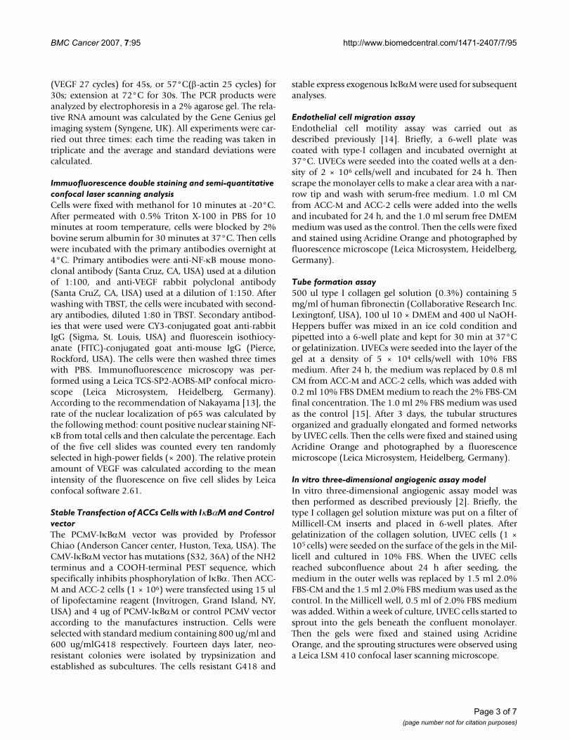
ResultsNF-κBp65 activity and mRNA expression level of VEGF in ACCs cell linesNF-κBp65 DNA binding activity in the nuclear extractsfrom ACC-M and ACC-2 cell lines was showed in Figure 1,lane 1–2. There was a constitutive NF-κBp65 activity inACC-M and ACC-2 cell lines. In the ACC-M, the intensityof the shift band of p65/P50 was much stronger than thatin ACC-2.
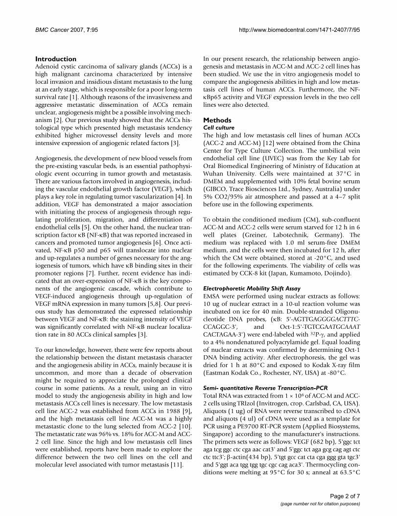
Figure 2 showed the mRNA expression band of VEGF inACC-2 and ACC-M. The mean level of VEGF mRNA inACC-M (0.575 ± 0.10) was about 2-fold than that in ACC-2 (0.309 ± 0.11), and the statistical difference was deemedsignificant (P < 0.01) [see additional file 1].
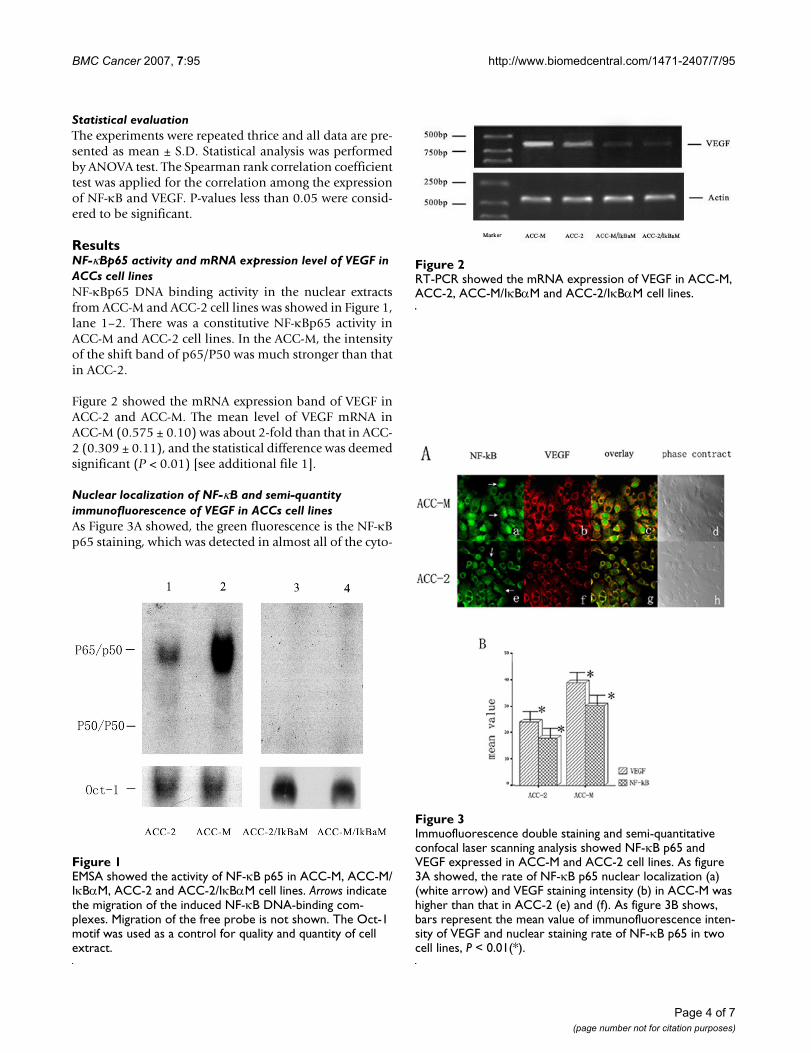
Nuclear localization of NF-κB and semi-quantity immunofluorescence of VEGF in ACCs cell linesAs Figure 3A showed, the green fluorescence is the NF-κBp65 staining, which was detected in almost all of the cyto-
Immuofluorescence double staining and semi-quantitative confocal laser scanning analysis showed NF-κB p65 and VEGF expressed in ACC-M and ACC-2 cell linesFigure 3Immuofluorescence double staining and semi-quantitative confocal laser scanning analysis showed NF-κB p65 and VEGF expressed in ACC-M and ACC-2 cell lines. As figure 3A showed, the rate of NF-κB p65 nuclear localization (a) (white arrow) and VEGF staining intensity (b) in ACC-M was higher than that in ACC-2 (e) and (f). As figure 3B shows, bars represent the mean value of immunofluorescence inten-sity of VEGF and nuclear staining rate of NF-κB p65 in two cell lines, P < 0.01(*).
EMSA showed the activity of NF-κB p65 in ACC-M, ACC-M/IκBαM, ACC-2 and ACC-2/IκBαM cell linesFigure 1EMSA showed the activity of NF-κB p65 in ACC-M, ACC-M/IκBαM, ACC-2 and ACC-2/IκBαM cell lines. Arrows indicate the migration of the induced NF-κB DNA-binding com-plexes. Migration of the free probe is not shown. The Oct-1 motif was used as a control for quality and quantity of cell extract.
RT-PCR showed the mRNA expression of VEGF in ACC-M, ACC-2, ACC-M/IκBαM and ACC-2/IκBαM cell linesFigure 2RT-PCR showed the mRNA expression of VEGF in ACC-M, ACC-2, ACC-M/IκBαM and ACC-2/IκBαM cell lines.
Page 4 of 7(page number not for citation purposes)
BMC Cancer 2007, 7:95 http://www.biomedcentral.com/1471-2407/7/95
plasm but only some of the nucleus in ACCs cells. Themean rate of NF-κB p65 nuclear staining detected in ACC-M and ACC-2 was 30.35 ± 2.52% and 17.97 ± 1.50%,respectively. The rate of NF-κB p65 nuclear localization inACC-M was significantly higher than that in ACC-2 (P <0.01).
The red fluorescence detected in the cytoplasms of ACCscells is the VEGF staining. The mean immunofluorescenceintensities of VEGF in ACC-M and ACC-2 cell lines were38.98 ± 4.98 and 24.10 ± 1.57, respectively (Figure 3). Theexpression level of VEGF in ACC-M was significantlyhigher than that in ACC2 (P < 0.01, Figure 3B).
The Spearman Correlation analysis was performed toquantitate the association between two variables. Theexpression level of VEGF was significantly correlated toNF-kB p65 nuclear localization in both of the ACC-M andACC-2 cell lines (P < 0.01).
Down regulation of VEGF mRNA expression by inhibition of NF-κBp65 activationACC-M and ACC-2 cells were transfected with the mutantIκBα expression vector. The constitutive NF-κBp65 activ-ity found in ACC-M and ACC-2 cell lines was completelyabolished in ACC-M/IκBαM and ACC-2/IκBαM cell lines(figure 1, lane 3–4). Furthermore, the constitutive mRNAexpression level of VEGF was effectively inhibited by over-expression of IκBαM in ACC-M/IκBαM and ACC-2/IκBαM cells (figure 2, lane 3–4).
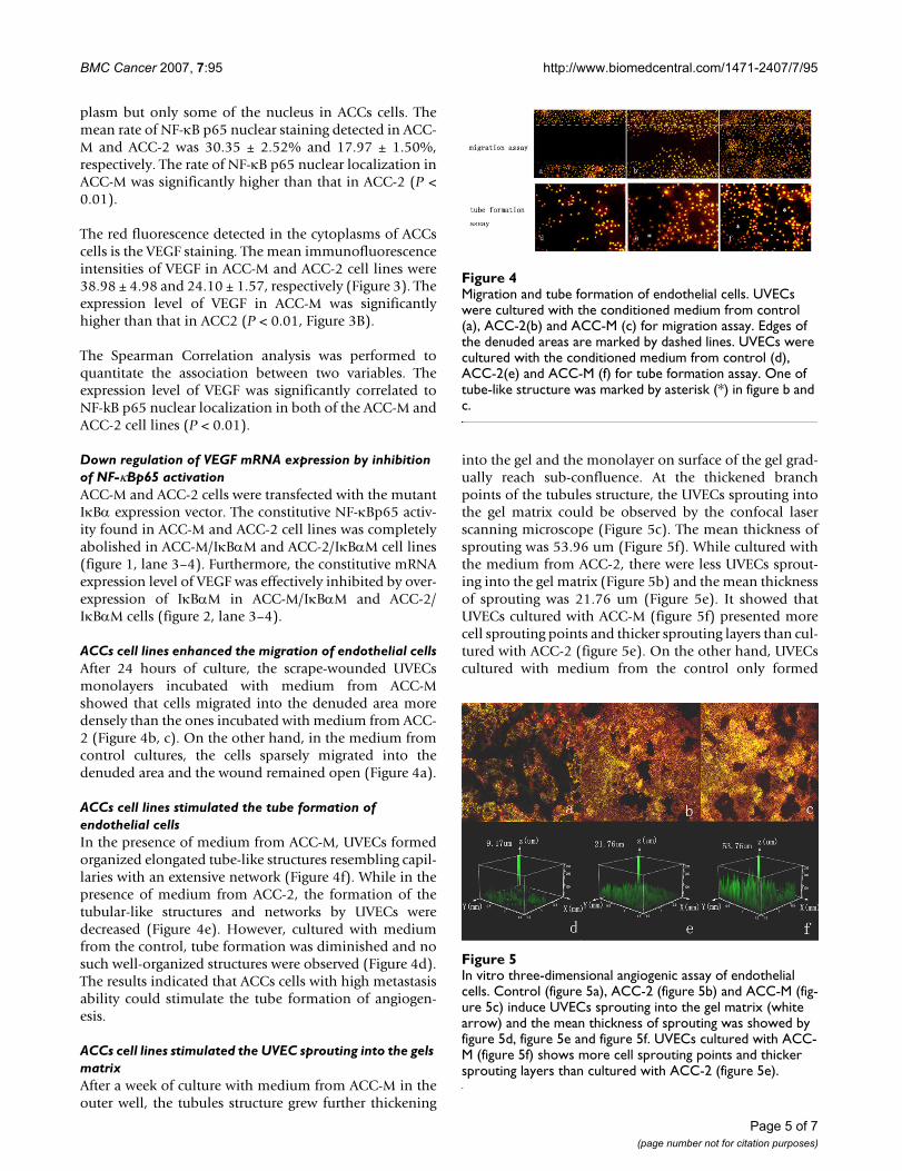
ACCs cell lines enhanced the migration of endothelial cellsAfter 24 hours of culture, the scrape-wounded UVECsmonolayers incubated with medium from ACC-Mshowed that cells migrated into the denuded area moredensely than the ones incubated with medium from ACC-2 (Figure 4b, c). On the other hand, in the medium fromcontrol cultures, the cells sparsely migrated into thedenuded area and the wound remained open (Figure 4a).
ACCs cell lines stimulated the tube formation of endothelial cellsIn the presence of medium from ACC-M, UVECs formedorganized elongated tube-like structures resembling capil-laries with an extensive network (Figure 4f). While in thepresence of medium from ACC-2, the formation of thetubular-like structures and networks by UVECs weredecreased (Figure 4e). However, cultured with mediumfrom the control, tube formation was diminished and nosuch well-organized structures were observed (Figure 4d).The results indicated that ACCs cells with high metastasisability could stimulate the tube formation of angiogen-esis.
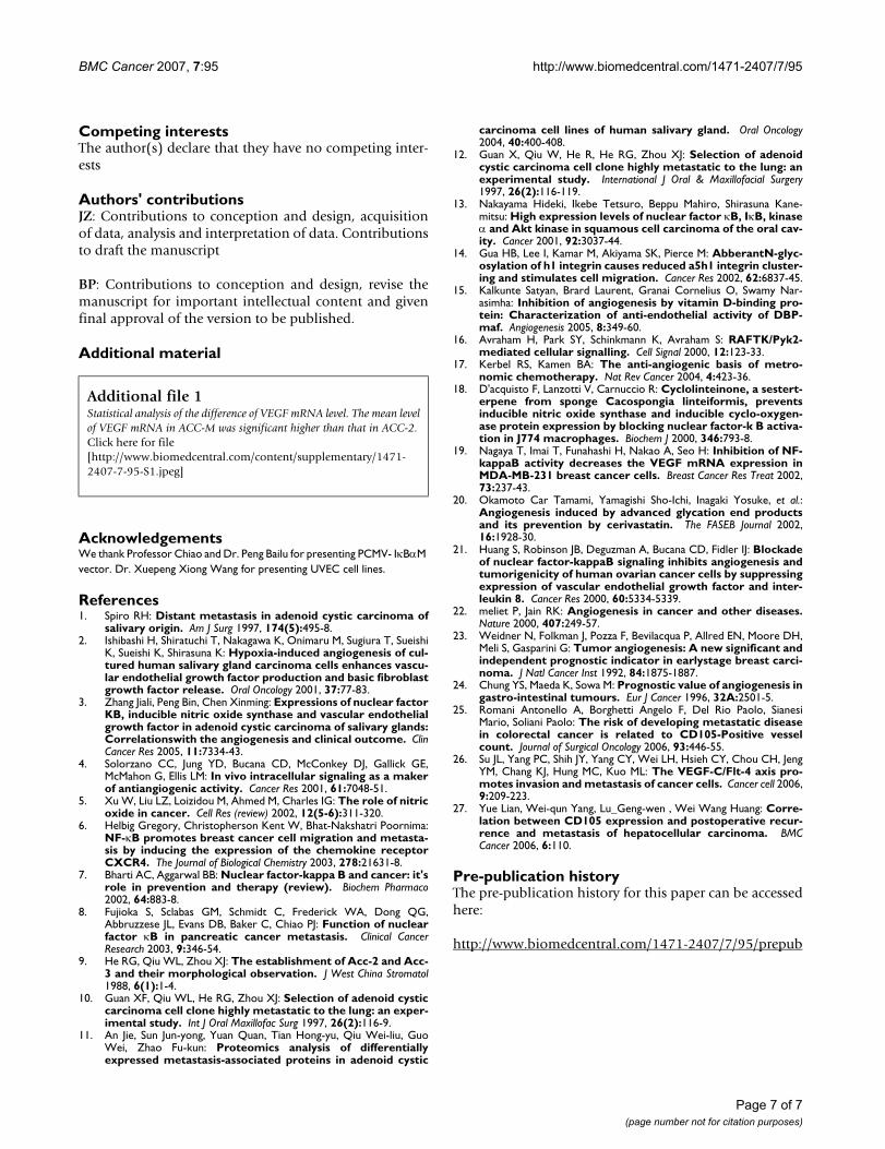
ACCs cell lines stimulated the UVEC sprouting into the gels matrixAfter a week of culture with medium from ACC-M in theouter well, the tubules structure grew further thickening
into the gel and the monolayer on surface of the gel grad-ually reach sub-confluence. At the thickened branchpoints of the tubules structure, the UVECs sprouting intothe gel matrix could be observed by the confocal laserscanning microscope (Figure 5c). The mean thickness ofsprouting was 53.96 um (Figure 5f). While cultured withthe medium from ACC-2, there were less UVECs sprout-ing into the gel matrix (Figure 5b) and the mean thicknessof sprouting was 21.76 um (Figure 5e). It showed thatUVECs cultured with ACC-M (figure 5f) presented morecell sprouting points and thicker sprouting layers than cul-tured with ACC-2 (figure 5e). On the other hand, UVECscultured with medium from the control only formed
In vitro three-dimensional angiogenic assay of endothelial cellsFigure 5In vitro three-dimensional angiogenic assay of endothelial cells. Control (figure 5a), ACC-2 (figure 5b) and ACC-M (fig-ure 5c) induce UVECs sprouting into the gel matrix (white arrow) and the mean thickness of sprouting was showed by figure 5d, figure 5e and figure 5f. UVECs cultured with ACC-M (figure 5f) shows more cell sprouting points and thicker sprouting layers than cultured with ACC-2 (figure 5e).
Migration and tube formation of endothelial cellsFigure 4Migration and tube formation of endothelial cells. UVECs were cultured with the conditioned medium from control (a), ACC-2(b) and ACC-M (c) for migration assay. Edges of the denuded areas are marked by dashed lines. UVECs were cultured with the conditioned medium from control (d), ACC-2(e) and ACC-M (f) for tube formation assay. One of tube-like structure was marked by asterisk (*) in figure b and c.
Page 5 of 7(page number not for citation purposes)
BMC Cancer 2007, 7:95 http://www.biomedcentral.com/1471-2407/7/95
uncompleted tubule structures on the surface of gel, andalmost no sprouting cells could be observed.
DiscussionThe great potential for hematogenous metastasis at anearly stage is one of the unique characters of ACCs. Ourprevious study showed that the high expressions of angio-genesis related factors NF-κB and VEGF were significantlycorrelated to lung metastasis and solid histotype, whichpresent high metastasis tendency [3]. In the current study,we used high and low metastasis cell lines of humanACCs-ACC-M and ACC-2- to compare the angiogenicrelated factor NF-κB and VEGF expression levels. We alsocompared the ability of angiogenesis between the two celllines by in vitro cell migration, tube formation andsprouting assay.
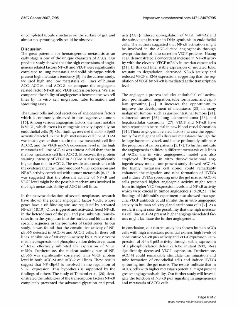
The tumor cells induced secretion of angiogenesis factorswhich is commonly observed in most aggressive tumors[16]. Among various angiogenic factors, the most notableis VEGF, which exerts its mitogenic activity especially onendothelial cells [5]. Our findings revealed that NF-κBp65activity detected in the high metastasis cell line ACC-Mwas much greater than that in the low metastasis cell lineACC-2, and the VEGF mRNA expression level in the highmetastasis cell line ACC-M was almost 2-fold than that inthe low metastasis cell line ACC-2. Moreover, the proteinstaining intensity of VEGF in ACC-M is also significantlyhigher than that in ACC-2. The results are consistent withthe evidence that the tumor-induced VEGF expression andNF-κB activity correlated with tumor metastasis [8,17]. Itwas suggested that the aberrant activity of NF-κB andVEGF level might be the possible mechanisms involved inthe high metastasis ability of ACC-M cell lines.
In the neovascularization of several neoplasms, researchhave shown the potent angiogenic factor VEGF, whosegenes have a κB binding site, are regulated by activatedNF-κB [18,19]. Once triggered and activated, freed NF-κB,in the heterodimer of the p65 and p50 subunits, translo-cates from the cytoplasm into the nucleus and binds to thespecific sequence in the promoter of target genes. In ourstudy, it was found that the constitutive activity of NF-κBp65 detected in ACC-M and ACC-2 cells. In these celllines, inhibition of NF-κBp65 activity by a PCMV vectormediated expression of phosphorylation defective mutantof IκBα effectively inhibited the expression of VEGFmRNA. Furthermore, the nuclear staining rate of NF-κBp65 was significantly correlated with VEGF proteinlevel in both ACC-M and ACC-2 cell lines. These resultssuggest that NF-κBp65 is involved in the regulation ofVEGF expression. This hypothesis is supported by thefindings of others. The study of Tamami et al. [20] dem-onstrated the inhibitors of the transcription factors NF-κBcompletely prevented the advanced glycation end prod-
ucts (AGE)-induced up-regulation of VEGF mRNAs andthe subsequent increase in DNA synthesis in endothelialcells. The authors suggested that NF-κB activation mightbe involved in the AGE-elicited angiogenesis throughoverproduction of auto-secretion VEGF proteins. Huanget al. demonstrated a concordant increase in NF-κB activ-ity with the elevated VEGF mRNA in ovarian cancer cells[21]. In this cell line, stable expression of mutated IκBαresistant to degradation, decreased NF-κB activity andreduced VEGF mRNA expression, suggesting that the reg-ulation of VEGF by NF-κB is mediated at the transcriptionlevel.
The angiogenic process includes endothelial cell activa-tion, proliferation, migration, tube formation, and capil-lary sprouting [22]. It increases the opportunity toimprove the development of metastases [23] in manymalignant tumors, such as gastro-intestinal tumors [24],colorectal cancer [25], lung adenocarcinoma [26], andhepatocellular carcinoma [27]. VEGF and NF-κB havebeen reported to be crucial in new blood vessel formation[14]. Those angiogenic-related factors increase the oppor-tunity for malignant cells distance metastases through theleakage basement vessel, and hence profoundly influencethe prognosis of cancer patients [5,17]. To further indicatethe angiogenesis abilities in different metastasis cells linesof ACCs, the in vitro angiogenesis model was alsoemployed. Through in vitro three-dimensional ang-iogenic assay model, our present study showed ACC-M,the highly metastasis cell clone, could remarkablyenhanced the migration and tube formation of UVECsand induce UVECs sprouting into the gel matrix. ACC-Mcells presented higher angiogenic activity might resultfrom its higher VEGF expression levels and NF-κB activitywhich were crucial in tumor angiogenesis [8,20,21]. Thefindings of Ishibashi's experiment also showed that spe-cific VEGF antibody could inhibit the in vitro angiogenicactivity in human salivary gland carcinoma cells [2]. As aresult, it might raise the possibility that the high metasta-sis cell line ACC-M present higher angiogenic-related fac-tors might facilitate the further angiogenesis.
In conclusion, our current study has shown human ACCscells with high metastasis potential express high levels ofconstitutive NF-κB p65 activity and VEGF expression. Sup-pression of NF-κB p65 activity through stable expressionof a phosphorylation defective IκBα mutant (S32, 36A)significantly decreased VEGF expression. Furthermore,ACC-M could remarkably stimulate the migration andtube formation of endothelial cells and induce UVECssprouting into the gel matrix. The results indicate that inACCs, cells with higher metastasis potential might presentgreater angiogenesis ability. Our further study will investi-gate the function of NF-κB p65 signaling in angiogenesisand metastasis of ACCs cells.
Page 6 of 7(page number not for citation purposes)
BMC Cancer 2007, 7:95 http://www.biomedcentral.com/1471-2407/7/95
Competing interestsThe author(s) declare that they have no competing inter-ests
Authors' contributionsJZ: Contributions to conception and design, acquisitionof data, analysis and interpretation of data. Contributionsto draft the manuscript
BP: Contributions to conception and design, revise themanuscript for important intellectual content and givenfinal approval of the version to be published.
Additional material
AcknowledgementsWe thank Professor Chiao and Dr. Peng Bailu for presenting PCMV- IκBαM vector. Dr. Xuepeng Xiong Wang for presenting UVEC cell lines.
References1. Spiro RH: Distant metastasis in adenoid cystic carcinoma of
salivary origin. Am J Surg 1997, 174(5):495-8.2. Ishibashi H, Shiratuchi T, Nakagawa K, Onimaru M, Sugiura T, Sueishi
K, Sueishi K, Shirasuna K: Hypoxia-induced angiogenesis of cul-tured human salivary gland carcinoma cells enhances vascu-lar endothelial growth factor production and basic fibroblastgrowth factor release. Oral Oncology 2001, 37:77-83.
3. Zhang Jiali, Peng Bin, Chen Xinming: Expressions of nuclear factorKB, inducible nitric oxide synthase and vascular endothelialgrowth factor in adenoid cystic carcinoma of salivary glands:Correlationswith the angiogenesis and clinical outcome. ClinCancer Res 2005, 11:7334-43.
4. Solorzano CC, Jung YD, Bucana CD, McConkey DJ, Gallick GE,McMahon G, Ellis LM: In vivo intracellular signaling as a makerof antiangiogenic activity. Cancer Res 2001, 61:7048-51.
5. Xu W, Liu LZ, Loizidou M, Ahmed M, Charles IG: The role of nitricoxide in cancer. Cell Res (review) 2002, 12(5-6):311-320.
6. Helbig Gregory, Christopherson Kent W, Bhat-Nakshatri Poornima:NF-κB promotes breast cancer cell migration and metasta-sis by inducing the expression of the chemokine receptorCXCR4. The Journal of Biological Chemistry 2003, 278:21631-8.
7. Bharti AC, Aggarwal BB: Nuclear factor-kappa B and cancer: it'srole in prevention and therapy (review). Biochem Pharmaco2002, 64:883-8.
8. Fujioka S, Sclabas GM, Schmidt C, Frederick WA, Dong QG,Abbruzzese JL, Evans DB, Baker C, Chiao PJ: Function of nuclearfactor κB in pancreatic cancer metastasis. Clinical CancerResearch 2003, 9:346-54.
9. He RG, Qiu WL, Zhou XJ: The establishment of Acc-2 and Acc-3 and their morphological observation. J West China Stromatol1988, 6(1):1-4.
10. Guan XF, Qiu WL, He RG, Zhou XJ: Selection of adenoid cysticcarcinoma cell clone highly metastatic to the lung: an exper-imental study. Int J Oral Maxillofac Surg 1997, 26(2):116-9.
11. An Jie, Sun Jun-yong, Yuan Quan, Tian Hong-yu, Qiu Wei-liu, GuoWei, Zhao Fu-kun: Proteomics analysis of differentiallyexpressed metastasis-associated proteins in adenoid cystic
carcinoma cell lines of human salivary gland. Oral Oncology2004, 40:400-408.
12. Guan X, Qiu W, He R, He RG, Zhou XJ: Selection of adenoidcystic carcinoma cell clone highly metastatic to the lung: anexperimental study. International J Oral & Maxillofacial Surgery1997, 26(2):116-119.
13. Nakayama Hideki, Ikebe Tetsuro, Beppu Mahiro, Shirasuna Kane-mitsu: High expression levels of nuclear factor κB, IκB, kinaseα and Akt kinase in squamous cell carcinoma of the oral cav-ity. Cancer 2001, 92:3037-44.
14. Gua HB, Lee I, Kamar M, Akiyama SK, Pierce M: AbberantN-glyc-osylation of h1 integrin causes reduced a5h1 integrin cluster-ing and stimulates cell migration. Cancer Res 2002, 62:6837-45.
15. Kalkunte Satyan, Brard Laurent, Granai Cornelius O, Swamy Nar-asimha: Inhibition of angiogenesis by vitamin D-binding pro-tein: Characterization of anti-endothelial activity of DBP-maf. Angiogenesis 2005, 8:349-60.
16. Avraham H, Park SY, Schinkmann K, Avraham S: RAFTK/Pyk2-mediated cellular signalling. Cell Signal 2000, 12:123-33.
17. Kerbel RS, Kamen BA: The anti-angiogenic basis of metro-nomic chemotherapy. Nat Rev Cancer 2004, 4:423-36.
18. D'acquisto F, Lanzotti V, Carnuccio R: Cyclolinteinone, a sestert-erpene from sponge Cacospongia linteiformis, preventsinducible nitric oxide synthase and inducible cyclo-oxygen-ase protein expression by blocking nuclear factor-k B activa-tion in J774 macrophages. Biochem J 2000, 346:793-8.
19. Nagaya T, Imai T, Funahashi H, Nakao A, Seo H: Inhibition of NF-kappaB activity decreases the VEGF mRNA expression inMDA-MB-231 breast cancer cells. Breast Cancer Res Treat 2002,73:237-43.
20. Okamoto Car Tamami, Yamagishi Sho-Ichi, Inagaki Yosuke, et al.:Angiogenesis induced by advanced glycation end productsand its prevention by cerivastatin. The FASEB Journal 2002,16:1928-30.
21. Huang S, Robinson JB, Deguzman A, Bucana CD, Fidler IJ: Blockadeof nuclear factor-kappaB signaling inhibits angiogenesis andtumorigenicity of human ovarian cancer cells by suppressingexpression of vascular endothelial growth factor and inter-leukin 8. Cancer Res 2000, 60:5334-5339.
22. meliet P, Jain RK: Angiogenesis in cancer and other diseases.Nature 2000, 407:249-57.
23. Weidner N, Folkman J, Pozza F, Bevilacqua P, Allred EN, Moore DH,Meli S, Gasparini G: Tumor angiogenesis: A new significant andindependent prognostic indicator in earlystage breast carci-noma. J Natl Cancer Inst 1992, 84:1875-1887.
24. Chung YS, Maeda K, Sowa M: Prognostic value of angiogenesis ingastro-intestinal tumours. Eur J Cancer 1996, 32A:2501-5.
25. Romani Antonello A, Borghetti Angelo F, Del Rio Paolo, SianesiMario, Soliani Paolo: The risk of developing metastatic diseasein colorectal cancer is related to CD105-Positive vesselcount. Journal of Surgical Oncology 2006, 93:446-55.
26. Su JL, Yang PC, Shih JY, Yang CY, Wei LH, Hsieh CY, Chou CH, JengYM, Chang KJ, Hung MC, Kuo ML: The VEGF-C/Flt-4 axis pro-motes invasion and metastasis of cancer cells. Cancer cell 2006,9:209-223.
27. Yue Lian, Wei-qun Yang, Lu_Geng-wen , Wei Wang Huang: Corre-lation between CD105 expression and postoperative recur-rence and metastasis of hepatocellular carcinoma. BMCCancer 2006, 6:110.
Pre-publication historyThe pre-publication history for this paper can be accessedhere:
http://www.biomedcentral.com/1471-2407/7/95/prepub
Additional file 1Statistical analysis of the difference of VEGF mRNA level. The mean level of VEGF mRNA in ACC-M was significant higher than that in ACC-2.Click here for file[http://www.biomedcentral.com/content/supplementary/1471-2407-7-95-S1.jpeg]
Page 7 of 7(page number not for citation purposes)