Embed Size (px)
Citation preview
Mendeleev Commun., 2013, 23, 316–318
– 316 –© 2013 Mendeleev Communications. All rights reserved.
MendeleevCommunications
Experimental data1,2 demonstrated that Alzheimer’s Ab amyloid peptide interacts with biomembranes, and this interaction modifies the properties of lipid bilayers, further disrupting the fluidity and functions of the membranes. Sphingomyelin (SM), phosphatidylcholine (PC) and cholesterol (CHOL) are important lipids of eukaryotic cellular membranes and neuronal tissues, and they presumably participate in the formation of membrane domains, known as ‘rafts’, through intermolecular interaction, lateral microphase decomposition and formation of SMdepleted (liquid dis ordered, ld) and SMenriched (liquid ordered, lo) liquid crystalline phases.3–5 Ab peptide induces a strong membrane destabilization in these lipid bilayers2,6 and binds to both phases but preferably to the lo phase. Lateral diffusion of lipids is an essential dynamic factor in the twodimensional lipid membrane system,7 which can be indicative of interlipid interactions and decomposition processes, particularly in ‘raft’forming membranes.4,5 In this work, we employed pulsed field gradient NMR to study the lateral diffusion of lipids in the macroscopically oriented bilayers of raft compositions [containing SM, dioleoylPC (DOPC) and CHOL] with and without added Ab(140) peptide.
Human wildtype Alzheimer’s Ab(140) peptide with an amino acid sequence (DAEFRHDSGYEVHHQKLVFFAEDVGSNKGAIIGLMVGGVV) was synthesized by a standard solidphase FMOC procedure with HBTU activation and subsequently purified by HPLC as described previously.8 The purity of the final product was 95%, as found by MALDITOF mass spectrometry. Commercial DOPC, egg yolk SM and CHOL (Avanti Polar Lipids, Alabaster, AL) were used. Macroscopically oriented multi bilayers were prepared following reported pro ce dure.9 The Ab(140) peptide was initially used in monomeric form. The lipid/peptide molar ratio was 700:1. Hydration (D2O) was performed during five days, and the final water content of the test samples was 50 wt%; pH 4.4–5.2. To elucidate how the lipid composition influences the secondary structure of the peptide, UV circular dichroism (CD) spectra were measured in the same systems at a 5000fold dilution because of too high absorptivity, and the final concentration of Ab(140) was 12.5 mmol dm–3. A Jasco J810 spectropolarimeter equipped with a quartz cuvette having a sample volume of 370 ml and an optical path length of 1 mm was used in the CD measurements. The experiments were carried out in a wavelength range of 190–250 nm. Each sample was measured eight times, and the CD spectra were averaged;
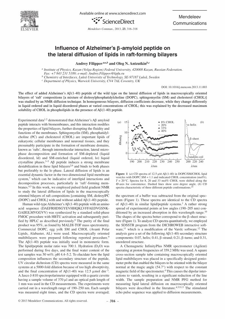
the spectrum of a buffer was subtracted from the original spectrum (Figure 1). These spectra are identical to the CD spectra of Ab(140) in similar lipid/peptide systems.1 A rather strong spread of experimental points at low angles (190–205 nm) conditioned by an increased absorption in this wavelength range.10 The shapes of the spectra better correspond to the bsheet structure (Figure 1). To analyze CD spectra quantitatively, we employed the SDSSTR program from the DICHROWEB interactive software,11 which is a modification of the Varslc software.12 The analysis gave a set of the following Ab(140) secondary structure components: 0.07, helix; 0.41, bstrand; 0.21, bturns; and 0.31, unordered structure.
A Chemagnetic InfinityPlus NMR spectrometer (Agilent) operating at proton frequencies of 359.2 MHz was used. A square crosssection sample tube containing macroscopically oriented lipid multibilayers was placed in a specifically designed goniometer probe that enabled the bilayers to be oriented with the bilayer normal at the magic angle (54.7°) with respect to the constant magnetic field of the spectrometer.4 This causes the dipolar interactions to vanish, resulting in a significant reduction of the line width. The sample preparation and NMR PFG method for measuring lipid lateral diffusion on macroscopically oriented bilayers were described in the literature.4,5,9,13 The stimulated echo pulse sequence was applied to diffusion measurements.
Influence of Alzheimer’s bbb-amyloid peptide on the lateral diffusion of lipids in raft-forming bilayers
Andrey Filippov*a,b and Oleg N. Antzutkinb,c
a Institute of Physics, Kazan (Volga Region) Federal University, 420008 Kazan, Russian Federation. Fax: +7 843 231 5189; e-mail: [email protected]
b Chemistry of Interfaces, Luleå University of Technology, SE-97187 Luleå, Swedenc Department of Physics, Warwick University, CV4 7AL Coventry, UK
11.003DOI: 10.1016/j.mencom.2013.
The effect of added Alzheimer’s Ab(1-40) peptide of the wild type on the lateral diffusion of lipids in macroscopically oriented bilayers of ‘raft’ compositions [a mixture of dioleoylphosphatidylcholine (DOPC), sphingomyelin (SM) and cholesterol (CHOL)] was studied by an NMR-diffusion technique. In homogeneous bilayers, diffusion coefficients decrease, while they change differently in liquid ordered and in liquid disordered phases at varied concentrations of CHOL; this was explained by the decreased maximum solubility of CHOL in phospholipids in the presence of Ab(1-40) peptide.
190 200 200210 220 220230 240 240250
0
5
10
15Q
/mde
g
l/nm l/nm
8% CHOL20%33%40%
(a) (b)α-helix
β-sheet
random coil
(Figure 1 a) CD spectra of 12.5 mm Ab(140) in DOPC/SM/CHOL lipid vesicles with DOPC:SM = 1.1 and indicated CHOL con centration (mol%). T = 20 °C. Spectra for 8, 20 and 33 mol% CHOL were shifted along the Qaxis for convenience. Dashed lines show zero degree angle. (b) CD spectra characteristic of three different peptide conformations.24
Mendeleev Commun., 2013, 23, 316–318
– 317 –
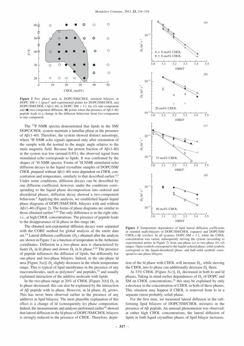
The 31P NMR spectra demonstrated that lipids in the SM/DOPC/CHOL system maintain a lamellar phase in the presence of Ab(140). Therefore, the system showed distinct anisotropy, where 1H NMR echo signals appeared only after orientation of the sample with the normal to the magic angle relative to the main magnetic field. Because the proton fraction of Ab(140) in the system was low (around 0.8%), the observed signal from stimulated echo corresponds to lipids. It was confirmed by the shapes of 1H NMR spectra. Forms of 1H NMR stimulated echo diffusion decays in the liquid crystalline samples of DOPC/SM/CHOL prepared without Ab(140) were dependent on CHOL concentration and temperature, similarly to that described earlier.4,5 Under some conditions, diffusion decays can be described by one diffusion coefficient; however, under the conditions corresponding to the liquid phase decomposition into ordered and disordered phases, diffusion decay showed a twocomponent behaviour.4 Applying this analysis, we established liquid–liquid phase diagrams of DOPC/SM/CHOL bilayers with and without Ab(140) (Figure 2). The forms of phase diagrams are similar to those obtained earlier.4,10 The only difference is in the right side, i.e., at high CHOL concentrations. The presence of peptide leads to the disappearance of ld phase in this range ( ).
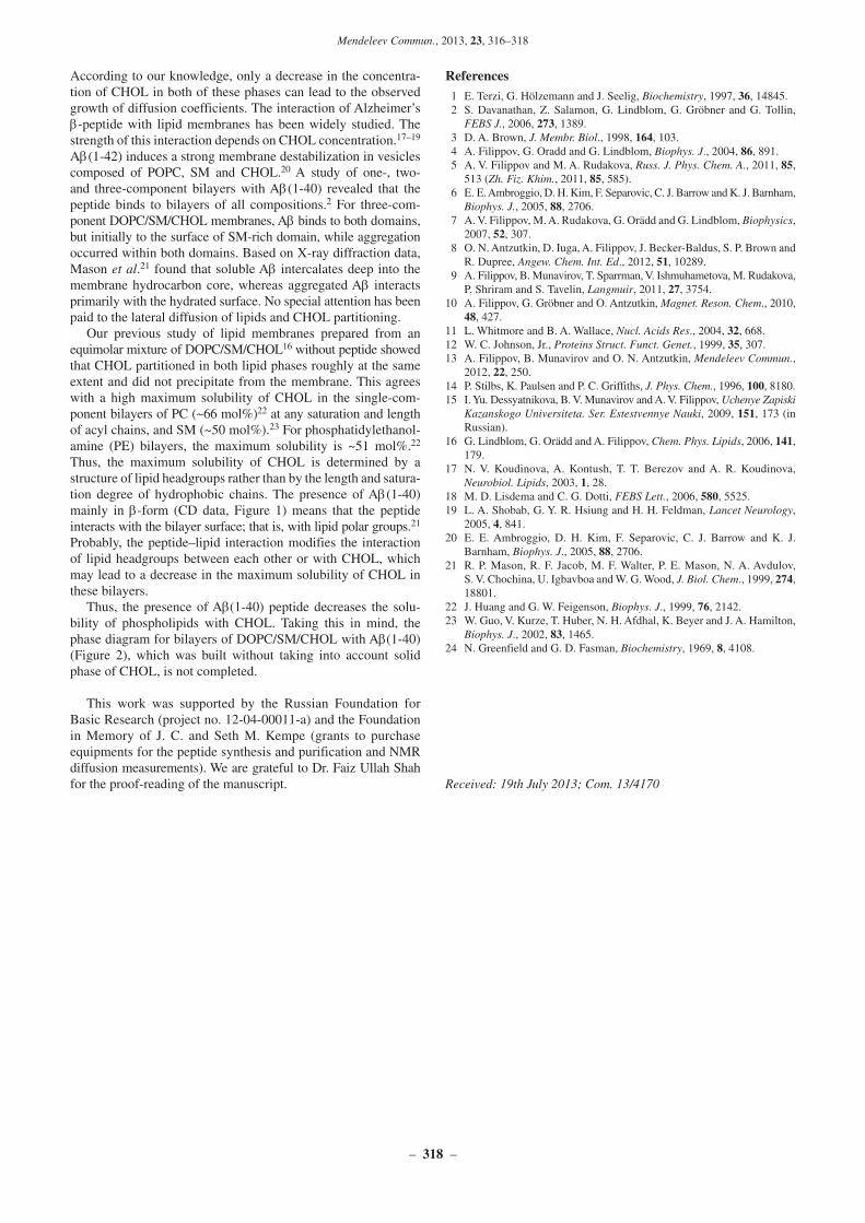
The obtained nonexponential diffusion decays were separated with the CORE method for global analysis of the entire data set.14 Lateral diffusion coefficients (DL) obtained after the analysis are shown in Figure 3 as a function of temperature in the Arrhenius coordinates. Diffusion in a twophase area is characterized by faster DL in ld phase and slower DL in lo phase.4,14 The presence of peptide influences the diffusion of lipids, but differently for onephase and twophase bilayers. Indeed, in the onephase ld area [Figure 3(a)], DL slightly decreases in the whole temperature range. This is typical of lipid membranes in the presence of any macromolecules, such as polymers9 and peptides,15 and usually explained interaction of the additive molecule with lipids.
In the twophase range at 20% of CHOL [Figure 3(b)] DL in lo phase decreased; this can also be explained by the interaction of Ab peptide with lo phase. However, in ld phase, DL grows. This has never been observed earlier in the presence of any additives in lipid bilayers. The most plausible explanation of this effect is a change of ld (consequently lo) phase composition. Indeed, the measurements and analyses performed earlier16 showed that lateral diffusion in the ld phase of DOPC/SM/CHOL bilayers is strongly reduced in the presence of CHOL. Therefore, deple
tion of the ld phase with CHOL will increase DL, while moving the CHOL into lo phase can additionally decrease DL there.
At 33% CHOL [Figure 3(c)], DL decreased in both lo and ld phases. Taking in mind earlier dependences of DL of DOPC and SM on CHOL concentrations,16 this may be explained by only a decrease in the concentration of CHOL in both of these phases. This situation may happen if CHOL is removed from lo in a separate (most probably, solid) phase.
For the first time, we measured lateral diffusion in the raftforming lipid bilayers of DOPC/SM/CHOL mixtures in the presence of Ab peptide. An unusual phenomenon was observed; at rather high CHOL concentrations, the lateral diffusion of lipids in both liquid crystalline phases of lipid bilayer increases.
0 10 20 30 40 50
20
30
40
50
60T
/°C
CHOL (mol%)
Two phase area in DOPC/SM/CHOL oriented bilayers at Figure 2 DOPC:SM = 1 (gray)4 and experimental points for DOPC/SM/CHOL and DOPC/SM/CHOL + Ab(140) at DOPC:SM = 1.1 for ( ) onecomponent and ( ) twocomponent diffusion; ( ) points where the presence of Ab(140) peptide leads to a change in the diffusion behaviour from twocomponent to onecomponent.
3.0 3.1 3.2 3.3 3.4 3.5
1000/T
, 0 mol% CHOL, 8 mol% CHOL
3.0 3.1 3.2 3.3 3.4 3.5
1000/T
20 mol% CHOL
3.0 3.1 3.2 3.3 3.4 3.5
1000/T
33 mol% CHOL
3.0 3.1 3.2 3.3 3.4 3.5
10–12
10–11
DL/m
2 s–1
10–12
10–11
DL/m
2 s–1
10–12
10–11
DL/m
2 s–1
10–11
DL/m
2 s–1
1000/T
40 mol% CHOL
(a)
(b)
(c)
(d)
Temperature dependence of lipid lateral diffusion coefficients Figure 3 in oriented multibilayers of DOPC/SM/CHOL (squares) and DOPC/SM/CHOL + Ab (circles). In all systems, DOPC:SM = 1.1, while the CHOL concentration was varied, subsequently moving the system (according to experimental points in Figure 2) from onephase (a) to twophase (b)–(d) ranges. Open symbols correspond to the liquidordered phase; solid symbols correspond to the liquiddisordered phase, and halfsolid symbols correspond to onephase bilayers.
Mendeleev Commun., 2013, 23, 316–318
– 318 –
According to our knowledge, only a decrease in the concentration of CHOL in both of these phases can lead to the observed growth of diffusion coefficients. The interaction of Alzheimer’s bpeptide with lipid membranes has been widely studied. The strength of this interaction depends on CHOL concentration.17–19 Ab(142) induces a strong membrane destabilization in vesicles composed of POPC, SM and CHOL.20 A study of one, two and threecomponent bilayers with Ab(140) revealed that the peptide binds to bilayers of all compositions.2 For threecomponent DOPC/SM/CHOL membranes, Ab binds to both domains, but initially to the surface of SMrich domain, while aggregation occurred within both domains. Based on Xray diffraction data, Mason et al.21 found that soluble Ab intercalates deep into the membrane hydrocarbon core, whereas aggregated Ab interacts primarily with the hydrated surface. No special attention has been paid to the lateral diffusion of lipids and CHOL partitioning.
Our previous study of lipid membranes prepared from an equimolar mixture of DOPC/SM/CHOL16 without peptide showed that CHOL partitioned in both lipid phases roughly at the same extent and did not precipitate from the membrane. This agrees with a high maximum solubility of CHOL in the singlecomponent bilayers of PC (~66 mol%)22 at any saturation and length of acyl chains, and SM (~50 mol%).23 For phosphatidyl ethanolamine (PE) bilayers, the maximum solubility is ~51 mol%.22 Thus, the maximum solubility of CHOL is determined by a structure of lipid headgroups rather than by the length and saturation degree of hydrophobic chains. The presence of Ab(140) mainly in bform (CD data, Figure 1) means that the peptide interacts with the bilayer surface; that is, with lipid polar groups.21 Probably, the peptide–lipid interaction modifies the interaction of lipid headgroups between each other or with CHOL, which may lead to a decrease in the maximum solubility of CHOL in these bilayers.
Thus, the presence of Ab(140) peptide decreases the solubility of phospholipids with CHOL. Taking this in mind, the phase diagram for bilayers of DOPC/SM/CHOL with Ab(140) (Figure 2), which was built without taking into account solid phase of CHOL, is not completed.
This work was supported by the Russian Foundation for Basic Research (project no. 120400011a) and the Foundation in Memory of J. C. and Seth M. Kempe (grants to purchase equipments for the peptide synthesis and purification and NMR diffusion measurements). We are grateful to Dr. Faiz Ullah Shah for the proofreading of the manuscript.
ReferencesE. Terzi, G. Hölzemann and J. Seelig, 1 Biochemistry, 1997, 36, 14845.S. Davanathan, Z. Salamon, G. Lindblom, G. Gröbner and G. Tollin, 2 FEBS J., 2006, 273, 1389.D. A. Brown, 3 J. Membr. Biol., 1998, 164, 103.A. Filippov, G. Oradd and G. Lindblom, 4 Biophys. J., 2004, 86, 891.A. V. Filippov and M. A. Rudakova, 5 Russ. J. Phys. Chem. A., 2011, 85, 513 (Zh. Fiz. Khim., 2011, 85, 585).E. E. Ambroggio, D. H. Kim, F. Separovic, C. J. Barrow and K. J. Barnham, 6 Biophys. J., 2005, 88, 2706.A. V. Filippov, M. A. Rudakova, G. Orädd and G. Lindblom, 7 Biophysics, 2007, 52, 307.O. N. Antzutkin, D. Iuga, A. Filippov, J. BeckerBaldus, S. P. Brown and 8 R. Dupree, Angew. Chem. Int. Ed., 2012, 51, 10289.A. Filippov, B. Munavirov, T. Sparrman, V. Ishmuhametova, M. Rudakova, 9 P. Shriram and S. Tavelin, Langmuir, 2011, 27, 3754.A. Filippov, G. Gröbner and O. Antzutkin, 10 Magnet. Reson. Chem., 2010, 48, 427.L. Whitmore and B. A. Wallace, 11 Nucl. Acids Res., 2004, 32, 668.W. C. Johnson, Jr., 12 Proteins Struct. Funct. Genet., 1999, 35, 307.A. Filippov, B. Munavirov and O. N. Antzutkin, 13 Mendeleev Commun., 2012, 22, 250.P. Stilbs, K. Paulsen and P. C. Griffiths, 14 J. Phys. Chem., 1996, 100, 8180.I. Yu. Dessyatnikova, B. V. Munavirov and A. V. Filippov, 15 Uchenye Zapiski Kazanskogo Universiteta. Ser. Estestvennye Nauki, 2009, 151, 173 (in Russian).G. Lindblom, G. Orädd and A. Filippov, 16 Chem. Phys. Lipids, 2006, 141, 179.N. V. Koudinova, A. Kontush, T. T. Berezov and A. R. Koudinova, 17 Neurobiol. Lipids, 2003, 1, 28.M. D. Lisdema and C. G. Dotti, 18 FEBS Lett., 2006, 580, 5525.L. A. Shobab, G. Y. R. Hsiung and H. H. Feldman, 19 Lancet Neurology, 2005, 4, 841.E. E. Ambroggio, D. H. Kim, F. Separovic, C. J. Barrow and K. J. 20 Barnham, Biophys. J., 2005, 88, 2706.R. P. Mason, R. F. Jacob, M. F. Walter, P. E. Mason, N. A. Avdulov, 21 S. V. Chochina, U. Igbavboa and W. G. Wood, J. Biol. Chem., 1999, 274, 18801.J. Huang and G. W. Feigenson, 22 Biophys. J., 1999, 76, 2142.W. Guo, V. Kurze, T. Huber, N. H. Afdhal, K. Beyer and J. A. Hamilton, 23 Biophys. J., 2002, 83, 1465.N. Greenfield and G. D. Fasman, 24 Biochemistry, 1969, 8, 4108.
19th July 2013; Com. 13/4170Received: