Embed Size (px)
Citation preview
1
Internal Organization of
Macromonomers and Dendronized
Polymers based on Thiophene Dendrons
Esther Córdova-Mateo,1,2
Oscar Bertran,1 A. Dieter Schlüter,
3 Martin
Kröger,3 and Carlos Alemán
2,4,*
1 Departament de Física Aplicada, Escola d’Enginyeria d’Igualada, Universitat
Politècnica de Catalunya, Pça Rei 15, Igualada 08700, Spain
2 Departament d’Enginyeria Química, E. T. S. d’Enginyeria Industrial de Barcelona,
Universitat Politècnica de Catalunya, Diagonal 647, Barcelona E-08028, Spain
3 Department of Materials, Institute of Polymers, Swiss Federal Institute of Technology,
ETH Zurich, Vladimir-Prelog-Weg 5, 8093 Zurich, Switzerland
4 Centre for Research in Nano-Engineering, Universitat Politècnica de Catalunya,
Edifici C’, C/Pasqual i Vila s/n, Barcelona E-08028, Spain
2
ABSTRACT
The internal organization of macromonomers (MGs) consisting of all-thiophene
dendrons of generation g= 2 and 3 attached to a phenyl core, as well as of the
dendronized polymers resulting from polymerization of these macromonomers (PG2
and PG3, respectively), has been investigated using theoretical methods. The
conformational preferences of the MGs, determined using density functional theory
calculations, are characterized by the relative orientation between dendrons and core.
We find that the strain of the MGs increases with the generation number and is
alleviated by small conformational re-arrangements of the peripheral thiophene rings.
The conformations obtained for the MGs have subsequently been used to construct
models for the dendronized polymers. Classical molecular dynamics simulations have
evidenced that the interpenetration of dendrons belonging to different repeat units is
very small for PG2. In contrast, the degree of interpenetration is found to be very high
for PG3, which also shows a significant degree of backfolding (i.e. occurrence of
peripheral methyl groups approaching the backbone). Consequently, PG2 behaves as a
conventional linear flexible polymer bearing bulk pendant groups, whereas PG3 is
better characterized as a semirigid homogenous cylinder. The two polymers are
stabilized by - stacking interactions, even though these are significantly more
abundant for PG3 than for PG2; the average number of interactions per repeat unit is 3.0
and 8.8 for PG2 and PG3, respectively. While in these interactions the thiophene rings
can adopt either parallel (sandwich) or perpendicular (T-shaped) dispositions, the
former scenario turns out to be the most abundant.
3
INTRODUCTION
Organic molecules with dendritic architectures are typically classified in dendrimers
and dendronized polymers (DPs). Dendrimers are the more widely investigated
polymers1-18
and consist of perfectly branched molecules made of tree-like fragments
(dendrons) attached to a central core. The internal organization and size of these well-
defined, often monodisperse, molecules depends on the generation number, g, of
dendrons. Thus, dendrimers of lower generation have relatively loose inner structure
while higher generations are densely packed and organized.1-4
These particular
organizations result in unusual properties, as for example liquid-crystalline behavior at
high concentrations,5,6
anomalous intrinsic viscosity,7,8
and a multivalent molecular
surface.9,10
As a consequence, dendrimers have been proposed for very useful
applications, as for example, drug-delivery systems,11,12
gene vectors,13,14
catalysts15,16
and organic light-emitting diodes.17,18
DPs can be seen as wormlike macromolecular objects of cylindrical cross section.19-
21 The mass per repeat unit of the polymer backbone increases nonlinearly with the
generation number (g), which allows to modulate properties like the rigidity, diameter
and concentration of functionalities.21-24
Due to this particular architecture, DPs
currently represent a class of single molecular nanomaterials with several potential
applications. Among the most promising applications of DPs are nanoscopic building
blocks,25,26
functional materials,27,28
organic optoelectronic materials,29,30
self-
assembling vectors for complexation with DNA,31,32
and nanomaterials to stabilize
therapeutic proteins in the gastrointestinal tract33
and both to copy34
and to immobilize
enzymes.35
Furthermore, the structure of different families of DPs has been investigated
using atomistic simulations, results providing microscopic understanding of the physical
properties of these nanomaterials.36-41
Importantly, these microscopic studies have

4
revealed that the internal organization and properties are intimately related not only with
g but also with the chemical nature of the dendrons. For example, the stability of the
right-handed helical conformation found for neutral and charged dendronized
polymetylmethacrylates carrying chiral 4-aminoproline based dendrons is essentially
due to the formation of networks of specific interactions.36,37
In contrast, the interactions
defined by the backfolding of the external dendrons dominates the properties of
dendronized polymethacrylates made of tree-like fragments with amide and aromatic
groups separated by polymethylenic segments.38,39
These results supported the idea of
treating these DPs as soft elongated colloidal objects.40,41
Thiophene (Th) based dendrimers and DPs with a fully -conjugated core are
considered as very promising kinds of conducting materials.42
Since Advincula and co-
workers reported on the first Th dendrimer synthesis,43,44
several other Th dendrimers
for different energy-related applications have been described.42
For example, Bäuerle
and co-workers45
synthesized different all-Th dendrimers containing up to 90 Th rings
with a divergent/convergent approach to facilitate the inclusion of functionalities in the
external surface of the conducting dendrimer. These Th dendrimers were used as
entangled photon sensors.46
Mitchell et al.47
prepared phenyl-cored Th dendrimers for
organic photovoltaic devices, their power-conversion efficiency being recently
overtaken by hexaperi-hexabenzocornene-cored Th dendrimers described by Wong et
al.48
and the hybrid gold-nanoparticle-cored dendrimers of Deng et al.49.
Furthermore,
valuable microscopic and electronic information was derived from quantum mechanical
studies on several Th-based dendrimers.50-52
In contrast, studies devoted to Th-based DPs are very scarce because of the intrinsic
complexity associated to this kind of macromolecular objects. In a pioneering work, a
few years ago Schlüter and co-workers53
reported the synthesis of Th-containing second

5
and third generation dendronized macromonomers with methacrylate polymerizable
units as well as their corresponding DPs. More recently, Kimura et al.54
prepared novel
all-Th dendritic macromonomers that were subsequently polymerized. The electronic
and electrical properties of the resulting DPs, which showed enhanced conductivity
upon doping, were attributed to the spatial overlapping of the Th dendrons through -
interactions. Very recently, Griffin et al.55
reported the synthesis and characterization of
a benzodithiophene/Th alternating copolymers decorated with rigid, singly branched
pendant side chains. Photoexcitation of these copolymers resulted in excited states
primarily localized on the pendant side chains that excitations were rapidly transferred
to the polymer backbone (i.e. in less than 250 fs).
In this work we use a multi-scale theoretical approach to characterize at the
microscopic level the internal organization of macromonomers and DPs made of
branched Th dendrons. More specifically, the molecular and electronic structures of two
macromonomers, MG2 and MG3 in Figure 1a, have been studied using quantum
mechanical methods based on density functional theory (DFT) calculations. After this,
in a second step, DPs derived from the polymerization of MG2 and MG3 have been
investigated using molecular dynamics (MD) simulations based on classical force-
fields. The chemical structure of these DPs, named PG2 and PG3 (with g= 2 and 3,
respectively), is depicted in Figure 1b. It is worth noting that MG2, MG3, PG2 and PG3
are practically identical to the macromonomers and their DPs synthesized by Schlüter
and co-workers.53
The only difference involves the alkyl groups attached to the Th
rings, where we have replaced the hexyl groups of the experimental systems by methyl
groups for simplicity.
METHODS

6
Quantum mechanical calculations.
All calculations were performed using the Gaussian 09 computer program.56
The
conformational preferences of MG2 and MG3 were examined using DFT calculations
with the wB97X-D functional57
combined with the 6-311+G(d,p) basis set,58,59
(i.e.
wB97X-D /6-311+G(d,p) level).
The ionization potential (IP) was estimated using the Koopman’s theorem (KT),60
according to which the IP was taken as the negative of the highest occupied molecular
orbital (HOMO) energy (IP= -HOMO). It is worth noting that KT is not applied to DFT
methodologies since energies of Kohn-Shan orbitals do not involve any physical
meaning. However, Janak’s theorem61
was used by Perdew62
to show the connection
between IP and the HOMO energy.
The lowest -* transition energy (g) was derived from the excitation energies
calculated using time-dependent density functional theory (TD-DFT).63
This
methodology provides a robust and efficient description of the low-lying molecular
states and is widely applied to study the UV-vis spectra of conjugated organic
compounds.64-66
Electronic excitations were evaluated using the PB067,68
and
B3LYP69,70
functionals, which are known to be very reliable for the calculation of
electronic transitions,71,72
combined with the 6-311+G(d,p) basis set and employing
geometries fully optimized at the wB97X-D /6-311+G(d,p) level.
Electron densities of the most stable conformations identified for MG2 and MG3
were determined at the wB97X-D /6-311+G(d,p) level using the Merz-Kollman (MK)
scheme,73,74
which assigns point charges to fit the computed electrostatic potential to
points on nested Connolly surface with a density of 1 point/Å2.
Classical force field simulations.
7
The most stable structures of MG2 and MG3 obtained by DFT calculations were
used to build the starting geometries for PG2 and PG3. The stability of the resulting
structures was investigated in vacuum considering DP chains with N= 150 repeat units.
Generally speaking, this solvent-free model corresponds to the situation encountered in
the poor solvent experiments as was proved in previous studies of other non-charged
DPs.38,39
The backbone conformation of PG2 and PG3 was determined by applying a
systematic search strategy. More specifically, 144 trial backbone conformations were
constructed for each DP varying the dihedral angles and (Figure 1b) in steps of 30º.
The number of backbone conformations without backbone–backbone, backbone–side
group and side group–side group steric clashes, hereafter denoted feasible
conformations, was approximately 30% and 20% of the initial trial conformation for
PG2 and PG3, respectively. The rest of the conformations (i.e. those with atomic
overlaps) were directly discarded without performing any calculation.
Energy minimizations and MD simulations of the feasible conformations were
performed using the NAMD program.75
The energy was calculated using the AMBER
force-field.76
All bonding and van der Waals parameters required for PG2 and PG3 were
taken from the Generalized AMBER force-field77
(GAFF) while atomic charges were
computed at the wB97X-D /6-311+G(d,p) level using the Restrained ElectroStatic
Potential (RESP) strategy (Figure 2).78
Geometry optimizations of all feasible conformations were performed by applying
the conjugate gradient method during 5000 steps. After that, only 15% and 16% of
feasible conformations were kept for PG2 and PG3, respectively. Such structures were
pre-equilibrated by heating up the system from 0 to 298 K using a rate of 1 K each 1.5
ps. Visual inspection of the structures obtained after such short simulation time (i.e. 447
ps) indicated that many of them lost the initial helical regularity during the
8
thermalization process. Thus, only 6 and 4 configurations remained regular for PG2 and
PG3, respectively. These structures were submitted to 5 ns of MD for equilibration.
Finally, a 20 ns production (“relaxation”) trajectory was carried out for the structure of
lowest energy of each DP, which corresponds to that started using {,}= 180º, 60º and
-150º,-60º for PG2 and PG3, respectively. Data were saved every 8 ps for subsequent
analysis (i.e. 2500 snapshots).
Atom-pair distance cut-offs were applied at 14 Å to compute van der Waals and
electrostatic interactions. Bond lengths involving hydrogen atoms were constrained
using the SHAKE algorithm with a numerical integration step of 1 fs.79
The temperature
was controlled by a weak coupling method, the Berendsen thermostat80
with a time
constant for heat-bath coupling of 1 ps.
RESULTS AND DISCUSSION
MG2 and MG3 macromonomers
The conformational preferences of the dendron used to prepare MG2,53
which
consists of three Th rings linked by - and - linkages (3T; Scheme 1), was studied
in a previous work.52
More specifically, the potential energy surface derived from the
systematic variation of the inter-ring dihedral angles was calculated at the B3LYP/6-
31G(d) level. The most stable arrangement reported for 3T, with {,’}= 125º,-38º,52
has been used in this work as starting point for the construction of MG2.
Scheme 1: Chemical structure of the dendron (3T) used to construct MG2
SS
S
3T
9
The conformational preferences of MG2 were calculated using a systemic strategy.
For this purpose, after constructing the starting geometry, the potential energy surface
defined by dihedral angles associated to the linkage between 3T and the phenyl core (
and ’ in Figure 1a) was determined at the wB97X-D/6-311++G(d,p) level.
Specifically, and ’ were varied between 0º and 360º in steps of 30º, the resulting 144
structures being optimized using a flexible rotor approximation (i.e. each structure was
submitted to a constrained geometry optimization in which the inter-ring dihedral angles
θ and θ’ were kept fixed at their initial values).
Figure 3 displays the potential energy surface E=E(,’) obtained for MG2, which
was calculated without imposing any symmetry constraint. As it can be seen, the low-
energy regions, which are indicated by blue colors in the map, are located at {,’}
30º,150º, 150º,30º, 150º,150º and 30º,30º, where all combinations of signs
are possible for each pair of values (e.g. {,’} 30º,150º refers to the following four
pairs: +30º,+150º; +30º,–150º; –30º,–150º; and –150º,–150º). The geometry of these 16
conformations was re-optimized without any constraint in and ’. The dihedral angles
and relative energies of the completely optimized representative conformations are
displayed in Table 1. It should be pointed out that, although the four minima obtained
for each pair of {,’} values are not formally equivalent because of the lack of
molecular symmetry (Figure 1a), they are very similar in terms of energy and geometric
properties. Accordingly, only one of four minima detected for each {,’} pair (that of
lowest energy) has been explicitly included in Table 1. On the other hand, as the
dihedrals and ’, which refer to the α-α and α-β linkages of the two peripheral Th
rings to the central one (Figure 1a), are very similar for the two dendrons contained in
MG2, Table 1 lists the average values and the corresponding standard deviations rather

10
than the explicit values for each dendron. Table 1 also includes the average values of α-
α and α-β bond lengths (denoted R and R, respectively, in Scheme 1).
As it can be seen, the disposition of the peripheral Th rings is very similar for all
conformations included in Table 1. This feature indicates that the two 3T dendrons used
to prepare MG2 react upon the attachment to the phenyl core, provoking a significant
conformational change in one of the peripheral Th rings. Thus, the conformation found
for the individual 3T dendron, {,’}= 125º,-38º,52
evolves towards {,’} 120º,140º.
As occurred for {,’}, the R and R values obtained for the different optimized
conformations do not show appreciable differences. These observations are consistent
with the very low relative energies (E) separating the 16 optimized conformations, the
energy gap between the most and the least stable conformation being of only 0.6
kcal/mol. The scarce influence of {,’} values on the relative stability and geometries
facilitates the study and interpretation of MG2 properties, which have been analyzed for
the lowest energy conformation only (MG2-1 in Table 1).
In the MG2-1 (Figure 4a) repulsive S···S interactions involving the Th rings directly
attached to the phenyl core are strictly minimized by the dihedrals ,’= 144.9º,-147.7º.
The g and IP values calculated at the PB0/6-311+G(d,p) and B3LYP/6-311+G(d,p)
levels for such conformation, which are practically identical to those obtained the rest of
minimized conformations, are in excellent agreement (i.e. g= 3.65 eV and IP= 5.76 eV
at the former level, and g= 3.48 eV and IP= 5.56 eV at the latter level). The g values
derived from TD-DFT calculations slightly overestimate the experimental estimation,
3.00 eV, which was determined by absorption and emission spectroscopy.53
However,
our g estimates are significantly lower than the value calculated for the individual 3T
dendron (i.e. the theoretical g obtained using DFT calculations at the B3LYP/6-31G(d)
level for 4.13 eV52
while the experimental measure was 3.33 eV81
), which is fully

11
consistent with experimental observations. On the other hand, the IP predicted for MG2
by the KT, 0.97 eV per thiophene ring, is lower than that obtained at the B3LYP/6-
31G(d) for the individual 3T dendron (i.e. 3.21 eV per thiophene ring).52
Inspection of
the topology of the highest occupied molecular orbital (HOMO) and the lowest
unoccupied molecular orbital (LUMO), which are displayed in Figures 4b and 4c,
evidences that these frontier orbitals are distributed through the aromatic rings of the
two 3T dendrons and the phenyl core. This is fully consistent the homogeneous
distribution of the electron density displayed in Figure 4d.
The conformational preferences of MG3 were evaluated using the following strategy.
The four structures of lower energy identified in a previous study for the all-thiophene
dendron used to prepare MG3, which has been denoted 7T (Scheme 2),52
were attached
to the phenyl core considering the following pairs of values for the dihedral angles ,’:
150º,-150º; 150º, 30º; 30º, 30º; and 30º,-150º. The 44= 16 starting structures were
subjected to complete geometry optimization at the wB97X-D/6-311++G(d,p) level,
resulting in 12 different conformations. Interestingly, the structures with ,’ 150º,-
150º are not stable when the generation number g increases from 2 to 3, reverting in
conformations similar to those achieved after optimize the starting points with ,’
30º,-150º.
Scheme 2 Chemical structure of the dendron (7T) used to construct MG3
12
The three conformations of lower energy, which are listed in Table 2, are separated
by an energy gap of 0.2 kcal/mol only, whereas the relative energy of the remaining
optimized structures (not shown) was higher than 0.5 kcal/mol. The most stable
conformation (G3-1 in Table 2) is displayed in Figure 4a. As occurred for MG2-1, S···S
repulsive interactions are minimized in MG3-1. Also, Table 3 reflects that the strain of
the macromonomer increases the generation number g. In order to alleviate such strain,
inter-ring dihedral angles of different dendrons present larger deviations than in MG2,
as is evidenced by the standard deviations of the corresponding averages. In spite of
this, it is worth noting that the disposition of the peripheral Th rings is similar for MG2
and MG3, the dihedrals {,’} of the minima identified for each macromonomer
differing in 10º only.
The g values derived for MG3-1 from TD-DFT calculations at the PB0/6-
311+G(d,p) and B3LYP/6-311+G(d,p) levels are 3.50 and 3.31 eV, respectively. which
represents a slight reduction with respect to MG2-1. This is fully consistent with the g
values experimentally determined for MG2 and MG3, which reflected a reduction of
0.35 eV.53
The HOMO extends over two dendrons located of the same branch (Figure
4b), which represents a difference with respect to MG2-1. This provokes a slight
reduction in the predicted IP values, which are 5.62 and 5.42 eV at the PB0/6-
311+G(d,p) and B3LYP/6-311+G(d,p) levels, respectively. In contrast, the
delocalization of the LUMO (Figure 4c) is similar to that observed for MG2-1. As
occurred for MG2, the electron density is homogeneously distributed through the whole
molecule (Figure 4d).
PG2 and PG3 dendronized polymers: Structural characterization
13
The conformational search strategy explained in the Methods section was applied to
PG2 and PG3 chains made of N= 150 repeat units. Figure 5 represents a complete view
of the final atomistic conformations obtained for PG2 and PG3 at the end of the MD
production phase as well as details on both the backbone conformation and the inter-
dendron interactions.
Table 3 displays the values of the average end-to-end distance, Lav, and the radius, R,
derived from MD simulations for PG2 and PG3. As it can be seen, the chain length of
the two DPs differs in 100 Å, even though the same number of repeat units was
considered in both cases. This should be attributed to the backbone flexibility, which is
significantly higher for PG2 than for PG3. Analysis of the recorded snapshots evidences
that, as the interpenetration of dendrons belonging to different repeat units is practically
null in PG2, the backbone undergoes some irregularities (e.g. kinks and folds) (Figure 5,
left) provoking a reduction of the molecular length. Thus, the conformational behavior
of PG2 resembles that of a conventional linear flexible polymer bearing bulk pendant
groups. In contrast, the interpenetration of dendrons belonging to different repeat units
is very significant in PG3, which results in a significant degree of backbone stiffness
(Figure 5, right). Thus, PG3 molecules can be viewed as semirigid homogenous
cylinders. Similar features were obtained for bottle-brush polymers with high grafting
density.82-85
More specifically, the backbone end-to-end distance of bottle-brush
macromolecules in solution and, especially, adsorbed onto surfaces was found to
increase with growing length of the side chains, which was attributed to the fact that
side chains progressively repel and stretch the backbone into an extended conformation.
However, in DPs steric repulsions are provoked not only by the size of the side chains
but also bytheir particular architectures, which promote interpenetration (see below).

14
In order to provide a quantitative estimation of the differences between the two DPs,
interpenetrations have been defined as intertwined dendrons with uncrossability
constraints. Accordingly, interpenetrations have been quantified through a geometric
descriptor defined as the number of dendron – dendron crossing in a regular 2D
projection of the DP chain averaged over all possible projections and calculated on the
recorded snapshots. Thus, this geometric descriptor explains the occurrence of
entanglements (i.e. overcrossings) that cause geometrically constrained motion in PG3.
The mean number of dendron – dendron overcrossings is 2310 and 10816 for PG2
and PG3, respectively. These structural differences explain the drastic enlargement of
the end-to-end distance experienced by PG3 with respect to PG2.
The radius R of each DP was determined considering a proportionality between the
radial probability distribution and radial density profiles, ( ) ( )p r r , and that the
density profile, before it approaches zero is approximately constant as for a
homogeneous cylinder of yet unspecified radius R. This case satisfies 2( ) 1/p r r
subject to normalization, 2
01
R
p r dr , with 2 2dr rdr , and thus
1/2
2 0.712
Rr R (1)
Although the persistence length of flexible PG2 cannot be derived from the present
atomistic simulations due to the limited length of the model, cylindrical tracts are
representative enough to obtain R values comparable to those calculated for PG3. As it
was expected, R increases with increasing g. Thus, the thickness is 6.6 Å larger for PG3
than for PG2.
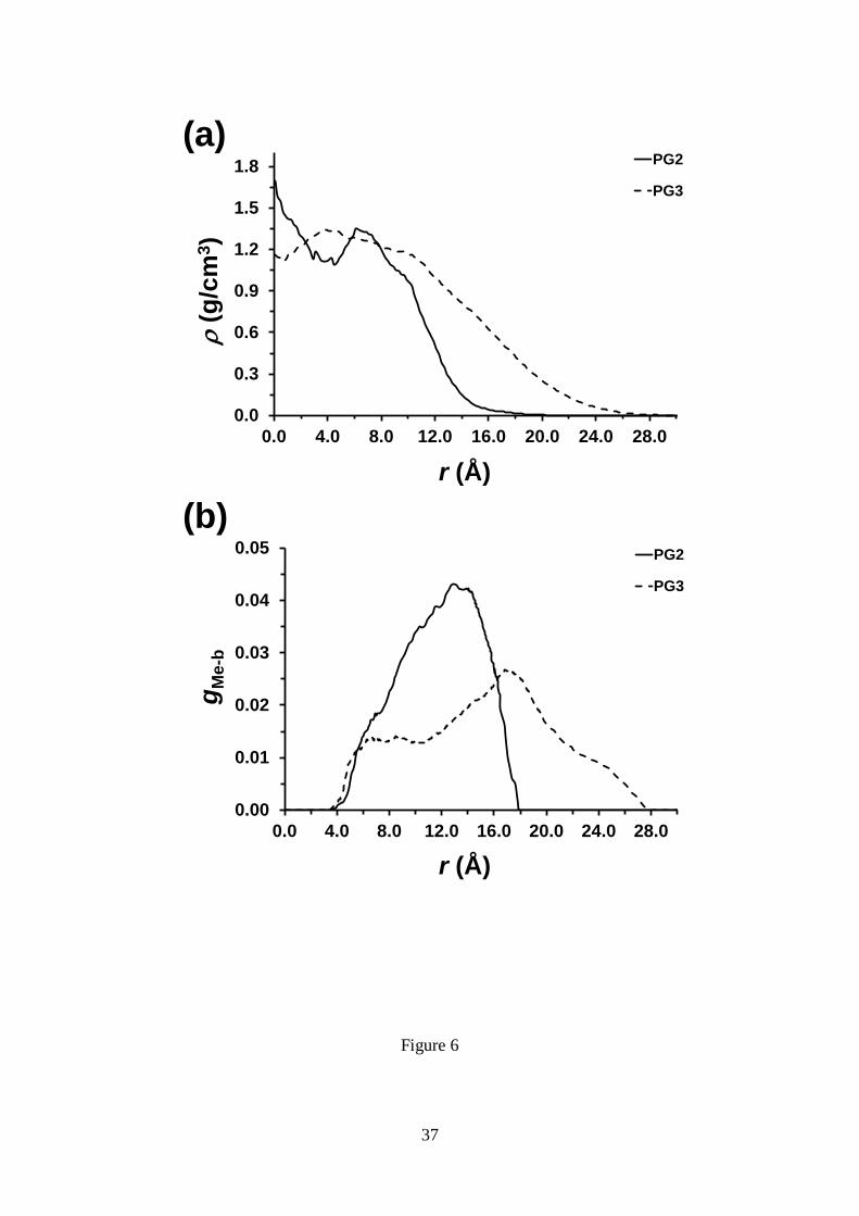
Figure 6a represents the variation of the density as function of radial distance r from
the macromolecular backbone (MB) for PG2 and PG3, obtained by averaging over
different cylindrical cross-sections of these macromolecules, while the average values
15
of the density (av) are listed in Table 3. It is worth noting that the density profiles
reflect all the effects associated with the influence of g on the spatial distribution of the
atoms (e.g. changes in backbone conformation). Figure 6a indicates that for PG2 the
highest density is localized at the region close to the backbone, reaching a value of 1.6
g/cm3. After this, the density of PG2 fluctuates between 1.4 and 1.1 g/cm
3 due to
conformational irregularities previously mentioned and, finally, it decreases
progressively. For PG3 the density remains relatively constant from the backbone to a
distance of approximately 9.9 Å and, hereafter, decreases slowly until the external layer
of the cylinder section is reached. As it can be seen in Table 3, av is around 1 g/cm3 for
both DPs.
Figure 6b depicts the radial probability distribution of the peripheral methyl groups
(see Figure 1b) as a function of the distance from the MB, gMe-b(r), for the two
examined DPs. PG2, whose repeat unit presents two external dendrons, shows a single
wide peak centered at around 12.9 Å, which corresponds to the value of R listed in
Table 3. The peak observed for PG3, centered at approximately 17.3 Å, is smaller and
broader than that observed for PG2. In addition of the position of the peak, which is
located at a distance 2.2 Å smaller than R (Table 3), the gMe-b profile calculated for PG3
shows inner and outer tails. The inner tail is related with the backfolding or looping
phenomenon, which refers to the probability of a peripheral methyl groups to be located
at distances lower than R= 19.5 Å. For PG3 the broad inner tail indicates that some
peripheral methyl groups are very close to the MB (4 Å) and, therefore, evidencing
that there must be parts of the same dendron residing at larger radial distances. In
contrast, for PG2 the shape of the gMe-b profile indicates that the backfolding is
practically null, the wideness of the peak being essentially due to the conformational
flexibility of the backbone.
16
- Stacking interactions in PG2 and PG3
In a recent study we examined intramolecular and intermolecular interactions in DPs
composed of a poly(methacrylic acid) backbone and repeat units with regularly
branched dendrons of generation four containing amide and aromatic groups separated
by a flexible segment (Scheme 3).40
After analyzing several complexes formed by two
interacting macromolecular chains, we concluded that intramolecular interactions are
significantly more abundant than intermolecular ones. Thus, this DP forms rigid
cylindrical-like molecules stabilized by intramolecular N–H···O hydrogen bonds and -
stacking interactions between two aromatic rings arranged in a sandwich or T-shaped
configuration, intermolecular interactions being only detected when two molecules
interpenetrate considerably. The results supported the scenario put forth in previous
structural and rheological studies,23,41
which evidenced the colloidal-filament nature and
associated solid-like viscoelastic response in the melt of such DP
Scheme 3: Chemical structure of the DP with g= 4 studied in reference 40.
PG4: Y= Boc

17
In an earlier study, the helical arrangement identified for the DP obtained from the
spontaneous polymerization of a chiral 4-aminoproline-based second generation
macromonomer (Scheme 4) was found to be stabilized by intramolecular hydrogen
bonding networks that extend along the whole polymer chain.36,37
Thus, the NH groups
of the 4-aminoprolines were found to form this kind of interaction with the amide
oxygen atoms of either the neighbor or the same repeat unit, enhancing the stability and
stiffness of the right-handed helical conformation.
Scheme 4: DP studied in references 36 and 37.
According to these antecedents, intramolecular interactions are also expected to play
a fundamental role in the stability of PG2 and PG3. Based on the molecular details
given in Figure 1b, the only specific interactions that may exhibit the two DPs examined
in this work correspond to the --stacking of Th rings. The presence of these
interactions is clearly reflected in Figure 7, which represents the partial radial
distribution function for pairs of centers of masses of Th rings, gTh-Th(r). The very
narrow and sharp peaks at r= 3.7, 4.1 and 4.8 Å (see insets) reflect the three inter-ring
distances at 3T units (Scheme 1) contained in the side groups of both PG2 and PG3.
Some other relatively narrow and sharp peaks are detected for PG2 at r= 7.3, 8.8 and
10.9 Å, which also correspond to regular distributions of 3T units. This is fully
consistent with the absence of backfolding in PG2. The shoulder and the small peak
centered at 4.4 and 5.2 Å (marked with arrows in Figure 7a), respectively, has been
N
HN
N
NHBoc
O
O O
NHBoc
O
NBoc Boc
n
N
HN
+H2N
NH3+
O
O O
NH3+
O
NH2+
de-PG2
nc1
c2
4 CF3CO2-
18
attributed to two different types of - stacking interactions. More specifically, the
shoulder at r= 4.4 Å has been attributed to - interactions in which the two aromatic
rings are coplanar (sandwich configuration) while the two Th rings are perpendicular
(T-shaped configuration) in the interaction associated to the peak at r=5.2 Å.82
In order to count the number of interactions of each type in PG2, the following
cutoffs were considered. For the sandwich configuration the separation between the two
Th rings (D in Scheme 5) is 4.5 Å and the degree of tilting (d in Scheme 5) is < 45º
or > 135º, whereas for the T-shaped configuration D 5.5 Å and d ranges from 45º to
135º. Results obtained from all the snapshots saved during the production run, which
are included in Table 3, indicate that the sandwich configuration is 3.4 times more
frequent than the T-shaped one in PG2. Accordingly, the average number of -
interactions per repeat unit of PG2 is 3 (i.e. 2.3 and 0.7 participate in sandwich and T-
shaped configurations, respectively).
Scheme 5: Parameters used to define the sandwich and T-shaped configurations
The gTh-Th profile calculated for PG3 show broad peaks at r= 6.5, 7.8 and 8.9 Å
(Figure 7b), which represents a significant difference with respect to PG2. Thus, the
regularity in the position of the Th rings is lost in PG3 due to the backfolding
phenomenon discussed above. The shoulder and peak centered at 4.4 and 5.2 Å (marked
with arrows in Figure 7b) have been related with - stacking interactions, as occurred
CM
S
CM
S
D
CM
S
CM
S
C
S
C
S d

19
for PG2. Quantitative analysis indicates that the total number of - stacking
interactions for PG3 is around three times higher than for PG2 (Table 3). Thus, the
average number of interactions counted per repeat unit of PG3 is 8.8 (i.e. 6.4 and 2.4
participate in sandwich and T-shaped configurations, respectively). According to these
values, the relative frequency of the sandwich configuration with respect to the T-
shaped one is lower for PG3 (2.7 : 1) than for PG2 (3.4 : 1). This should be attributed to
the interpenetration and backfolding phenomena discussed above, which restricts the
ability of the dendrons to rearrange and adopt T-shaped configurations.
CONCLUSIONS
The potential energy surface E=E(,’) calculated for MG2 reveals that the well-
defined conformational preferences of the macromonomers are essentially defined by
the relative orientation between the all-thiophene dendrons and the phenyl core. The
disposition of the peripheral Th rings, which is associated to the dihedral angles and
’, is similar for MG2 and MG3. However, small re-arrangements at these dihedrals
allow to alleviate the conformational strain, which increases with g. The calculated
electronic properties indicate that both the g and the IP decrease with increasing g,
which is in good agreement with experimental observations.
Atomistic models of PG2 and PG3, which were constructed using the most favored
conformations of MG2 and MG3, evidenced very different internal organizations. PG2
behaves as a flexible linear polymer bearing bulk side group while PG3 is a semirigid
cylinder. The particular behavior of the latter, which affects the molecular length, is due
to both the interpenetration of dendrons belonging to different repeat units and presence
of backfolding phenomena. In contrast, these effects are practically inexistent for PG2.

20
The different behaviors of PG2 and PG3 also affect the radial density profiles. The more
uniform profile is obtained for PG3.
Analysis of the inter-dendron interactions in PG2 and PG3 reveals that - stacking
interactions are significantly more abundant for latter than for the former. This is
consistent with the semirigidity and backfolding of PG3 and the flexibility of PG2.
Deeper analysis reveals that the interacting Th rings prefer the sandwich configuration
with respect to the T-shaped one, this effect being more pronounced for PG2 than PG3.
ACKNOWLEDGEMENTS
Financial support from the MINECO and FEDER (MAT2012-34498) and
Generalitat de Catalunya (Research group 2009 SGR 925 and XRQTC) is gratefully
acknowledged. EC-M is thanked for the financial support through a FPI grant.
REFERENCES
1. G. R. Newkome and C. Shreiner, C., Chem. Rev., 2010, 110, 6338-6442.
2. M. Ballauff and C. N. Likos, Angew. Chem., Int. Ed., 2004, 43, 2998-3020.
3. P. K. Maiti, T. Cagin, G. Wang and W. A. Goddard III, Macromolecules, 2004, 37,
236-6254.
4. Y. Liu, C. Y. Chen, H. L. Chen, K. L. Hong, C. Y. Shew, X. Li, L. Liu, Y. B.
Melnichenko, G. S. Smith, K. W. Herwig, L. Porcar and W. R. Chen, J. Phys. Chem.
Lett., 2010, 1, 2020-2024.
5. X. F. Li, T. Imae, D. Leisner and M. A. Lopez-Quintela, J. Phys. Chem. B, 2002, 106,
12170-12177.
6. R. Mezzenga, J. Ruokolainen, N. Canilho, E. Kasemi, A. D. Schlüter, W. B. Lee and
G. H. Fredrickson, Soft Matter, 2009, 5, 92-97.
21
7. Y. Lu, T. Shi, L. An, L. Jin and Z.-G. Wang, Soft Matter, 2010, 6, 2619-2622
8. T. H, Mourey, S. R. Turner, M. Rubinstein, J. M. J. Frechet, C. J. Hawker and K. L.
Wooley, Macromolecules, 1992, 25, 2401-2406.
9. D. Astruc, E. Boisselier and C. Ornelas, Chem. Rev., 2010, 110, 1857-1959.
10. G. R. Newkome, C. N. Moorefield and F. Vögtle, Dendrimers and Dendrons:
Concepts, Syntheses, Applications, Wiley-VCH, Weinheim, Germany, 2001.
11. A.-M. Caminade and C.-O. Turrin, J. Mater. Chem. B, 2014, 2, 4055-4066.
12. G. M. Soliman, A. Sharma, D. Maysinger and A. Kakkar, Chem. Commun., 2011,
47, 9572-9587.
13. N. K. Voulgarakis, K. O. Rasmussen and P. M. Welch, J. Chem. Phys., 2009, 130,
155101.
14. H. M. Liu, Y. Wang, M. M. Wang, J. R. Xiao and Y. Y. Cheng, Biomaterials, 2014,
35, 5407-5413.
15. L. Ropartz, R. E. Morris, D. F. Foster and D. J. Cole-Hamilton, Chem. Commun.,
2001, 361-362.
16. E. Karakhanov, A. Maximov, Y. Kardasheva, V. Semernina, A. Zolotukhina, A.
Ivanov, G. Abbott, E. Rosenberg and V. Vinokurov, ACS Mater. Interfaces, 2014, 6,
8807-8816.
17. J. Y. Li, Q. Li and D. Liu, ACS Mater. Interfaces, 2011, 3, 2099-2107.
18. J. Y. Li, T. Zhang, Y. J. Liang and R. X. Yang, Adv. Funct. Mater., 2013, 23, 619-
628.
19. A. D. Schlu ̈ter and J. P. Rabe, Angew. Chem., Int. Ed., 2000, 39, 864-883.
20. B. M. Rosen, C. J. Wilson, D. A. Wilson, M. Peterca, M. R. Imam and V. Percec,
Chem. Rev., 2009, 109, 6275-6540.

22
21. Y. Guo, J. D. van Beek, B. Zhang, M. Colussi, P. Walde, A. Zhang, M. Kröger, A.
Halperin and A. D. Schlüter, J. Am. Chem. Soc., 2009, 131, 11841-11854.
22. B. Zhang, R. Wepf, K. Fischer, M. Schmidt, S. Besse, P. Lindner, B. T. King, R.
Sigel, P. Schurtenberger, Y. Talmon, Y. Ding, M. Kröger, A. Halperin and A. D.
Schlüter, Angew. Chem. Int. Ed., 2011, 50, 763-766.
23. A. Kroeger, B. Zhang, C. Rosenauer and A. D. Schlüter, Colloid Polym. Sci., 2013,
291, 2879-2892.
24. B. Zhang, R. Wepf, M. Kröger, A. Halperin and A. D. Schlüter, Macromolecules,
2011, 44, 6785-6792.
25. Z. S. Bo, J. P. Rabe and A. D. Schlüter, Angew. Chem. Int. Ed., 1999, 38, 2370.
26. V. Percec, C. H. Ahn, T. K. Bera, G. Ungar, D. J. P. Yeardley, Chem. Eur. J., 1999,
5, 1070-1083.
27. C. O. Liang, B. Helms, C. J. Hawker and J. M. J. Frechet, Chem. Commun., 2003,
2524-2525.
28. B. M. J. M. Suijkerbuijk, L. J. Shu, R. J. M. K. Gebbink, A. D. Schlüter and G. van
Koten, Organometallics, 2003, 22, 4175-4177.
29. Z. N. Bao, K. R. Amundson and A. J. Lovinger, Macromolecules, 1998, 31, 8647-
8649.
30. T. Sato, D. L. Jiang and T. Aida, J. Am. Chem. Soc., 1999, 121, 10658-10659.
31. D. Joester, M. Losson, R. Pugin, H. Heinzelmann, E. Walter, H. P. Merkle and F.
Diederich, Angew. Chem. Int. Ed., 2003, 42, 1486-1490.
32. I. Gossl, L. J. Shu, A. D. Schlüter and J. P. Rabe, J. Am. Chem. Soc., 2002, 124,
6860-6865.
33. G. Fuhrmann, A. Grotzky, R. Lukic, S. Matoori, P. Luciani, H. Yu, B. Zhang, P.
Walde, A. D. Schlüter and J.-C. Leroux, Nature Chem., 2013, 5, 582-589.

23
34. A. Grotzky, T. Nauser, H. Erdogan, A. D. Schlüter and P. Walde, J. Am. Chem.
Soc., 2012, 134, 11392-11395.
35. S. Fornera, T. E. Balmer, B. Zhang, A. D. Schlüter and P. Walde, Macromol.
Biosci., 2011, 11, 1052-1067.
36. A. Zhang, F. Rodríguez-Ropero, D. Zanuy, C. Alemán, E. W. Meijer and A. D.
Schlüter, Chem. Eur. J., 2008, 14, 6924-6934.
37. F. Rodríguez-Ropero, M. Canales, D. Zanuy, A. Zhang, A. D. Schlüter and C.
Alemán, J. Phys. Chem. B, 2009, 113, 14868-14876.
38. O. Bertran, B. Zhang, A. D. Schlüter, A. Halperin, M. Kröger and C. Aleman, RSC
Adv., 2013, 3, 126-140.
39. O. Bertran, B. Zhang, A. D. Schlüter, M. Kröger and C. Alemán, J. Phys. Chem. B,
2013, 117, 6007-6017.
40. E. Córdova-Mateo, O. Bertran, B. Zhang, D. Vlassopoulos, R. Pasquino, A. D.
Schlüter, M. Kröger and C. Alemán, Soft Matter, 2014, 10, 1032-1044.
41. R. Pasquino, B. Zhang, R. Sigel, H. Yu, M. Ottiger, O. Bertran, C. Aleman, A. D.
Schlüter and D. Vlassopoulos, Macromolecules, 2012, 45, 8813-8823.
42. A. Mishra, C. Q. Ma and P. Baeuerle, Chem. Rev., 2009, 109, 1141-1276.
43. C. Xia, X. Fan, J. Locklin and R. C. Advincula, Org. Lett., 2002, 4, 2067-2070.
44. C. Xia, X. Fan, J. Locklin, R. C. Advincula, A. Gies and W. Nonidez, J. Am. Chem.
Soc., 2004, 126, 8735-8743.
45. C.-Q. Ma, E. Mena-Osteritz, T. Debaerdemaeker, M. M. Wienk, R. A. Janssen and
P. Bäuerle, Angew. Chem. Int. Ed., 2007, 46, 1679-1683.
46. M. R. Harpham, Ö. Süzer, C. Q. Ma, P. Bäuerle and T. Goodson III, J. Am. Chem.
Soc., 2009, 131, 973-979.
24
47. W. J. Mitchell, N. Kopidakis, G. Rumbles, D. S. Ginley and S. E. Shaheen, J.
Mater. Chem., 2005, 15, 4518-4528.
48. W. W. H. Wong, C. Q. Ma, W. Pisula, C. Yan, X. Feng, D. J. Jones, K. Müllen, R.
A. J. Janssen, P. Bäuerle and A. B. Holmes, Chem. Mater. 2010, 22, 457-466.
49. S. Deng, G. Fulghum, Krueger, D. Patton, Y. Park and R. C. Advincula, Chem. Eur.
J., 2011, 17, 8929-8940.
50. E. Córdova-Mateo, F. Rodríguez-Ropero, O. Bertran and C. Alemán, Chem. Phys.
Chem., 2012, 13, 1354-1362.
51. E. Badaeva, M. R. Harpham, R. Guda, O. Suzer, C. Q. Ma, P. Bäuerle, T. Goodson
and S. Tretiak, J. Phys. Chem. A, 2010, 114,15808-15817.
52. F. Rodríguez-Ropero, D. Zanuy and C. Alemán, Polymer, 2010, 51, 308-315.
53. P. Sonar, H. Benmansour, T. Geiger and A. D. Schlüter, Polymer, 2007, 48, 4996-
5004.
54. M. Kimura, A. Kitao, R. Fukawa and H. Shirai, Chem. Eur. J., 2011, 17, 6821-6829.
55. G. B. Griffin, P. M. Lundin, B. S. Rolczynski, A. Linkin, R. D. McGillicuddy, Z.
Bao and G. S. Engel, J. Chem. Phys., 2014, 140, 034903.
56. M. J. Frisch, G. W. Trucks, H. B. Schlegel, G. E. Scuseria, M. A. Robb, J. R.
Cheeseman, G. Scalmani, V. Barone, B. Mennucci, G. A. Petersson, H. Nakatsuji, M.
Caricato, X. Li, H. P. Hratchian, A. F. Izmaylov, J. Bloino, G. Zheng, J. L. Sonnenberg,
M. Hada, M. Ehara, K. Toyota, R. Fukuda, J. Hasegawa, M. Ishida, T. Nakajima, Y.
Honda, O. Kitao, H. Nakai, T. Vreven, J. A. Montgomery Jr., J. E. Peralta, F. Ogliaro,
M. Bearpark, J. J. Heyd, E. Brothers, K. N. Kudin, V. N. Staroverov, R. Kobayashi, J.
Normand, K. Raghavachari, A. Rendell, J. C. Burant, S. S. Iyengar, J. Tomasi, M.
Cossi, N. Rega, J. M. Millam, M. Klene, J. E. Knox, J. B. Cross, V. Bakken, C. Adamo,
J. Jaramillo, R. Gomperts, R. E. Stratmann, O. Yazyev, A. J. Austin, R. Cammi, C.
25
Pomelli, J. W. Ochterski, R. L. Martin, K.Morokuma, V. G. Zakrzewski, G. A. Voth, P.
Salvador, J. J. Dannenberg, S. Dapprich, A. D. Daniels, O. Farkas, J. B. Foresman, J. V.
Ortiz, J. Cioslowski and D. J. Fox, Gaussian 09, revision A.01, Gaussian, Inc.,
Wallingford, CT, 2009.
57. J. D. Chai and M. Head-GordonPhys. Chem. Chem. Phys., 2008, 10, 6615-6620.
58. A. D. McLean and G. S. Chandler, J. Chem. Phys., 1980, 72, 5639-5648.
59. M. J. Frisch, J. A. Pople and J. S. Binkley, J. Chem. Phys., 1984, 80, 3265-3269.
60. T. Koopmans, Physica, 1934, 1, 104-113.
61. J. D. Janak, Phys. Rev. B, 1978, 18, 7165-7168.
62. R. M. Dreizler and J. da Providência, Density Functional Methods in Physics,
Springer Dordrecht, 1985.
63. E. Runge and E. K. U. Gross, Phys. Rev. Lett., 1984, 52, 997-1000.
64. D. Jacquemin, J. Preat, E. A. Perpete and C. Adamo, Int. J. Quatum Chem., 2010,
110, 2121-2129.
65. A. Laurent and D. Jacquemin, Int. J. Quatum Chem., 2013, 113, 2019-2039.
66. C. Adamo and D. Jacquemin, Chem. Rev., 2013, 42, 845-856.
67. J. P. Perdew, M. Ernzerhof and K. Burke, J. Chem. Phys. 1996, 105, 9982-9985.
68. C. Adamo and V. Barone, J. Chem. Phys., 1999, 110, 6158-6170.
69. A. D. Becke, J. Chem. Phys. 1993, 98, 5648-5652.
70. C. Lee, W. Yang and R. G. Parr, Phys. Rev. B, 1988, 37, 785-789.
71. J. Torras, J. Casanovas and C. Alemán, J. Phys. Chem. A, 2012, 116, 7571-7583.
72. D. Jacquemin, E. A. Perpete, I. Ciofini and C. Adamo, Acc. Chem. Res., 2009, 42,
326-334.
73. U. C. Singh and P. A. Kollman, J. Comput. Chem., 1984, 5, 129-145.
74. B. H. Besler, K. M. Merz and P. A. Kollman, J. Comput. Chem., 1990, 11, 431-439.
26
75. J. C. Phillips, R. Braun, W. Wang, J. Gumbart, E. Tajkhorshid, E. Villa, C. Chipot,
R. D. Skeel, L. Kale and K. Schulten, J. Comput. Chem., 2005, 26, 1781-1802.
76. W. D. Cornell, P. Cieplak, C. I. Bayly, I. R. Gould, K. M. Merz, D. M. Ferguson, D.
C. Spellmeyer, T. Fox, J. W. Caldwell and P. A. Kollman, J. Am. Chem. Soc., 1995,
117, 5179-5197.
77. J. Wang, R. M. Wolf, J. W. Caldwell and D. A. Case, J. Comput. Chem., 2004, 15,
1157-1174.
78. P. Cieplak, W. Cornell, C. I. Bayly and P. A. Kollman, J. Comput. Chem., 1995, 16,
1357-1377.
79. J. P. Ryckaert, G. Ciccotti and H. J. C. Berendsen, J. Comput. Phys., 1977, 23, 327-
341.
80. H. J. C. Berendsen, J. P. M. Postma, W. F. van Gunsteren, A. Di Nola and J. R.
Haak, J. Chem. Phys., 1984, 81, 3684-3690.
81. C. Xia, X. Fan, J. Locklin, R. C. Advincula, A. Gies and W. Nonidez, J. Am. Chem.
Soc., 2004, 126, 8735-8743.
82. S. S. Sheiko, F. C. Sun, A. Randall, D. Shirvanyants, M. Rubinstein, H-I Lee and K.
Matyjaszewski, Nature, 2006, 440, 191-194.
83. A. Milchev, J. Paturej, V. G. Tostiashvili and T. A. Vilgis, Macromolecules, 2011,
44, 3981.3987.
84. S. Panyukov, E. B. Zhulina, S. S. Sheiko, G. C. Randall, J. Brock, M. Rubinstein, J.
Phys. Chem. B, 2009, 113, 3750–3768
85. J. Paturej, L. Kuban, A. Milchev and T. A. Vilgis, Eurphys. Lett., 2012, 97, 58003.
86. F. Rodriguez-Ropero, J. Casanovas and C. Alemán, J. Comput. Chem., 2008, 29,
69-78.
27
CAPTIONS TO FIGURES
Figure 1. Chemical structure of: (a) MG2 and MG3 macromonomers; and (b) PG2 and
PG3 DPs.
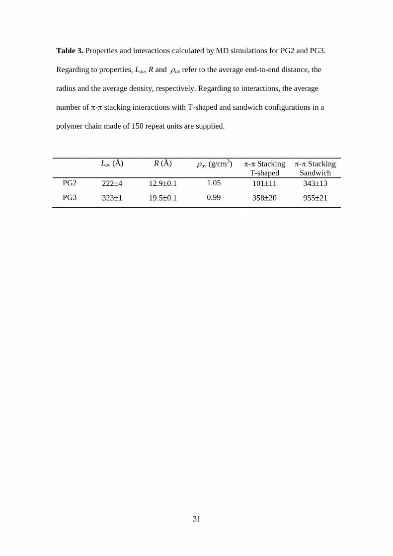
Figure 2. Electrostatic parameters determined for the repeat unit of (a) PG2 and (b)
PG3. Charges in parenthesis correspond to hydrogen atoms, where n refer to the
number n of equivalent hydrogens, while charges for carbon, oxygen and sulfur atoms
are out of the parenthesis. Charges for equivalent thiophene rings are omitted for clarity.
In the repeat unit of PG3, equivalent pairs of thiophene rings have been labelled using
letters.
Figure 3. Potential energy surface E=E(,’) calculated for MG2. The dihedral angles
and ’ are displayed in Figure 1a.
Figure 4. (a) Molecular representation, (b) HOMO, (c) LUMO and (d) electron density
of the MG2-1 (left) and MG3-1 (right) structures.
Figure 5. Atomistic conformations for PG2 (left) and PG3 (right). The complete axial
projections represent the whole calculated systems, the number of repeat units being N=
150. The magnified axial projection involves 20 repeat units in all cases, whereas the
equatorial projection involves 10 repeat units.
Figure 6. (a) Density profile for PG2 and PG3 representing the density () against the
distance to the backbone measured using the vector perpendicular to the helical axis (r).
The profile displayed for each DP corresponds to an average considering different
cross-sections within a given snapshot. (b) Distribution of peripheral methyl groups
(gMe-b) as a function of the distance from the backbone for PG2 and PG3. All data were
obtained by averaging over 2500 snapshots taken during the last 20 ns of the MD
relaxation runs.

28
Figure 7. Partial radial distribution functions for the pairs of centers of masses of Th
rings of (a) PG2 and (b) PG3. Data in were obtained by averaging over 2500 snapshots
taken during the last 20 ns of the MD relaxation runs.

29
Table 1. Representative minimum energy conformations calculated for MG2 at the
wB97X-D/6-311++G(d,p) level. Dihedral angles (, ’, and ’; in degrees), bond
lengths (R and R; in Å) and relative energy (E; in kcal/mol) are displayed.
# ’ ’ R R E
MG2-1a
144.9 -147.7 118.90.4 141.20.6 1.4610.000 1.4630.002 0.0
MG2-2b
147.6 33.6 119.50.6 140.50.1 1.4610.000 1.4650.000 0.0
MG2-3c
34.5º 33.0 120.90.9 140.00.1 1.4610.000 1.4650.000 0.0
MG2-4d 31.1º -148.5 119.30.4 142.20.8 1.4610.000 1.4650.000 0.2
a The E of minima with ,’ = 144.9º,-147.7º; -147.9º,-146.1º; 147.6º,146.3º; and -
145.9º,150.0º is lower than 0.2 kcal/mol. Differences in the rest of the geometric
parameter are practically inexistent. b
The E of minima with ,’ = 147.6º,33.6º;-
145.9º,-34.4º; -147.4º,33.2º; and 146.3º,-32.4º is lower than 0.2 kcal/mol. Differences in
the rest of the geometric parameter are practically inexistent. c
The E of minima with
,’ = 34.5º,33.0º; 34.0º,-34.3º; -35.9º,31.8º; and -30.5º,-32.0º is lower than 0.6
kcal/mol. Differences in the rest of the geometric parameter are practically inexistent. d
The E of minima with ,’ = 34.1º,-148.5º; -36.2º,-148.3º; 35.2º,-147.9º; and -34.8º,-148.6º is lower than 0.4 kcal/mol. Differences in the rest of the geometric parameter are
practically inexistent.

30
Table 2. Representative minimum energy conformations calculated for MG3 at the
wB97X-D/6-311++G(d,p) level. Dihedral angles (, ’, and ’; in degrees), bond
lengths (R and R; in Å) and relative energy (E; in kcal/mol) are displayed.
# ’ ’ R R E
MG3-1
148.8 31.5 110.14.4 148.78.3 1.4630.002 1.4650.001 0.0
MG3-2
33.6 33.7 110.95.1 149.07.6 1.4630.002 1.4640.001 0.0
MG3-3
34.6º -146.7 111.94.1 147.58.4 1.4630.002 1.4640.001 0.2
31
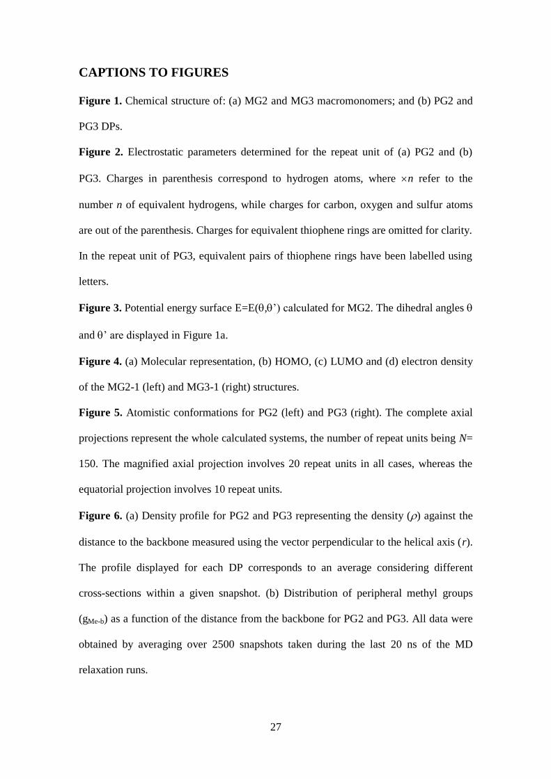
Table 3. Properties and interactions calculated by MD simulations for PG2 and PG3.
Regarding to properties, Lav, R and av refer to the average end-to-end distance, the
radius and the average density, respectively. Regarding to interactions, the average
number of - stacking interactions with T-shaped and sandwich configurations in a
polymer chain made of 150 repeat units are supplied.
Lav (Å) R (Å) av (g/cm3) - Stacking
T-shaped
- Stacking
Sandwich
PG2 2224 12.90.1 1.05 10111 34313
PG3 3231 19.50.1 0.99 35820 95521

32
Figure 1
O
S
S
SS
S
S
O
OH
SS
S
S
S
S
SS
SS
S
S
S
SOH
S
S
SS
S
S
SS
S
S
S
S
SS
SS
S
S
S
S
O O
'
n
'
'
'
n
'
'
PG2 PG3
MG3MG2
a)
b)
33
Figure 2
(a)
(b)

34
Figure 3
-180 -150-120 -90 -60 -30 0 30 60 90 120 150 180
-180
-150
-120
-90
-60
-30
0
30
60
90
120
150
180
0.0 kcal/mol
0.5 kcal/mol
1.0 kcal/mol
1.5 kcal/mol
2.0 kcal/mol
2.5 kcal/mol
3.0 kcal/mol
3.5 kcal/mol
4.0 kcal/mol
(º)
’ (º
)

35
Figure 4
(a)
(b)
(c)
(d)

36
Figure 5
PG2 PG3
37
Figure 6
0.00
0.01
0.02
0.03
0.04
0.05
0.0 4.0 8.0 12.0 16.0 20.0 24.0 28.0
PG2
PG3
r (Å)
0.0
0.3
0.6
0.9
1.2
1.5
1.8
0.0 4.0 8.0 12.0 16.0 20.0 24.0 28.0
PG2
PG3
(g
/cm
3)
r (Å)
gM
e-b
(a)
(b)

38
Figure 7
0.0 3.0 6.0 9.0 12.0 15.0 18.0
0.0 3.0 6.0 9.0 12.0 15.0 18.0
0.0 3.0 6.0 9.0 12.0 15.0 18.0
0.0 3.0 6.0 9.0 12.0 15.0 18.0
r (Å)
gT
h-T
h
(a)
r (Å)
gT
h-T
h
(b)

39
GRAPHICAL ABSTRACT
Multi-scale simulations reveal the organization of macromonomers and dendronized
polymers derived from all-thiophene dendrons attached to a phenyl core