Embed Size (px)
Citation preview
Intrinsic Disorder Mediates CooperativeSignal Transduction in STIM1
Yukio Furukawa1, †, Shunsuke Teraguchi 2, 3, †, Takahisa Ikegami4, Onur Dagliyan5,Lin Jin6, Damien Hall 6, 7, Nikolay V. Dokholyan5, Keiichi Namba1, Shizuo Akira2,Tomohiro Kurosaki 8, Yoshihiro Baba8 and Daron M. Standley6
1 - Nanobiology Laboratories, Protonic NanoMachine Group, Graduate School of Frontier Biosciences, Osaka University,Osaka 565–0871, Japan2 - Laboratory of Host Defense, WPI Immunology Frontier Research Center, Osaka University, Osaka 565–0871, Japan3 - Quantitative Immunology Research Unit, WPI Immunology Frontier Research Center, Osaka University, Osaka 565–0871, Japan4 - Laboratory of Advanced Protein Characterization, Research Center for State-of-the-Art Functional Protein Analysis,Institute for Protein Research, Osaka University, Osaka 565–0871, Japan5 - Department of Biochemistry and Biophysics, School of Medicine, University of North Carolina at Chapel Hill, Chapel Hill,NC 27599, USA6 - Laboratory of Systems Immunology, WPI Immunology Frontier Research Center, Osaka University, Osaka 565–0871, Japan7 - Research School of Chemistry, Australian National University Section on Biophysical Chemistry, Acton, 0200 ACT, Australia8 - Laboratory of Lymphocyte Differentiation, WPI Immunology Frontier Research Center, Osaka University,Osaka 565–0871, Japan
Correspondence to Daron M. Standley: [email protected]://dx.doi.org/10.1016/j.jmb.2014.03.006Edited by R. W. Kriwacki
Abstract
Intrinsically disordered domains have been reported to play important roles in signal transduction networks byintroducing cooperativity into protein–protein interactions. Unlike intrinsically disordered domains that becomeordered upon binding, the EF-SAM domain in the stromal interaction molecule (STIM) 1 is distinct in that it isordered in the monomeric state and partially unfolded in its oligomeric state, with the population of the twostates depending on the local Ca2+ concentration. The oligomerization of STIM1, which triggers extracellularCa2+ influx, exhibits cooperativity with respect to the local endoplasmic reticulum Ca2+ concentration.Although the physiological importance of the oligomerization reaction is well established, the mechanism ofthe observed cooperativity is not known. Here, we examine the response of the STIM1 EF-SAM domain tochanges in Ca2+ concentration using mathematical modeling based on in vitro experiments. We find that theEF-SAM domain partially unfolds and dimerizes cooperatively with respect to Ca2+ concentration, with Hillcoefficients and half-maximal activation concentrations very close to the values observed in vivo for STIM1redistribution and extracellular Ca2+ influx. Our mathematical model of the dimerization reaction agreesquantitatively with our analytical ultracentrifugation-based measurements and previously published freeenergies of unfolding. A simple interpretation of these results is that Ca2+ loss effectively acts as a denaturant,enabling cooperative dimerization and robust signal transduction. We present a structural model of theCa2+-unbound EF-SAM domain that is consistent with a wide range of evidence, including resistance toproteolytic cleavage of the putative dimerization portion.
© 2014 The Authors. Published by Elsevier Ltd. This is an open access article under the CC BY-NC-ND license(http://creativecommons.org/licenses/by-nc-nd/3.0/).
Introduction
It is well known that intrinsically disordereddomains (IDDs) are common in eukaryotic proteins
[1], in particular in protein–protein interaction (PPI)hubs [2]. The functional role of IDDs in PPIs hasbeen explained in terms of versatility: their flexibilityallows them to interact with multiple binding partners
0022-2836/$ - see front matter © 2014 The Authors. Published by Elsevier Ltd. This is an open access article under the CC BY-NC-NDlicense (http://creativecommons.org/licenses/by-nc-nd/3.0/). J. Mol. Biol. (2014) 426, 2082–2097
Article

[3]. In addition, since order/disorder reactions canbe triggered by changes in the local chemicalenvironment, phosphorylation, or ligand binding,IDDs play important roles as switches in signalingpathways [4]. Moreover, several recent studies [5–8]have shed light on another function of IDDs thatunderlies their role in PPIs: cooperativity. By foldingupon binding, IDDs can mediate allosteric couplingbetween spatially distant domains. For example, thebinding of tetracycline (Tc) to the Tc repressor (TetR)causes the dynamics of the intrinsically disorderedDNA-binding domain to become strongly coupled tothe more structured Tc binding/dimerization domains[8]. Another example of IDD-mediated cooperativity isthe phd antitoxin of the phd/doc toxin–antitoxinoperon, which is an extrachromosomal addictionmodule located on the P1 prophage and is responsi-ble for the postsegregational killing effect (death ofplasmid-free cells) of Escherichia coli [7]. The IDD inphd was shown to be stabilized by binding to doc,which, in turn, increased or decreased phd's affinity forits target DNA sequence, depending on the relativephd/doc ratio [7]. In a third example, the IDD-containing
adenovirus protein E1A was shown to mediate eitherpositive or negative cooperativity between host pro-teins TAZ2 and pRB, depending on the availability ofE1A-binding sites [5]. More recently, ligand-dependentfolding was found to regulate the activity of thehomodimeric enzyme aminoglycoside N-(6)-acetyl-transferase-Ii [6]. Interestingly, Hilser and Thompsonshowed earlier that an IDD in a multi-domain systemcould maximize allosteric coupling between ligand-binding sites [9]. The above are just four examples ofsuch “IDD-mediated allostery”, but the abundance ofIDDs in signaling molecules and nuclear proteinssuggests that there may be a greater link betweenintrinsic disorder and the cooperativity of intermolecularinteractions than previously realized.Here, we describe yet another variation on IDD-
mediated cooperativity. In the examples above, theIDD in question becomes ordered upon ligand binding,and this ordering facilitates energetic coupling be-tween spatially separated binding sites. In contrast, theendoplasmic reticulum (ER) luminal domain in the ERmembrane-bound stromal interactionmolecule (STIM)1 equilibrates between a foldedmonomeric state and a
SP
TM
hel
ix
SA
M
cEF
-han
d
hEF
-han
d
Coi
led-
coil
Luminal region Cytosolic region
P/S
ric
h
K r
ich
EF-SAM domain
Orai1 (closed) Orai1 (open)
ER Lumen
PM
[Ca2+] high
[Ca2+] low
Cytoplasm
A
B
Fig. 1. Overview of STIM1 function. (A) Domain architectures of luminal and cytosolic regions are indicated to scale.(B) Schematic illustration of CRAC channel opening.
2083Intrinsic Disorder and Signal Transduction in STIM
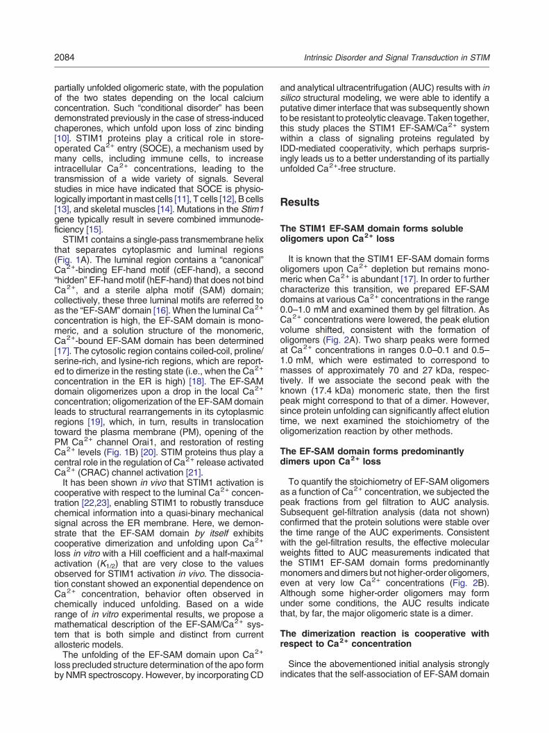
partially unfolded oligomeric state, with the populationof the two states depending on the local calciumconcentration. Such “conditional disorder” has beendemonstrated previously in the case of stress-inducedchaperones, which unfold upon loss of zinc binding[10]. STIM1 proteins play a critical role in store-operated Ca2+ entry (SOCE), a mechanism used bymany cells, including immune cells, to increaseintracellular Ca2+ concentrations, leading to thetransmission of a wide variety of signals. Severalstudies in mice have indicated that SOCE is physio-logically important inmast cells [11], T cells [12], B cells[13], and skeletal muscles [14]. Mutations in the Stim1gene typically result in severe combined immunode-ficiency [15].STIM1 contains a single-pass transmembrane helix
that separates cytoplasmic and luminal regions(Fig. 1A). The luminal region contains a “canonical”Ca2+-binding EF-hand motif (cEF-hand), a second“hidden” EF-handmotif (hEF-hand) that does not bindCa2+, and a sterile alpha motif (SAM) domain;collectively, these three luminal motifs are referred toas the “EF-SAM” domain [16]. When the luminal Ca2+
concentration is high, the EF-SAM domain is mono-meric, and a solution structure of the monomeric,Ca2+-bound EF-SAM domain has been determined[17]. The cytosolic region contains coiled-coil, proline/serine-rich, and lysine-rich regions, which are report-ed to dimerize in the resting state (i.e., when the Ca2+
concentration in the ER is high) [18]. The EF-SAMdomain oligomerizes upon a drop in the local Ca2+
concentration; oligomerization of the EF-SAM domainleads to structural rearrangements in its cytoplasmicregions [19], which, in turn, results in translocationtoward the plasma membrane (PM), opening of thePM Ca2+ channel Orai1, and restoration of restingCa2+ levels (Fig. 1B) [20]. STIM proteins thus play acentral role in the regulation of Ca2+ release activatedCa2+ (CRAC) channel activation [21].It has been shown in vivo that STIM1 activation is
cooperative with respect to the luminal Ca2+ concen-tration [22,23], enabling STIM1 to robustly transducechemical information into a quasi-binary mechanicalsignal across the ER membrane. Here, we demon-strate that the EF-SAM domain by itself exhibitscooperative dimerization and unfolding upon Ca2+
loss in vitro with a Hill coefficient and a half-maximalactivation (K1/2) that are very close to the valuesobserved for STIM1 activation in vivo. The dissocia-tion constant showed an exponential dependence onCa2+ concentration, behavior often observed inchemically induced unfolding. Based on a widerange of in vitro experimental results, we propose amathematical description of the EF-SAM/Ca2+ sys-tem that is both simple and distinct from currentallosteric models.The unfolding of the EF-SAM domain upon Ca2+
loss precluded structure determination of the apo formby NMR spectroscopy. However, by incorporating CD
and analytical ultracentrifugation (AUC) results with insilico structural modeling, we were able to identify aputative dimer interface that was subsequently shownto be resistant to proteolytic cleavage. Taken together,this study places the STIM1 EF-SAM/Ca2+ systemwithin a class of signaling proteins regulated byIDD-mediated cooperativity, which perhaps surpris-ingly leads us to a better understanding of its partiallyunfolded Ca2+-free structure.
Results
The STIM1 EF-SAM domain forms solubleoligomers upon Ca2+ loss
It is known that the STIM1 EF-SAM domain formsoligomers upon Ca2+ depletion but remains mono-meric when Ca2+ is abundant [17]. In order to furthercharacterize this transition, we prepared EF-SAMdomains at various Ca2+ concentrations in the range0.0–1.0 mM and examined them by gel filtration. AsCa2+ concentrations were lowered, the peak elutionvolume shifted, consistent with the formation ofoligomers (Fig. 2A). Two sharp peaks were formedat Ca2+ concentrations in ranges 0.0–0.1 and 0.5–1.0 mM, which were estimated to correspond tomasses of approximately 70 and 27 kDa, respec-tively. If we associate the second peak with theknown (17.4 kDa) monomeric state, then the firstpeak might correspond to that of a dimer. However,since protein unfolding can significantly affect elutiontime, we next examined the stoichiometry of theoligomerization reaction by other methods.
The EF-SAM domain forms predominantlydimers upon Ca2+ loss
To quantify the stoichiometry of EF-SAM oligomersas a function of Ca2+ concentration, we subjected thepeak fractions from gel filtration to AUC analysis.Subsequent gel-filtration analysis (data not shown)confirmed that the protein solutions were stable overthe time range of the AUC experiments. Consistentwith the gel-filtration results, the effective molecularweights fitted to AUC measurements indicated thatthe STIM1 EF-SAM domain forms predominantlymonomers and dimers but not higher-order oligomers,even at very low Ca2+ concentrations (Fig. 2B).Although some higher-order oligomers may formunder some conditions, the AUC results indicatethat, by far, the major oligomeric state is a dimer.
The dimerization reaction is cooperative withrespect to Ca2+ concentration
Since the abovementioned initial analysis stronglyindicates that the self-association of EF-SAM domain
2084 Intrinsic Disorder and Signal Transduction in STIM
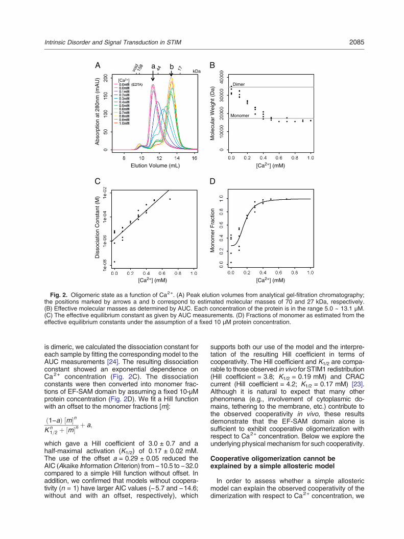
is dimeric, we calculated the dissociation constant foreach sample by fitting the corresponding model to theAUC measurements [24]. The resulting dissociationconstant showed an exponential dependence onCa2+ concentration (Fig. 2C). The dissociationconstants were then converted into monomer frac-tions of EF-SAM domain by assuming a fixed 10-μMprotein concentration (Fig. 2D). We fit a Hill functionwith an offset to the monomer fractions [m]:
1!a! " m# $n
K n1=2 % m# $n
% a;
which gave a Hill coefficient of 3.0 ± 0.7 and ahalf-maximal activation (K1/2) of 0.17 ± 0.02 mM.The use of the offset a = 0.29 ± 0.05 reduced theAIC (Akaike InformationCriterion) from !10.5 to !32.0compared to a simple Hill function without offset. Inaddition, we confirmed that models without coopera-tivity (n = 1) have larger AIC values (!5.7 and !14.6;without and with an offset, respectively), which
supports both our use of the model and the interpre-tation of the resulting Hill coefficient in terms ofcooperativity. The Hill coefficient and K1/2 are compa-rable to those observed in vivo for STIM1 redistribution(Hill coefficient = 3.8; K1/2 = 0.19 mM) and CRACcurrent (Hill coefficient = 4.2; K1/2 = 0.17 mM) [23].Although it is natural to expect that many otherphenomena (e.g., involvement of cytoplasmic do-mains, tethering to the membrane, etc.) contribute tothe observed cooperativity in vivo, these resultsdemonstrate that the EF-SAM domain alone issufficient to exhibit cooperative oligomerization withrespect to Ca2+ concentration. Below we explore theunderlying physical mechanism for such cooperativity.
Cooperative oligomerization cannot beexplained by a simple allosteric model
In order to assess whether a simple allostericmodel can explain the observed cooperativity of thedimerization with respect to Ca2+ concentration, we
[Ca2+] (mM)
[Ca2+] (mM)[Ca2+] (mM)
Monomer
Dimer
Mol
ecul
ar W
eigh
t (D
a)
B
Mon
omer
Fra
ctio
n
C
Elution Volume (mL)
Abs
orpt
ion
at 2
80nm
(m
AU
) [Ca2+]
A
Dis
soci
atio
n C
onst
ant (
M)
D
kDaa b
Fig. 2. Oligomeric state as a function of Ca2+. (A) Peak elution volumes from analytical gel-filtration chromatography;the positions marked by arrows a and b correspond to estimated molecular masses of 70 and 27 kDa, respectively.(B) Effective molecular masses as determined by AUC. Each concentration of the protein is in the range 5.0 ~ 13.1 μM.(C) The effective equilibrium constant as given by AUC measurements. (D) Fractions of monomer as estimated from theeffective equilibrium constants under the assumption of a fixed 10 μM protein concentration.
2085Intrinsic Disorder and Signal Transduction in STIM
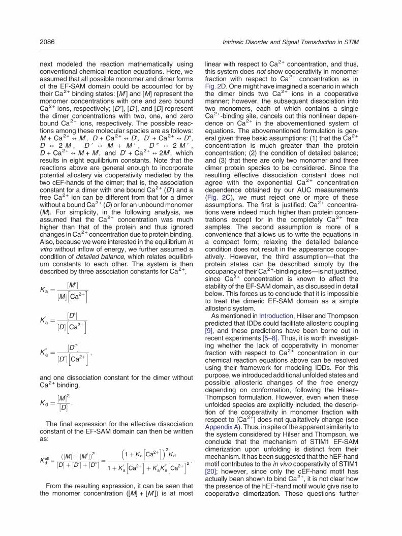
next modeled the reaction mathematically usingconventional chemical reaction equations. Here, weassumed that all possible monomer and dimer formsof the EF-SAM domain could be accounted for bytheir Ca2+ binding states: [M!] and [M] represent themonomer concentrations with one and zero boundCa2+ ions, respectively; [D"], [D!], and [D] representthe dimer concentrations with two, one, and zerobound Ca2+ ions, respectively. The possible reac-tions among these molecular species are as follows:M + Ca2+ ! M!, D + Ca2+ ! D!, D! + Ca2+ ! D",D ! 2 M , D ! ! M + M ! , D " ! 2 M ! ,D + Ca2+ ! M + M!, and D! + Ca2+ ! 2M!, whichresults in eight equilibrium constants. Note that thereactions above are general enough to incorporatepotential allostery via cooperativity mediated by thetwo cEF-hands of the dimer; that is, the associationconstant for a dimer with one bound Ca2+ (D!) and afree Ca2+ ion can be different from that for a dimerwithout a boundCa2+ (D) or for an unboundmonomer(M). For simplicity, in the following analysis, weassumed that the Ca2+ concentration was muchhigher than that of the protein and thus ignoredchanges in Ca2+ concentration due to protein binding.Also, because we were interested in the equilibrium invitro without inflow of energy, we further assumed acondition of detailed balance, which relates equilibri-um constants to each other. The system is thendescribed by three association constants for Ca2+,
K a &M 0# $
M# $ Ca2%h i
K0
a &D0# $
D# $ Ca2%h i
K00
a &D00# $
D0# $ Ca2%h i ;
and one dissociation constant for the dimer withoutCa2+ binding,
K d &M# $2
D# $:
The final expression for the effective dissociationconstant of the EF-SAM domain can then be writtenas:
K effd "
M# $ % M 0# $! "2
D# $ % D0# $ % D00# $&
1% K a Ca2%h i! "2
K d
1% K0
a Ca2%h i
% K0
aK00
a Ca2%h i2 :
From the resulting expression, it can be seen thatthe monomer concentration ([M] + [M!]) is at most
linear with respect to Ca2+ concentration, and thus,this system does not show cooperativity in monomerfraction with respect to Ca2+ concentration as inFig. 2D. Onemight have imagined a scenario in whichthe dimer binds two Ca2+ ions in a cooperativemanner; however, the subsequent dissociation intotwo monomers, each of which contains a singleCa2+-binding site, cancels out this nonlinear depen-dence on Ca2+ in the abovementioned system ofequations. The abovementioned formulation is gen-eral given three basic assumptions: (1) that the Ca2+
concentration is much greater than the proteinconcentration; (2) the condition of detailed balance;and (3) that there are only two monomer and threedimer protein species to be considered. Since theresulting effective dissociation constant does notagree with the exponential Ca2+ concentrationdependence obtained by our AUC measurements(Fig. 2C), we must reject one or more of theseassumptions. The first is justified: Ca2+ concentra-tions were indeed much higher than protein concen-trations except for in the completely Ca2+ freesamples. The second assumption is more of aconvenience that allows us to write the equations ina compact form; relaxing the detailed balancecondition does not result in the appearance cooper-atively. However, the third assumption—that theprotein states can be described simply by theoccupancy of their Ca2+-binding sites—is not justified,since Ca2+ concentration is known to affect thestability of the EF-SAM domain, as discussed in detailbelow. This forces us to conclude that it is impossibleto treat the dimeric EF-SAM domain as a simpleallosteric system.As mentioned in Introduction, Hilser and Thompson
predicted that IDDs could facilitate allosteric coupling[9], and these predictions have been borne out inrecent experiments [5–8]. Thus, it is worth investigat-ing whether the lack of cooperativity in monomerfraction with respect to Ca2+ concentration in ourchemical reaction equations above can be resolvedusing their framework for modeling IDDs. For thispurpose,we introducedadditional unfolded states andpossible allosteric changes of the free energydepending on conformation, following the Hilser–Thompson formulation. However, even when theseunfolded species are explicitly included, the descrip-tion of the cooperativity in monomer fraction withrespect to [Ca2+] does not qualitatively change (seeAppendix A). Thus, in spite of the apparent similarity tothe system considered by Hilser and Thompson, weconclude that the mechanism of STIM1 EF-SAMdimerization upon unfolding is distinct from theirmechanism. It has been suggested that the hEF-handmotif contributes to the in vivo cooperativity of STIM1[20]; however, since only the cEF-hand motif hasactually been shown to bind Ca2+, it is not clear howthe presence of the hEF-hand motif would give rise tocooperative dimerization. These questions further
2086 Intrinsic Disorder and Signal Transduction in STIM
motivated us to analyze the structural diversity ofEF-SAM domain as a function of Ca2+ concentration.
The EF-SAM domain cooperatively unfolds inresponse to Ca2+ depletion
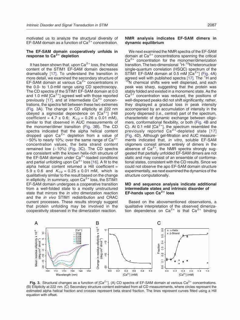
It has been shown that, upon Ca2+ loss, the helicalcontent of the STIM1 EF-SAM domain decreasesdramatically [17]. To understand the transition inmore detail, we examined the secondary structure ofEF-SAM domain at various Ca2+ concentrations inthe 0.0- to 1.0-mM range using CD spectroscopy.The CD spectra of the STIM1 EF-SAM domain at 0.0and 1.0 mM [Ca2+] agreed well with those reportedpreviously [17], and at intermediate Ca2+ concen-trations, the spectra fell between these two extremes(Fig. 3A). The change in CD ellipticity at 222 nmobeyed a sigmoidal dependence on [Ca2+] (Hillcoefficient = 4.7 ± 0.6; K1/2 = 0.26 ± 0.01 mM),similar to that observed in AUC measurements ofthe monomer/dimer transition (Fig. 3B). The CDspectra indicated that the alpha helical contentdropped upon Ca2+ depletion from a value ofN50% to nearly 10%; over the same range of Ca2+
concentration values, the beta strand contentremained low (~10%) (Fig. 3C). The CD spectraare consistent with the known helix-rich structure ofthe EF-SAM domain under Ca2+-loaded conditionsand partial unfolding upon Ca2+ loss [16]. A fit to thealpha helical content returned a Hill coefficient =5.9 ± 0.6 and K1/2 = 0.25 ± 0.01 mM, which isqualitatively similar to the result based on the changein ellipticity. In summary, upon Ca2+ loss, the STIM1EF-SAM domain undergoes a cooperative transitionfrom a well-folded state to a mostly unstructuredstate that mirrors the in vitro dimerization reactionand the in vivo STIM1 redistribution and CRACcurrent processes. These results strongly suggestthat protein unfolding may be involved in thecooperativity observed in the dimerization reaction.
NMR analysis indicates EF-SAM dimers indynamic equilibrium
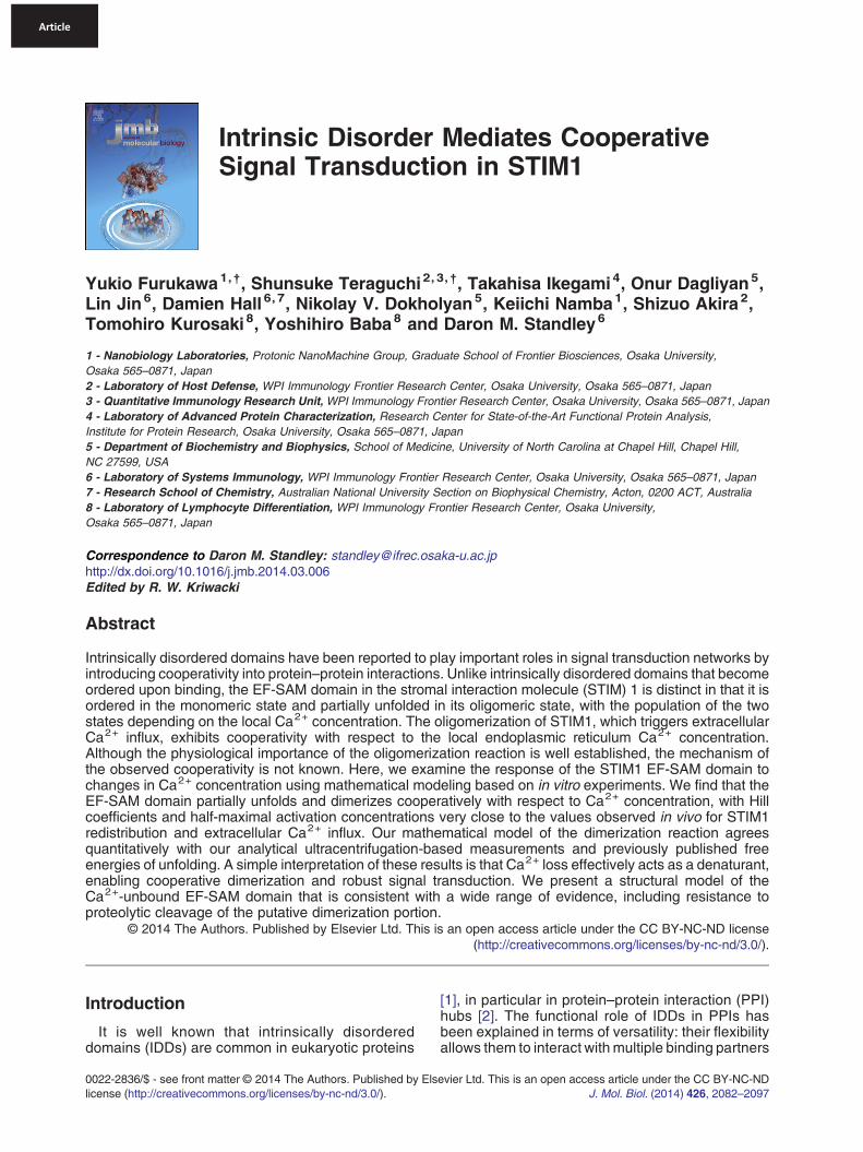
We next examined the NMR spectra of the EF-SAMdomain at Ca2+ concentrations spanning the criticalCa2+ concentration for the monomer/dimerizationtransition. The two-dimensional 1H-15N heteronuclearsingle-quantum correlation (HSQC) spectrum of theSTIM1 EF-SAM domain at 0.5 mM [Ca2+] (Fig. 4A)agreed well with published spectra [17]. The 1H and15N chemical shifts were well dispersed, and eachpeak was sharp, suggesting that the protein wasstably folded and existed in a monomeric state. As theCa2+ concentration was reduced, the positions ofwell-dispersed peaks did not shift significantly; rather,they displayed a gradual loss in peak intensityaccompanied by an accumulation of intensity in thepoorly dispersed (i.e., central) part of the spectrum,characteristic of dynamic exchange between oligo-mers, conformational flexibility, or both (Fig. 4B andC). At 0.1 mM [Ca2+], the spectrum resembled thepreviously reported Ca2+-depleted state [17](Fig. 4D). Although gel-filtration and AUC measure-ments indicated that, in vitro, soluble EF-SAMoligomers consist almost entirely of dimers in theabsence of Ca2+, the NMR spectra strongly sug-gested that partially unfolded EF-SAM dimers are notstatic and may consist of an ensemble of conforma-tional states, consistent with the CD results. Since wecould not observe the apo EF-SAM domain structureexperimentally, we next examined the dynamics of thestructure computationally.
MD and sequence analysis indicate additionalintermediate states and intrinsic disorder ofEF-hands upon Ca2+ loss
Based on the abovementioned observations, aqualitative interpretation of the observed dimeriza-tion dependence on Ca2+ is that Ca2+ binding
Fra
ctio
n
C
Wavelength (nm)
Mea
n re
sidu
e el
liptic
ity x
10-3
(deg
cm
2 dm
ol-1
)
[Ca2+]
Ao -Helixx -Strand
B
Cha
nge
in r
esid
ue e
llipt
icity
x 1
0-3
(deg
cm
2 dm
ol-1
)
[Ca2+] (mM) [Ca2+] (mM)
Fig. 3. Structural changes as a function of [Ca2+]. (A) CD spectra of EF-SAM domain at various Ca2+ concentrations.(B) Ellipticity at 222 nm. (C) Secondary structure content estimated from all CD measurements, where circles represent theestimated alpha helical fraction and crosses represent beta strand fraction. The lines represent curves fitted using a Hillequation with offset.
2087Intrinsic Disorder and Signal Transduction in STIM

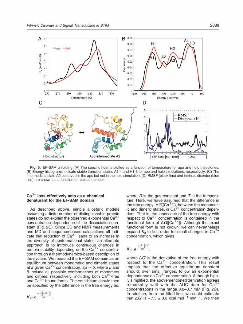
requires a well-folded protein, which, in turn, requireshigh Ca2+ concentrations. Conversely, dimerizationof the EF-SAM domain is likely to occur byinteraction between exposed hydrophobic residues[16], which occurs only when Ca2+ concentrationsare low enough to promote unfolding. In order tobetter understand the structural response of theEF-SAM domain to Ca2+ loss, we carried outreplica exchange simulations of the Ca2+-boundand Ca2+-free STIM1 EF-SAM domain. A heatcapacity–temperature diagram was used to find thetransitions (Fig. 5A) from which energy histogramsand intermediate representative conformations wereidentified (Fig. 5B). To compare the stability of theapo and holo EF-SAM monomers, we calculatedtheir specific heats (Cv) at different temperatures byapplying the weighted histogram analysis method[25]. The sharp peaks in the Cv–T diagram indicateunfolding transition events. The temperature of thefirst transition of the apo EF-SAM domain was atapproximately 295 K, which was reported as 21 °Cbased on CD thermal unfolding experiments [26].
The corresponding temperature in the holo formshifted to 314 K. Interestingly, we observed anintermediate state, which features a shoulder at322 K in the apo form that is not observed in the holounfolding simulations (Fig. 5C). The existence of anadditional intermediate state may be one reason whythe apo form unfolds more easily compared to theholo EF-SAM domain. The apo molecular dynamics(MD) trajectory in explicit water indicated thatelectrostatic repulsion between negatively chargedresidues in the EF-hands promoted unfolding underconditions of reduced Ca2+ concentration. TheEF-hands are predicted to become intrinsicallydisordered in the absence of Ca2+, as indicated byboth the RMSF (root-mean-square fluctuation) com-puted from the explicit water MD simulation andsequence-based intrinsic disorder prediction [27](Fig. 5D), consistent with both CD and NMRmeasurements. From these simulations, we cansee that the hEF-hand, while not explicitly involved inbinding Ca2+, contributes significantly to the insta-bility of the EF-SAM domain.
[Ca2+] = 0.5 mM [Ca2+] = 0.3 mM
[Ca2+] = 0.2 mM [Ca2+] = 0.1 mM
1H chemical shift (ppm)
15N
che
mic
al s
hift
(ppm
)
A B
C D
Fig. 4. 1H-15N HSQC spectra. NMR spectra were acquired at four Ca2+ concentrations, as indicated. Red peaks are ofthe resonances of 1Hε-15Nε, which appear at aliased positions.
2088 Intrinsic Disorder and Signal Transduction in STIM
Ca2+ loss effectively acts as a chemicaldenaturant for the EF-SAM domain
As described above, simple allosteric modelsassuming a finite number of distinguishable proteinstates do not explain the observed exponential Ca2+
concentration dependence of the dissociation con-stant (Fig. 2C). Since CD and NMR measurementsand MD and sequence-based calculations all indi-cate that reduction of Ca2+ leads to an increase inthe diversity of conformational states, an alternateapproach is to introduce continuous changes inprotein stability depending on the Ca2+ concentra-tion through a thermodynamics-based description ofthe system. We modeled the EF-SAM domain as anequilibrium between monomeric and dimeric statesat a given Ca2+ concentration, 2μ ! δ, where μ andδ include all possible conformations of monomersand dimers, respectively, including both Ca2+-freeand Ca2+-bound forms. The equilibrium should thenbe specified by the difference in the free energy as:
K d#e!ΔG Ca2%# $! "
RT ;
where R is the gas constant and T is the tempera-ture. Here, we have assumed that the difference inthe free energy, ΔG([Ca2+]), between the monomer-ic and dimeric states, is Ca2+ concentration depen-dent. That is, the landscape of the free energy withrespect to Ca2+ concentration is contained in thefunctional form of ΔG([Ca2+]). Although the exactfunctional form is not known, we can neverthelessexpand Kd to first order for small changes in Ca2+
concentration, which gives
K d#e! Ca2%# $ΔG0
RT ;
where ΔG! is the derivative of the free energy withrespect to the Ca2+ concentration. This resultimplies that the effective equilibrium constantshould, over small ranges, follow an exponentialdependence on Ca2+ concentration. Although high-ly simplified, the abovementioned derivation agreesremarkably well with the AUC data for Ca2+
concentrations in the range 0.0–0.7 mM (Fig. 2C).In addition, from the fitted line, we could estimatethat ΔG! is !7.5 ± 0.8 kcal mol!1 mM!1. We then
AH1
H2H3
A1 A2A3
A4B
D
cEF-hand hEF-hand SAM
C
Holo structure Apo intermediate A2
Energy (kcal/mol)
Fre
quen
cy
Temperature (K)
CV (
kcal
/mol
K)
Fig. 5. EF-SAM unfolding. (A) The specific heat is plotted as a function of temperature for apo and holo trajectories.(B) Energy histograms indicate stable transition states A1-4 and H1-3 for apo and holo simulations, respectively. (C) Theintermediate state A2 observed in the apo but not in the holo simulation. (D) RMSF (black line) and intrinsic disorder (blueline) are shown as a function of residue number.
2089Intrinsic Disorder and Signal Transduction in STIM
estimated change in free-energy difference withrespect to the monomer between 0.0 and 0.5 mM[Ca 2 +] at 4 °C by |0.5 mM ! ΔG ! |/2 = 1.9 ±0.2 kcal mol!1. This value is of the same order asthe experimentally measured value (3.2 kcal mol!1)at 10 °C [17].The exponential dependence of the effective
equilibrium constant with respect to the decrease inCa2+ concentration is analogous to that oftenobserved in unfolding by chemical denaturation, inwhich the free energy shows a linear dependence ondenaturant concentration [28,29]. Taken together,these results strongly indicate that Ca2+-dependentprotein unfolding drives the cooperative dimerizationof STIM1 EF-SAM domain.
Proposed model of STIM1 EF-SAM dimerization
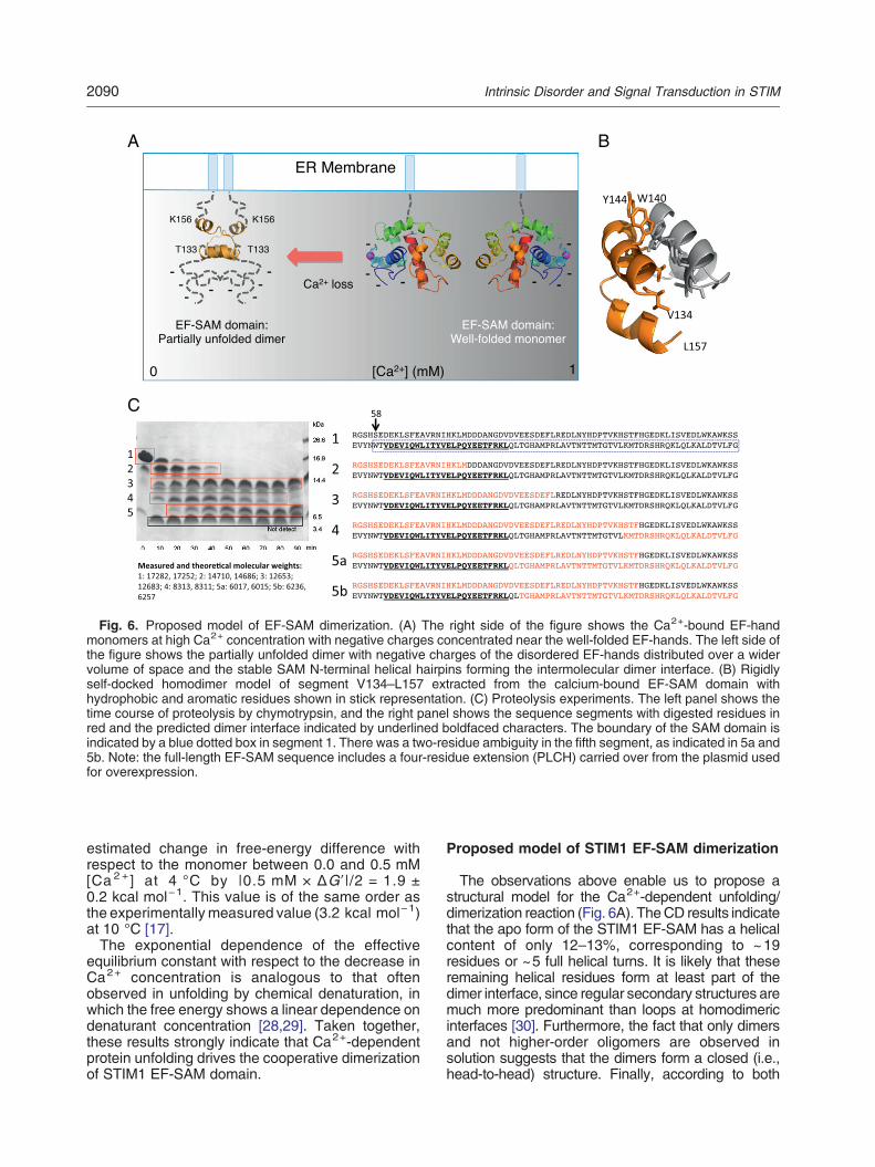
The observations above enable us to propose astructural model for the Ca2+-dependent unfolding/dimerization reaction (Fig. 6A). TheCD results indicatethat the apo form of the STIM1 EF-SAM has a helicalcontent of only 12–13%, corresponding to ~19residues or ~5 full helical turns. It is likely that theseremaining helical residues form at least part of thedimer interface, since regular secondary structures aremuch more predominant than loops at homodimericinterfaces [30]. Furthermore, the fact that only dimersand not higher-order oligomers are observed insolution suggests that the dimers form a closed (i.e.,head-to-head) structure. Finally, according to both
C
A B
Fig. 6. Proposed model of EF-SAM dimerization. (A) The right side of the figure shows the Ca2+-bound EF-handmonomers at high Ca2+ concentration with negative charges concentrated near the well-folded EF-hands. The left side ofthe figure shows the partially unfolded dimer with negative charges of the disordered EF-hands distributed over a widervolume of space and the stable SAM N-terminal helical hairpins forming the intermolecular dimer interface. (B) Rigidlyself-docked homodimer model of segment V134–L157 extracted from the calcium-bound EF-SAM domain withhydrophobic and aromatic residues shown in stick representation. (C) Proteolysis experiments. The left panel shows thetime course of proteolysis by chymotrypsin, and the right panel shows the sequence segments with digested residues inred and the predicted dimer interface indicated by underlined boldfaced characters. The boundary of the SAM domain isindicated by a blue dotted box in segment 1. There was a two-residue ambiguity in the fifth segment, as indicated in 5a and5b. Note: the full-length EF-SAM sequence includes a four-residue extension (PLCH) carried over from the plasmid usedfor overexpression.
2090 Intrinsic Disorder and Signal Transduction in STIM
sequence-based analysis and MD simulations, themost stable regions in the EF-SAM domain consist ofhelices located in the SAM domain. The above-mentioned observations imply that EF-SAM dimeriza-tion is likely to occur through interaction ofapproximately 5 helical turns in the SAM domain.There are two candidate regions that contain theappropriate number of helical turns in the SAMdomainthat are stable upon unfolding simulations: the SAMC-terminal helix (D183–F200) and the SAMN-terminalhelical hairpin (V134–L157). To assess the relativelikelihood of the SAM C-terminal helix or the SAMN-terminal helical hairpin to homodimerize, we extract-ed each from the native Ca2+-bound structure andself-docked. High-scoring docked homodimers werethen subjected to explicit water MD simulations.Asymmetric dimer structures were excluded in con-sideration of the transmembrane helix that wouldconstrain dimerization in vivo; however, we cannotrule out the possibility that asymmetric dimers mightform. The dimer composed of two SAM C-terminalhelices was found to be highly unstable due toelectrostatic repulsion between positively chargedresidues (R184, R187, K189, K193). In contrast, theSAM N-terminal helical hairpin was stabilized throughpairing of a hydrophobic face (V134, V137, L141,V145) and stacking between aromatic groups W140and Y144 (Fig. 6B). The proposed dimer interface ofthe SAM N-terminal helical hairpin model was subse-quently shown to be resistant to proteolysis bychymotrypsin (Fig. 6C). In fact, the proteolysis resultsshow very close agreement with the sequence-basedintrinsic disorder prediction and the MD-based RMSFcalculation (Fig. 5D), which both indicate that, inaddition to the SAMdomain, a small C-terminal portionof the hEF-hand is stable.The EF-hands are rich in Asp/Glu compared with
Arg/Lys (21 versus 8 residues, respectively). At highCa2+ concentrations, the folded EF-SAM domainburies hydrophobic groups and concentrates nega-tively charged residues at the surface of a compactglobular domain. This high surface charge densityis likely to prevent EF-SAM dimerization underCa2+-loaded conditions. Upon Ca2+ depletion, theEF-hand domains become disordered and distributetheir charges over a larger volume of space.Dimerization via pairing of exposed hydrophobicresidues in the SAM N-terminal helices can thenproceed.
Discussion
Recently, a number of reports have demonstratedthe important role of conditional disorder in signalingmolecules [4]. Since, by definition, disordered regionsin proteins are difficult to observe, it is likely that theirimportance has been underappreciated compared tothe role of ordered domains. Similarly, the role of
disorder-mediated cooperativity has only recentlybeen experimentally studied [5–8]. The cooperativeoligomerization of STIM1 is important for its role inresponding to Ca2+ depletion within the ER. It haspreviously been shown that oligomerization of theSTIM1 luminal region is necessary for translocation tothe ER–PM junction and subsequent opening of theOrai1 channel [23]. However, the direct relationshipbetween STIM1 oligomerization and Ca2+ bindinghas not been described. Our in vitro measurementsindicate that the dominant oligomerization reaction ofthe EF-SAM domain is that of monomer/dimerequilibration and that the dimerization is cooperativewith respect to Ca2+ concentration. Interestingly, theHill coefficient and K1/2 of the in vitro reaction are veryclose to the in vivo values for STIM1 redistribution andCRAC current intensity as a function of ER Ca2+
concentration [23].In order to deepen our understanding of the
underlying mechanism of this cooperativity, weinvestigated the conformational changes in theEF-SAM domain that occur upon Ca2+ depletion.TheCDandNMRspectra presented here agreedwithprevious reports [17] and indicated that loss of definedstructure and dimerization occur concurrently; theNMR results also strongly suggested that EF-SAMoligomers exist in dynamic equilibrium. MD simula-tions and sequence analysis were consistent withthese observations and indicated that the order-to-di-sorder transition is driven by unfolding of the EF-handmotifs. We observed an additional intermediate statein the apo MD simulation that was not present in theholo simulation that may be important for rapidunfolding.In order to quantify the dependence on Ca2+
concentration without having to treat each micro-scopic conformational state explicitly, we expandedthe free energy to first order and obtained anexponential dependence of the monomer/dimerequilibrium constant on Ca2+ concentration. Thisrelationship is far more sensitive than what would beobtained from treating the single Ca2+-binding siteexplicitly with classical chemical reaction equationsand agrees remarkably well with the AUC resultsover a wide range of Ca2+ concentrations. Interest-ingly, this result implies that, to first order, the freeenergy depends linearly on Ca2+ concentration, aresult commonly observed in equilibrium unfoldingby chemical denaturation [28,29]. This interpretationsuggests that although the hEF-hand domain is not aclassical allosteric site, or even a Ca2+-binding site,it is highly prone to disorder and thus plays animportant role in coordinating EF-SAM responses toCa2+ loss.It has recently been shown that dimerization of the
STIM1 luminal region results in conformationalchanges in the cytoplasmic region that lead to Orai1channel opening [19]. In this same study, the authorsargue that, since a STIM1 dimer contains only two
2091Intrinsic Disorder and Signal Transduction in STIM
Ca2+-binding sites, the observed cooperativity ofOrai1 activation in vivo requires high-order oligomer-ization [19]. Here, we present an alternative view inwhich EF-SAM dimerization is itself cooperative withrespect to Ca2+ concentration owing to intrinsicdisorder rather than higher-order effects. If we acceptthat the dimerization of the EF-SAM domain iscooperative, it is reasonable to assume that alldownstream events, including CRAC channel open-ing, will be cooperative as well. This is not to say thatcytoplasmic PPIs or higher-order oligomerization donot contribute quantitatively to the observed coopera-tivity in vivo. Rather, what the results presented hereimply is that the EF-SAM dimerization reaction is itselfsufficient formediating cooperativity to a degree that isobserved in vivo. In this respect, it is worth noting thatthe Hill coefficients measured in two different in vivostudies of STIM1 activation [22,23] differed byapproximately a factor of 2.The functional importance of the order-to-disorder
transition in STIM1 has been discussed previously[16]. Since protein folding in general [31] and foldingof multiple EF-hand domains in particular [32] hasbeen shown to be a cooperative process, and sincedimerization of STIM1 depends on unfolding, it isperhaps not surprising that STIM1 exhibits cooper-ative dimerization. Nevertheless, to our knowledge,even a qualitative model of STIM1 cooperativitybased on Ca2+ dependent unfolding of the EF-SAMdomain has not been proposed previously. Here, wepresent a mathematical model that both agrees withexperimental observations and describes the coop-erativity quantitatively. From a biological point ofview, this model is interesting because it implies thatthe stability of the EF-hands precisely and robustlydetermines the Ca2+ concentration threshold forEF-SAM dimerization.We further show that the exponential dependence
of the dimerization dissociation constant on Ca2+
concentration does not follow from modeling by eitherchemical reaction equations or the Hilser–Thompsonmodel of IDD-mediated allostery. The STIM1EF-SAM/Ca2+ system is clearly allosteric in theconventional sense that Ca2+ binding drasticallyinfluences the EF-SAM homodimerization dissocia-tion constant. However, since, strictly speaking, alldynamic proteins are allosteric [33], and since currentallostery models do not capture the Ca2+ concentra-tion dependence, the label “allosteric” would seem tobe insufficient to describe the STIM1 EF-SAM/Ca2+
system. Moreover, this may not be an isolated case,given the abundance of EF-hand domains in signalingproteins [34].Our interpretation of the cooperativity mechanism is
consistent with the observation that the homologousprotein STIM2, which is more stable than STIM1 [35],exhibits lower cooperativity with respect to Ca2+
concentration [22]. This, in turn, implies that proteinstability might tune the kinetics and thermodynamics
of other importantPPI networks, an intriguing prospectgiven the abundance of disorder-containing proteinsin eukaryotes.The in vitro experiments and simulations discussed
here allowed us to propose a structural model of theCa2+-dependent dimerization reaction (Fig. 6). In thismodel, dimerization is predicted to occur via pairing ofexposed hydrophobic residues in the SAM domain.We identified two potential dimerization interfaces,one that involves the SAM C-terminal helix and theother involving the SAM N-terminal hairpin. Ourflexible docking simulations indicated that the SAMN-terminal hairpin dimer is much more likely to formdue to packing of hydrophobic residues and stackingof aromatic residues; in contrast, strong electrostaticrepulsions in the hypothetical SAM C-terminal dimerrender this model infeasible. This SAM N-terminalhairpin dimer model is admittedly speculative but issupported by experiments showing that the predicteddimer interface corresponds exactly to the portion ofthe SAM domain that is protected upon proteolysis.The dimermodel suggests a novel route for controllingSTIM1 activation by dimer inhibition, which is impor-tant for activation in immune cells. Although STIM1and Orai1 are broadly expressed in mammaliantissues, the clinical phenotype of severe combinedimmunodeficiency patients is predominantly that ofimmunodeficiency. Given that the inhibition of SOCEby attenuation of STIM1 or Orai1 function results inamelioration of allergy [11], autoimmunity [36], andinflammation [37] in animal models, artificial inhibitionof the partially unfolded EF-SAMdimermay open newavenues to the development of beneficial therapies.
Materials and Methods
Purification of EF-SAM fragment
AHis-taggedEF-SAM fragment fromSTIM1 inmousewasoverproduced in E. coli Rosetta2 (DE3) pLysS carryingpET28a. Cells were grown in L broth medium containing30 mg l!1 chloramphenicol and 20 mg l!1 kanamycin at37 °C. When the optical density at 600 nm reached 0.5–0.6,isopropyl-beta-D-thiogalactopyranoside was added to a finalconcentration of 1 mM for induction of the expression. Afteradditional 27 h growth, cells were harvested by centrifugationat 7000g for 10 min. The cells were suspended in buffer A(10 mM Tris–HCl at pH 8.0) with a tablet of protease inhibitorcocktail (Boehringer Mannheim). The cell suspension wassonicated (AsTRASON model XL2020 sonicator; MisonixInc.) on ice and centrifuged at 80,000g for 20 min.His-EF-SAM was found in the precipitant. After suspendingthe precipitant in buffer B (buffer A supplemented with100 mMNaCl) containing 1%TritonX-100,we sonicated andcentrifuged the suspensions. The precipitant was thensuspended in buffer B containing 2% beta-octylglucosideand centrifuged. The precipitant was further suspended inbuffer B containing 6.0 M urea and centrifuged. Thesupernatant was loaded onto a Q-Sepharose FF column
2092 Intrinsic Disorder and Signal Transduction in STIM
(5 ml) (GEHealthcare) equilibrated and washedwith buffer Acontaining 6.0 M urea. After the elution of unbound species,His-EF-SAMwas eluted with an NaCl gradient of 50%/10 cv.The solution diluted twice with buffer A was loaded onto aQ-Sepharose HP column (5 ml) (GE Healthcare), equilibrat-ed, and washed with buffer A containing 6.0 M urea. After theelution of unbound species, His-EF-SAM was eluted with anNaCl gradient of 50%/5 cv. Then, the sample fraction wasloaded onto a gel-filtration column, Superose 12 HR 10/30column (GEHealthcare), equilibratedwith buffer A containing150 mM NaCl and 6.0 M urea. For concentration of theprotein-containing fraction, the sample was loaded onto aQ-Sepharose HP column (5 ml) and eluted with an NaClgradient of 50%/5 cv. The fraction containing His-EF-SAMwas concentrated with buffer C (buffer B supplemented with5 mM CaCl2) containing 20% polyethylene glycol 20,000.The His-tag was removed using proteolytic cleavage byadding thrombin (GE Healthcare). The reaction was carriedout at room temperature for 100 min in buffer C at thrombinandprotein concentrationsof 25 units ml!1 and0.5 mg ml!1,respectively. To remove the N-terminally His-tagged peptide,non-cleaved His-EF-SAM, and thrombin, we loaded thesample solution onto a gel-filtration column, Superdex 75 10/30 column (GE Healthcare), and equilibrated it with buffer C.
Analytical ultracentrifugation
Sedimentation equilibrium AUC was performed using aBeckman Optima XL-A analytical ultracentrifuge with anAn60Ti rotor (BeckmanCoulter). The proteinswere dissolvedin buffer B containing various concentrations of CaCl2 (0.0–1.0 mM). Measurements were conducted at 4 °C at threedifferent speeds using charcoal-filled Epon centerpieces andQuartz windows. Concentration profiles of the proteinsamples were monitored by absorbance at a wavelength of280 nm and recorded at a spacing of 0.001 cm in the stepmode, with 20 averages per step, for 10, 14, and 18 h aftereach rotor speed was reached. Equilibrium data wereanalyzed using BeckmanOptimaTMXL-A/XL-I data analysissoftware, version 6.04, provided as an add-on to Originversion 6.0 (MicroCal Inc.). The partial specific volume ofEF-SAM at 4 °C, 0.721 ml g!1, was estimated based on theamino acid composition of each protein.
Analytical gel-filtration chromatography
Analytical gel-filtration chromatography of EF-SAM con-taining several CaCl2 concentrations was performed withSuperdex 75 HR 10/30 column (GE Healthcare) with a pathlength of 0.5 cm. Gamma-globulin (158 kDa), ovalbumin(44 kDa), myoglobin (17 kDa), and vitamin B12 (1.4 kDa,data not shown) were used as size markers.
Circular dichroism
Far-UV CD spectra (200–250 nm) were measured on aJasco-720 spectropolarimeter with a Peltier type cell holder,which allows for temperature control (JASCO InternationalCo., Tokyo, Japan). Cylindrical fused quartz cells with 1 mmpath length were filled with 0.06 mg ml!1 protein dissolvedin buffer B and various concentrations of CaCl2. Spectrawere obtained by averaging five successive accumulations
with a wavelength step of 0.5 nmat a rate of 20 nm min!1, aresponse time of 8 s, and a bandwidth of 2.0 nm. The CDspectra were analyzed with the K2D2 program for predictionof secondary structure [38].
NMR spectroscopy
EF-SAM labeled uniformly with a 15N stable isotope wasexpressed by culturing the transformed bacteria in an M9minimal medium containing 1.0 g l!1 15NH4Cl as the solenitrogen source. The protein was purified as described aboveand prepared at a protein concentration between 11.0 and12.2 μM dissolved in buffer B containing 10% (v/v) D2O forthe NMR lock. NMR spectra were acquired at 293 K with aBruker Avance III 950 spectrometer equipped with a tripleresonance (1H, 15N, and 13C) cryogenic TCI probe with az-axis gradient coil. For the two-dimensional 1H-15N HSQCspectroscopy experiments, the 1H carrier was set at thefrequency of the water resonance (4.821 ppm), and the 15Ncarrier, at 119.63 ppm. The spectral widths (samplingcomplex numbers) were set at 2994 Hz (150*) and18,939 Hz (1536*) for the 15N and 1H dimensions, respec-tively. For each free-induction decay, 128 scans wereaccumulated. Measurement of each spectrum took 22 h.
Proteolysis
The calcium-free EF-SAM domain was subjected todigestion by chymotrypsin in buffer B at 25 °C. Chymotryp-sin was added to an EF-SAM domain solution at a proteinconcentration of 1.0 mg ml!1, at a ratio of chymotrypsin toEF-SAM domain of 1:500 (w/w). Samples were collectedevery 10 min, followed by SDS-PAGE analysis to observethe state of digestion. We then analyzed the samples bymass spectrometry acquired with an Autoflex TOF instru-ment (Bruker Daltonics) to identify the amino acid sequenceof proteolytic products.
Biochemical reaction equations
Canonical biochemical reaction equations with massaction were used to model the EF-SAM dimerization. Inthis analysis, the change in the Ca2+ concentration due tobinding to the EF-SAM domain was ignored since the Ca2+
concentration was much higher than the EF-SAM concen-tration in the range of interest.
Molecular dynamics
Discrete molecular dynamics (DMD) simulations [39,40]were carried out using the holo SAM-EF domain structure(Protein Data Bank identifier 2k60) [16] as a startingconformation. For apo SAM-EF, the Ca2+ atom wasremoved from the structure. Both apo and holo structureswere optimized by minimizing clashes using short all-atomDMDsimulations at high temperature (~350 K). To compareunfolding transitions of apo and holo EF-SAM domains, weperformed 50-ns replica exchange DMD simulations [41,42]using 18 replicas with temperatures ranging from ~240 K to~380 K. The weighted histogram analysis method [25] wasused to calculate specific heats (Cv) at different
2093Intrinsic Disorder and Signal Transduction in STIM

temperatures. Representative intermediate states wereselected by extracting conformations ±2 kcal mol!1 fromeachpeak in the energy histogram, clusteringwith respect topairwise root-mean-square deviation and retaining thecentroid conformation of each cluster. RMSF calculationswere based on an 80-ns MD simulation in explicit waterusing the cosgene/myPresto package [43] with AMBERpotential and TIP3P water model.
Flexible docking
The surFit server‡ was used to generate rigidly dockedinitial structures, which were then refined by explicit waterMD simulations, as described above.
Acknowledgments
The authors would like to thank Ms. Yuriko Suenarifor her useful advice and Ms. Aya Takamori for helpwith mass spectrometry measurements. This workwas supported by a combined research grant,provided by IFReC toD.M.S. andY.B., by the Platformfor Drug Discovery, Informatics and Structural LifeScience, and by the Grant-in-Aid for ScientificResearch on Innovative Areas “Harmonized Supra-molecular Motility Machinery and Its Diversity” (No.25117501) to Ms. Aya Takamori.
Appendix A. Inclusion of distinguishable unfolded states
In the text,weshow that a simplemodel basedonchemical reactionequationsdoesnot recapitulate theobservedcooperative Ca2+ dependence of the monomer fraction (Fig. 2D). We then remarked that even when additionalunfolded states are considered, as in the Hilser–Thompson framework, the cooperativity in the monomer fractiondoes not appear. In this appendix, we will clarify this point by including distinguishable unfolded state a la Hilser–Thompson in our system.First, let us review the original Hilser–Thompson framework briefly. In their work, the authors considered a
protein with two binding sites, I and II with the corresponding ligands A and B. They assumed that thesebinding sites can take both folded and unfolded forms whose free-energy differences are ΔGI and ΔGII. Inaddition they introduced an interaction energy Δgint between the folded binding sites. Then, in equilibriumwithout ligand A, the probability of having a folded binding site for ligand B is given by:
PB;Folded & N# $ % H2# $N# $ % H1# $ % H2# $ % U# $
& 1% K Iϕint
1% K IIϕint % K Iϕint % K IK IIϕint:
Here, [N] and [U] represent the concentrations of the nativeand completely unfoldedproteins, respectively. [H1]and [H2] correspond to partially unfolded proteins with folded binding sites I and II, respectively. We have alsodefined the following symbols following Hilser and Thompson:
K II & exp !ΔGII
RT
# $;K I & exp !
ΔGI
RT
# $;ϕint & exp !
Δg int
RT
# $:
The existence of ligand A introduces additional states [N!] and [H1!], which correspond to states N and H1,respectively, bound by ligand A. Then, in the presence of ligand A, the probability of having a folded binding sitefor ligand B is shifted, as given by
PB;Folded &N# $ % N
0h i
% H2# $
N# $ % N0% &% H1# $ % H
0
1
% &% H2# $ % U# $
& 1% K a A# $ % K Iϕint
1% K a A# $! " 1% K IIϕint! " % K Iϕint % K IK IIϕint;
whereKa is the intrinsic association constant for ligandA. This is the definition of allostery in their paper. Theynextinvestigated when such a shift is maximized.Now, let us assume that the abovementioned protein is an EF-SAM dimer and both ligands A and B are
identified as Ca2+. For simplicity, we will assume it to be a symmetric dimer so that it has two identicalCa2+-binding cEF-hands with the same intrinsic association constantKa, andKunfold " KI = KII. This assumptionsimplifies the mathematics but does not affect the result qualitatively. Then, we can express the concentration ofeach state in terms of the concentration of native protein, [N], as in the following table:
2094 Intrinsic Disorder and Signal Transduction in STIM
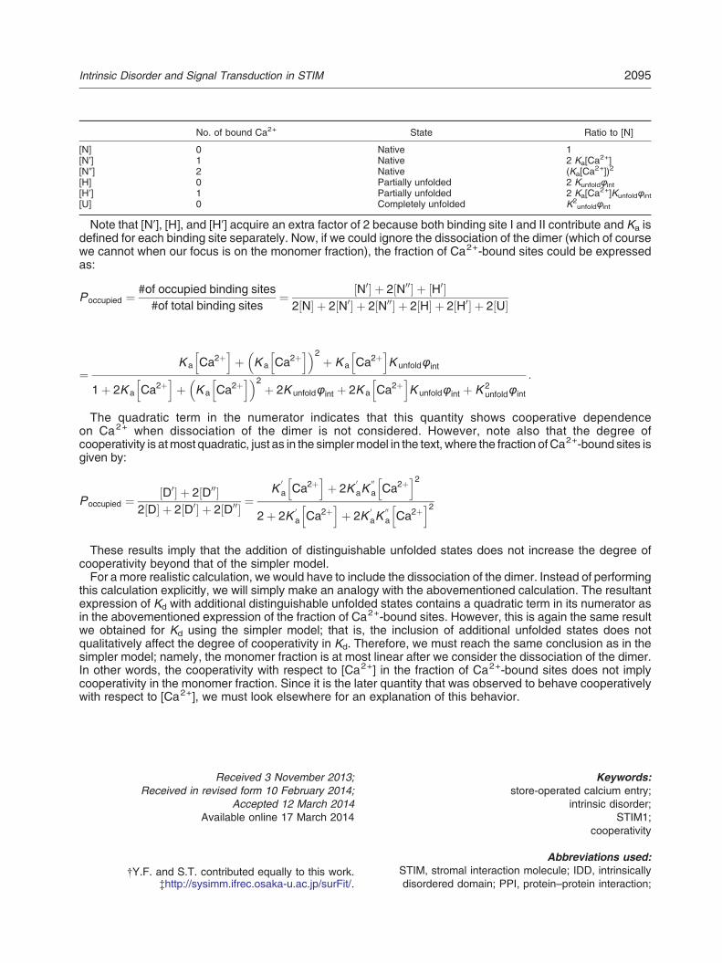
No. of bound Ca2+ State Ratio to [N]
[N] 0 Native 1[N!] 1 Native 2 Ka[Ca
2+][N"] 2 Native (Ka[Ca
2+])2
[H] 0 Partially unfolded 2 Kunfoldϕint[H!] 1 Partially unfolded 2 Ka[Ca
2+]Kunfoldϕint[U] 0 Completely unfolded K2
unfoldϕint
Note that [N!], [H], and [H!] acquire an extra factor of 2 because both binding site I and II contribute and Ka isdefined for each binding site separately. Now, if we could ignore the dissociation of the dimer (which of coursewe cannot when our focus is on the monomer fraction), the fraction of Ca2+-bound sites could be expressedas:
Poccupied & #of occupied binding sites#of total binding sites
& N0# $ % 2 N00# $ % H0# $2 N# $ % 2 N0# $ % 2 N00# $ % 2 H# $ % 2 H0# $ % 2 U# $
&K a Ca2%
h i% K a Ca2%
h i! "2% K a Ca2%
h iK unfoldϕint
1% 2K a Ca2%h i
% K a Ca2%h i! "2
% 2K unfoldϕint % 2K a Ca2%h i
K unfoldϕint % K 2unfoldϕint
:
The quadratic term in the numerator indicates that this quantity shows cooperative dependenceon Ca2+ when dissociation of the dimer is not considered. However, note also that the degree ofcooperativity is atmost quadratic, just as in the simplermodel in the text, where the fraction of Ca2+-bound sites isgiven by:
Poccupied &D0# $ % 2 D00# $
2 D# $ % 2 D0# $ % 2 D00# $&
K0
a Ca2%h i
% 2K0
aK00
a Ca2%h i2
2% 2K0
a Ca2%h i
% 2K0
aK00
a Ca2%h i2
These results imply that the addition of distinguishable unfolded states does not increase the degree ofcooperativity beyond that of the simpler model.For a more realistic calculation, we would have to include the dissociation of the dimer. Instead of performing
this calculation explicitly, we will simply make an analogy with the abovementioned calculation. The resultantexpression of Kd with additional distinguishable unfolded states contains a quadratic term in its numerator asin the abovementioned expression of the fraction of Ca2+-bound sites. However, this is again the same resultwe obtained for Kd using the simpler model; that is, the inclusion of additional unfolded states does notqualitatively affect the degree of cooperativity in Kd. Therefore, we must reach the same conclusion as in thesimpler model; namely, the monomer fraction is at most linear after we consider the dissociation of the dimer.In other words, the cooperativity with respect to [Ca2+] in the fraction of Ca2+-bound sites does not implycooperativity in the monomer fraction. Since it is the later quantity that was observed to behave cooperativelywith respect to [Ca2+], we must look elsewhere for an explanation of this behavior.
Received 3 November 2013;Received in revised form 10 February 2014;
Accepted 12 March 2014Available online 17 March 2014
Keywords:store-operated calcium entry;
intrinsic disorder;STIM1;
cooperativity
Abbreviations used:STIM, stromal interaction molecule; IDD, intrinsicallydisordered domain; PPI, protein–protein interaction;
†Y.F. and S.T. contributed equally to this work.‡http://sysimm.ifrec.osaka-u.ac.jp/surFit/.
2095Intrinsic Disorder and Signal Transduction in STIM
ER, endoplasmic reticulum; SOCE, store-operated Ca2+
entry; PM, plasma membrane; CRAC, Ca2+ releaseactivated Ca2+; AUC, analytical ultracentrifugation;
HSQC, heteronuclear single-quantum correlation; MD,molecular dynamics; DMD, discrete molecular dynamics.
References
[1] Dyson HJ, Wright PE. Intrinsically unstructured proteins andtheir functions. Nat Rev Mol Cell Biol 2005;6:197–208.
[2] Haynes C, Oldfield CJ, Ji F, Klitgord N, Cusick ME, RadivojacP, et al. Intrinsic disorder is a common feature of hubproteins from four eukaryotic interactomes. PLoS ComputBiol 2006;2:e100.
[3] Dunker AK, Cortese MS, Romero P, Iakoucheva LM,Uversky VN. Flexible nets. The roles of intrinsicdisorder in protein interaction networks. FEBS J2005;272:5129–48.
[4] Mitrea DM, Kriwacki RW. Regulated unfolding of proteins insignaling. FEBS Lett 2013;587:1081–8.
[5] Ferreon AC, Ferreon JC, Wright PE, Deniz AA. Modula-tion of allostery by protein intrinsic disorder. Nature2013;498:390–4.
[6] Freiburger LA, Baettig OM, Sprules T, Berghuis AM, AuclairK, Mittermaier AK. Competing allosteric mechanisms mod-ulate substrate binding in a dimeric enzyme. Nat Struct MolBiol 2011;18:288–94.
[7] Garcia-Pino A, Balasubramanian S, Wyns L, Gazit E, DeGreve H, Magnuson RD, et al. Allostery and intrinsic disordermediate transcription regulation by conditional cooperativity.Cell 2010;142:101–11.
[8] Reichheld SE, Yu Z, Davidson AR. The induction of foldingcooperativity by ligand binding drives the allosteric responseof tetracycline repressor. Proc Natl Acad Sci U S A2009;106:22263–8.
[9] Hilser VJ, Thompson EB. Intrinsic disorder as a mechanismto optimize allosteric coupling in proteins. Proc Natl Acad SciU S A 2007;104:8311–5.
[10] Reichmann D, Jakob U. The roles of conditional disorder inredox proteins. Curr Opin Struct Biol 2013;23:436–42.
[11] Baba Y, Nishida K, Fujii Y, Hirano T, Hikida M, Kurosaki T.Essential function for the calcium sensor STIM1 in mast cellactivation and anaphylactic responses. Nat Immunol2008;9:81–8.
[12] Oh-Hora M, Yamashita M, Hogan PG, Sharma S, Lamperti E,Chung W, et al. Dual functions for the endoplasmic reticulumcalcium sensors STIM1 and STIM2 in T cell activation andtolerance. Nat Immunol 2008;9:432–43.
[13] Matsumoto M, Fujii Y, Baba A, Hikida M, Kurosaki T, Baba Y.The calcium sensors STIM1 and STIM2 control B cellregulatory function through interleukin-10 production. Immu-nity 2011;34:703–14.
[14] Stiber J, Hawkins A, Zhang ZS, Wang S, Burch J, Graham V,et al. STIM1 signalling controls store-operated calcium entryrequired for development and contractile function in skeletalmuscle. Nat Cell Biol 2008;10:688–97.
[15] Picard C, McCarl CA, Papolos A, Khalil S, Luthy K, Hivroz C,et al. STIM1 mutation associated with a syndrome ofimmunodeficiency and autoimmunity. N Engl J Med2009;360:1971–80.
[16] Stathopulos PB, Zheng L, Li GY, PlevinMJ, IkuraM. Structuraland mechanistic insights into STIM1-mediated initiation ofstore-operated calcium entry. Cell 2008;135:110–22.
[17] Stathopulos PB, Li GY, Plevin MJ, Ames JB, Ikura M. StoredCa2+ depletion-induced oligomerization of stromal interactionmolecule 1 (STIM1) via the EF-SAM region: an initiationmechanism for capacitive Ca2+ entry. J Biol Chem2006;281:35855–62.
[18] Yang X, Jin H, Cai X, Li S, Shen Y. Structural and mechanisticinsights into the activation of stromal interaction molecule 1(STIM1). Proc Natl Acad Sci U S A 2012;109:5657–62.
[19] Zhou Y, Srinivasan P, Razavi S, Seymour S, Meraner P,Gudlur A, et al. Initial activation of STIM1, the regulator of store-operated calcium entry. Nat Struct Mol Biol 2013;20:973–81.
[20] Soboloff J, Rothberg BS, Madesh M, Gill DL. STIM proteins:dynamic calcium signal transducers. Nat Rev Mol Cell Biol2012;13:549–65.
[21] Hogan PG, Lewis RS, Rao A. Molecular basis of calciumsignaling in lymphocytes: STIM and ORAI. Annu RevImmunol 2010;28:491–533.
[22] BrandmanO, Liou J, ParkWS, Meyer T. STIM2 is a feedbackregulator that stabilizes basal cytosolic and endoplasmicreticulum Ca2+ levels. Cell 2007;131:1327–39.
[23] Luik RM, Wang B, Prakriya M, Wu MM, Lewis RS.Oligomerization of STIM1 couples ER calcium depletion toCRAC channel activation. Nature 2008;454:538–42.
[24] McRorie DK, Voelker PJ. Self-associating systems in theanalytical ultracentrifuge. Palo Alto: Beckman Instruments,Inc.; 1993.
[25] Kumar S, Rosenberg JM, Bouzida D, Swendsen RH,Kollman PA. THE weighted histogram analysis method forfree-energy calculations on biomolecules. I. The method. JComput Chem 1992;13:1011–21.
[26] Zheng L, Stathopulos PB, Li GY, Ikura M. Biophysicalcharacterization of the EF-hand and SAM domain containingCa2+ sensory region of STIM1 and STIM2. Biochem BiophysRes Commun 2008;369:240–6.
[27] Ward JJ, McGuffin LJ, Bryson K, Buxton BF, Jones DT. TheDISOPRED server for the prediction of protein disorder.Bioinformatics 2004;20:2138–9.
[28] Schellman JA. The thermodynamics of solvent exchange.Biopolymers 1994;34:1015–26.
[29] Myers JK, PaceCN,Scholtz JM.Denaturantm valuesand heatcapacity changes: relation to changes in accessible surfaceareas of protein unfolding. Protein Sci 1995;4:2138–48.
[30] Guharoy M, Chakrabarti P. Secondary structure basedanalysis and classification of biological interfaces: identifica-tion of binding motifs in protein–protein interactions. Bioin-formatics 2007;23:1909–18.
[31] OlivebergM,Wolynes PG. The experimental survey of protein-folding energy landscapes. Q Rev Biophys 2005;38:245–88.
[32] Heidarsson PO, Otazo MR, Bellucci L, Mossa A, Imparato A,Paci E, et al. Single-molecule folding mechanism of an EF-hand neuronal calcium sensor. Structure 2013;21:1812–21.
[33] Gunasekaran K, Ma B, Nussinov R. Is allostery an intrinsicproperty of all dynamic proteins? Proteins 2004;57:433–43.
[34] Lewit-Bentley A, Rety S. EF-hand calcium-binding proteins.Curr Opin Struct Biol 2000;10:637–43.
[35] Zheng L, Stathopulos PB, Schindl R, Li GY, Romanin C, IkuraM. Auto-inhibitory role of the EF-SAM domain of STIMproteins in store-operated calcium entry. Proc Natl Acad SciU S A 2011;108:1337–42.
[36] Schuhmann MK, Stegner D, Berna-Erro A, Bittner S, BraunA, Kleinschnitz C, et al. Stromal interaction molecules 1 and 2are key regulators of autoreactive T cell activation in murineautoimmune central nervous system inflammation. J Immu-nol 2010;184:1536–42.
2096 Intrinsic Disorder and Signal Transduction in STIM
[37] McCarl CA, Khalil S, Ma J, Oh-hora M, Yamashita M, RoetherJ, et al. Store-operated Ca2+ entry through ORAI1 is criticalfor T cell-mediated autoimmunity and allograft rejection. JImmunol 2010;185:5845–58.
[38] Greenfield NJ. Using circular dichroism spectra to estimateprotein secondary structure. Nat Protoc 2006;1:2876–90.
[39] Dokholyan NV, Buldyrev SV, Stanley HE, Shakhnovich EI.Discrete molecular dynamics studies of the folding of aprotein-like model. Folding Des 1998;3:577–87.
[40] Shirvanyants D, Ding F, Tsao D, Ramachandran S,Dokholyan NV. Discrete molecular dynamics: an efficient
and versatile simulation method for fine protein characteri-zation. J Phys Chem B 2012;116:8375–82.
[41] Ding F, Tsao D, Nie H, Dokholyan NV. Ab initio folding ofproteins with all-atom discrete molecular dynamics. Structure2008;16:1010–8.
[42] Dagliyan O, Proctor EA, D'Auria KM, Ding F, Dokholyan NV.Structural and dynamic determinants of protein–peptiderecognition. Structure 2011;19:1837–45.
[43] FukunishiY,MikamiY,NakamuraH.Similarities among receptorpockets and among compounds: analysis and application toin silico ligand screening. JMol GraphicsModell 2005;24:34–45.
2097Intrinsic Disorder and Signal Transduction in STIM