Embed Size (px)
Citation preview
lable at ScienceDirect
Journal of Orthopaedics, Trauma and Rehabilitation 19 (2015) 60e65
Contents lists avai
Journal of Orthopaedics, Trauma and Rehabilitation
Journal homepages: www.e- jotr .com & www.ejotr .org
Orthopaedic Rehabilitation
The Effect of Low Dose Extracorporeal ShockWave Therapy (ESWT) onPlantar Fasciitis: A Trial Study in Queen Mary Hospital低劑量體外衝擊波治療(ESWT)對足底筋膜炎的影響 - 在瑪麗醫院的試用
研究
Wan Yik-Cheung Samuel a, Lie Wai Hung Chester b, *, Pun Cheuk Ting Terence a,Lam Yuen Ha Rita c, Ng Chui San Maggie c, Ng Tze Pui d
a Department of Orthopaedics and Traumatology, Queen Mary Hospital, Hong Kongb Department of Orthopaedics and Traumatology, Kwong Wah Hospital, Hong Kongc Department of Physiotherapy, Queen Mary Hospital, Hong Kongd Private Practice, Hong Kong
a r t i c l e i n f o
Article history:Received 6 June 2014Received in revised form13 October 2014Accepted 17 October 2014
Keywords:extracorporeal shockwave therapylow energyplantar fasciitisshockwave
* Corresponding author. E-mail: chesterliewh@gma
http://dx.doi.org/10.1016/j.jotr.2014.10.0052210-4917/Copyright © 2015, The Hong Kong Orthopaedic As
a b s t r a c t
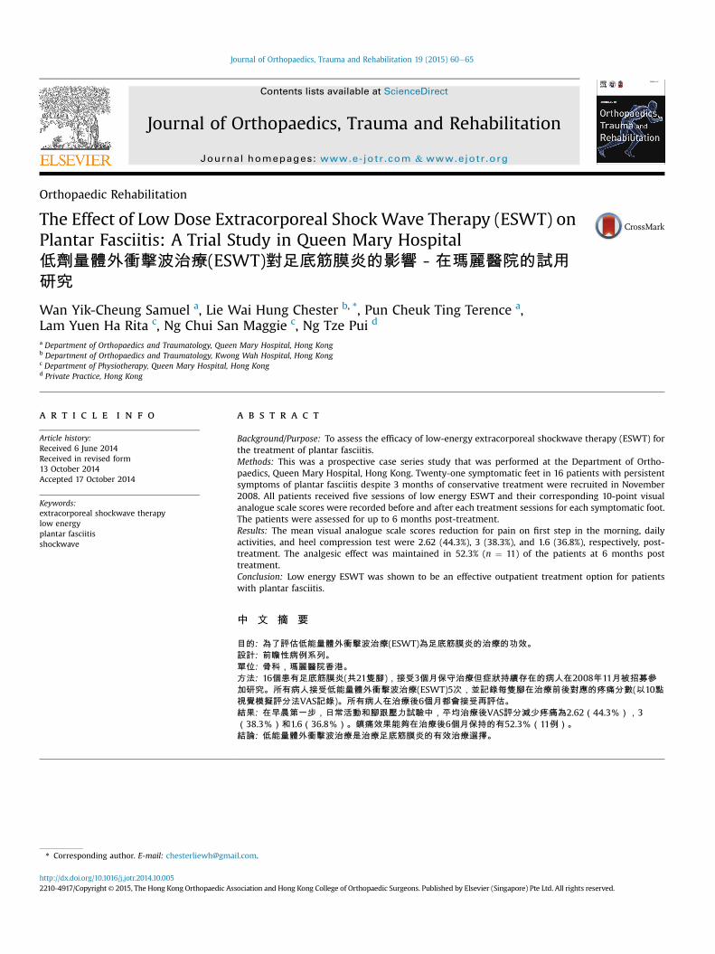
Background/Purpose: To assess the efficacy of low-energy extracorporeal shockwave therapy (ESWT) forthe treatment of plantar fasciitis.Methods: This was a prospective case series study that was performed at the Department of Ortho-paedics, Queen Mary Hospital, Hong Kong. Twenty-one symptomatic feet in 16 patients with persistentsymptoms of plantar fasciitis despite 3 months of conservative treatment were recruited in November2008. All patients received five sessions of low energy ESWT and their corresponding 10-point visualanalogue scale scores were recorded before and after each treatment sessions for each symptomatic foot.The patients were assessed for up to 6 months post-treatment.Results: The mean visual analogue scale scores reduction for pain on first step in the morning, dailyactivities, and heel compression test were 2.62 (44.3%), 3 (38.3%), and 1.6 (36.8%), respectively, post-treatment. The analgesic effect was maintained in 52.3% (n ¼ 11) of the patients at 6 months posttreatment.Conclusion: Low energy ESWT was shown to be an effective outpatient treatment option for patientswith plantar fasciitis.
中 文 摘 要
目的: 為了評估低能量體外衝擊波治療(ESWT)為足底筋膜炎的治療的功效。
設計: 前瞻性病例系列。
單位: 骨科,瑪麗醫院香港。
方法: 16個患有足底筋膜炎(共21隻腳),接受3個月保守治療但症狀持續存在的病人在2008年11月被招募參
加研究。所有病人接受低能量體外衝擊波治療(ESWT)5次,並記錄每隻腳在治療前後對應的疼痛分數(以10點視覺模擬評分法VAS記錄)。所有病人在治療後6個月都會接受再評估。
結果: 在早晨第一步,日常活動和腳跟壓力試驗中,平均治療後VAS評分減少疼痛為2.62(44.3%),3(38.3%)和1.6(36.8%)。鎮痛效果能夠在治療後6個月保持的有52.3%(11例)。
結論: 低能量體外衝擊波治療是治療足底筋膜炎的有效治療選擇。
il.com.
sociation and Hong Kong College of Orthopaedic Surgeons. Published by Elsevier (Singapore) Pte Ltd. All rights reserved.
Y.-C.S. Wan et al. / Journal of Orthopaedics, Trauma and Rehabilitation 19 (2015) 60e65 61
Introduction
Shockwaves are energy waves generated in water mediumcharacterized by rapid expansion and vaporisation. The highlyamplified soundwave expands in three dimensions, forming a conewith themaximum pressure at thewave front.1,2 The attenuation ofwave fronts subsequently causes an abrupt rise and fall of airpressure that can causes high tension at the surface of calculus,resulting in structural crack.1e4 The application of shockwaves wasused successfully for the treatment of urinary and biliary calculi inthe 1980s but it was not until the 1990s that the use of extracor-poreal shockwave therapy (ESWT) was adopted for the treatmentof various tendinopathies.1,2,5
The exact mechanism by which ESWT relieves tendon-associated pain is not known. It is postulated that shockwavesmay have an initial analgesic effect by altering the permeability ofneuron cell membranes. A higher stimulus is required to trigger anaction potential in the sensory neuron.1,2,4,5
Another postulated theory is that ESWT may cause increasedblood flow to the treated site and induce an inflammatory-mediated healing process in damaged tendons by disruptingavascular, damaged tissues, and encouraging revascularization,release of local growth factor, and the recruitment of appropriatestem cells to the area.2,4,6
Plantar fasciitis is the most common cause of heel pain in theadult population and a small subset of patients has symptoms thatare refractory to conservative management for months to years.Patients with such disease typically report a sharp heel pain that isworse on the first step in the morning and towards the end of theday.7,8 Many studies in the past involving ESWT on plantar fasciitiswere highly variable in their study designs and treatment regimensand thus conclusive evidence regarding the optimal treatmentprotocol is still lacking.9e18 Most studies involving ESWTon plantarfasciitis employ high-energy shockwaves that are believed to have ahigher clinical efficacy. However, the associated discomfort in high-energy ESWT often requires the use of local anaesthesia that couldbe more cumbersome to the patient.13,14,18 The present study aimsat studying the effect of a low dose ESWT system on plantar fasciitisin a local population.
Methodology
Patients
Patients with clinical diagnosis of plantar fasciitis that have failedconservative management (nonsteroidal anti-inflammatory drugs,
Table 1Recruitment criteria
Inclusion criteria
Age >18 yHistory of plantar heel pain for >3 moFailed conservative treatment including physiotherapy treatment
stretchingexercises, heel pads, andnight splints) of at least 3monthswere recruited from the specialist clinic of the Orthopaedics andTraumatology unit of Queen Mary Hospital, Hong Kong, between 1November 2008 and 30 November 2008. For our study, plantarfasciitis was clinically defined as moderate to severe heel pain uponthe first few steps in the morning that gets worse with continuedweight bearing. The pain and tenderness should be located in themedial tubercle of the calcaneus without obvious signs and symp-toms of other differentials of heel pain such as stress fractures,osteomyelitis or hindfoot arthritis. Radiographs of the involved footwere taken for patients whose clinical diagnosis was uncertain toexclude other causes of heel pain. The daily activities are our patientsall required walking on level ground, stairs, and slopes for �1 hourdaily. Those who met the specific inclusion and exclusion criterialisted in Table 1 were referred to the Physiotherapy Department ofthe hospital for assessment of feasibility of treatment.
Informed consent was obtained for all patients who met theinclusion and exclusion criteria, prior to the start of treatment.Possible adverse effects were explained to them.
The machine
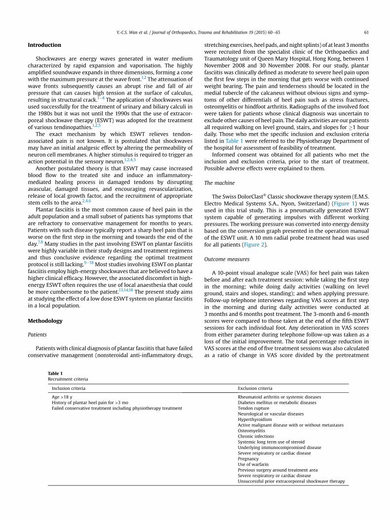
The Swiss DolorClast® Classic shockwave therapy system (E.M.S.Electro Medical Systems S.A., Nyon, Switzerland) (Figure 1) wasused in this trial study. This is a pneumatically generated ESWTsystem capable of generating impulses with different workingpressures. Theworking pressure was converted into energy densitybased on the conversion graph presented in the operation manualof the ESWT unit. A 10 mm radial probe treatment head was usedfor all patients (Figure 2).
Outcome measures
A 10-point visual analogue scale (VAS) for heel pain was takenbefore and after each treatment session: while taking the first stepin the morning; while doing daily activities (walking on levelground, stairs and slopes, standing); and when applying pressure.Follow-up telephone interviews regarding VAS scores at first stepin the morning and during daily activities were conducted at3 months and 6 months post treatment. The 3-month and 6-monthscores were compared to those taken at the end of the fifth ESWTsessions for each individual foot. Any deterioration in VAS scoresfrom either parameter during telephone follow-up was taken as aloss of the initial improvement. The total percentage reduction inVAS scores at the end of five treatment sessions was also calculatedas a ratio of change in VAS score divided by the pretreatment
Exclusion criteria
Rheumatoid arthritis or systemic diseasesDiabetes mellitus or metabolic diseasesTendon ruptureNeurological or vascular diseasesHyperthyroidismActive malignant disease with or without metastasesOsteomyelitisChronic infectionsSystemic long term use of steroidUnderlying immunocompromised diseaseSevere respiratory or cardiac diseasePregnancyUse of warfarinPrevious surgery around treatment areaSevere respiratory or cardiac diseaseUnsuccessful prior extracorporeal shockwave therapy
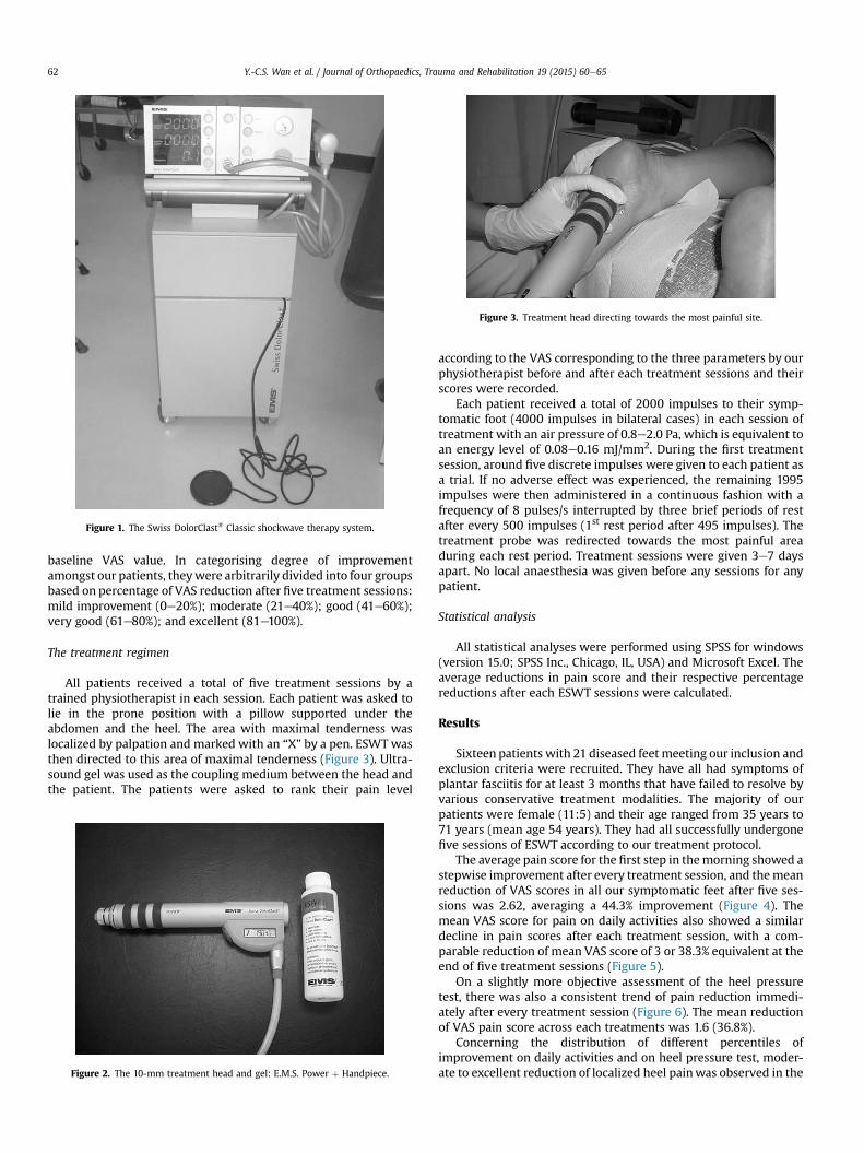
Figure 1. The Swiss DolorClast® Classic shockwave therapy system.
Figure 3. Treatment head directing towards the most painful site.
Y.-C.S. Wan et al. / Journal of Orthopaedics, Trauma and Rehabilitation 19 (2015) 60e6562
baseline VAS value. In categorising degree of improvementamongst our patients, theywere arbitrarily divided into four groupsbased on percentage of VAS reduction after five treatment sessions:mild improvement (0e20%); moderate (21e40%); good (41e60%);very good (61e80%); and excellent (81e100%).
The treatment regimen
All patients received a total of five treatment sessions by atrained physiotherapist in each session. Each patient was asked tolie in the prone position with a pillow supported under theabdomen and the heel. The area with maximal tenderness waslocalized by palpation and marked with an “X” by a pen. ESWT wasthen directed to this area of maximal tenderness (Figure 3). Ultra-sound gel was used as the coupling medium between the head andthe patient. The patients were asked to rank their pain level
Figure 2. The 10-mm treatment head and gel: E.M.S. Power þ Handpiece.
according to the VAS corresponding to the three parameters by ourphysiotherapist before and after each treatment sessions and theirscores were recorded.
Each patient received a total of 2000 impulses to their symp-tomatic foot (4000 impulses in bilateral cases) in each session oftreatment with an air pressure of 0.8e2.0 Pa, which is equivalent toan energy level of 0.08e0.16 mJ/mm2. During the first treatmentsession, around five discrete impulses were given to each patient asa trial. If no adverse effect was experienced, the remaining 1995impulses were then administered in a continuous fashion with afrequency of 8 pulses/s interrupted by three brief periods of restafter every 500 impulses (1st rest period after 495 impulses). Thetreatment probe was redirected towards the most painful areaduring each rest period. Treatment sessions were given 3e7 daysapart. No local anaesthesia was given before any sessions for anypatient.
Statistical analysis
All statistical analyses were performed using SPSS for windows(version 15.0; SPSS Inc., Chicago, IL, USA) and Microsoft Excel. Theaverage reductions in pain score and their respective percentagereductions after each ESWT sessions were calculated.
Results
Sixteen patients with 21 diseased feet meeting our inclusion andexclusion criteria were recruited. They have all had symptoms ofplantar fasciitis for at least 3 months that have failed to resolve byvarious conservative treatment modalities. The majority of ourpatients were female (11:5) and their age ranged from 35 years to71 years (mean age 54 years). They had all successfully undergonefive sessions of ESWT according to our treatment protocol.
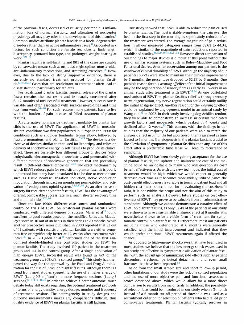
The average pain score for the first step in themorning showed astepwise improvement after every treatment session, and themeanreduction of VAS scores in all our symptomatic feet after five ses-sions was 2.62, averaging a 44.3% improvement (Figure 4). Themean VAS score for pain on daily activities also showed a similardecline in pain scores after each treatment session, with a com-parable reduction of mean VAS score of 3 or 38.3% equivalent at theend of five treatment sessions (Figure 5).
On a slightly more objective assessment of the heel pressuretest, there was also a consistent trend of pain reduction immedi-ately after every treatment session (Figure 6). The mean reductionof VAS pain score across each treatments was 1.6 (36.8%).
Concerning the distribution of different percentiles ofimprovement on daily activities and on heel pressure test, moder-ate to excellent reduction of localized heel painwas observed in the
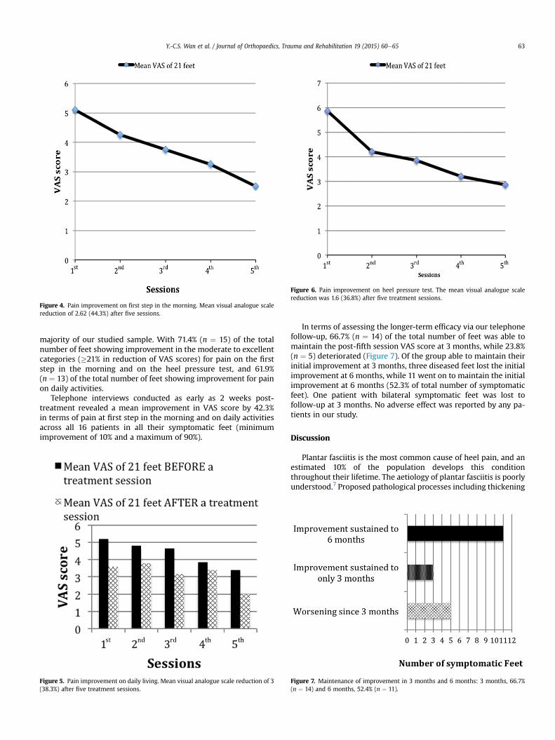
Figure 4. Pain improvement on first step in the morning. Mean visual analogue scalereduction of 2.62 (44.3%) after five sessions.
Figure 6. Pain improvement on heel pressure test. The mean visual analogue scalereduction was 1.6 (36.8%) after five treatment sessions.
Y.-C.S. Wan et al. / Journal of Orthopaedics, Trauma and Rehabilitation 19 (2015) 60e65 63
majority of our studied sample. With 71.4% (n ¼ 15) of the totalnumber of feet showing improvement in the moderate to excellentcategories (�21% in reduction of VAS scores) for pain on the firststep in the morning and on the heel pressure test, and 61.9%(n ¼ 13) of the total number of feet showing improvement for painon daily activities.
Telephone interviews conducted as early as 2 weeks post-treatment revealed a mean improvement in VAS score by 42.3%in terms of pain at first step in the morning and on daily activitiesacross all 16 patients in all their symptomatic feet (minimumimprovement of 10% and a maximum of 90%).
Figure 5. Pain improvement on daily living. Mean visual analogue scale reduction of 3(38.3%) after five treatment sessions.
In terms of assessing the longer-term efficacy via our telephonefollow-up, 66.7% (n ¼ 14) of the total number of feet was able tomaintain the post-fifth session VAS score at 3 months, while 23.8%(n ¼ 5) deteriorated (Figure 7). Of the group able to maintain theirinitial improvement at 3 months, three diseased feet lost the initialimprovement at 6 months, while 11 went on to maintain the initialimprovement at 6 months (52.3% of total number of symptomaticfeet). One patient with bilateral symptomatic feet was lost tofollow-up at 3 months. No adverse effect was reported by any pa-tients in our study.
Discussion
Plantar fasciitis is the most common cause of heel pain, and anestimated 10% of the population develops this conditionthroughout their lifetime. The aetiology of plantar fasciitis is poorlyunderstood.7 Proposed pathological processes including thickening
Figure 7. Maintenance of improvement in 3 months and 6 months: 3 months, 66.7%(n ¼ 14) and 6 months, 52.4% (n ¼ 11).
Y.-C.S. Wan et al. / Journal of Orthopaedics, Trauma and Rehabilitation 19 (2015) 60e6564
of the proximal fascia, decreased vascularity, peritendinus inflam-mation, loss of normal elasticity, and alteration of nociceptorphysiology all may play roles in the development of this disorder.8
Common studies attribute plantar fasciitis to a fascial degenerativedisorder rather than an active inflammatory cause.8 Associated riskfactors for such condition are female sex, obesity, limb-lengthdiscrepancy, pronated foot type, and prolonged weight bearing atwork.7,19e21
Plantar fasciitis is self-limiting and 90% of the cases are curableby conservative means such as orthotics, night splints, nonsteroidalanti-inflammatory medications, and stretching exercises.7,8 How-ever, due to the lack of strong supportive evidence, there iscurrently no standard treatment protocol for plantar fascii-tis.12,18,22,23 Cases that are recalcitrant to treatment often lead todissatisfaction, particularly for athletes.
For recalcitrant plantar fasciitis, surgical release of the plantarfascia remains the last resort and its usually considered after6e12 months of unsuccessful treatment. However, success rate isvariable and often associated with surgical morbidities and timeloss from work.24e26 For such reasons many patients have to livewith the burden of pain in cases of failed treatment of plantarfasciitis.
An alternative noninvasive treatment modality for plantar fas-ciitis is the use of ESWT. The application of ESWT in musculo-skeletal conditions was first popularized in Europe in the 1990s forconditions such as shoulder tendinitis, tennis elbow, followed byfracture nonunions, and plantar fasciitis.1,2,4,5 The device is a de-rivative of devices similar to that used for lithotripsy and relies ondelivery of shockwave energy in soft tissues to produce its clinicaleffect. There are currently four different groups of devices (elec-trohydraulic, electromagnetic, piezoelectric, and pneumatic) withdifferent methods of shockwave generation that can potentiallyresult in different clinical efficacies.1,4,23 The exact mechanism bywhich ESWT reduces pain for recalcitrant plantar fasciitis is poorlyunderstood but many have postulated it to be due to mechanismssuch as tissue neovascularization induction, nerve conductionmodulation through impact on membrane permeability, and acti-vation of endogenous opioid system.1,2,6,27,28 As an alternative tosurgery for recalcitrant plantar fasciitis, ESWT has the advantage ofoffering comparable success rate in a much shorter recovery timeand minimal risks.5,23,24
Since the late 1990s, different case control and randomizedcontrolled trials of ESWT on recalcitrant plantar fasciitis wereconducted with different degrees of success. Maier et al29 foundexcellent to good results based on the modified Roles and Mauds-ley's score in 36 out of 48 heels in their series at 29 months. Whileanother prospective series published in 2000 reported that 33 outof 41 patients with recalcitrant plantar fasciitis were either symp-tom free or significantly better at 12 weeks after treatment withESWT.30 In 2002 Ogden et al18 performed one of the first ran-domized double-blinded case controlled studies on ESWT forplantar fasciitis. The study involved 119 patient in the treatmentgroup and 114 in the control group. After a single application ofhigh energy ESWT, successful result was found in 47% of thetreatment group vs. 30% of the control group.18 This study had thenpaved the way for the approval by the Food and Drug Adminis-tration for the use of ESWT on plantar fasciitis. Although there is atrend from most studies suggesting the use of a higher energy ofESWT (i.e., �0.2 mJ/mm2) in more frequent sessions (i.e., �3sessions)5,11,14,16e18,31 in order to achieve a better outcome, muchdebate today still exists regarding the optimal treatment protocolsin terms of energy density, energy dosage, number and frequencyof treatment sessions. The heterogeneity in study designs andoutcome measurements makes any comparisons difficult, thusquality evidence of ESWT on plantar fasciitis is still lacking.
Our study showed that ESWT is able to reduce the pain causedby plantar fasciitis. The most irritable symptoms, the pain over theheel in the first step in the morning, is significantly reduced afterthe treatment was started. The average magnitude of pain reduc-tion in all our measured categories ranges from 38.6% to 44.3%,which is similar to the magnitude of pain reductions reported inestablished studies.11,12,14,16,18,29,31,32 However, direct comparison ofour findings to major studies is difficult at this point without theuse of similar scoring systems such as RoleseMaudsley and FootFunctional Scores. Another observation among our patients is thewindow of clinical durability of ESWT. Although the majority of ourpatients (66.7%) were able to maintain their clinical improvementby 3 months, the percentage dropped to 52.3% by 6 months. Onepossible reason for thiswearing off effect of the initial improvementmay be the regeneration of sensory fibres as early as 3 weeks in ananimal study after treatment with ESWT.33,34 As one postulatedmechanism of ESWT on plantar fasciitis relief is through sensorynerve degeneration, any nerve regeneration could certainly nullifythe initial analgesic effect. Another reason for the wearing off effectcould be explained by angiogenesis model in an animal study byWang et al35 in 2002. In their study involving dog Achilles tendon,they were able to demonstrate an increase in certain medicatedgrowth factors and neovessels, which peaked at 4 weeks anddeclined after 12 weeks.35 This concurs with the findings in ourstudies that the majority of our patients were able to retain theanalgesic effect in 3months but a portion of them regressed as timelapsed to 6 months. If angiogenesis in the plantar fascia is crucial tothe alleviation of symptoms in plantar fasciitis, then any loss of thisaffect after a predictable time lapse will lead to recurrence ofsymptoms.
Although ESWT has been slowly gaining acceptance for the useof plantar fasciitis, the upfront and maintenance cost of the ma-chine could be an obstacle to many who will question its cost/benefit effectiveness. As with any new technique, the initial cost oftreatment would be high, which we would expect to generallydecrease over time as it becomes more widely utilized. Since thecost-benefit effectiveness is variable in terms of patient load and allhidden cost must be accounted for in evaluating the cost/benefitratio, it is not within the scope and not the aim of this study toperform such an analysis. However, future studies on cost effec-tiveness of ESWT may prove to be valuable from an administrativestandpoint. Although we cannot demonstrate a curative effect ofESWT on plantar fasciitis, as only 52.4% of the patients in our studywere shown to have a sustainable analgesic effect at 6 months, it isnevertheless shown to be a viable form of treatment for symp-tomatic control in plantar fasciitis. Furthermore, most our patientsincluding those who deteriorated at 6 months were generallysatisfied with the initial improvement and indicated that theywould prefer additional ESWT treatments again if offered thechance.
As opposed to high-energy shockwaves that have been used inmost studies, we believe that the low-energy shock waves used inour study are effective in symptomatic alleviation of plantar fasci-itis, with the advantage of minimizing side effects such as patientdiscomfort, erythema, periosteal detachment, and even smallfractures that have been reported.1,2
Aside from the small sample size and short follow-up period,other limitations of our study were the lack of a control populationand the use of more objective pain and functional assessmentscores described above, which would allow for a more directcomparison to results from major trials. In addition, the possibilityof selection bias could be introduced in our study when a 3-monthinstead of a 6-month cut-off period of threshold was used as arecruitment criterion for selection of patients who had failed priorconservative treatments. Plantar fasciitis typically resolves in
Y.-C.S. Wan et al. / Journal of Orthopaedics, Trauma and Rehabilitation 19 (2015) 60e65 65
6e18months, and an earlier cut-off time-line for recruitment in ourstudy could introduce a self-improvement effect, enrolling patientswith symptoms that would otherwise improvewith time. However,as surgical release for plantar fasciitis is usually not considered untilat least 6months of failed treatment (i.e., recalcitrant cases), the useof ESWT can help patients alleviate symptoms and reduce thechance of development into recalcitrant cases that may in turnrequire more invasive treatment. Although radiographs were ob-tained for most of our studied patients, another improvement inour study would be to obtain radiographs for all patients duringrecruitment, as it is necessary and good clinical practice to excludeother conditions of heel pain thoroughly before properly diag-nosing and initiating the treatment of plantar fasciitis. In terms ofoutcome measurement, the use of VAS score alone in our case inour assessment criteria certainly has its limitations as it is oftenconsidered nonlinear and subjective, opening the possibility ofrecall bias. However, the VAS is a simple measurement especiallyfor the elderly and has been validated as a reliable measurement forpain in musculoskeletal disorders. Although almost all of our pa-tients with documented VAS improvement were satisfied with theoutcome, the addition of other assessment tools such as theRoleseMaudsley score, Foot Functional Index, and SF-36 healthsurvey can certainly be considered in future studies for a morecomprehensive outcomemeasure, as such tools can allow for betterassessment of symptomatic improvement and overall level of pa-tient satisfaction than VAS score alone.
Conclusion
Our study has illustrated the positive efficacy of ESWT intreatment of plantar fasciitis. The use of low energy pneumaticESWT over five sessions of 2000 impulses of 0.16 mJ/mm2 appearsto be a safe alternative to other conservative modalities in symp-tomatic control of plantar fasciitis. Its use demonstrated a time-dependent relationship with an effective durability period, withan early positive clinical effect seen as early as 2 weeks in themajority of the symptomatic feet that can be sustained to at least6 months in 52.4% of our series. The positive results of our studysupport its use as a viable noninvasive treatment modality forplantar fasciitis and may reduce the chance of development ofrecalcitrant plantar fasciitis.
Conflicts of interest
All authors declare no conflicts of interest.
References
1. Clement DB, Taunton JE. Extracorporeal shock wave therapy in the manage-ment of plantar fasciitis. Br Colombia Med J 2004;46:174e8.
2. Chung B, Wiley JP. Extracorporeal shockwave therapy: a review. Sports Med(Auckland) 2002;32:851e65.
3. Halliday D, Resnick R, Walker J. Fundamentals of physics. Oxford: Wiley; 2001.4. Ogden JA, T�oth-Kischkat A, Schultheiss R. Principles of shock wave therapy. Clin
Orthop Relat Res 2001;387:8e17.5. Ogden JA, Alvarez RG, Levitt R, et al. Shock wave therapy (Orthotripsy) in
musculoskeletal disorders. Clin Orthop Relat Res 2001;387:22e40.6. Wang FS, Wang CJ, Huang HJ, et al. Physical shock wave mediates membrane
hyperpolarization and Ras activation for osteogenesis in human bone marrowstromal cells. Biochem Biophys Res Commun 2001;287:648e55.
7. Tahririan MA, Motififard M, Tahmasebi MN, et al. Plantar fasciitis. J Res Med Sci2012;17:799e804.
8. Lemont H, Ammirati KM, Usen N. Plantar fasciitis: a degenerative process(fasciosis) without inflammation. J Am Podiatr Med Assoc 2003;93:234e7.
9. Gollwitzer H, Diehl P, von Korff A, et al. Extracorporeal shock wave therapy forchronic painful heel syndrome: a prospective, double blind, randomized trialassessing the efficacy of a new electromagnetic shock wave device. J Foot AnkleSurg 2007;46:348e57.
10. Kudo P, Dainty K, Clarfield M, et al. Randomized, placebo-controlled, double-blind clinical trial evaluating the treatment of plantar fasciitis with anextracoporeal shockwave therapy (ESWT) device: a North American confir-matory study. J Orthop Res 2006;24:115e23.
11. Rompe JD, Meurer A, Nafe B, et al. Repetitive low-energy shock wave appli-cation without local anesthesia is more efficient than repetitive low-energyshock wave application with local anesthesia in the treatment of chronicplantar fasciitis. J Orthop Res 2005;23:931e41.
12. Wang CJ, Wang FS, Yang KD, et al. Long-term results of extracorporealshockwave treatment for plantar fasciitis. Am J Sports Med 2006;34:592e6.
13. Haake M, Buch M, Schoellner C, et al. Extracorporeal shock wave therapy forplantar fasciitis: randomised controlled multicentre trial. BMJ 2003;327:75.
14. Ogden JA, Alvarez RG, Levitt RL, et al. Electrohydraulic high-energy shock-wavetreatment for chronic plantar fasciitis. J Bone Joint Surg Am 2004;86-A:2216e28.
15. Rompe JD, Schoellner C, Nafe B. Evaluation of low-energy extracorporealshock-wave application for treatment of chronic plantar fasciitis. J Bone JointSurg Am 2002;84-A:335e41.
16. Lee SJ, Kang JH, Kim JY, et al. Dose-related effect of extracorporeal shock wavetherapy for plantar fasciitis. Ann Rehab Med 2013;37:379e88.
17. Chow IH, Cheing GL. Comparison of different energy densities of extracorporealshock wave therapy (ESWT) for the management of chronic heel pain. ClinRehab 2007;21:131e41.
18. Ogden JA, Alvarez RG, Marlow M. Shockwave therapy for chronic proximalplantar fasciitis: a meta-analysis. Foot Ankle Int 2002;23:301e8.
19. Irving DB, Cook JL, Young MA, et al. Obesity and pronated foot type may in-crease the risk of chronic plantar heel pain: a matched case-control study. BMCMusculoskel Disord 2007;8:41.
20. Mahmood S, Huffman LK, Harris JG. Limb-length discrepancy as a cause ofplantar fasciitis. J Am Podiatr Med Assoc 2010;100:452e5.
21. Riddle DL, Pulisic M, Pidcoe P, et al. Risk factors for plantar fasciitis: a matchedcase-control study. J Bone Joint Surg Am 2003;85-A:872e7.
22. Martin RL, Irrgang JJ, Conti SF. Outcome study of subjects with insertionalplantar fasciitis. Foot Ankle Int 1998;19:803e11.
23. Weil Jr LS, Roukis TS, Weil LS, et al. Extracorporeal shock wave therapy for thetreatment of chronic plantar fasciitis: indications, protocol, intermediate re-sults, and a comparison of results to fasciotomy. J Foot Ankle Surg 2002;41:166e72.
24. Davies MS, Weiss GA, Saxby TS. Plantar fasciitis: how successful is surgicalintervention? Foot Ankle Int 1999;20:803e7.
25. Gentile AT, Zizzo CJ, Dahukey A, et al. Traumatic pseudoaneurysm of the lateralplantar artery after endoscopic plantar fasciotomy. Foot Ankle Int 1997;18(12):821e2.
26. Tomczak RL, Haverstock BD. A retrospective comparison of endoscopic plantarfasciotomy to open plantar fasciotomy with heel spur resection for chronicplantar fasciitis/heel spur syndrome. J Foot Ankle Surg 1995;34:305e11.
27. Kaulesar Johannes EJ, Sukul DM, Bijma AM, et al. Effects of high-energyshockwaves on normal human fibroblasts in suspension. J Surg Res 1994;57:677e81.
28. Haake M, Thon A, Bette M. Absence of spinal response to extracorporeal shockwaves on the endogenous opioid systems in the rat. Ultrasound Med Biol2001;27:279e84.
29. Maier M, Steinborn M, Schmitz C, et al. Extracorporeal shock wave applicationfor chronic plantar fasciitis associated with heel spurs: prediction of outcomeby magnetic resonance imaging. J Rheumatol 2000;27:2455e62.
30. Wang CJ, Chen HS, Chen WS, et al. Treatment of painful heels using extracor-poreal shock wave. J Formos Med Assoc 2000;99:580e3.
31. Theodore GH, Buch M, Amendola A, et al. Extracorporeal shock wave therapyfor the treatment of plantar fasciitis. Foot Ankle Int 2004;25:290e7.
32. Rompe JD. Repetitive low-energy shock wave treatment is effective forchronic symptomatic plantar fasciitis. Knee Surg Sports Traumatol Arthrosc2007;15:107.
33. Takahashi N, Ohtori S, Saisu T, et al. Second application of low-energy shockwaves has a cumulative effect on free nerve endings. Clin Orthop Relat Res2006;443:315e9.
34. Takahashi N, Wada Y, Ohtori S, et al. Application of shock waves to rat skindecreases calcitonin gene-related peptide immunoreactivity in dorsal rootganglion neurons. Auton Neurosci 2003;107:81e4.
35. Wang CJ, Huang HY, Pai CH. Shock wave-enhanced neovascularization at thetendon-bone junction: an experiment in dogs. J Foot Ankle Surg 2002;41:16e22.