Embed Size (px)
Citation preview

Jun Mo, M.D.
Rady Children’ Hospital
Department of Pathology
University of California, San Diego

Introduction
Brief overview/classification of histiocytic disorders
Focus on the most common entities
Format: real case presentation
Emphasis on issues relevant to our daily practice
Briefly touch on recent biologic and genetic insights

Derived from bone marrow hematopoietic stem cells
Part of the immune system with two main functions.
Antigen processing and presentation
Phagocytosis
Two subtypes:
Dendritic cells- antigen processing and presentation
( CD1a+, CD14-)
Monocytes/macropahges - phagocytosis
(CD1a-, CD14+ CD68+, CD163+)
Immune function and phenotype
of histiocytes

Classification of Histiocytic Disorders
Class I - Dendritic cell related
Langerhans cell histiocytosis(LCH)
Secondary dendritic cell processes
Juvenile xanthogranuloma(JXG)
Solitary histiocytomas of various dendritic cell phenotypes
Class II - Monocyte/macrophage related
Hemophagocytic lymphohistiocytosis(HLH)
Rosai-Dorfman Disease
Solitary histiocytoma
Lipid Storage Disorders (storage histiocytosis)
Class III -Malignant histiocytic disorders
Monocytes –related: AML M4/5, CMML
Dendritic cell related histiocytic sarcoma
Case 1
A male infant delivered by C-section at 35 weeks
At birth, had hepatosplenomegaly and mild ascites.
Shortly developed thrombocytopenia and anemia,
required multiple transfusions
FH: a F. sibling died of fulminant liver failure of
unknown etiology on DOL 17

Case 1
Extensive infection and other work up were all negative
Blood and stool cultures for bacterial pathogens
Serology workup for viral infections and others
CMV, Rubella, herpes I and II, parvovirus B19, Toxoplasma gondii
Anticardiolipin antibodies
Lupus anticoagulative antibodies
Metabolic screen
Urine for bile acid synthesis
Hereditary tyrosiemia

Case 1
Had two biopsies:
liver biopsy
bone marrow biopsy

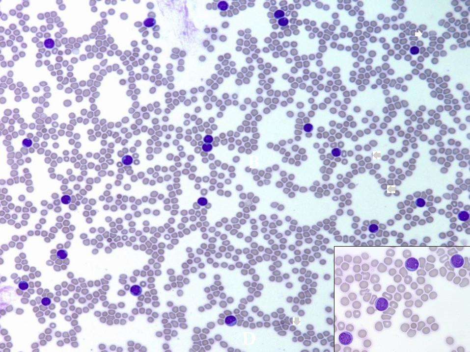
Bone marrow aspirate smear-patient

Bone marrow aspirate smear-patient

Liver biopsy-patient

Liver biopsy-patient

Liver biopsy-patient
CD-68 CD 68

Case 1
Dx of HLH was highly suspected.
Studies for natural killer (NK) and cytotoxic T-
cell function and perforin expression were
initiated
The patient deteriorated rapidly with metabolic
acidosis, fulminate liver failure and DIC.
Died on DOL 25. An autopsy was performed.

Autopsy liver-patient

Autopsy brain-patient

Autopsy findings-patient
Leptomeninges
Liver
Spleen
Lymph nodes
Bone marrow
Lungs
Kidneys
Adrenal glands
Pancreas
Testicles
Systemic lymphohistiocytic infiltrate with remarkable hemophagocytosis, organs involvement:

Further evaluation and follow up
Patient:
lack cytolytic function in NK cells (0, NL > 3.2 LU).
Normal NK cell number with NL perforin expression.
No mutation detected by PRF1 gene sequencing.
Mother:
Decreased perforin expression and cytolytic function in
cytotoxic T cells, and normal in NK cells.
Father: Normal

Further evaluation and follow up
F. Sibling :
hydrops fetalis at birth
Hepatosplenomegaly
Pancytopenia
Triglyceride 184 mg/dl (normal <150 mg/dL)
liver transaminases (AST 8897 IU/L; ALT 1770 IU/L)
Ferritin level 63100 ng/ml (normal 3 to 244 ng/ml)
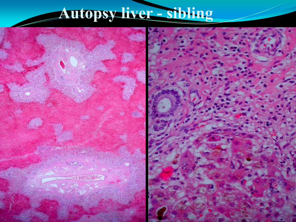
Autopsy liver - sibling

Case 1 – Diagnosis
Familial hemophagocytic lymphohistiocytosis
Pediatr Dev Pathol. 2006; 9(3):239-44.

Case 2
A 22 y/o F. had kidney transplant 5 years ago due to focal segmental glomerulosclerosis (FSGS)
On immunosuppression with stable renal function.
Presented with fever, headache, vomiting and back pain for 1½ months.
Extensive workup, all negative Negative Blood PCRs for EBV, CMV, adenovirus, parvovirus, enterovirus,
and BK virus.
No organisms isolated from cultures.
Normal chest X-ray.
Normal renal ultrasound
Empirically given antibiotics, but with persistent daily spiking fevers
Due to her persistent fever and progressive pancytopenia, a bone marrow specimen was obtained.


Case 2 LAB tests:
CBC: wbc3.8, Hg8.5, Plt 93
Fibrinogen 135 (normal 150-300mg/dL)
Triglycerides: 512 (normal <199mg/dL)
Ferritin: 14,094 (normal 3-105ng/mL)
Soluble IL-2 receptor: 28,245 (normal 45-1105 unit/mL)
No HLH associated genetic defects were detected.
Diagnosis: Secondary HLH associated with histoplasmosis
Treated with antifungal agent; in a few days patient’s fever and hematologic abnormalities were resolved.
American Journal of Transplantation, 2010, 10(3): 687-91

Hemophagocytic lymphohistiocytosis(HLH)
A rare, potentially fatal clinical syndrome caused by
hyperinflammatory reaction due to defective cytotoxic function
Excessive activation of cytotoxic T lymphocytes and histiocytes Overproduction of inflammatory cytokines
IL-1, IL-2Rs(sCD25), IL-6, TNF, and IFN.
Main clinical features
fever
hepatosplenomegaly
pancytopenia
coagulative abnormalities
neurological symptoms
skin rash

Hemophagocytic lymphohistiocytosis(HLH)
Familial or primarily HLH (~25%) Due to genetic defects
Mostly in infancy and early childhood(70-80% cases, < 1 year old)
First case reported by Farquhar and Claireux in 1952
Sporadic or secondary HLH (~75%) Associated with predisposing factors/underline
conditions
Affects any age, tends to occur in older children and adults.

Genetic (primary)HLH FHLH, subtype and associated genes
FHLH1, Gene unknown, 9q21.3-22
FHLH2: PFR1(perforin), account for ~20%-30% worldwide, 1999
FHLH3: UNC13 D(Munc13-4), account for ~20%-30% worldwide, 2003
FHLH4: STX11(syntaxin), ~20% of Turkish/Kurdish families, 2005
FHLH5 : STXBP2(syntaxin binding protein 2)/UNC18-2, 2009
Inherited immune disorders associated HLH
X-linked lymphoproliferative disease (XLP) SAP deficiency-SH2D1A mutations.
XIAP deficiency-XIAP (aka BIRC4) mutations
Chediak-Higashi syndrome(CHS)
CHS1 mutations
Griscelli syndrome type 2 (GS2) RAB27A mutations.
SCID, Wiskott-Aldrich Syndrome
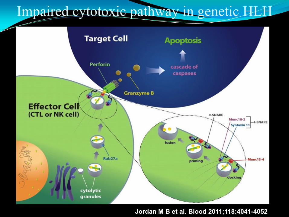
Impaired cytotoxic pathway in genetic HLH
Jordan M B et al. Blood 2011;118:4041-4052

Secondary HLH
Underline conditions associated with secondary HLH Infection
Virus: EBV, CMV, HHV6, HHV8, HSV
Bacteria, fungi, parasites
Autoimmune disorder:
macrophage activation syndrome(MAS) associated with systemic juvenile rheumatoid arthritis(sJRA)
Malignancy
Leukemia/lymphoma
solid tumors( Ewing Sarcoma, RMS, mediastinal germ cell tumor)
Immunodeficiency
Transplant, chemotherapy…

Diagnostic Criteria for HLH- 2004
A. Familial disease/known genetic defect.
or
B. Clinical and laboratory criteria (fulfill 5/8 criteria)
1. Fever
2. Splenomegaly
3. Cytopenias (affecting 2 of 3 lineages in the peripheral blood):
Hemoglobin <90 g/L (in infants <4 weeks: hemoglobin <100 g/L)
Platelets <100 x 109 /L
Neutrophils < 1.0 109 /L
4. Hypertriglyceridemia (fasting ≥265 mg/dl) and/or hypofibrinogenemia(≤1.5 g/L)
5. Hemophagocytosis in bone marrow, CSF, lymph nodes, or liver
6. Low or absent NK-cell activity
7. Ferritin ≥ 500 mg/L
8. Soluble CD25 (soluble IL-2 receptor) ≥ 2,400 U/ml

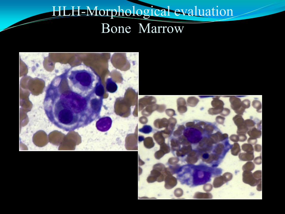
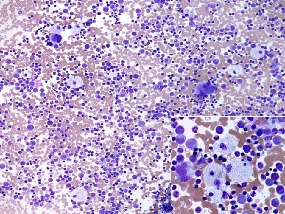
HLH-Morphological evaluation
Hemophagocytosis–the morphological hallmaker of HLH
Missed in 40% of cases at first examination
Tips in morphologic evaluation
Be alert, keep a high index of suspicion
Preferred material/tissue: bone marrow, liver tissue
Look for clues other than hemophagocytosis
Active looking histiocytes
“Friendly” histiocytes
Small sneaky histiocytes
Immunohistochemical staining CD163 can be helpful.
HLH-Morphological evaluation
Bone Marrow

Active looking histiocytes Irregular cytoplasmic contours
cytoplasmic vacuoles, blebs,
projects
free fragments

“Friendly” histiocytes
• kissing, hugging, wrapping
“target” cells
• perinuclear halo
• bag of cells

Small, “sneaky” hemophagocytic histiocytes

Mimics of hemaphagocytic histiocytes
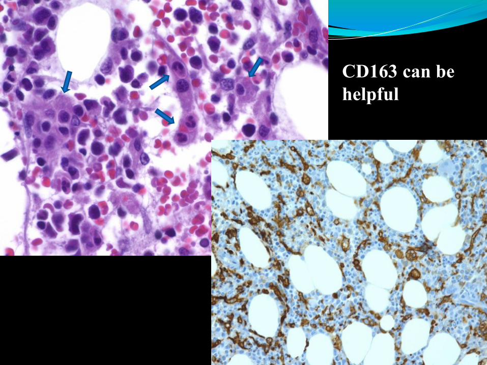
CD163 can be
helpful

Case 3
A previous healthy 9 year old girl presented
with recurrent epistaxis
Lab: Leukopenia and thrombocytopenia
Hematology consultation: A bone marrow
sample was obtained

A B
C D

A B
C D

Case 3
Molecular study detected two different
mutations in Acid-Glucosidase (GBA) that are
associated with Gaucher disease.
Dx: Gaucher disease

Case 4
3 yr old boy was found splenomegaly on
routine well check up.
He was asymptomatic and followed for 1.5
years
A BM sample was obtained.
A B
C D

A B
C D

Case 4
Two mutations in NPC1 gene detected
by molecular study
DX: Niemann–Pick disease type C
(subacute/juvenile)

Lysosomal storage diseases
Both Gaucher and Niemann-Pick diseases belong to a
group of lysosomal storage disease.
GD, deficiency of glucocerebrosidase
NPD, deficiency of sphingomyelinase
Rare genetic metabolic disorders with lysosomal
enzymes deficiencies caused by gene mutation
A diverse group of approximately 50 diseases

Case 5
A 2 year old boy with skin rashes,
multiple lytic bone lesions, and
abnormal bone marrow signals
A bone marrow specimen was obtained

CD1a
S100

Langerhans cell histiocytosis
Other names
Histiocytosis-X
Eosinophilic granuloma ( unifocal)
Hand-Schüller-Christian syndrome (multifocal)
Letterer-Siwe disease (disseminated)

Langerhans cell histiocytosis
One of the most common dendritic cell disorders in children
incidence: 3-5/million children
Average age at presentation: 2.4 yrs
M:F=1.3:1
Characterized by the pathologic accumulation of LC and other
inflammatory cells in organs. Birbeck granules in EM study
Single lesion, multiple focal, or systemic disease
Organs commonly involved: skin, bone, liver, lungs, bone
marrow, and brain.
Epidemiology: increased incidence in patients with
thyroid/autoimmune disease in family

LCH - Etiology and pathophysiology
Reactive or neoplastic ?

New insights into pathogenesis of LCH
Badalian-Very G. and colleagues reported the first
recurrent mutation of LCH in 2010.
BRAF V600E mutation detected in 35 of 61 (57%) LCH.
Brown NA et. al reported mutations in MAP2K1 in
27.5% of LCH cases
Both BRAF mutations and MAP2K1 mutations are part of
the RAS-MAPK pathway, they are mutually exclusive.
Strongly support: LCH is neoplastic
Clinical implication: BRAF-directed therapies, especially
in the systemic and aggressive form of LCH
Badalian V.G. Blood. 2010;116(11):1919–1923 ; Brown NA, Blood. 2014;124(10):1655–1658.

Case 6
2 year old boy presented with a two weeks
history of fever and cough.
On further evaluation, he had WBC 67000,
mediastinal mass, and hepatosplenomegaly.
B
D

Case 6
Immunophenotyping by flow cytometry:
Positive for CD2, surface CD3, cyt CD3, CD5, CD7, CD11b, and T-cell receptor γδ.
Negative for CD1a, CD4, CD8, CD25, CD34, TdT, and B or myeloid markers.
Cytogenetics: 46,XY,t(8;14)(q24;q11.2),der(12)t(12;20)(q11;q13.3),
der(20)t(12;20)(q21;q13.3)[9]/46,XY[5]
FISH: MYC (8q24) rearrangement in 13% of cells
Dx: T lymphoblastic leukemia with t(8;14)(q24;q11)
T(8;14)(q24;q11) only detectable in ~1% of T-ALL, a highly aggressive type
leukemia
Refractory to standard therapy AALL0434, switch to AALL0031
Received MUD cord blood transplantation five months after diagnosis
At day +110 post-BMT, a routine BM was obtained

A B
D

A B
C D

MYC (8q24) rearrangement in 15.6% of the cells on direct BMA smear

Juvenile Xanthogranuloma
A non-Langerhans cell dendritic histiocytic disorder
Occur in all ages, but mainly affects infants and young children
Mostly present as a single skin lesion
Biological behavior
usually benign and self-limiting, especially solitary skin lesions
systemic JXG
can virtually involves any organs
may cause serious morbidity or even death
Etiology: unknown.
Well-described association with neurofibromatosis, juvenile
chronic myeloid leukemia.

JXG and related hematopoietic
neoplasms
Dr. W. Klapper et al(2011) reported a very similar case as this case
A 5-year-old F. with diagnosis of T-ALL
Five months later presented with an aggressive systemic JXG
Clonal relationship between T-ALL and JXG
T-cell receptor gamma rearrangement in T-ALL blasts
Micro-dissected histiocytes from JXG lesion in a lymph
node revealed an identical bi-allelic TCR-γ rearrangement
Pediatr Blood Cancer 2011;56:859–862

Histiocytic disorders and related hematopoietic
neoplasm and clonality
In 2010 Dr. Ronald Jaffe, et al reviewed 15 patients who had histiocytic lesions followed ALL
All patients were in ALL remission while developed histiocytic lesions
( 5 JXG, 1 LCH, 4 Langhans’ cell sarcoma, 1 Rosai-Dorfman disease,
4 histiocytic scarcoma)
Clonal relationship with leukemia (Ig H or monoclonal TCR γ gene
rearrangements)
The post ALL histiocytic lesions are more aggressive than their native
lesions
Generally favorable prognosis
( 4 died of progressive histiocytic lesion, 1 died of recurrent ALL, 10 survived)
Pediatric and Developmental Pathology: May 2010, Vol. 13, No. 3, pp. 225-237

Case 6
Follow up CT/PET scan showed disseminated
JXG in his marrow, spleen, and mediastinum.
Treated per LCH III with partially response
Switch to thalidomide treatment
Patient eventually died of disseminated JXG

Histiocytic Disorders in Pediatric
Hematopathology
Monocyte/macrophage related Hemophagocytic lymphohistiocytosis(HLH)
Primary HLH
Secondary HLH
Lipid Storage Disorders (storage histiocytosis) Gaucher disease
Niemann–Pick disease
Dendritic cell related: Langerhans cell histiocytosis(LCH)
BRAF gene mutation
MAP2K1 gene mutation
Juvenile xanthogranuloma(JXG) Associated disorders and clonality
Class III -Malignant histiocytic disorders


![Proteinüri.ppt [Uyumluluk Modu] proteinüri nedenleri Primer glomerülopati MDH Membranöz GN FSGS IgA nefropati MPGN Sekonder glomerülopati APSGN Malignite İlaçlar (altın, NSAID,](https://img.pdfslide.tips/doc/110x75/5d0235f288c9932c7a8bfccf/proteinuerippt-uyumluluk-modu-proteinueri-nedenleri-primer-glomeruelopati-mdh.jpg)