Embed Size (px)
Citation preview
Fakultät für Medizin
Institut für Molekulare Immunologie
IKKα links autophagy, ER stress and caspase-12 function in a mouse
model of acute colitis
Michaela Alexandra Diamanti
Vollständiger Abdruck der von der Fakultät für Medizin der Technischen Universität
München zur Erlangung des akademischen Grades eines
Doctor of Philosophy (Ph.D.)
genehmigten Dissertation
Vorsitzende: Univ.-Prof. Dr. Dr. St. Engelhardt
Betreuer: Univ.-Prof. Dr. F. R. Greten
Prüfer der Dissertation:
1. Univ.-Prof. Dr. J. Ruland
2. Univ.-Prof. Dr. M. Heikenwälder
Die Dissertation wurde am 14.11.2014 bei der Fakultät für Medizin der Technischen
Universität München eingereicht und durch die Fakultät für Medizin am 16.02.2015
angenommen.
i
Abstract
Inflammatory bowel diseases (IBDs), manifesting as Crohn’s disease (CD) and Ulcerative
colitis (UC), are chronic conditions of the gastrointestinal tract and show a rapidly
increasing incidence in industrialized nations. Inhibitor of nuclear factor kappa-B kinase
subunit alpha (IKKα), a member of the IκB kinase (IKK) / nuclear factor-kappa B (NF-κB)
pathway, is involved in many inflammatory-mediated human diseases. Although the role
of IKKβ kinase in IBD has been described, that of IKKα remains currently unknown. Here,
the role of IKKα in intestinal homeostasis is investigated. Mice deficient in IKKα kinase
activity (IkkαAA/AA) exhibit defects in mucosa tissue repair, massive leukocyte infiltration
and elevated chemokine production in the colon, resulting in an exacerbated
inflammatory response when challenged with dextran sulfate sodium (DSS), causing
Ulcerative colitis. Defective IKKα signaling causes enhanced caspase-12 activation leading
to the inhibition of inflammasome-dependent IL-18 production by the intestinal epithelial
cells (IECs). Loss of caspase-12 or exogenous administration of recombinant IL-18
alleviates the severe colitis phenotype of IkkαAA/AA mice. Importantly, impaired IKKα
activation results in defective autophagic protein degradation causing p62 accumulation
and elevated endoplasmic reticulum (ER) stress, which is responsible for the elevated
caspase-12 activity. The present study demonstrates that IKKα, which might control the
autophagic process through its interaction with the Crohn’s disease variant, Atg16l1, is an
essential regulator of inflammatory responses in the large intestine and is required for
epithelial regeneration. Thus, IKKα is identified as a novel link between autophagy, ER
stress and inflammasome pathway, which are often deregulated in IBD.
ii
Zusammenfassung
Chronisch-entzündliche Darmerkrankungen (CED), die sich als Morbus Crohn oder Colitis
Ulcerosa manifestieren, sind chronische Erkrankungen des Magen-Darm-Traktes und
zeigen eine rapide ansteigende Inzidenz in Industrieländern. Inhibitor of nuclear factor
kappa-B kinase subunit alpha (IKKα), eine Komponente des IκB kinase (IKK) / nuclear
factor-kappa B (NF-κB) Signalwegs, ist an vielen entzündlichen Erkrankungen beteiligt.
Während die Bedeutung von IKKβ in der Entstehung von CED bereits beschrieben wurde,
ist die Rolle von IKKα dabei unbekannt. In der vorliegenden Arbeit wurde die Funktion
von IKKα in der Homöostase des Darms untersucht. Transgene Mäuse (IkkαAA/AA) mit
IKKα-Kinase-Defizienz zeigen gestörte Regeneration der Darmschleimhaut im Kolon,
massive Infiltration von Leukozyten sowie gesteigerte Zytokinproduktion. Dies führt zu
einer verstärkten Entzündungsreaktion bei Dextran-Sodium-Sulfat (DSS) induzierter
Colitis. Fehlregulierte Signaltransduktion von IKKα bewirkt eine verstärkte Aktivierung
von Caspase-12 und in Folge eine Hemmung der Inflammasom-abhängigen IL-18
Produktion in Enterozyten. Deletion von Caspase-12 oder Gabe von rekombinantem IL-
18 mildert den starken Kolitis-Phänotyp in IkkαAA/AA Mäusen. Die gestörte Aktivierung
von IKKα resultiert in defekter autophagosomaler Proteindegradation, welche
Akkumulation von p62 und verstärkten ER (Endoplasmatisches Retikulum)-Stress
hervorruft. Der gesteigerte ER-Stress wiederum ist ursächlich für die erhöhte Aktivität
von Caspase 12. Der autophagische Prozess wird möglicherweise durch Interaktion mit
der Morbus Crohn Mutante Atg16l1 kontrolliert. Die vorliegende Arbeit demonstriert,
dass IKKα einen essentieller Regulator von Entzündungsreaktion im Dickdarm darstellt
und für die epitheliale Regeneration benötigt wird. Folglich wurde IKKα als neues
Bindeglied zwischen Autophagie, ER-Stress und Inflammasom-Aktivierung identifiziert.

Acknowledgements
iii
Acknowledgments
I would like now to thank all those who each in their own special way supported me in
this work and without them would have been hard to achieve.
First of all, my sincerest gratitude is extended to my thesis supervisor Prof. Dr. F. Greten
for entrusting me with one of his research projects, for his unconditional support,
patience, guidance and valuable discussions during the entire course of my Ph.D.
Furthermore, I would like to deeply thank him for being instrumental in keeping this
project alive, despite the numerous hurdles, and for offering me the chance to publish this
work.
I am sincerely grateful to my advisor Dr. M. C. Arkan for her support and inestimable
advice and discussions during the entire course of the Ph.D and for her kind collaboration
and suggestions concerning this project during the committee meetings.
I am very thankful to my advisor Prof. Dr. J. Ruland for his inspirational supervision,
appreciated comments and insights.
Million thanks to my dear friends and colleagues Dr. Özge Canli and Dr. Tiago de Oliveira
for teaching and supporting me at the very first steps and course of this ‘Journey’ and
simply for being there for me at tough and joyful moments. Many thanks also to my
colleagues, Julia Varga, Dr. Mallika Ramakrishna, Tobias Neumann, Paul Ziegler, Dr.
Marina Pesic, Olga Goncharova and Charles Pallangyo for the nice working atmosphere,
the joy and excitement we shared and for being wonderful lab mates.
I would also want to thank former members of the lab, especially Dr. Moritz Bennecke for
initiating this project, Begüm Alankus, Dr. HsinYu Fang, Dr. Sarah Schwitalla, Dr. Arun
Mankan, Dr. Julia Bollrath, Dr. Tim Nebelsiek, and former members of the Arkan group,
Dr. Cigdem Atay, Dr. Manon Schultz, Dr. Jessica Heringer and Dr. Franciscka Romrig for
their companionship and support.

Acknowledgements
iv
I am really grateful to our technical assistants Natalia Delis, Christine Danneil, Eva Rudolf,
Kathleen Moos and former assistants, especially Kerstin Burmeister, one of the nicest
person I have ever met, Saskia Ettl and Kristin Retzlaff for their excellent work and honest
support.
Moreover, I wish to thank the PhD program coordinator Dr. Katrin Offe and Dessislava
Slatanova for excellent PhD program organization and their precious help and guidance.
Special thanks to Dr. Michael Aichler, Dr Jörn Lausen and Dr. Stefan Stein for the
conductive instruction during my rotation projects.
I would like to deeply thank my dear friend Dieter Weislmaier for his understanding and
support and for always caring for me.
I want to thank my very good friends Nikos Giannakos and Christina Kamilari, Pavlina
Gerontari for standing by me all this time and also Sevi Vletsi and Chrysi Petraki for the
fun and joy we had together throughout this work.
Last but not least, I deeply want to express my gratitude to my lovely family, my sister
Christina Maria Diamanti, my mother Elena Diamanti, my father Zisis Diamantis, my
grandfather Radu Cutieru and grandmother Michaela Cutieru for their understanding,
encouragement, and patience and for being always there for me. Thank you for believing
and trusting me. ‘Σας αγαπάω όλους πάρα πολύ και σας έχω ιδιαίτερη ευγνωμοσύνη.

Table of Contents
v
Abstract ................................................................................................................................................. i
Zusammenfassung ............................................................................................................................ ii
Acknowledgments .......................................................................................................................... iii
Table of Figures .............................................................................................................................. vii
List of Tables .................................................................................................................................. viii
Abbreviations.................................................................................................................................... ix
1.Introduction .................................................................................................................................... 1
1.1 The gastrointestinal tract ................................................................................................................................... 1
1.1.1 Morphology and function of the intestinal tract ................................................................................................... 2
1.2 Inflammatory bowel diseases (IBDs) ............................................................................................................. 4
1.3 Nuclear Factor-κB (NF-κB)................................................................................................................................. 9
1.3.1 NF-κB signaling in IBD ................................................................................................................................................... 11
1.3.2 IKKα: a multifunction protein kinase ...................................................................................................................... 12
1.3.2.1 The anti-inflammatory role of IKKα ................................................................................................................. 13
1.4 The key players in unfolded protein response (UPR) .......................................................................... 15
1.4.1 ER stress and inflammation......................................................................................................................................... 17
1.5 Autophagy ............................................................................................................................................................. 19
1.5.1 Role of autophagy in intestinal inflammation ..................................................................................................... 20
1.6 p62: a multimodule scaffold protein ........................................................................................................... 21
1.7 Microbial recognition by the innate immune system ........................................................................... 23
1.7.1 Inflammasomes ................................................................................................................................................................. 25
1.7.2 Role of the inflammasomes in intestinal homeostasis .................................................................................... 26
1.7.3 IL-1β/IL-18/caspase-1 axis in intestinal homeostasis .................................................................................... 28
1.8 Caspase-12 expression and activation ....................................................................................................... 31
1.8.1 The role of caspase-12 in inflammatory responses .......................................................................................... 32
Aim of Study ..................................................................................................................................... 35
2. Material and Methods .............................................................................................................. 36
2.1 Materials ................................................................................................................................................................ 36
2.1.1 Mouse models .................................................................................................................................................................... 36
2.1.2 Chemicals and reagents ................................................................................................................................................. 37
2.2 Methods .................................................................................................................................................................. 38
2.2.1 Animal experiments ........................................................................................................................................................ 38
2.2.2 Primary cell isolation and culture ............................................................................................................................ 41
2.2.3 Histology .............................................................................................................................................................................. 44
2.2.4 RNA Analysis ...................................................................................................................................................................... 47
2.2.5 Protein Analysis ................................................................................................................................................................ 49
3. Results ........................................................................................................................................... 55
3.1 IKKα is essential for mucosa healing after DSS-induced injury ........................................................ 55
Table of Contents
vi
3.1.1 Increased inflammation in the colons of IkkαΑΑ/AA mice ................................................................................. 56
3.2 IKKα signaling in IECs is crucial for tissue repair .................................................................................. 57
3.3 Severity of colitis is independent of alternative NF-κB activation ................................................... 60
3.3.1 IkkαAA/AA IECs display defective NF-κB activation ............................................................................................. 61
3.4 Impaired IKKα activation enhances IECs cell death .............................................................................. 62
3.5 IKKα mutant IECs display enhanced caspase-12 activation ............................................................... 63
3.6 IkkαAA/AA mutants display decreased IL-18 serum levels .................................................................... 65
3.6.1 IL-18 mediates tissue repair after injury .............................................................................................................. 66
3.6.2 IECs comprise the main source of IL-18 production during DSS colitis .................................................. 68
3.7 Deficiency of caspase-12 rescues the phenotype of IkkαAA/AA mice ................................................. 70
3.7.1 Caspase-12 ablation attenuates colitis in IkkαAA/AA mutants ........................................................................ 71
3.8 Impaired IKKα activation results in enhanced ER stress .................................................................... 72
3.9 Caspase-12 ablation does not affect cell death ........................................................................................ 74
3.10 Defective IKKα activation impairs autophagy ...................................................................................... 75
4. Discussion .................................................................................................................................... 78
4.1 Activation of IKKα suppresses inflammation in the large intestine ................................................ 78
4.1.1 Pathology in IkkαAA/AA mice is independent of the alternative NF-κB pathway................................... 80
4.2 IKKα activates inflammasome function and IL-18 production ......................................................... 81
4.3 Defective IKKα activation results in ER stress ......................................................................................... 84
4.4 IKKα controls autophagy ................................................................................................................................. 86
5. Conclusion ................................................................................................................................... 90
6. References ................................................................................................................................... 92
Appendix ......................................................................................................................................... 101
A.1 Chemicals, Reagents & Kits ...........................................................................................................................101
A.2 Genotyping of mice ..........................................................................................................................................102
A.3 Antibodies ...........................................................................................................................................................103
A.3.1 Antibodies used for western blot .......................................................................................................................... 103
A.3.2 Antibodies used for IHC/Immunofluorescence .............................................................................................. 103
A.3.3 Antibodies used for cell sorting .............................................................................................................................. 104
A.4 Real-time PCR primers ...................................................................................................................................104
Table of Figures
vii
Table of Figures
Figure 1.1 Picture depicting the main layers of the GI tract……………………………………………………………………...2
Figure 1.2 Architecture of the colonic crypt and the small intestinal crypt-villus……………………………………... 4
Figure 1.3 Canonical and alternative NF-κB activation…………………………………………………………………………..11
Figure 1.4 Figure depicting the mammalian unfolded protein response (UPR) cascade…………………………..17
Figure 1.5 Model for autophagy in mammalian cells…………………………………………………………………………….. 20
Figure 1.6 The interaction domains of the p62 protein…………………………………………………………....................... 22
Figure 1.7 The Inflammasome-IL-1β-IL-18 axis mediates tissue repair in the intestine………………………….. 30
Figure 3.1. Increased epithelial damage and infiltration of immune cells in IkkαΑΑ/AA colons after DSS
challenge……………………………………………………………………………………………………………………………………………. 56
Figure 3.2 Enhanced inflammation in the colons of IkkαΑΑ/AA mice………………………………………………………… 57
Figure 3.3 Impaired IKKα activation in IECs mediates the severe pathology………………………………………….. 58
Figure 3.4 IKKα activation in IECs protects against colitis…………………………………………………………………….. 59
Figure 3.5 IKKα protects the intestinal epithelium from DSS-induced colitis independent of the alternative
NF-κΒ activation………………………………………………………………………………………………………………………………….60
Figure 3.6 DSS activates NF-κB in Ikkαwt/wt but not in IkkαAA/AA IECs………..……………………………………………. 62
Figure 3.7 Massive cell death in IkkαAA/AA mutant intestinal epithelium…………………………………………………. 63
Figure 3.8 Increased epithelial cell death is independent of the intrinsic apoptotic pathway………………….. 64
Figure 3.9 Caspase-12 is activated in IkkαΑΑ/AA IECs after DSS administration………………………………………... 65
Figure 3.10 DSS-challenged IkkαΑΑ/ΑΑ mice fail to secrete active IL-18…………………………………………………… 66
Figure 3.11 Administration of recombinant IL-18 rescues severity of colitis…………………………........................68
Figure 3.12 IECs produce IL-18 during DSS colitis………………………………………………………………......................... 69
Figure 3.13 Caspase-12 deletion alleviates severity of colitis…………………………………………………………………71
Figure 3.14 Deletion of caspase-12 reduces the expression of proinflammatory factors………………………… 72
Figure 3.15 ER stress is aggravated in DSS-treated IkkaAA/AA mice………………………………………….......................74
Figure 3.16 Caspase-12 deletion does not affect cell death……………………………………………………..……………..75
Figure 3.17 IKKα inactivation results in defective autophagic protein degradation and p62
accumulation……………………………………………………………………………………………………………………………………… 77
Figure 5.1 Summary of the suggested model depicting the function of IKKα in the colonic
epithelium………………………………………………………………………………………………………………………………………….. 91
List of Tables
viii
List of Tables
Table 2:1 Tail lysis buffer recipe…………………………………………………………………………………………… 39
Table 2:2 General PCR reaction mix recipe……………………………………………………………………………. 40
Table 2:3 Agarose gel buffer recipe……………………………………………………………………………………….. 40
Table 2:4 Rehydration procedure…………………………………………………………………………………………. 44
Table 2:5 Dehydration procedure…………………………………………………………………………………………. 44
Table 2:6 Reverse transcription master mix…………………………………………………………………………..48
Table 2:7 Real-Time PCR master mix…………………………………………………………………………………….. 48
Table 2:8 Real-Time PCR program………………………………………………………………………………………… 49
Table 2:9 Protein lysis buffer recipe……………………………………………………………………………………… 50
Table 2:10 Laemmli buffer recipe…………………………………………………………………………………………. 50
Table 2:11 Resolving gel recipes…………………………………………………………………………………………… 51
Table 2:12 Resolving and stacking gel buffer recipes………………………………………………………………51
Table 2:13 10X and 1X Running buffer recipes……………………………………………………………………….51
Table 2:14 10X and 1X transfer buffer recipes………………………………………………………………………..52
Abbreviations
ix
Abbreviations
ASC Apoptosis-associated speck-like protein containing a caspase recruitment domain
ATF6a Activating transcription factor 6a
ATG Autophagy protein
ATG16L1 Autophagy-related protein 16 like-1 protein
BCL2 Pro apoptotic B cell lymphoma 2
Bcl-xl B-cell lymphoma-extra large
BiP Binding immunoglobulin protein
CARD Caspase activation and recruitment domain
CCL2 Chemokine C-C motif ligand 2
CD Crohn's Disease
CHOP CCAAT/enhancer-binding protein homologous protein
COX2 Cyclooxygenase 2
CXCL Chemokine ligand C-X-C motif
DAMPs Damage-associated molecular patterns
DC Dendritic cell
DD Death domain
DEPC Diethylpyrocarbonate
DMEM Dulbecco's modified eagle medium
DSS Dextran sulfate sodium
DTT Dithiothreitol
ECM1 Extracellular matrix protein 1
EDTA Ethylenediaminetetraacetic acid
EIF2α Eukaryotic translation-initiation factor 2
ELISA Enzyme-linked immunosorbent assay
ER Endoplasmic reticulum
ERAD ER-associated degradation
GRP78 78 kDa Glucose regulated protein
HRP Horseradish peroxidase
IBD Inflammatory bowel disease
IECs Intestinal epithelial cells
IFNγ Interferon γ
IKK Inhibitor of nuclear factor kappa-B kinase subunit
IL Interleukin
iNOS Inducible nitric oxide
IRE1α Inositol-requiring kinase 1α
IκB Nuclear factor of kappa light polypeptide gene enhancer in B-cells inhibitor
JNK C-Jun-N-terminal kinase
LC3 Microtubule-associated protein 1A/1B light chain 3
Abbreviations
x
LIR LC3-interacting region
LRR C-terminal leucine-rich repeats
MAPK Mitogen-activated protein kinase
MIP Macrophage inflammatory protein
MMP Matrix metalloproteinase
mRNA Messenger RNA
MUC2 Mucin 2
NaCl Sodium chloride
NF-κB Nuclear factor kappa-light-chain-enhancer of activated B cells
NIK NF-κB inducing kinase
NLRP3 NOD-like receptor family pyrin domain containing 3
NLRs NOD-like receptors
NOD Nucleotide-binding oligomerization domain-containing protein
PAMPS Pathogen-associated molecular patterns
PB1 Phox and Bem1
PBS Phosphate buffered saline
PERK Eukaryotic translation initiation factor 2-alpha kinase 3
PFA Paraformaldehyde
PMSF Phenylmethylsulfonyl fluoride
PRRs Pattern recognition receptors
PYD Pyrin domain
RelB V-rel avian reticuloendotheliosis viral oncogene homolog B
RIP Receptor interacting protein
ROS Reactive oxygen species
RT-PCR Real time-polymerase chain reaction
SEM Standard error of the mean
SNPs Single nucleotide polymorphisms
TEMED Tetramethylethylenediamine
TLR Toll-like receptor
TNFα Tumor necrosis factor α
TRAF2 Tumor necrosis factor receptor-associated factor 2
TUNEL TdT-mediated dUTP-biotin nick end labeling
UC Ulcerative Colitis
UPR Unfolded protein response
VEGF Vascular endothelial growth factor
Wnt Mouse homolog of wingless
XBP1 X-box binding protein 1
ZZ ZZ-type zinc finger domain
Introduction
1
1.Introduction
1.1 The gastrointestinal tract
The gastrointestinal tract (GI) is a hollow muscular tube that consists of many organs
beginning with the oral cavity where food enters the mouth, followed by the pharynx,
esophagus, stomach, small intestine, large intestine, rectum and ending with the anus
where food is expelled. The primary function of this system is the transport and
breakdown of food into basic nutrients that can be absorbed in the body to provide
energy. There are various organs, like pancreas, liver, gall bladder that assist in this
process by secreting digestive enzymes. The main digestion though takes place in the
stomach and small intestine where fat, carbohydrates and proteins are being broken
down into their main building blocks [1]. Small molecules pass across the epithelium of
the small intestine and then enter the circulation, while reabsorption of excess water,
sugar, salts, vitamins and fecal formation takes place in the large intestine. Finally, the
undigested material and waste products are excluded from the body [1]. This muscular
tube is lined up by a specialized layer of epithelial cells. Despite the fact that each part of
the GI system has distinct functions, the entire tract has a similar basic architecture
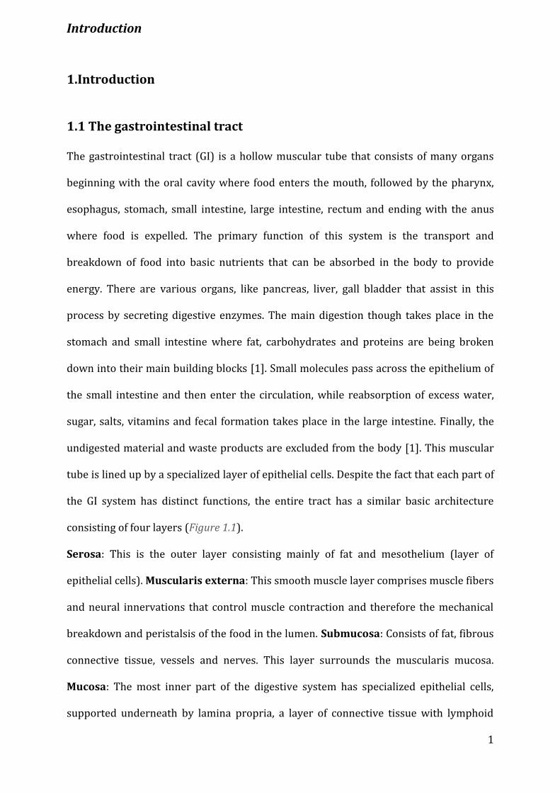
consisting of four layers (Figure 1.1).
Serosa: This is the outer layer consisting mainly of fat and mesothelium (layer of
epithelial cells). Muscularis externa: This smooth muscle layer comprises muscle fibers
and neural innervations that control muscle contraction and therefore the mechanical
breakdown and peristalsis of the food in the lumen. Submucosa: Consists of fat, fibrous
connective tissue, vessels and nerves. This layer surrounds the muscularis mucosa.
Mucosa: The most inner part of the digestive system has specialized epithelial cells,
supported underneath by lamina propria, a layer of connective tissue with lymphoid

Introduction
2
tissue, blood vessels, nerves and glands. Epithelium can be either stratified (flat)
squamous such as in the mouth or esophagus, standing the wear and tear caused by the
passing food, or columnar (tall)-like in the stomach and the intestine to help secretion and
absorption. Beneath lamina propria is the muscularis mucosa, a layer with smooth
muscles that contracts in order to change the shape of the lumen.
Figure 1.1: Picture depicting the main layers of the GI tract.
1.1.1 Morphology and function of the intestinal tract
The mouse and human intestines share great similarity concerning morphology,
physiology and development. The gut is anatomically divided into two parts, the small
intestine, which is further subdivided into duodenum, jejunum and ileum, and large
intestine/colon. The absorptive surface area of the small intestine is greatly enlarged by
finger-like protrusions that point into the lumen, otherwise known as villi. The opposite
end of the villi invaginates into the underlying connective tissue, and are referred to as
the crypts of Lieberkühn [2]. The crypts comprise the proliferative compartment, while
the villi in the small intestine and remainder of the crypt (2/3 of the crypt) in the large
intestine, is occupied by transit-amplifying (TA) progenitor cells, which are estimated to
Introduction
3
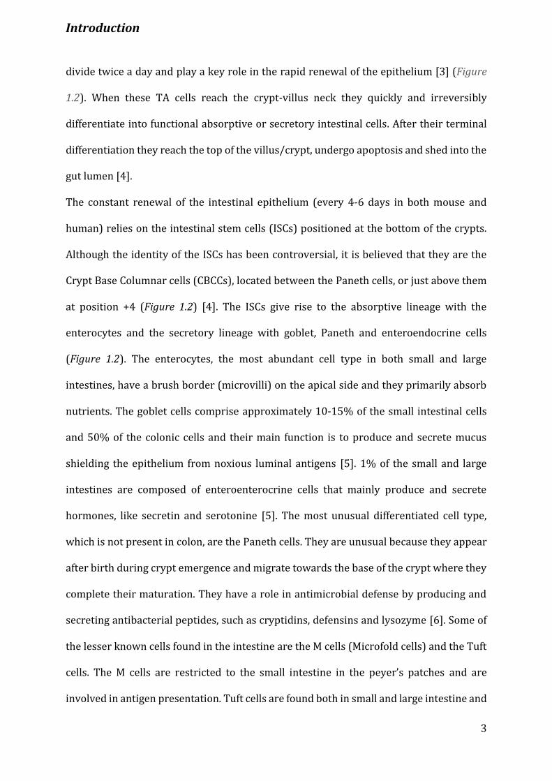
divide twice a day and play a key role in the rapid renewal of the epithelium [3] (Figure
1.2). When these TA cells reach the crypt-villus neck they quickly and irreversibly
differentiate into functional absorptive or secretory intestinal cells. After their terminal
differentiation they reach the top of the villus/crypt, undergo apoptosis and shed into the
gut lumen [4].
The constant renewal of the intestinal epithelium (every 4-6 days in both mouse and
human) relies on the intestinal stem cells (ISCs) positioned at the bottom of the crypts.
Although the identity of the ISCs has been controversial, it is believed that they are the
Crypt Base Columnar cells (CBCCs), located between the Paneth cells, or just above them
at position +4 (Figure 1.2) [4]. The ISCs give rise to the absorptive lineage with the
enterocytes and the secretory lineage with goblet, Paneth and enteroendocrine cells
(Figure 1.2). The enterocytes, the most abundant cell type in both small and large
intestines, have a brush border (microvilli) on the apical side and they primarily absorb
nutrients. The goblet cells comprise approximately 10-15% of the small intestinal cells
and 50% of the colonic cells and their main function is to produce and secrete mucus
shielding the epithelium from noxious luminal antigens [5]. 1% of the small and large
intestines are composed of enteroenterocrine cells that mainly produce and secrete
hormones, like secretin and serotonine [5]. The most unusual differentiated cell type,
which is not present in colon, are the Paneth cells. They are unusual because they appear
after birth during crypt emergence and migrate towards the base of the crypt where they
complete their maturation. They have a role in antimicrobial defense by producing and
secreting antibacterial peptides, such as cryptidins, defensins and lysozyme [6]. Some of
the lesser known cells found in the intestine are the M cells (Microfold cells) and the Tuft
cells. The M cells are restricted to the small intestine in the peyer’s patches and are
involved in antigen presentation. Tuft cells are found both in small and large intestine and
Introduction
4
are believed to be the fourth secretory lineage involved in chemical sensation of luminal
antigens [5].
Renewal system and crypt homeostasis are tightly controlled through signaling between
the supporting mesenchymal cells and the differentiated epithelial progeny. Wnt pathway
has a unique and central role in the (patho) physiology of the intestine, controlling
intestinal proliferation and stem cell maintenance, while Notch signaling at the crypt is
involved in cell fate determination between absorptive and secretory lineage [3, 5].
Figure 1.2: Architecture of the colonic crypt and the small intestinal crypt-villus. Both the colon
crypt and small intestine crypt-villus contain a stem cell compartment at the bottom of the crypt. The
crypt base columnar cells, located in between the Paneth cells and/or the +4 cells located above the
Paneth cells, have been suggested to be the ISCs. Paneth cells are not detected in the colon and they
migrate downwards, whereas the other differentiated progenitors migrate upwards [3]. ISC,
intestinal stem cells.
1.2 Inflammatory bowel diseases (IBDs)
The two major clinically defined forms of IBD manifesting as Crohn’s Disease (CD) and
Ulcerative Colitis (UC), are chronic relapsing inflammatory disorders of the
gastrointestinal tract, which are strongly associated with an increased risk of colon

Introduction
5
cancer. Crohn’s Disease can affect any part of the gastrointestinal tract, especially the
ileum and colon, potentially involving all layers of the tissue. Unlike CD, pathology in UC
is restricted mainly to the colon and rectum affecting the whole mucosa [7]. Although the
primary causative factor for IBD pathogenesis remains unknown, recent research
indicates that the intestinal microbial communities (microbiota), host’s genetic
susceptibility, exaggerated immune responses and the external environment are linked and
functionally integrated to the pathogenesis of IBDs [8].
1.2.1 Microbiota
Mammalian hosts have coevolved to exist with the gut microbiota in a mutual symbiotic
relationship where the host provides a suitable environment in return for benefits
provided to it by the gut microbiota. For instance, break-down of indigestible
carbohydrates to short chain fatty acids, synthesis of certain vitamins, degradation of
dietary oxalates and education of mucosal immune system [9]. The intestinal tract
comprises the largest reservoir of microorganisms (Bacteria, Archea, Eukarya and
Viruses) in the human body, where their density increases gradually from the proximal to
the distal parts of the tract [10]. Commonly identified commensal bacteria include the
phyla of Firmicutes (Lactobacillus, Clostridium, Enterococcus), Bacteroidetes
(Bacteroides), Actinobacteria (Bifidobacteria) and Proteobacteria (Escherichia coli and
Helicobacter) [10]. Dysbiosis of the gut microbiota, an alteration of the microbial
community structure associated with disease, has been observed in patients with IBD.
Many studies have examined the microbiota in UC and CD and found reduced biodiversity
in fecal microbiome and higher numbers of mucosa-associated bacteria in IBD patients
compared to healthy individuals [11]. For instance, in patients with CD or UC the number
of bacteria with anti-inflammatory properties such as, Bacteroidetes and Firmicutes, are
Introduction
6
reduced, while a greater abundance of Enterobacteria, mostly Escherichia coli, is observed
especially in the mucosa and not the fecal samples [11]. The breakthrough finding which
implicated the role of gut microflora on disease pathogenesis was that antibiotic
treatment or germ-free breeding of several mouse models of IBD including IL-10 deficient
or/and other transgenic mice resulted in less pronounced intestinal inflammation [12,
13].
1.2.2 Genetic susceptibility
Both forms of IBD arise in genetically susceptible individuals through interplay with
poorly comprehended environmental factors [7]. Although both diseases have
overlapping genetic factors, hereditability in CD is more important than in UC, as
monozygotic twins exhibit 50-75% phenotypic concordance in CD compared to only 10-
20% observed in UC [7]. Mutations in autophagy-associated genes (Atg16l1, Irgm) and
NOD-like receptors (Nod2) are quite specific for CD, whereas those in loci related to
regulatory pathways (Il10) and intestinal epithelial (IEC) function (Ecm1, Cadm2) appear
to be more specific for UC [7]. Polymorphisms in Atg16l11 and Nod2 genes interfere with
the secretion of antimicrobial peptides by Paneth cells [14, 15], while Xbp1 variants,
present in UC and CD patients, associate with endoplasmic reticulum (ER) stress and
affect Paneth cell function [16]. Therefore, patients with mutations in genes involved in
autophagy (an innate defence mechanism involved in the elimination of intracellular
pathogens), such as Atg16l1, Irgm or Nod2, in ER stress (Xbp1) or innate immunity (Nod2)
might have altered microbial composition due to defects in defensin secretion [7]. Hence,
the genetic variants that confer risk to IBD reflect the importance of autophagy, innate
immunity and ER stress in the pathogenesis of these disorders.
Introduction
7
1.2.3 Innate and adaptive immunity
Apart from the composition of intestinal microbiota and genetic susceptibility, IBD
pathogenesis has been strongly linked to dysfunctions of innate and adaptive pathways
that result in aberrant inflammatory responses in patients with CD or UC.
Innate immunity, the first line of defence against pathogens, is mediated by various cell
types and is initiated by the recognition of microbial antigens by pattern recognition
receptors (PRR) on the cell surface (Toll-like receptors (TLRs)) and in the cytoplasm
(NOD-like receptors (NLRs)) [17]. Accumulating evidence suggests that loss of PRR can
lead to changes of the microbial composition and intestinal barrier defects allowing
microbial invasion of systemic organs. Specifically, deficiency in TLR5, which recognizes
bacterial flagellin, leads to the translocation of bacteria to the spleen and liver followed
by spontaneous colitis and metabolic syndrome in mice [18, 19]. Furthermore, mice
lacking MyD88, a common adaptor for most of the TLR family members, have increased
numbers of commensal bacteria in the liver and spleen [20] and specific expression of
MyD88 in Paneth cells limits the microbial invasion in those tissues [21]. 30-40% of
patients with CD exhibit hypomorphic NOD2 (an intracellular NLR) variants [7] and
therefore decreased α-defensin expression [22]. In addition, autophagy is compromised
in CD. This can be seen in CD patients who are impaired in their ability to mediate bacterial
capture by autophagy due to polymorphisms in the autophagy-related gene Atg16l1 [23].
Patients with CD and UC display intestinal permeability, which could reflect mucosal
barrier defects that allow bacterial translocation through the intestinal mucosa. In healthy
intestines, goblet cells secrete mucin glycoproteins (mucus) that generate a two-layer
substructure extending up to 150 μm from the epithelium: the outer is loosely adherent,
suitable for bacterial growth, while the inner layer is more compact and sterile [24]. The
Introduction
8
importance of this mucinous deposit in maintaining the symbiotic relationship with the
microbiota is emphasized by the fact that mice deficient in mucin glycoprotein Muc2, the
most abundant constituent of the mucus layer, ultimately develop spontaneous chronic
colitis and colorectal cancer [25, 26]. Likewise, aberrant mucin assembly causes ER stress
and spontaneous inflammation that resembles UC in mice [27]. Defects in the mucus
composition can also influence the pattern of microbial colonization and maintenance of
microbial community and structure [11]. Furthermore, altered function of antimicrobial
peptides (including α/β-defensins, lysozyme, C-type lectins (RegIIIγ), cathelicidins and
lipocalins) are also involved in IBD [28]. For example, reduced expression of β-defensins
by enterocytes or α-defensins by Paneth cells is associated with colonic and ileal CD,
respectively [22, 29]. C-type lectin RegIIIγ, secreted by Paneth cells in response to
bacterial signals, was found to be a key component in sensing and restricting the bacterial
community near the mucosal surface of the small intestine [30], while enhanced secretion
of RegIIIγ into the intestinal lumen conferred resistance to L.monocytogenes infection via
MyD88 signaling emanating from IECs [31].
One of the defence mechanisms of the adaptive immune system in sequestering symbiotic
bacteria involves the secretion of IgA by B cells. IgA, which is transcytosed through the
epithelium [32], was shown to bind to bacteria and regulate their composition and access
to the mucosa [33].
Furthermore, it is believed that intestinal inflammation is triggered by imbalances in Th1
(Type 1 helper cells) and Th2 (Type 2 helper cells) cytokine responses that take place in
the lamina propria, mesenteric lymph nodes (MLN) and isolated lymphoid follicles (ILFs).
CD is mainly mediated by Th1 cells (via IFNγ and IL-12), whereas UC by Th2 cells (via IL-
4 and IL-13). Recently, another subset of effector Th cells, the IL-17-producing Th17 cells,
was described [34]. This cell type was shown to be induced by IL-6 and transforming

Introduction
9
growth factor β (TGFβ) and expanded by IL-23 [35, 36]. The elevated Th1 and Th17 cells
in the lamina propria of patients with CD suggested that both IL-12/IFNγ-producing and
Th17-associated-IL-23/IL-17 pathways might be involved in disease pathogenesis [37].
However, blockade or genetic ablation of IL-23 demonstrated that IL-23, and not IL-12, is
essential in promoting chronic intestinal inflammation via IL-17 [38, 39]. Additionally,
antibodies against IL-12p40 (the common p40 subunit of IL-12 and IL-23) induced clinical
responses and remissions in patients with active CD [40], demonstrating the prominent
role of IL-23/IL-17 in disease development. Contrary to the pathogenic role of Th17,
regulatory T cells (Treg) characterized by the expression of the transcription factor FOXP3
and secretion of regulatory cytokines such as TGFβ and IL-10, have been reported to have
immunosuppressive functions during intestinal inflammation. This is evident by the fact
that both IL-10 [41] and TGFβ [42] deficient mice develop spontaneous intestinal
inflammation and co-transfer of Treg in mouse models of T-cell mediated chronic intestinal
inflammation rescues the severe pathology [35].
Thus, IBD is a multifactorial complex disorder whose pathogenesis is influenced by the
genetic make-up of the individual, the diversity of the intestinal microbiome and the
interplay of both the adaptive and innate immune system. This array of factors might be
also influenced by the extrinsic environment, such as diet, drugs, social stress, geography,
smoking and psychological elements which are also considered as prominent risk factors
for IBD [8].
1.3 Nuclear Factor-κB (NF-κB)
One of the most important and devastating consequence of long-term and unresolved
inflammation in IBD is the development of colorectal cancer and one of the best studied
transcription factors involved in both processes is NF-κB. NF-κB plays vital role in many
Introduction
10
biological processes, including development, immune responses, cell proliferation and
survival, whereas deregulated NF-κB signaling has been linked to many human diseases,
including chronic inflammatory disorders and cancer [43, 44].
In mammals the NF-κB family consists of five related transcription factors, p65 (RelA),
RelB, c-Rel, p52 and p50 where they bind as, either hetero - or –homodimers, to κB sites
in the target genes and can either activate or suppress gene expression [45]. A hallmark
of the NF-κB pathway is its regulation by the IκB proteins, which function as inhibitors for
the NF-κB dimers. There are various human IκB proteins such as, IκBα, IκBβ, IκBε, IκBζ,
BCL-3, IκBns. In addition the p105 and p100, the respective precursors of the processed
p50 and p52 can also function as IκB inhibitors [45]. The prototypical NF-κB complex
under steady-state conditions is the p50-p65-IκBα trimer. In the canonical or classic
pathway IκBα is phosphorylated by the IκBα kinase (IKK) complex, which consists of two
catalytically active kinases IKKα/1 and IKKβ/2 and a regulatory scaffold protein NEMO
(NF-κB essential modulator)/IKKγ. Phosphorylation of IκBα at serines 32 and 36 targets
it for ubiquitin-dependent proteasomal degradation thereby liberating active NF-κB
dimers to enter the nucleus and activate the transcription of responsive genes. Target
elements of the canonical pathway include genes encoding inflammatory mediators,
chemokines, cytokines, inhibitors of apoptosis, proteases and adhesion molecules [43].
The alternative or non-canonical NF-κB pathway, on the other hand, is governed
exclusively by the IKKα kinase. Activation of IKKα by the upstream NF-κB inducing kinase
(NIK) results in the phosphorylation and processing of p100 generating the active
p52/RelB heterodimers (Figure 1.3). Active p52/RelB dimers translocate to the nucleus
where they turn on the transcription of genes involved in lymphoid organogenesis, B cell
survival and maturation as well as dendritic cell (DC) activation [46].

Introduction
11
Figure 1.3: Canonical and alternative NF-κB activation. IKK complex and primarily IKKβ mediate
activation of the canonical pathway through phosphorylation and proteasomal degradation of the
IκB inhibitors in the cytoplasm. On the other hand, IKKα dimers control exclusively the alternative
pathway by phosphorylating and targeting p100 for proteasomal degradation [47].
1.3.1 NF-κB signaling in IBD
NF-κB, the master transcriptional regulator of inflammation, governs the expression of
various cytokines involved in IBDs, such as Tumor necrosis factor α (TNFα), IL-6 and IL-
1β. Accordingly, increased NF-κB-dependent expression of TNFα, IL-6 and IL-1β in IECs
and macrophages of inflamed mucosa of patients with CD and UC has been reported more
than a decade ago [48]. Moreover, rectal or oral administration of p65 antisense
oligonucleotides decreased severity of experimental colitis or colitis observed in Il10
deficient mice [49]. Additionally, deletion of Ikkβ in myeloid cells ameliorated chronic
colitis observed in Il10 knockout mice [50], suggesting that targeting NF-κB could be a
possible strategy in IBD therapy. However, defective NF-κB activation in the intestinal
epithelium, as a result of specific ablation of NEMO/Ikkγ [51] or RelA/p65 [52] causes
spontaneous inflammation and sensitizes the epithelium to radiation-induced apoptosis
[53]. Also, loss of IKKβ-dependent NF-κB activation in IECs results in exacerbated DSS-

Introduction
12
induced acute colitis due to increased apoptosis and reduced recruitment of inflammatory
cells that secreted cytoprotective factors [50, 54]. In addition, epithelial IKKβ can control
adaptive immune functions in the lamina propria via distal effects on DCs. More specific,
mice lacking Ikkβ in the intestinal epithelial cells are characterized by diminished
expression of thymic stromal lymphopoietin in the gut and fail to develop specific CD4+
Th2 responses after infection with the parasite Trichuris muris [55]. Instead, mucosal DC
secrete elevated TNFα and IL-12/23p40 levels and CD4+ cells produce increased amount
of IL-17 and IFNγ resulting in severe intestinal inflammation [55].
Thus, inflammatory factors such as NF-κB, can accelerate intestinal epithelial cell
proliferation and apoptosis, nevertheless under conditions of impaired replacement of
apoptotic enterocytes a breach in the intestinal barrier will allow bacteria translocation
that will eventually cause a strong local inflammatory response. It seems that on one hand
NF-κB promotes survival of cells by regulating anti-apoptotic genes and on the other hand
regulates the expression of many cytokines and other modulators of the inflammatory
processes in IBD. Therefore, it is an absolute requirement that NF-κB signaling and the
genes induced by this signaling cascade are under tight regulation.
1.3.2 IKKα: a multifunction protein kinase
Despite the fact that IKKα and IKKβ share structural and biochemical similarities the
different phenotypes observed in the respective knockouts suggests that they regulate
distinct targets and they have non-redundant functions. For instance, Ikkβ deficient mice
die embryonically due to massive liver apoptosis [56], whereas Ikkα knockout mice die
soon after birth due to limb and skin abnormalities [57-59]. Interestingly, Ikkα knockout
mice exhibit normal NF-κB activation demonstrating for the first time IKKβ/γ/NF-κB-
independent functions [59]. Another specific function found for IKKα is its involvement
Introduction
13
in the alternative NF-κB pathway by phosphorylating the NF-κB2/p100 and generating
the active p52 member [60]. The authors demonstrated that the IKKα kinase activity is
required for the maturation and activation of B cells as well as for the development of
secondary lymphoid organs (Peyer’s patches and germinal centers) [60]. Cao et.al.
presented that IKKα is essential for mammary gland development, and adult female mice
bearing an inactive IKKα kinase mutant are deficient in milk production owing to
impaired cyclin D1 activation [61]. With this study the authors showed that IKKα activity
is required for NF-κB activation in mammary epithelial cells in response to specific
signals, such as RANK-L, whereas TNFα-induced NF-κB activation remains intact [61].
1.3.2.1 The anti-inflammatory role of IKKα
IKKα has been suggested as a negative regulator of inflammatory immune responses. It
was demonstrated that though IKKα kinase-dead (IkkαAA/AA) mutant mice when
challenged with bacteria, show better bacterial clearance, they are more prone to septic
shock due to elevated inflammatory response [62]. The authors presented that IKKα is
important in limiting macrophage activation by preventing prolonged promoter binding
of RelA/p65 and c-Rel [62]. Similarly, embryonic liver-derived macrophages deficient in
Ikkα display elevated production of proinflammatory cytokines and chemokines
associated with increased NF-κB activity [63]. Moreover, the role of this kinase in turning
off inflammatory pathways was demonstrated by its ability to phosphorylate TAX1BP1
protein in response to TNF and IL-1 stimuli and thus terminating NF-κB signaling [64].
Remarkably, the suggested IKKα-mediated anti-inflammatory effect is not restricted to
NF-κB. The alternative mechanism by which IKKα acts to limit inflammation is through
phosphorylation and thereby activation of the protein inhibitor of STAT1 (PIAS1). In
response to different proinflammatory stimuli, such as TNFα or LPS, IKKα-induced PIAS1
Introduction
14
phosphorylation is required for the transcriptional repression of NF-κB/STAT1
dependent genes, with a notable preference for proinflammatory cytokines and
chemokines, such as Mip2, Tnfα, Irf1 and Iκbα [65]. A recent study supported the fact that
deletion of IKKα in pancreas associates with increased acinar cell death, fibrosis,
inflammation and release of pancreatic enzymes, clinical signs resembling those of human
chronic pancreatitis [66]. The pathology depended on the protein scaffold p62, as deletion
of p62 attenuated pancreatitis [66].
IKKα has acquired a unique role in bridging innate and adaptive immunity. It was
demonstrated that IKKα is essential for the induction of genes (Blc, sdf-1, Elc and Slc)
encoding chemokines crucial for spleen organogenesis and maintenance of tissue
architecture [67]. LTβR engagement triggers IKKα-dependent RelB:p52 nuclear
translocation and selective recruitment of those dimers to the promoters of genes whose
consensus sequence was distinct from that of the classical κB sites. Thus, the authors
proposed that IKKα optimizes adaptive immunity through proper organization of
secondary lymphoid organs by regulating the production of organogenic chemokines,
such as Blc and Elc [67]. Additionally, IKKα kinase activity is required to limit
inflammation while promoting acquired antigen-specific immunity [68]. IKKα kinase-
dead mutant mice challenged with the human pathogen Listeria monocytogenes display
efficient pathogen clearance from the spleen and liver in the acute phase but they are
highly susceptible upon secondary infection. This is attributed to the impaired Th1 cell
priming and acquired immunity dependent on the development of protective CD8+
memory T cells [68]. In vitro assays demonstrated that IKKα activation in DCs is required
for T cell priming, whereas the kinase dead mutants (IkkαAA/ΑΑ mice) are unable to prime
naïve CD4+ T cells to produce IFNγ [68].
Introduction
15
Thus, this protein kinase has the uncommon property of coupling innate and adaptive
immunity. It inhibits innate nonspecific immunity and at the same time enhances antigen-
acquired immunity making it an attractive molecule to study in inflammatory conditions.
IKKα and in general NF-κB signaling have been considered essential regulators in many
inflammatory conditions, including IBDs. In addition to NF-κB, three interacting pathways
have recently gained more attention due to their implication in IBD. These pathways,
which associate with unfolded protein response (UPR), autophagy and intracellular
bacteria sensing will be elaborated in the next sections.
1.4 The key players in unfolded protein response (UPR)
An important pathway that has emerged in IBD pathophysiology is the unfolded protein
response (UPR), which is induced by endoplasmic reticulum (ER) stress.
The ER in all eukaryotic cells provides a unique environment essential for proper protein
folding and post-translational modifications, storage of free calcium and energy and
synthesis of lipid and sterols [69]. Since protein folding is a key process for protein
function, all cells have evolved sophisticated mechanisms that authorise only properly
folded proteins to exit the ER on their way either to the cell surface or other intracellular
organelles. Misfolded proteins are retained in the ER lumen bound to molecular
chaperones or directed for degradation through the proteasome, a process called ER-
associated degradation (ERAD) or through autophagy [69]. Primary-genetic factors
(mutated proteins) or secondary-environmental factors (glucose or calcium deprivation)
or decreased proteasomal or autophagy function can interfere with the capacity of the ER
to fold proteins properly, thus causing ER stress [69]. To sense and respond to ER stress
eukaryotic cells activate the UPR signaling cascade which either protects the cell or
alternatively promotes cell death. The UPR cascade consists of a group of transmembrane
Introduction
16
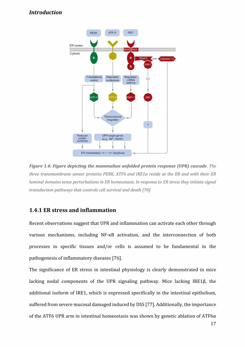
ER-resident proteins including inositol-requiring protein 1 (IRE1α), PKR-like
endoplasmic reticulum kinase (PERK) and activating transcription factor (ATF)-6 (Figure
1.4) [70]. This cascade is further regulated by glucose-regulated protein 78 (GRP78), an
ER-associated chaperone that binds to all three ER-transmembrane proteins holding
them in an inactive state. Binding of misfolded or unfolded proteins to GRP78 releases
each of these factors resulting in the characteristic UPR-related behaviours.
The most immediate response, following the release of BiP from PERK, is the
homodimerization and trans-phosphorylation of PERK, allowing it to phosphorylate the
eukaryotic translation initiator factor 2α (eIF2α). Phosphorylation of eIF2α inhibits the
assembly of 80S ribosome and consequently leads to attenuation of mRNA translation.
ATF6 upon migration to the Golgi becomes transcriptionally active by undergoing
proteolytic cleavage of its cytosolic tail by site 1 and 2 proteases. Finally, IRE1α, which
governs the most conserved UPR pathway, possesses both kinase and specific
endoribonuclease (RNase) activity. When ER stress occurs IRE1α undergoes
homodimerization and trans-autophosphorylation turning on the c-Jun N-terminal kinase
(JNK) and NF-κB pathways, mediated by its physical and functional interaction with
tumor necrosis factor-receptor associated factor 2 (TRAF2) [71, 72]. TRAF2 is essential
in regulating ER-induced cell death and its absence reduces NF-κB activation and
sensitizes cells to death [73, 74]. Autophosphorylation of IRE1α activates its RNase
activity to initiate nonconventional splicing of the Xbp1 (X-box binding protein 1) mRNA,
its only known target so far [75]. Only XBP1 protein translated from spliced Xbp1 mRNA
(Xbp1s) is transcriptionally active because it contains a C-terminus trans-activating
domain, enabling the transcription of target genes encoding proteins that either degrade
misfolded proteins or enhance ER-protein folding capacity [75] (Figure 1.4).
Introduction
17
Figure 1.4: Figure depicting the mammalian unfolded protein response (UPR) cascade. The
three transmembrane sensor proteins PERK, ATF6 and IRE1α reside at the ER and with their ER
luminal domains sense perturbations in ER homeostasis. In response to ER stress they initiate signal
transduction pathways that controls cell survival and death [70].
1.4.1 ER stress and inflammation
Recent observations suggest that UPR and inflammation can activate each other through
various mechanisms, including NF-κB activation, and the interconnection of both
processes in specific tissues and/or cells is assumed to be fundamental in the
pathogenesis of inflammatory diseases [76].
The significance of ER stress in intestinal physiology is clearly demonstrated in mice
lacking nodal components of the UPR signaling pathway. Mice lacking IRE1β, the
additional isoform of IRE1, which is expressed specifically in the intestinal epithelium,
suffered from severe mucosal damaged induced by DSS [77]. Additionally, the importance
of the ATF6 UPR arm in intestinal homeostasis was shown by genetic ablation of ATF6α
Introduction
18
and P58IPK ER chaperone, which exacerbated symptoms of DSS-induced colitis due to
hyperactivated proapoptotic (IRE1α/JNK) UPR signaling [78]. Recently, Heijmans and
colleagues unraveled that ER stress is low in intestinal stem cells compared to transit
amplifying cells and ER stress, induced by GPR78 deletion, causes loss of stem cell
markers (Lgr5, Olfm4 and Ascl2) in a PERK-eIF2α-dependent manner [79]. It was thus
suggested, by means of active PERK-eIF2α signaling, that under homeostatic conditions
ER stress is both sufficient and critical for stem cell differentiation [79].
Cells employed to secrete high amounts of proteins (plasma cells, hepatocytes, pancreatic
acinar cells, plasmacytoid dendritic cells), either for their basal or their induced functions,
dependent on a normal UPR and as such are more prone to environmental and/or genetic
factors that induce ER stress [80]. Consistent with this, IECs, especially Paneth and goblet
cells, rely on an intact UPR for their normal function. The association of XBP1 as a genetic
risk factor for IBD originated from a mouse model with partial or complete genetic
deletion of Xbp1 specifically in the intestinal epithelium [16]. These mice develope
spontaneous inflammation in the small intestine and increased susceptibility to DSS-
induced acute colitis. Deep sequencing in more than 1000 patients with IBD identified
rare non-synonymous SNPs (leading to amino-acid exchange) in the Xbp1 gene that
exhibited hypomorphic UPR induction, consistent with the mouse model [16]. Finally,
mice with mutations in Muc2, develop accumulation of abnormal MUC2 protein in goblet
cells, spontaneous ulcerative colitis-like phenotype in association with severe ER stress
[27].
These studies indicate the reciprocal interactions between the UPR and innate immunity
and suggest that ER stress affects the survival of secretory epithelial cell types, such as
Paneth and goblet cells, the composition of intestinal microbiota and susceptibility to IBD.
Introduction
19
1.5 Autophagy
Closely intertwined with ER stress and highly implicated in IBD pathogenesis is
autophagy. Implication of autophagy in IBD originated from polymorphisms identified in
the autophagy-related 16-like protein 1 (ATG16L1) [81] and immunity related-GTPase
family M (IRGM) [82] that predispose individuals to CD.
Autophagy (originating from the Greek word for ‘self-eating’) refers to the cellular
degradation pathway involving the delivery of cytoplasmic cargo to the lysosomes. Basal
levels of autophagy occurs in all cells as a homeostatic function in the process of
macromolecule and organelle turnover [83]. It is substantially elevated in response to
starvation (nonselective process), when cells need to produce intracellular nutrients and
energy, due to oxidative stress. It is also elevated when cells need to remove damaged
organelles (selective process), for instance during infection, or accumulation of misfolded
proteins and protein aggregates [84]. Therefore, is not surprising that autophagy is
involved in various essential processes, such as embryogenesis, tissue remodelling and
immune function (elimination of microbes, antigen presentation via MHC class II etc.) as
well as in many disease processes including cancer, neurodegeneration and inflammation
[84]. This catabolic process occurs through distinct steps with the aid of autophagy-
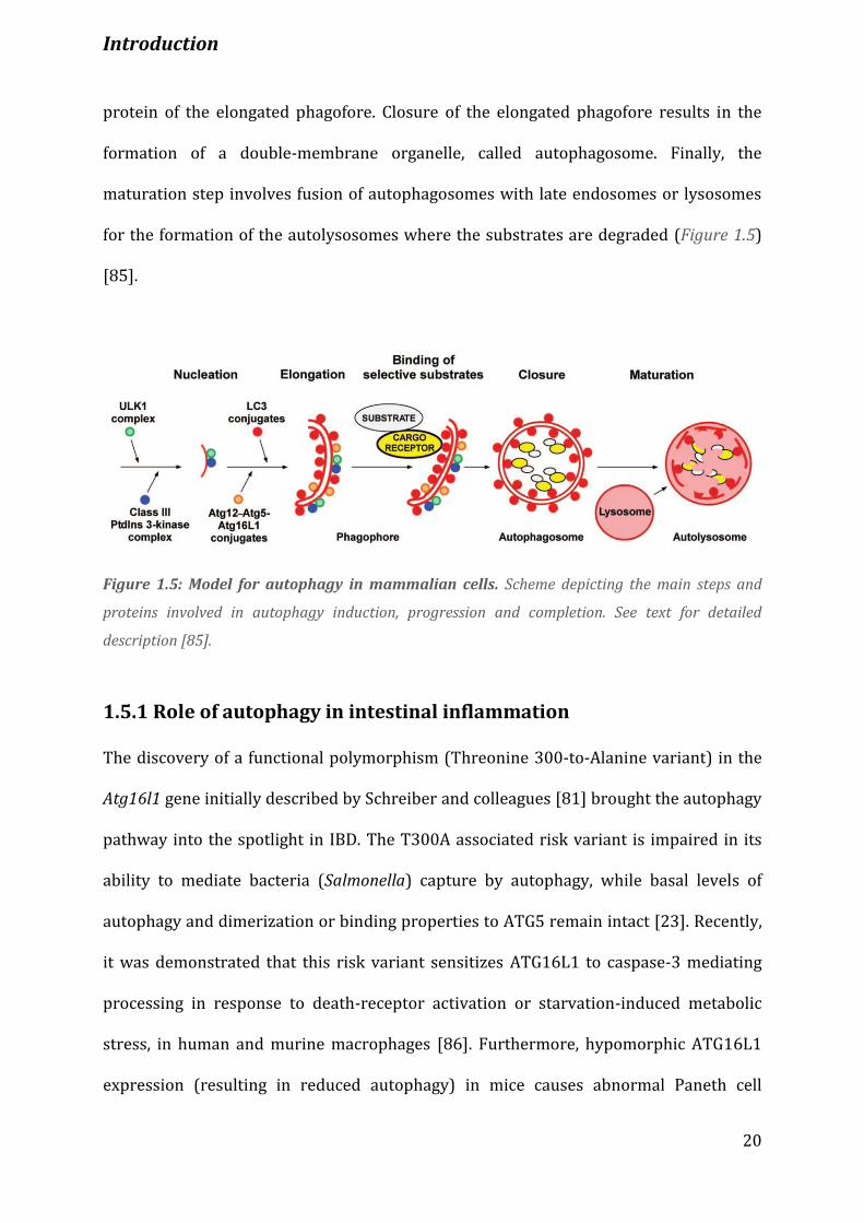
related proteins (ATG) as depicted in Figure 1.5. Autophagy is initiated by the formation
of the phagofore or isolation membrane, which in mammalian cells is regulated by the
serine/threonine protein kinase, ULK1, and the class III phosphatidylinositol (PtdIns)-3
complex. Additional ATG proteins are required for the elongation step, the growth of the
phagofore, such as ATG12-ATG5-ATG16L1 forming complex, ATG7, ATG10, ATG12 and
the recruitment of ATG8 (LC3-I) to the phagofore through a membrane anchoring,
phosphatidylethanolamine modification of ATG8 (LC3-II). Selectivity of autophagy is
achieved by cargo receptors that bind to both substrates and lipidated ATG8 (LC3II)
Introduction
20
protein of the elongated phagofore. Closure of the elongated phagofore results in the
formation of a double-membrane organelle, called autophagosome. Finally, the
maturation step involves fusion of autophagosomes with late endosomes or lysosomes
for the formation of the autolysosomes where the substrates are degraded (Figure 1.5)
[85].
Figure 1.5: Model for autophagy in mammalian cells. Scheme depicting the main steps and
proteins involved in autophagy induction, progression and completion. See text for detailed
description [85].
1.5.1 Role of autophagy in intestinal inflammation
The discovery of a functional polymorphism (Threonine 300-to-Alanine variant) in the
Atg16l1 gene initially described by Schreiber and colleagues [81] brought the autophagy
pathway into the spotlight in IBD. The T300A associated risk variant is impaired in its
ability to mediate bacteria (Salmonella) capture by autophagy, while basal levels of
autophagy and dimerization or binding properties to ATG5 remain intact [23]. Recently,
it was demonstrated that this risk variant sensitizes ATG16L1 to caspase-3 mediating
processing in response to death-receptor activation or starvation-induced metabolic
stress, in human and murine macrophages [86]. Furthermore, hypomorphic ATG16L1
expression (resulting in reduced autophagy) in mice causes abnormal Paneth cell
Introduction
21
exocytosis [14]. Strikingly, patients homozygous for the Atg16l1 CD risk allele exhibit
comparable Paneth cell abnormalities and a similar distortion was also noted in mice
lacking ATG5 in the IECs [14]. In addition, mice expressing hypomorphic ATG16L1 do not
develop spontaneous intestinal inflammation but they are more prone to DSS-induced
colitis ascribed to overactivation of the NLRP3 inflammasome [87]. These studies suggest
a concept that a specific genotype is transformed into a clinically relevant phenotype only
when a set of environmental factors are present.
Polymorphisms found in NOD2-encoding Card15 gene were the first decisive risk factors
identified for Crohn’s disease [88]. Defects in NOD2 signaling were reported to lead to CD
pathology through inefficient induction of autophagy and handling of enteric bacteria [89,
90]. Interestingly, CD-associated NOD2 variants are impaired in their ability to induce
autophagy and to recruit ATG16L1 to the bacteria entry sites at the plasma membrane,
while their interaction remains intact. The authors suggested a potential mechanism
wherein mutant NOD2 impairs autophagy by retaining ATG16L1 to the cytosol
suppressing its function [90].
Collectively, autophagy and intracellular sensing of pathogens seems to be part of a major
shared mechanism in the development of CD. This is supported by the fact that
polymorphisms in Nod2 receptor (sensing intracellular bacteria) account for the vast
majority of genetic heritability of CD and mutations in the Atg16l1 gene are one of the
most prevalent risk factors for CD.
1.6 p62: a multimodule scaffold protein
p62 (also known as sequestosome 1, SQSTM1) is a multimodule adaptor protein
implicated in selective signal transduction and degradation of proteins and organelles.
The recent generation of p62 knockout mice revealed the role that p62 holds in a number
Introduction
22
of cellular functions including, bone remodelling, autophagy, obesity, aging, inflammation
and cancer [91]. The structure of p62 shows a high potential for interaction with many
proteins consistent with its role as a signaling hub. The main interacting domains are, the
PB1 domain (important for oligomerization and interaction with other PB1-containing
proteins), the ZZ domain (for interaction with the RIP1 protein kinase), the TB domain
(for association with TRAF6), the LIR domain (important for binding to the LC3 protein)
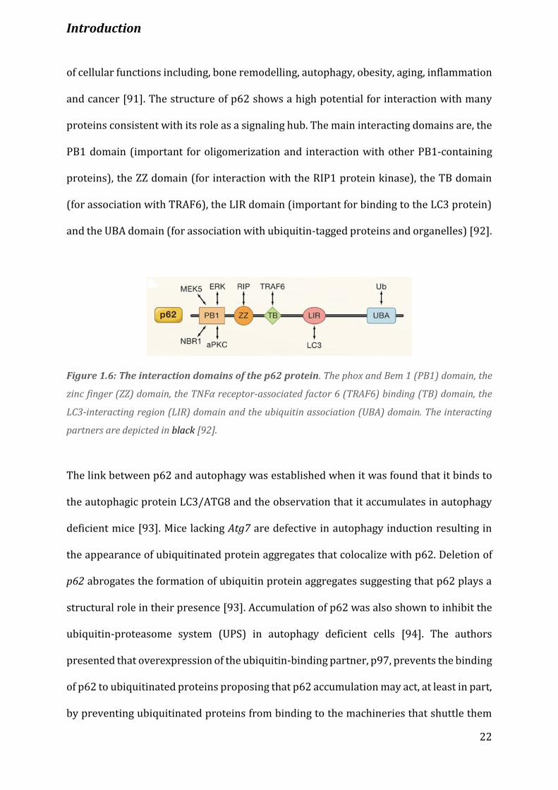
and the UBA domain (for association with ubiquitin-tagged proteins and organelles) [92].
Figure 1.6: The interaction domains of the p62 protein. The phox and Bem 1 (PB1) domain, the
zinc finger (ZZ) domain, the TNFα receptor-associated factor 6 (TRAF6) binding (TB) domain, the
LC3-interacting region (LIR) domain and the ubiquitin association (UBA) domain. The interacting
partners are depicted in black [92].
The link between p62 and autophagy was established when it was found that it binds to
the autophagic protein LC3/ATG8 and the observation that it accumulates in autophagy
deficient mice [93]. Mice lacking Atg7 are defective in autophagy induction resulting in
the appearance of ubiquitinated protein aggregates that colocalize with p62. Deletion of
p62 abrogates the formation of ubiquitin protein aggregates suggesting that p62 plays a
structural role in their presence [93]. Accumulation of p62 was also shown to inhibit the
ubiquitin-proteasome system (UPS) in autophagy deficient cells [94]. The authors
presented that overexpression of the ubiquitin-binding partner, p97, prevents the binding
of p62 to ubiquitinated proteins proposing that p62 accumulation may act, at least in part,
by preventing ubiquitinated proteins from binding to the machineries that shuttle them
Introduction
23
to or into the proteasome [94]. Although p62 is primarily degraded by the autolysosome,
a recent study showed that it can also be degraded by the proteasome [95]. The authors
demonstrated that cells deficient in the autophagy protein ATG16L1 have enhanced IL-1β
signaling due to p62 accumulation and knockdown or overexpression of p62 suppresses
or enhances IL-1β signaling, respectively [95].
Apart from being an autophagy target, p62 can determine cell survival or apoptosis. In
response to cell stimulation by IL-1 [96], RANK ligand [97] or nerve growth factor (NGF)
[98] p62 was shown to interact with the atypical protein kinase C (aPKC) through the PB1
domain and to induce NF-κB activation. Interestingly, in response to those stimuli p62
associates with TRAF6, resulting in NF-κB activation [96, 99]. On the other hand, p62 can
promote apoptosis through the extrinsic pathway by interacting with caspase-8 [100].
Recently, a link between p62 accumulation and inflammasome activation was described.
Human macrophages infected with Mycobacterium abscessus (Mabc) display elevated p62
levels, which result in NLRP3/ASC inflammasome-dependent caspase-1 activation and IL-
1β release [101]. Additionally, murine or human macrophages stimulated with
monosodium urate (MSU) crystals exhibit impaired proteasome function that causes
accumulation of p62 and caspase-1-dependent IL-1β production [102].
Altogether, p62 is a multidomain scaffold protein that not only interacts with the
autophagy machinery targeting proteins for degradation but is also implicated in many
signaling pathways determining cell fate.
1.7 Microbial recognition by the innate immune system
The presence of microbiota is beneficial for the host immune maturation as it can be
viewed that germ-free mammals lack a fully mature immune system and are unable to
secrete mucus, antimicrobial peptides and circulating antibodies [28]. The complex
Introduction
24
interaction between the intestinal epithelium and commensal bacteria is mediated by
innate immune receptors, called pattern recognition receptors (PRRs). They are
expressed constitutively by many cells at the front line of defence against infection, such
as macrophages, DCs, neutrophils, monocytes, epithelial cells, B and T cells. PRRs include
Toll-like receptors (TLRs) and C-type lectin receptors (CTLs) which scan the extracellular
milieu and endosomal compartments for microbial components known as pathogen
associated molecular patterns (PAMPs). Intracellular PRRs include the nucleic acid
sensors, such as the RNA-sensing RIG-I-like receptors (RIGs) and RIG-like helicases
(RLHs) and DNA sensors, like AIM2 [17]. According to the PAMP engagement and nature
of the responding cell, PRRs can trigger different signaling pathways leading to diverse
immune responses. Their signaling cascades converge however, on common
inflammatory pathways including NF-κB, caspase-1 and mitogen-activated protein kinase
(MAPK) signaling molecules [103]. Another set of PRRs, different from those mentioned
above, are the nucleotide and oligomerization domain (NOD)-like receptors (NLRs),
which are capable of recognizing PAMPs, such as lipopolysaccharide, peptidoglycan or
host-derived endogenous damage-associated molecular patterns (DAMPs), released
during cellular or tissue injury [103].
The NLR family is characterized by a central NOD (referred to as NACHT or NBD) domain,
flanked by a C-terminal leucine-rich repeats (LRR) and an N-terminal caspase recruitment
domain (CARD) or pyrin domain (PYD) [103]. The CARD/PYD domains mediate protein-
protein interactions, whereas LRRs determine ligand specificity. NOD/NACHT, which is
essential for activation via ATP-dependent oligomerization, is the only domain common
to all NLRs. Based on similarities in the NOD domain the NLR family is divided into 3
distinct subfamilies: the NOD (NOD1-2, NOD3/NLRC3, NOD4/NLRC5, NOD5/NLRX1), the
NLRP (NLRP1-14 also called NALPs) and the IPAF (NLRC4/IPAF and NAIP) subfamily
Introduction
25
[104]. NLRPs, for instance, contain PYD, NOD and LRR domains, with the exception of
NLPR10 that lacks LRRs, while NOD1-2 consist of NOD, LRR and CARD instead of PYD
domains [104]. Although they are primarily expressed in immune cells, some NLRs are
also expressed in non-immune cells and activation of each NLR member is induced by
specific microbial or endogenous components. For example, NOD1 and NOD2 recognize
products of bacterial cell wall (mesodiaminopimelic and muralmyl dipeptide (MDP),
respectively) and, upon ligand sensing, they oligomerize, and through CARD-CARD
interactions recruit RIP2 to the complex. Assembly of NOD1-2 signalosome results in NF-
κB activation and proinflammatory gene regulation [104]. Contrary to NOD1 and NOD2,
which are well studied, the rest of NODs and most of the NLR family members are poorly
characterized. However, the diverse functions of NLR members that regulate caspase-1
activity in anti-microbial responses, as well as in multifactorial diseases such as IBDs, have
started to be revealed.
1.7.1 Inflammasomes
Inflammasomes are a group of proteins that assemble huge complexes upon recognition
of a diverse set of inflammation-inducing stimuli, including PAMPs and DAMPs, and
control the production of important proinflammatory cytokines, such as IL-1β and IL-18
[104]. These multi-protein complexes include a sensor protein (NLR family), an adaptor
protein (the apoptosis-associated speck-like protein (ASC) containing a CARD domain)
and an inflammatory caspase. Activation of the NLR by exogenous or endogenous signals
results in direct recruitment of pro-caspase-1 through CARD-CARD interaction or
indirectly through PYD domain by means of the adaptor protein ASC, which contains both
a CARD and a PYD domain [104]. Activation and assembly of the inflammasome requires
simultaneously expression of all ‘players’ within the same cell type. Caspase-1 and ASC

Introduction
26
are usually expressed in many cell types, while the sensor proteins have a more restrictive
pattern of expression suggesting cell type-specific mechanisms of sensing tissue
perturbation [105]. Many inflammasomes within different cell types, (e.g. epithelial,
hematopoietic) display different but often complementary functions during mucosal
immune responses. This is more apparent in the intestinal tract where many cell types
have to come in contact with the tremendous load of commensal and pathogenic bacteria.
A tight regulation of inflammasome signaling is critical to maintain a beneficial level of
homeostatic interactions with the gut microflora and inflammasome hyper-activation or
absence could be deleterious leading to inflammation and cancer.
1.7.2 Role of the inflammasomes in intestinal homeostasis
It is becoming more apparent that inflammasomes play a crucial role in maintaining
intestinal homeostasis and healthy intestinal microflora. NLRP3 is the most widely
studied inflammasome which regulates immune responses to microbial infection and
auto-inflammatory diseases. NLRP3 is expressed in both hematopoietic and non-
hematopoietic cells and requires two signals for its full activation. The priming signal,
provided by microbial molecules and cytokines, activates the NF-κB or NOD1/2 receptors
resulting in pro-Il1β and Nlrp3 transcriptional induction. The activation signals,
potassium efflux, ROS generation, microbial-pore forming toxins or lysosomal membrane
damage, trigger inflammasome assembly, caspase-1 activation and subsequent cleavage
of pro-IL-1β and pro-IL-18 into their active cytokines [103]. Much attention has been paid
to NLPR3 owing to the possible genetic association of the Nlpr3 locus with IBDs [106].
There are multiple reports on how NLRP3 inflammasome confers protection to the
intestinal epithelium upon DSS-induced injury [107-110]. Similarly, Asc and Caspase-1/-
11 knockout mice also display high susceptibility to DSS-induced injury. Furthermore,
Introduction
27
bone marrow transplantation experiments suggested that NLRP3 expression in the non-
hematopoietic compartment, most likely the epithelium, is required for protection.
Furthermore, Asc and Caspase-1 knockout mice exhibit reduced IL-18 production by the
IECs and administration of recombinant IL-18 protects the knockout mice against colitis
(Figure 1.7) [107]. NLPR3 was shown to protect the intestinal epithelium from DSS-
induced injury by regulating the production of antimicrobial peptides [110]. Nlrp3
deficient mice exhibit impaired β-defensin production associated with more pathogenic
microflora distinct from that of wild-type mice [110]. Additionally, the increased
inflammation observed in Nlrp3 deficient mice results in increased tumor burden in colitis
associated cancer that correlates with decreased levels of IL-18 and IL-1β (Figure 1.7)
[108, 109]. However, despite the proposed role of NLPR3 in the negative regulation of
inflammation and inflammation-induced tumorigenesis there is one study showing that
Nlrp3-null mice are protected against DSS-induced colitis. The protection is related to the
decreased IL-1β secretion from Nlrp3 deficient macrophages, as inhibition of caspase-1
with pranaclasan,a well known caspase-1 inhibitor, results in attenuated colitis [111].
Another group also demonstrated reduced colitis pattern in Nlrp3 knockout mice [112].
The dissimilar phenotypes of Nlrp3 deficient mice could be attributed not only to
differences in the genetic background of mice or protocols used but most importantly to
differences in the gut microbiota.
Recently another inflammasome, NLRP6, was shown to be highly expressed in intestinal
epithelial cells and hematopoietic cells regulating colonic homeostasis [103]. In many
studies Nlrp6 deficient mice were shown to suffer from severe inflammation induced by
DSS. One of these studies demonstrated that the development of acute colitis and
increased AOM/DSS-induced tumorigenesis in Nlrp6 knockout mice is due to the deficient
function of the NLRP6 inflammasome in the hematopoietic cells [113]. A novel role of the
Introduction
28
NLPR6 inflammasome as a promising regulator of colonic microbial ecology was recently
proposed. Elinav et.al revealed that the Nlrp6 knockout mice are characterized by
spontaneous intestinal hyperplasia and develop severe colitis induced by DSS.
Interestingly, the microbiota of the knockout mice is distinct from that of the wild-type
and more importantly transferrable (colitogenic) to the co-housed wild-type mice. This
phenomenon is also observed in Asc and Caspase-1/-11 knockout mice but does not occur
in those deficient in other inflammasome components, Nlrp3, Nlrc4 and Nlrp12 [112].
Additionally, absence of NLRP6 from colonic epithelial cells results in impaired
production of IL-18 within the non-hematopoietic compartment and altered micobiota
characterized by colonisation of the epithelium by the bacterial phyla Bacteroidetes
(Prevotellaceae) and TM7 [112]. Recently, a link between autophagy and NLRP6
inflammasome function was described. Wlodarska et.al reported that the intestinal
epithelium of Nlrp6, Asc, Caspase-1/-11 deficient mice is devoid of the inner mucus layer
due to impaired mucus secretion. The impaired mucin granule exocytosis renders the
NLRP6 inflammasome deficient mice unable to effectively clear colonic and systemic C.
rodentium colonization resulting eventually in increased pathology [114]. It was further
suggested that Nlrp6 deficient intestinal epithelium exhibits defective autophagy, a
critical process required for goblet cell function and mucus secretion in the intestine
[114].
1.7.3 IL-1β/IL-18/caspase-1 axis in intestinal homeostasis
The influence of inflammasomes in IBDs received significant attention due to the
observation that single nucleotide polymorphisms (SNPs) in the gene encoding the IL-18
and IL-18 receptor accessory protein are associated with both UCs and CDs [115]. In
addition, impaired production of IL-1β in patients carrying SNPs in the Nlrp3 gene
Introduction
29
associate with increased susceptibility to CD [106]. These genetic linkage studies
designated a role for the inflammasome and IL-1β/IL-18 production in the development
of IBDs. IL-18 was found to be elevated in the inflamed intestine of CDs patients, localized
in the IECs, macrophages and dendritic cells in the lamina propria [116]. Similarly,
decreased production of IL-1β and IL-18 in mice lacking Caspase-1/-11 during DSS-
induced acute colitis related with decreased disease severity [111, 117]. Moreover, mice
lacking Atg16l1 in hematopoietic cells develop increased DSS-induced injury and more
importantly antibodies against IL-1β or IL-18 ameliorates disease progression [87]. The
anti-inflammatory effect of neutralizing IL-18 was first seen in the lamina propria. In this
study the authors confirmed that IL-18 is primarily expressed by macrophages of the
lamina propria and depletion of macrophages or neutralizing antibodies against IL-18
prevents TNBS-induced colitis, an intrarectal model directly targeting intestinal
permeability [118].
However, recent studies have hypothesized a negative regulatory role of the
inflammasome signaling in the development of colitis. DSS-challenged mice deficient in
Nlrp3 [107-110], Asc [107-109, 112, 119] and Caspase-1/-11 [107-109, 112, 119] display
increased mortality and colitis as opposed to challenged wild-type mice. It is not really
surprising that IL-18 is upregulated in wild-type DSS-treated animals at site of
inflammation but not in Nlrp3, Asc, Caspase-1/-11 deficient mice because NLRP3
inflammasome activation is essential for both IL-1β and IL-18 production. IL-18 could be
of major significance since disease severity could be reversed by administration of
recombinant IL-18 [107, 119]. Furthermore, the main source of IL-18 was proved the
intestinal epithelium based on in vitro IEC cultures [107, 119]. Additionally, IL-18
production by the NLPR6 inflammasome is critical for the regulation of colitogenic
properties of the microbiota and thus disease severity induced by DSS. Again, the main
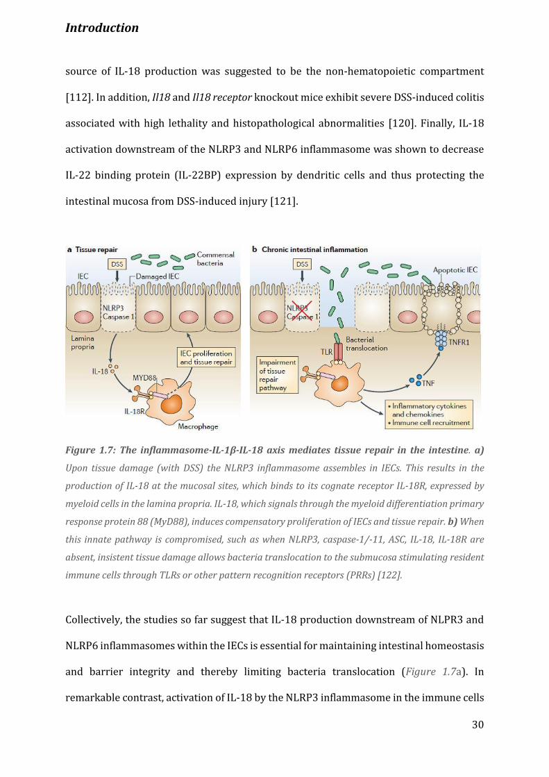
Introduction
30
source of IL-18 production was suggested to be the non-hematopoietic compartment
[112]. In addition, Il18 and Il18 receptor knockout mice exhibit severe DSS-induced colitis
associated with high lethality and histopathological abnormalities [120]. Finally, IL-18
activation downstream of the NLRP3 and NLRP6 inflammasome was shown to decrease
IL-22 binding protein (IL-22BP) expression by dendritic cells and thus protecting the
intestinal mucosa from DSS-induced injury [121].
Figure 1.7: The inflammasome-IL-1β-IL-18 axis mediates tissue repair in the intestine. a)
Upon tissue damage (with DSS) the NLRP3 inflammasome assembles in IECs. This results in the
production of IL-18 at the mucosal sites, which binds to its cognate receptor IL-18R, expressed by
myeloid cells in the lamina propria. IL-18, which signals through the myeloid differentiation primary
response protein 88 (MyD88), induces compensatory proliferation of IECs and tissue repair. b) When
this innate pathway is compromised, such as when NLRP3, caspase-1/-11, ASC, IL-18, IL-18R are
absent, insistent tissue damage allows bacteria translocation to the submucosa stimulating resident
immune cells through TLRs or other pattern recognition receptors (PRRs) [122].
Collectively, the studies so far suggest that IL-18 production downstream of NLPR3 and
NLRP6 inflammasomes within the IECs is essential for maintaining intestinal homeostasis
and barrier integrity and thereby limiting bacteria translocation (Figure 1.7a). In
remarkable contrast, activation of IL-18 by the NLRP3 inflammasome in the immune cells
Introduction
31
of the lamina propria seems to be proinflammatory and thus promoting intestinal
inflammation (Figure 1.7b). Therefore tight control of the caspase-1/IL-1 axis of
inflammation is the key to achieve immune tolerance in the intestine and deregulation of
this pathway contributes to the development of IBDs.
1.8 Caspase-12 expression and activation
Caspases are cysteine proteases that initiate or execute cellular programs causing
inflammation or cell death. All caspases are produced in cells as inactive zymogens and
then undergo proteolytic processing upon activation. They are categorized, according to
the cellular program they are involved in, either as proinflammatory or proapoptotic. The
proinflammatory caspases comprise caspase-1,-11,-12 and -1, -4, -5 and -12 in humans,
and they are called so due to their ability to regulate the production of the
proinflammatory cytokines IL-1β and IL-18 [123]. They share a conserved common
structure composed of a CARD-containing prodomain found at the N-terminus, a large and
a small subunit at the C-terminus. Furthermore, they are able to segregate their large and
small subunits through autoproteolysis which also serves to remove their N-terminal
prodomains [124].
Caspase-12 is expressed in many tissues at the RNA level with the highest expression
observed in the lung, small intestine and stomach [125]. Its constitutive protein
expression is restricted to the brain, heart, skeletal muscle, eye, liver, testis [126] and in
intestinal epithelial cells [127]. Although its expression is low in the liver, thymus and
spleen [126] it can be highly induced by IFNγ and bacterial components in various cell
types [126, 127]. Moreover, caspase-12 was found to localize at the cytoplasmic site of the
endoplasmic reticulum (ER) [128, 129].

Introduction
32
Several mechanisms have been postulated for the processing and activation of caspase-
12. Calpains [130], calcium-activated cysteine peptidases, or caspase-7 [128] in response
to ER stress process caspase-12 at first at the N-terminus, between the prodomain and
the large subunit. This is followed by autoprocessing of caspase-12, between the large and
small subunits. Another mechanism proposes that caspase-12 is released from TRAF2
during ER stress and then is autoprocessed via oligomerization [131]. Also, caspase-12
was suggested to be activated by tunicamycin, an ER stress stimulus, by inducing its
processing at the N-terminal and C-terminal region [132]. Unlike the other inflammatory
caspases, caspase-12 is unique in the respect that it is catalytically competent but its
specificity is highly confined to the cleavage of its corresponding pro-enzyme [133].
1.8.1 The role of caspase-12 in inflammatory responses
Human and murine pro-caspase-12 were reported to have the highest homology (68%
sequence identity) among all human and murine members of the interleukin-1β-
converting enzyme (ICE) subfamily of caspases [125]. Initial studies in Caspase-12
deficient mice have shown that caspase-12 is involved in ER stress-induced apoptosis
[129]. Similarly, other in vitro studies are in agreement with a role of caspase-12 in ER
stress mediated apoptosis [128, 130, 132]. Although activation and proteolysis of
caspase-12 is a hallmark of ER stress its role in ER stress-induced apoptosis is challenged.
More recent studies suggest that Caspase-12 deficient cells, in both humans and mice, are
as sensitive to ER stress-induced apoptosis as Caspase-12 proficient cells [126, 127, 134-
136].
Despite the high sequence identity between human and murine caspase-12, in the
majority of human population caspase-12 is truncated due to a single nucleotide
polymorphism that introduces a premature stop codon [136]. However, 20% of

Introduction
33
individuals of African descent have a point mutation that replaces the stop codon resulting
in the synthesis of the full length caspase-12. African individuals diagnosed with severe
sepsis are homozygous for the SNP encoding the full length variant and consequently this
mutation correlates with increased mortality rates compared to the individuals harboring
the stop codon mutation. The full length caspase-12 is associated with attenuated
cytokine production (IL-1β, IFNγ, TNFα) in response to endotoxin and decreased NF-κB
activation in response to TNFα, strongly suggesting that human caspase-12 functions as
dominant regulator of inflammatory responses and innate immunity [136].
Likewise, murine caspase-12 appears to have an inflammatory role as human caspase-12.
Caspase-12 deficiency in mice results in enhanced parasite clearance during blood-stage
Plasmodium infection and hyper-production of NF-κB and IFNγ [137]. This response is
NF-κB and IFNγ-dependent as systemic inhibition of NF-κB or neutralizing antibodies
against IFNγ reversed the increased resistance of the knockout mice to malaria infection.
Mechanistically, caspase-12 was proposed to associate with the IKKα/β kinases, thus
preventing IKKγ from binding to the complex and ultimately inhibiting NF-κB signaling
[137]. Moreover, mice lacking Caspase-12 were shown to be more resistant to
polymicrobial sepsis than wild-type mice due to more efficient bacterial clearance [127].
Mechanistically, caspase-12 dampens the production of proinflammatory cytokines (IL-
1β and IL-18) by associating with and blocking caspase-1 activation concomitant with the
downregulation of downstream cytokine production [127]. Nevertheless, the inhibitory
effect is independent of caspase-12 catalytic activity, since catalytically inactive caspase-
12 associates and as well blocks caspase-1 activity [127].
Furthermore, caspase-12 has been proven to be an important modulator of intestinal
mucosa immunity. Caspase-12 proficient enterocytes are blunted in their ability to
activate NOD and downstream NF-κB signaling and therefore cannot produce
Introduction
34
antimicrobial peptides and cytokines in response to bacterial infection [138]. Caspase-12
ablation results in efficient bacterial clearance and less severe pathology, independently
of caspase-1 activity [138]. Thus, in addition to its role in inhibiting caspase-1 activity in
macrophages, in the intestinal epithelium caspase-12 functions as negative regulator of
defensin and cytokine production by suppressing NOD signaling [138]. Lastly, caspase-12
is critical for the induction of inflammation in the gut after injury [119]. Caspase-12
deficient mice are more resistant to acute and chronic colitis induced by DSS and show
exaggerated epithelial cell compensatory proliferation. However, the increased
inflammation and tissue repair renders them more vulnerable to colitis-associated cancer
induced by AOM/DSS [119].
Overall, the presence of caspase-12 is linked to immunosuppressive responses to
pathogenic bacteria and parasites rendering both mice and humans prone to infection.
However, in the intestinal tract and under conditions that trigger chronic inflammation
and inflammation-associated tumorigenesis, caspase-12 is required to restrict excessive
inflammation and proliferation in the colon and thus to maintain intestinal homeostasis
Aim of Study
35
Aim of Study
Inflammatory bowel diseases (IBD), including Crohn’s Disease and Ulcerative Colitis, are
chronic inflammatory conditions of the gastrointestinal tract and are strongly associated
with the development of colorectal cancer. A large amount of evidence corroborates the
existence of a major influence of innate immunity, proper activation of PRRs, autophagy
and UPR pathways in the development of IBDs. NF-κB pathway is the key regulator of
inflammation and is strongly associated with IBD. IKKα, one of the core elements of this
pathway, has been shown by several independent studies to suppress inflammation in
response to a variety of stimuli. This study aimed to investigate the role of IKKα in colonic
inflammation by using a mouse model where IKKα is inactive. It demonstrates that IKKα
activation is required to suppress excessive inflammation in response to chemical factors,
such as dextran sodium sulfate, that induce colon injury.
To functionally examine the role of IKKα activation during inflammatory conditions in the
colon, the response of Ikkα kinase-dead (IkkαAA/AA) knock-in mice to DSS-induced colitis
was studied. To investigate the cell type specific functions of this kinase tissue specific
Ikkα knockout (IkkαΔIEC and IkkαΔmye) mice were used.
Material and Methods
36
2. Material and Methods
2.1 Materials
2.1.1 Mouse models
IkkαAA/AA Knock-in mice
A targeting vector was designed to place a neomycin resistance cassette flanked by loxP
sites (floxed, fl) into the exons 176 and 180 of the Ikkα gene [61]. Serines 176 and 180
were converted to alanines and the targeted vector was introduced into ES (Embryonic
Stem) cells. Cre-expressing vector was brought into the IkkαAA positive clones to disrupt
the neomycin cassette and such clones were injected into female’s blastocysts to generate
chimeric mice. These females were then crossed with males to give rise to heterozygous
IkkαAA/+ mice that were then intercrossed to obtain the homozygous IkkαAA/AA mutants.
Ikkαfl/fl
Mice harboring a loxP flanked Ikkα allele were created by inserting a neomycin cassette
fragment with loxP sites into the exons 6 and 9 of the gene [139]. The neomycin cassette
was deleted by introducing a Cre-expressing vector and thus following Cre recombination
the exons 6 to 8 were deleted resulting in a frame shift for Ikkα.
NfκB2-deficient mice
These mice were generated by replacing the exons 1b to 9 of the NfκB2 gene, which
contains the translation site and part of the Rel homology domain, with a targeting vector
containing a phosphoglycerate kinase-promoter neomycin resistance cassette [140]. The
Material and Methods
37
resulting vector was introduced into ES cells and clones containing a single copy insertion
of the neomycin cassette were used to generate independent knockout lines.
Villin-Cre
The Cre recombinase is expressed under the control of the mouse Villin 1 promoter [141].
Intercrossing with a strain harboring a loxP-flanked gene of interest results in Cre-
mediated recombination and enterocyte-specific deletion of the targeted gene.
Lysozyme M-Cre
Mice expressing Cre in myeloid cells were generated by targeted insertion of a vector
expressing the Cre cDNA into exon 1 of the M Lysozyme locus [142]. When crossed with
mice harboring loxP site flanked gene of interest Cre-mediated recombination results in
excision of the trageted gene in the myeloid lineage (monocytes, macrophages and
granulocytes).
Caspase-12-Knockout mice
Caspase-12 knockout mice were generated by targeted replacement of exon 2 of Caspase-
12 gene by a cassette consisting of the β-galactosidase gene and the neomycin resistance
gene [127]. The resulting vector was introduced into ES cells and clones obtained with the
disrupting allele were injected into blastocysts to generate chimeric mice.
2.1.2 Chemicals and reagents
All chemicals and reagents used are listed in the Appendix A.1
Material and Methods
38
2.2 Methods
2.2.1 Animal experiments
Induction of experimental acute colitis
In order to induce acute colitis in mice dextran sulfate sodium (DSS, MP Biomedicals) was
administrated in the drinking water at concentrations between 2.5 and 3.5 % (w/v). The
mice were subjected to the DSS treatment for 5 days followed by 5 additional days of
normal drinking water.
IL-18 treatment
For IL-18 rescue experiments, recombinant IL-18 (MBL International) was injected intra-
peritoneally at a concentration of 0.5 g/mouse, dissolved in cold PBS (Invitrogen), every
day during the whole acute DSS treatment. 25 g of lyophilized IL-18 were reconstituted
with 250 μl of ice-cold distilled water on ice.
Bone marrow isolation
For isolation of the bone marrow donor mice with the same genetic background as the
recipient mice, were sacrificed and femoral bones were removed carefully and placed in
cold PBS. Then under sterile conditions, bones were cut on the top and bottom, flushed
with sterile PBS using a 25 G needle, resuspended and filtered with a 70 μm strainer to
get rid of the skeletal parts and stromal cells. Total cell number was counted and then the
cells were pelleted by 5 minutes centrifugation at 500 g 4°C. Resuspension was done in
the appropriate buffer according to the purpose of the isolation.
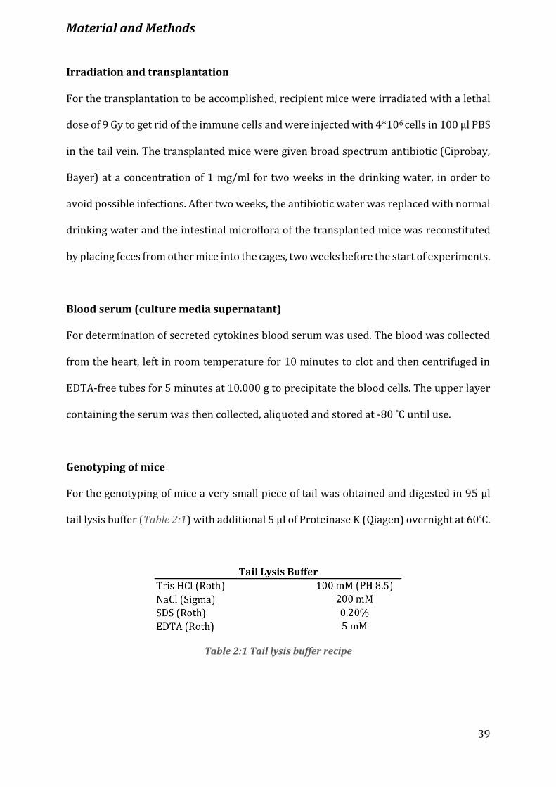
Material and Methods
39
Irradiation and transplantation
For the transplantation to be accomplished, recipient mice were irradiated with a lethal
dose of 9 Gy to get rid of the immune cells and were injected with 4*106 cells in 100 μl PBS
in the tail vein. The transplanted mice were given broad spectrum antibiotic (Ciprobay,
Bayer) at a concentration of 1 mg/ml for two weeks in the drinking water, in order to
avoid possible infections. After two weeks, the antibiotic water was replaced with normal
drinking water and the intestinal microflora of the transplanted mice was reconstituted
by placing feces from other mice into the cages, two weeks before the start of experiments.
Blood serum (culture media supernatant)
For determination of secreted cytokines blood serum was used. The blood was collected
from the heart, left in room temperature for 10 minutes to clot and then centrifuged in
EDTA-free tubes for 5 minutes at 10.000 g to precipitate the blood cells. The upper layer
containing the serum was then collected, aliquoted and stored at -80 °C until use.
Genotyping of mice
For the genotyping of mice a very small piece of tail was obtained and digested in 95 μl
tail lysis buffer (Table 2:1) with additional 5 μl of Proteinase K (Qiagen) overnight at 60°C.
Table 2:1 Tail lysis buffer recipe
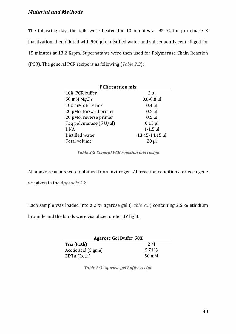
Material and Methods
40
The following day, the tails were heated for 10 minutes at 95 °C, for proteinase K
inactivation, then diluted with 900 μl of distilled water and subsequently centrifuged for
15 minutes at 13.2 Krpm. Supernatants were then used for Polymerase Chain Reaction
(PCR). The general PCR recipe is as following (Table 2:2):
Table 2:2 General PCR reaction mix recipe
All above reagents were obtained from Invitrogen. All reaction conditions for each gene
are given in the Appendix A.2.
Each sample was loaded into a 2 % agarose gel (Table 2:3) containing 2.5 % ethidium
bromide and the bands were visualized under UV light.
Table 2:3 Agarose gel buffer recipe
Material and Methods
41
2.2.2 Primary cell isolation and culture
Intestinal epithelial cell (IEC) isolation
After the mice were sacrificed, the intestine was divided into 4 parts (duodenum, jejunum,
ileum and colon) and the part/s of interest was/were cleaned from feces with PBS. The
intestinal parts were then cut-open longitudinally and divided in half. One half was used
for histological analysis, the other was rolled up to make a ‘swiss-roll’ [143], fixed over-
night at 4°C in 4 % paraformaldehyde (PFA), dehydrated and embedded in paraffin, while
the other half was used for epithelial cell isolation. For the cell isolation, the half of the
intestine was further cut in small pieces, incubated in 8 ml of pre-warmed HBSS
(Invitrogen) supplemented with 30 mM EDTA (Roth) and shacked for 10 minutes gently
at 37°C in order to remove epithelial cell physically. Next, the 50 ml falcon tube containing
the above tissue was strongly vortexed for 30 seconds, to detach epithelia and lamina
propria, and placed directly on ice. After 30 seconds approximately, lamina propria was
precipitated while epithelial cells remained in the supernatant. Supernatant was slowly
removed and transferred to a new 15 ml falcon tube for further centrifugation for 5
minutes at 1.5 Krpm at 4°C. The pellet was resuspended in cold 2 ml PBS and divided into
three 1.5 ml tubes that were subsequently centrifuged at 5 Krpm at 4°C for 5 minutes.
Washouts were discarded and pelleted epithelial cells were frozen in liquid nitrogen and
stored at -80°C until use.
For further analysis whole parts of intestinal mucosa (mostly distal part of each intestinal
part) were cut in small pieces, schock frozen in liquid nitrogen and stored as the epithelial
cells at -80°C.

Material and Methods
42
IEC isolation from colons and overnight culture
Following mice sacrifice, colons were removed and washed with RPMI1640 (GIBCO)
supplemented with penicillin/streptomycin and 50 μg/ml gentamicin. Colons were then
cut in small pieces and incubated at 37°C for 30 minutes with gentle shaking in pre-
warmed RPMI medium supplemented with penicillin, streptomycin, 50 μg/ml gentamicin,
5 mM EDTA pH:8, 3 % FCS, 0.145 mg/ml DTT (Sigma) (reffered as full RPMI medium).
Next, colons were vortexed for 30 seconds, to detach epithelia and lamina propria, placed
on ice for 30 seconds, to allow lamina propria to precipitate while the epithelial cells
remained in the supernatant. Supernatants were then filtered with 100 μM strainers
directly into a 50 ml falcon and centrifuged at 1.5 Krpm for 5 minutes at 4°C. IEC pellets
were then resuspended in 30 % Percoll (GE Healthcare), for efficient density separation,
and centrifuged at 500 g for 5 minutes at 4°C. The top layer containing the IECs was
removed carefully and washed 2 times in full RPMI medium to remove remaining Percoll.
Finally, pelleted IECs were resuspended in RPMI medium supplemented with 10 % FCS,
penicillin, streptomycin, gentamycin, L-glutamine, and 10 % FCS, counted and plated on
96-well plates pre-coated with collagen type I (GE Healthcare). 100.000 epithelial cells
were plated and left overnight in the incubator (37°C and 5 % CO2).
For coating 96-well plates with collagen type I (GE Healthcare), collagen was diluted to a
concentration of 10 μg/ml in 0.02 N acetic acid and 5 μg of collagen per cm2 was required
for the coating. The collagen-coated plates were left at 37°C for 30 minutes and then
washed 2 times with PBS. Supernatants and cells were collected 24 hours later and
analyzed by ELISA or immunobloting.
Material and Methods
43
Isolation of lamina propria cells
For isolation of lamina propria cells, the remaining colon pieces (after IEC isolation) were
recovered and incubated with RPMI supplemented with penicillin/streptomycin, + 0.1
mg/ml liberase TL (Roche) + 0.05 % DNase (Roche) for 30 minutes at 37°C with gentle
shaking. Supernatants were then filtered with 70 μm strainers and washed two times with
RPMI supplemented with penicillin/ streptomycin and 10 % FCS. Cells were then sorted
with BD FACS Aria into different cell populations.
Flow cytometry assisted cell sorting (FACS)
For cell sorting cells were washed in FACS buffer (5 % FCS/PBS). Before staining for the
cell surface markers, cells were blocked for unspecific binding with FcR block (diluted
1:100 in FACS buffer) for 10 minutes at 4°C and then washed once with PBS and pelleted
at 500 g for 5 minutes. Then cells were stained with the desired antibodies (Appendix
A.3.3) in FACS buffer at 4°C for 20 minutes, in the dark. Cells were washed 2 times in PBS,
pelleted at 500 g for 5 minutes, resuspended in FACS buffer and subjected for sorting with
BD FACS Aria.
Cell counts
Cells were harvested with 0.25 % Trypsin/EDTA (Invitrogen) and counted with a
hematocytometer in the presence of 1 % trypan blue (Sigma). Cells that were not stained
blue were considered alive.

Material and Methods
44
2.2.3 Histology
Hematoxylin & Eosin (H & E) staining
For closer examination of tissues H & E staining was performed. The organ/s was/were
isolated, fixed in 4 % PFA dehydrated and embedded in paraffin. The paraffin embedded
tissues were then cut 3 μm thick, fixed on glass slides (Thermo Scientific) and dry out
either over night at room temperature either at 37°C for 2 hours. The slides were
rehydrated following the procedure below (Table 2:4):
Table 2:4 Rehydration procedure
Slides were stained with ready to use Haematoxylin (Vector Laboratories) for 1 minute
and washed well with distilled water. Next, slides were stained with Eosin (Sigma) for 10
seconds, rinsed twice in distilled water and dehydrated with the following procedure
(Table 2:5):
Table 2:5 Dehydration procedure
Material and Methods
45
Afterwards, slides were air-dried, mounted with mounting medium (Vector Laboratories)
and then covered with cover slides.
Eosin solution: 1 % Eosin + 15 drops of acetic acid in 200 ml of dH2O
DNA fragmentation Assay
This assay is based on Terminal deoxynucleotidyl Transferase (TdT)-mediated dUTP
nick-end-labeling (TUNEL). This enzyme catalyses the incorporation of fluorescein
labelled dUTP at the free 3’-OH end of fragmented DNA. The fluorescein labelled DNA can
be visualised by fluorescence microscope equipped with FITC filters. The staining was
carried out using ApoAlert®DNA Fragmentation Assay (Clontech) in situ in tissue
sections. Tissue sections were deparaffinised and rehydrated as described in H & E
staining section. Sections were incubated for 5 minutes in 0.85 % sodium chloride
solution and afterwards washed with PBS. Then, the sections were fixed in 4 % PFA in 0.1
% Diethylpyrocarbonate (DEPC) (Sigma) distilled water for 15 minutes in room
temperature. After one more wash with PBS, samples were incubated in 4 % DEPC in
distilled water for 30 minutes at 4°C followed by a step of proteinase K (20 μg/ml)
digestion for 5 minutes at room temperature. Following PBS wash, tissues were incubated
with equilibration buffer for 15 minutes followed by another incubation with the
enzyme/buffer/nucleotide mix (30 μl equilibration buffer, 3.3 μl nucleotide mix , 0.33 μl
TdT enzyme), in the dark at 37°C for 1 hour. The reaction was stopped with 2x SSC buffer
for 15 minutes, in the dark and washed twice with PBS. Samples were then covered with
DAPI containing mounting medium (ProLong Gold, Invitrogen) and stored at 4°C in the
dark.
Material and Methods
46
Immunohistochemistry
After slides were deparaffinized and rehydrated (Table 2:4), permeabilization protocol
followed. For nuclear and cytoplasmic stainings the slides were heated up in the
microwave for 20 minutes in unmasking solution (VectorLab), cooled down to room
temperature and washed in PBS. Blocking of endogenous peroxidases was achieved by
incubating the slides in 3 % H2O2 / PBS for 5 minutes at room temperature. The slides
were then blocked with 3 % BSA / PBS + streptavidin (VectorLab) for 30 minutes at room
temperature in order to avoid unspecific binding of the primary antibody. After blocking
the slides were incubated with the primary antibody + 3 % BSA/PBS + biotin (VectorLab)
in different conditions (Appendix A.3.2). Subsequently, slides were washed 3 times in PBS,
5 minutes each washing step, and then incubated for 30 minutes at room temperature
with the appropriate secondary antibody conjugated with biotin molecule. Following 3
washing steps in PBS, slides were incubated with ABC (VectorLab) complex containing 2
drops of Avidin DH (Reagent A) and 2 drops of its paired biotinylated (horseradish
peroxidase) enzyme (Reagent B) for 30 minutes at room temperature. After washing with
PBS colour reaction with DAB kit (VectorLab) followed. The reaction was done by mixing
the three components of the kit (2 drops of buffer + 2 drops of hydrogen peroxide solution
+ 4 drops of DAB (3, 3'-diaminobenzidine)) just before starting the developing. The colour
reaction was observed under the microscope and was stopped with distilled water. Nuclei
were stained with haematoxylin for 1 minute (counter staining). After extensive washes
with distilled water sections were dehydrated (Table 2:5), air dried, mounted with
mounting medium (VectorLab) and covered with cover slides.

Material and Methods
47
Immunofluorescence
For immunofluorescence staining sections were first deparaffinized, rehydrated and then
tissues were permeabilized. For staining of surface antigens, tissues were incubated with
0.3 % Triton / PBS for 30 minutes at 4°C and for nuclear and cytoplasmic antigens tissues
were microwaved in antigen unmasking solution (VectorLab) for 20 minutes. Tissues
were then cooled down at room temperature, washed 3 times in PBS and blocked with 3
% BSA/PBS for 30 minutes to avoid unspecific binding of the antibody. Primary antibody
coupled with a fluorophore was incubated overnight at 4°C. Next day, tissues were washed
3 times with PBS and then covered with DAPI conservation medium (ProLong Gold,
Invitrogen) for counter staining of the nucleus. Tissues were left for 30 minutes at 4°C and
then used for imaging.
2.2.4 RNA Analysis
RNA isolation from cells and tissues
RNA from tissues or cells was obtained using the RNeasy Kit (Qiagen). Frozen tissue
samples or pelleted cells were directly placed in RLT buffer containing 1 % β-
mercaptoethanol and homogenized either using electronic homogenizer (for tissues) or
pistils (for cells). The procedure of RNA isolation with Qiagen RNeasy Kit is described in
the manufacturer’s manual. RNA was eluted in RNAase free water and stored in -80°C until
use.
cDNA Synthesis
For the synthesis of the first complementary DNA strand was performed using oligo dT
primer and reverse transcriptase. Upon isolation, purity and concentration of RNA was
determined. Generally, 1 μg of RNA was used or less according to the RNA concentration.

Material and Methods
48
RNA was incubated with 1 μl of Oligo dT (50 μM, Invitrogen) and 1 μl of dNTP-mix (10
mM, Invitrogen) and filled up to a total volume of 13 μl with RNase-free water for 5
minutes at 65°C for annealing of the dT primers to mRNAs. The samples were then shortly
spin-down and incubated on ice for 5 minutes. Master-mix (Table 2:6) of 7 μl was added
in every sample to make 20 μl total volume and incubated at 42°C for 1 hour. Finally, the
samples were diluted 1:4 with RNase-free water and stored at -20°C until use.
Table 2:6 Reverse transcription master mix
qRT - PCR (quantitative - real time - polymerase chain reaction)
Primers specific for each gene were designed using the Primer ExpressTM software
(primer sequences are listed in Appendix A.4). The lyophilized primers were reconstituted
with dH2O to a concentration of 100 μM and then combined to a final concentration of 20
μM to make a 10X solution. For the qRT-PCR reaction, SYBR Green (Roche) master mix
(Table 2:7) was used with the following mixture for every sample:
Table 2:7 Real-Time PCR master mix

Material and Methods
49
The analysis of the reaction was performed on a standard program (Table 2:8):
Table 2:8 Real-Time PCR program
The reaction was done on StepOnePlus Real Time PCR system (Applied Biosystems) and
the results were analyzed with StepOne Software v2.0.2 and normalized by the expression
levels of the housekeeping gene CyclophilinB, as an internal control, with the following
formula:
Expression = 2 ^ (CT (housekeeping) – CT (gene))
2.2.5 Protein Analysis
Protein extraction from IECs and total intestinal mucosa
Epithelial cell pellets removed from -80°C and directly placed in liquid nitrogen, were
lysed in 90-150 μl of ice-cold lysis buffer according to the amount of pelleted cells. The
pellet was mechanically homogenized with a micro-pistill, as well as up and down
pipetting, and left on ice for 15 minutes to ensure complete lysis. Subsequently, cell debris
was removed by 15 minutes centrifugation at 13.3 Krpm at 4°C and the proteins lysates
were removed and placed to into 1.5 ml eppi and stored at -80°C until further use.
For total mucosa samples the lysis was done with an electronic homogenizer within an
appropriate lysis buffer (Table 2:9) according to the size of the frozen intestinal piece. The
same procedure was followed also for the IEC isolation.
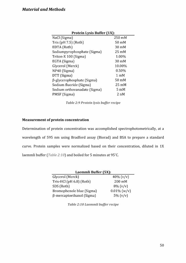
Material and Methods
50
Table 2:9 Protein lysis buffer recipe
Measurement of protein concentration
Determination of protein concentration was accomplished spectrophotometrically, at a
wavelength of 595 nm using Bradford assay (Biorad) and BSA to prepare a standard
curve. Protein samples were normalized based on their concentration, diluted in 1X
laemmli buffer (Table 2:10) and boiled for 5 minutes at 95°C.
Table 2:10 Laemmli buffer recipe

Material and Methods
51
Western blot analysis
For immunoblot analysis 6-15% SDS resolving gels were prepared depending on the size
of proteins to be analysed (Tables 2:11-2:12) and proteins were run in 1X running buffer
(Table 2:13).
Table 2:11 Resolving gel recipes
Table 12:12 Resolving and stacking gel buffer recipes
Table 2:13 10X and 1X Running buffer recipes
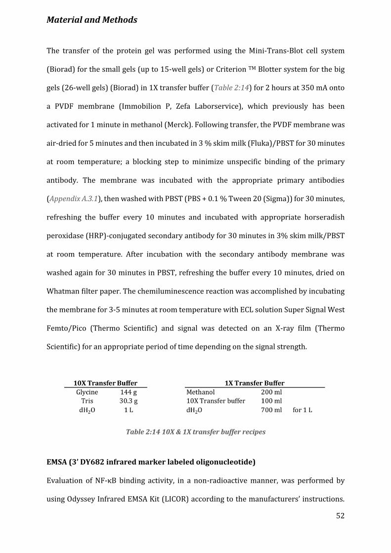
Material and Methods
52
The transfer of the protein gel was performed using the Mini-Trans-Blot cell system
(Biorad) for the small gels (up to 15-well gels) or Criterion TM Blotter system for the big
gels (26-well gels) (Biorad) in 1X transfer buffer (Table 2:14) for 2 hours at 350 mA onto
a PVDF membrane (Immobilion P, Zefa Laborservice), which previously has been
activated for 1 minute in methanol (Merck). Following transfer, the PVDF membrane was
air-dried for 5 minutes and then incubated in 3 % skim milk (Fluka)/PBST for 30 minutes
at room temperature; a blocking step to minimize unspecific binding of the primary
antibody. The membrane was incubated with the appropriate primary antibodies
(Appendix A.3.1), then washed with PBST (PBS + 0.1 % Tween 20 (Sigma)) for 30 minutes,
refreshing the buffer every 10 minutes and incubated with appropriate horseradish
peroxidase (HRP)-conjugated secondary antibody for 30 minutes in 3% skim milk/PBST
at room temperature. After incubation with the secondary antibody membrane was
washed again for 30 minutes in PBST, refreshing the buffer every 10 minutes, dried on
Whatman filter paper. The chemiluminescence reaction was accomplished by incubating
the membrane for 3-5 minutes at room temperature with ECL solution Super Signal West
Femto/Pico (Thermo Scientific) and signal was detected on an X-ray film (Thermo
Scientific) for an appropriate period of time depending on the signal strength.
Table 2:14 10X & 1X transfer buffer recipes
EMSA (3’ DY682 infrared marker labeled oligonucleotide)
Evaluation of NF-κB binding activity, in a non-radioactive manner, was performed by
using Odyssey Infrared EMSA Kit (LICOR) according to the manufacturers’ instructions.

Material and Methods
53
10-15 μg of proteins were incubated with 1 μM 3’ DY682 labeled NF-κB consensus oligos
(5’-GGA TCC TCA ACA GAG GGG ACT TTC CGA GGC CA-3’) for 30 minutes at room
temperature, protected from light. Then, samples were loaded in a 5 % acrylamide gel,
run for 2 hours and finally bands were visualized with Odyssey Infrared imaging system
(Li-COR Biosciences).
Co-immunoprecipitation
In order to identify protein-protein interaction co-immunoprecipitation was performed.
500 μg of total protein lysates were used to precipitate the protein of interest (X protein)
with a specific antibody against it. The antibody of interest and total protein lysates were
incubated in 500 μl PBS (low stringency buffer) total volume, overnight, rotating at 4°C to
allow for the possible complex to form. Next day, protein A sepharose beads (GE
Healthcare) were washed 3 times in cold PBS and protein lysates with the antibody were
applied to the beads. The above mixture was incubated at 4°C, rotating, for 1 hour (not
longer, as this might cause unspecific binding of the proteins to the beads). Beads-
antibody-protein lysates were then washed 3 times with 1X lysis buffer (high stringency
buffer, Table 2:9) and mixture was collected by 3 Krpm centrifugation for 5 minutes.
Afterwards, 30 μl of laemmli buffer + 5 % mercaptoethanol was added to the precipitated
beads, heated 10 minutes at 95°C and centrifuged maximum at 13 krpm for 5 minutes. The
supernatants were then subjected to immunobloting (as described above) for
investigation of potential protein-protein interaction.
ELISA
For analysis of cytokines in the blood serum or cell culture supernatants enzyme-linked
immunosorbent assay (ELISA) was used. The ELISA kits used are listed in the Appendix
Material and Methods
54
A.1. Briefly, ELISA plates coated with capture antibody were washed and blocked for 1
hour at room temperature. After blocking and washing steps, plate was incubated for 2
hours at room temperature with the samples and standards diluted in the dilution buffer.
Then, plates were washed and incubated with the detection antibody, streptavidin and
substrate mix. The colour reaction was detected by a spectrophotometer using a plate
reader at 450 nm.
Results
55
3. Results
3.1 IKKα is essential for mucosa healing after DSS-induced injury
Physiological levels of inflammation are protective, while extreme levels are deleterious
and can be the cause of Inflammatory Bowel Disease (IBD), such as Crohn’s Disease and
Ulcerative Colitis. In several studies, IΚΚα has been shown to confer resolution of
inflammation [62, 65]. To functionally investigate the role of IKKα in intestinal
homeostasis a knock-in mouse model that lacks inducible kinase activity due to the
presence of alanines instead of serines in the activation loop of IKKα was used [61]. Mice
homozygous for the mutated allele, IkkαΑΑ/AA knock-in mice, were subjected to acute
tissue injury and colitis by adding 3.5% dextran sulphate sodium (DSS) in the drinking
water for 5 days, followed by 4 days of recovery with normal drinking water (Figure 3.1
A). In sharp contrast to Ikkαwt/wt mice, which recovered once DSS was omitted, IkkαΑΑ/AA
mutants showed increased susceptibility to the treatment and suffered from continued
body weight loss, diarrhea and rectal bleeding, on day 7 until day 9 (Figure 3.1 B). Due to
the severe weight loss of IkkαΑΑ/AA mice and to ethical reasons both groups had to be
sacrificed on day 9. Hematoxylin & eosin (H & E)-stained colon sections from both
genotypes were evaluated for histological changes. Histolopathological alterations were
observed in DSS-treated colons from both Ikkαwt/wt and IkkαΑΑ/AA mice, characterized by
crypt loss, ulceration and infiltration of leukocytes (Figure 3.1 C-D). In marked contrast to
Ikkαwt/wt mice, which showed signs of re-epithelialisation, IkkαΑΑ/AA mutants displayed
severe signs of injury and defects in epithelial reconstruction. The IkkαΑΑ/AA mutant
epithelium was characterized by few if any crypts left throughout the entire colon,
extensive ulceration resulting in complete destruction of the normal architecture of the
tissue and massive infiltration of immune cells in the lamina propria and submucosa
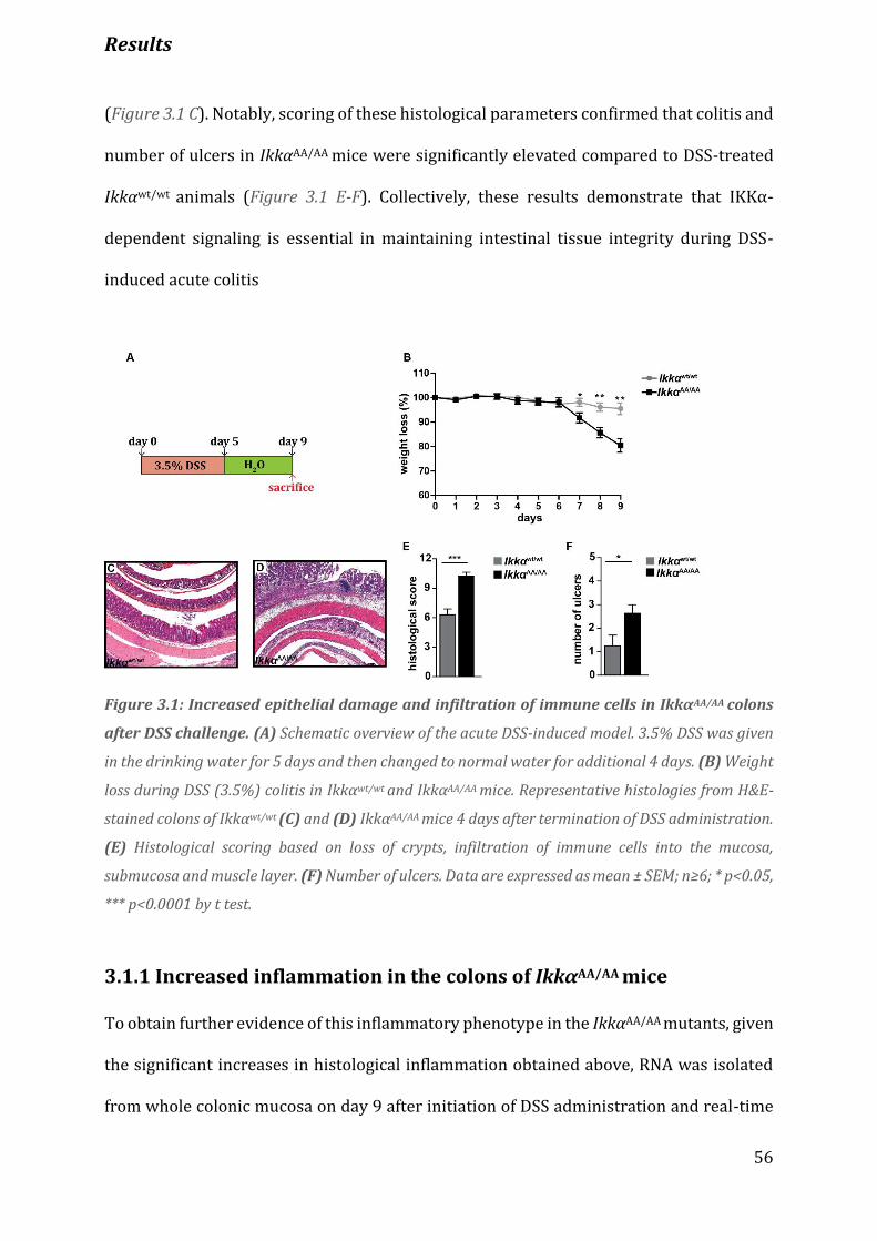
Results
56
(Figure 3.1 C). Notably, scoring of these histological parameters confirmed that colitis and
number of ulcers in IkkαΑΑ/AA mice were significantly elevated compared to DSS-treated
Ikkαwt/wt animals (Figure 3.1 E-F). Collectively, these results demonstrate that IKKα-
dependent signaling is essential in maintaining intestinal tissue integrity during DSS-
induced acute colitis
Figure 3.1: Increased epithelial damage and infiltration of immune cells in IkkαΑΑ/AA colons
after DSS challenge. (A) Schematic overview of the acute DSS-induced model. 3.5% DSS was given
in the drinking water for 5 days and then changed to normal water for additional 4 days. (B) Weight
loss during DSS (3.5%) colitis in Ikkαwt/wt and IkkαAA/AA mice. Representative histologies from H&E-
stained colons of Ikkαwt/wt (C) and (D) IkkαΑΑ/AA mice 4 days after termination of DSS administration.
(E) Histological scoring based on loss of crypts, infiltration of immune cells into the mucosa,
submucosa and muscle layer. (F) Number of ulcers. Data are expressed as mean ± SEM; n≥6; * p<0.05,
*** p<0.0001 by t test.
3.1.1 Increased inflammation in the colons of IkkαΑΑ/AA mice
To obtain further evidence of this inflammatory phenotype in the IkkαΑΑ/AA mutants, given
the significant increases in histological inflammation obtained above, RNA was isolated
from whole colonic mucosa on day 9 after initiation of DSS administration and real-time

Results
57
PCR was performed to examine the expression levels of proinflammatory genes including,
Il1β, Tnfα, Il6, Cxcl1, Cxcl2 and Ccl2. Consistent with the histological parameters discussed
above, whole mucosa from IkkαΑΑ/AA treated colons contained higher mRNA levels coding
for IL-1β, ΤΝFα, IL-6, CXCL1, CXCL2 and CCL2 compared to the Ikkαwt/wt treated colons
(Figure 3.2 A). Consistently, IkkαΑΑ/AA DSS-treated mice presented enhanced expression of
COX-2 and matrix metalloproteinase-9 (MMP-9) (Figure 3.2 B). Thus, the above data
demonstrate the fundamental role of IKKα activation in hindering excessive inflammatory
responses upon DSS-induced injury.
Figure 3.2: Enhanced inflammation in the colons of IkkαΑΑ/AA mice. (A) Expression of
inflammatory genes. Relative mRNA expression levels were quantified in whole colonic mucosa tissue
of mice on day 0 and 9 of DSS regimen. The mRNA levels were quantified by real-time PCR and
normalized to the level of cyclophilinB mRNA (n ≥ 4). (B) COX-2 and MMP-9 expression in whole
colonic extracts on day 0 and 9 of DSS regimen. COX-2 and MMP-9 expression were determined by
western blot (WB).
3.2 IKKα signaling in IECs is crucial for tissue repair
In order to distinguish whether IKKα functions in IEC or hematopoietic cells to control
DSS-induced tissue damage adoptive transfer experiments were performed (Figure 3.3 A-
B). Ikkαwt/wt and IkkαAA/AA bone marrow was transplanted into Ikkαwt/wt and IkkαAA/AA
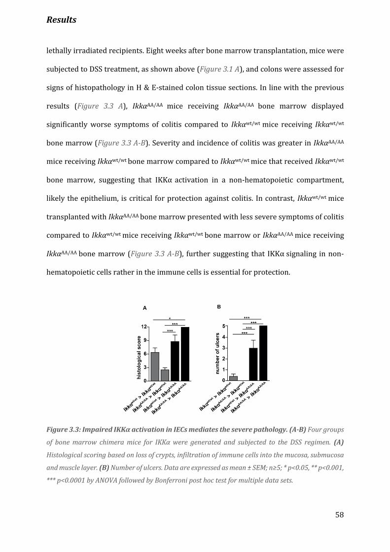
Results
58
lethally irradiated recipients. Eight weeks after bone marrow transplantation, mice were
subjected to DSS treatment, as shown above (Figure 3.1 A), and colons were assessed for
signs of histopathology in H & E-stained colon tissue sections. In line with the previous
results (Figure 3.3 A), IkkαΑΑ/AA mice receiving IkkαΑΑ/AA bone marrow displayed
significantly worse symptoms of colitis compared to Ikkαwt/wt mice receiving Ikkαwt/wt
bone marrow (Figure 3.3 A-B). Severity and incidence of colitis was greater in IkkαΑΑ/AA
mice receiving Ikkαwt/wt bone marrow compared to Ikkαwt/wt mice that received Ikkαwt/wt
bone marrow, suggesting that IKKα activation in a non-hematopoietic compartment,
likely the epithelium, is critical for protection against colitis. In contrast, Ikkαwt/wt mice
transplanted with IkkαΑΑ/AA bone marrow presented with less severe symptoms of colitis
compared to Ikkαwt/wt mice receiving Ikkαwt/wt bone marrow or IkkαΑΑ/AA mice receiving
IkkαΑΑ/AA bone marrow (Figure 3.3 A-B), further suggesting that IKKα signaling in non-
hematopoietic cells rather in the immune cells is essential for protection.
Figure 3.3: Impaired IKKα activation in IECs mediates the severe pathology. (A-B) Four groups
of bone marrow chimera mice for IKKα were generated and subjected to the DSS regimen. (A)
Histological scoring based on loss of crypts, infiltration of immune cells into the mucosa, submucosa
and muscle layer. (B) Number of ulcers. Data are expressed as mean ± SEM; n≥5; * p<0.05, ** p<0.001,
*** p<0.0001 by ΑΝΟVA followed by Bonferroni post hoc test for multiple data sets.

Results
59
Figure 3.4: IKKα activation in IECs protects against colitis. (A) Weight loss during DSS (3.5%)
colitis in Ikkafl/fl and IkkαΔIEC mice. Representative histologies from H&E-stained colons of (B) Ikkαf/f
and (C) IkkαΔIEC mice 4 days after termination of DSS administration. (D) Histological scoring based
on loss of crypts, infiltration of immune cells into the mucosa, submucosa and muscle layer. (E)
Number of ulcers. (F) Weight loss during DSS (3.5%) colitis in Ikkafl/fl and IkkαΔmye mice.
Representative histologies from H&E-stained colons of Ikkαf/f (G) and (H) IkkαΔmye mice 4 days after
termination of DSS administration. (I) Histological scoring and (J) number of ulcers in Ikkafl/fl and
IkkαΔmye 4 days after challenge with DSS (3.5%). Data are expressed as mean ± SEM; n≥5; * p<0.05,
** p<0.001, *** p<0.0001 by t test.
To further confirm that IKKα signaling in non-hematopoietic cells is essential for
protection, mice lacking Ikkα specifically in the intestinal epithelial cells (IkkαΔIEC) and
myeloid cells (IkkαΔmye) were challenged with the same DSS regimen as in Figure 3.1 A.
Mice deficient for IKKα in the IECs (IkkαΔIEC), displayed significant more body weight loss
compared to Ikkαfl/fl littermate controls (Figure 3.4 A) and consistently, they exhibited
increased sensitivity to the treatment reaching statistical significance on day 9 of DSS
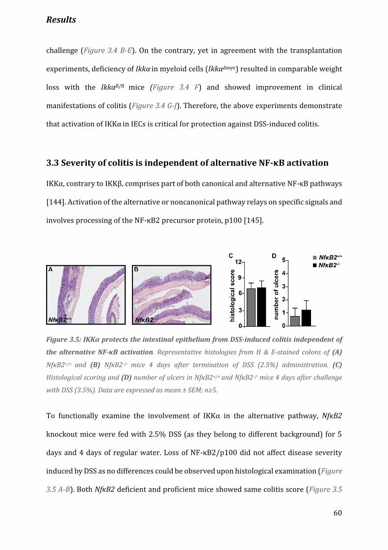
Results
60
challenge (Figure 3.4 B-E). On the contrary, yet in agreement with the transplantation
experiments, deficiency of Ikkα in myeloid cells (IkkαΔmye) resulted in comparable weight
loss with the Ikkαfl/fl mice (Figure 3.4 F) and showed improvement in clinical
manifestations of colitis (Figure 3.4 G-J). Therefore, the above experiments demonstrate
that activation of IKKα in IECs is critical for protection against DSS-induced colitis.
3.3 Severity of colitis is independent of alternative NF-κB activation
IKKα, contrary to IKKβ, comprises part of both canonical and alternative NF-κB pathways
[144]. Activation of the alternative or noncanonical pathway relays on specific signals and
involves processing of the NF-κB2 precursor protein, p100 [145].
Figure 3.5: IKKα protects the intestinal epithelium from DSS-induced colitis independent of
the alternative NF-κΒ activation. Representative histologies from H & E-stained colons of (A)
NfκΒ2+/+ and (B) NfκΒ2-/- mice 4 days after termination of DSS (2.5%) administration. (C)
Histological scoring and (D) number of ulcers in NfκΒ2+/+ and NfκΒ2-/- mice 4 days after challenge
with DSS (3.5%). Data are expressed as mean ± SEM; n≥5.
To functionally examine the involvement of IKKα in the alternative pathway, NfκB2
knockout mice were fed with 2.5% DSS (as they belong to different background) for 5
days and 4 days of regular water. Loss of NF-κB2/p100 did not affect disease severity
induced by DSS as no differences could be observed upon histological examination (Figure
3.5 A-B). Both NfκB2 deficient and proficient mice showed same colitis score (Figure 3.5
Results
61
C) and number of ulcers (Figure 3.5 D). These results suggest that the colitis phenotype in
IkkαΑΑ/AA mutants is not due to defective alternative NF-κB activation.
3.3.1 IkkαAA/AA IECs display defective NF-κB activation
As the above experiments demonstrated that IKKα acted independently of the alternative
NF-κB activation, its involvement in the canonical pathway was studied. Hence, intestinal
epithelial cells were examined for the main events that govern canonical NF-κB activation,
such as phosphorylation of IκBα or RelA/p65 subunit. In the DSS regimen the earliest
alterations observed are loss of crypts and separation of the crypt base from the
muscularis mucosa, which develop by day 3 post-administration of DSS [146]. Therefore,
day 3 was chosen as an early time-point to elucidate the mechanism behind the severe
acute colitis observed in IkkαAA/AA mice.
Both IKKα and IKKβ have been shown to phosphorylate IκBα at serine 32 [147] and
absence of IKKα resulted in diminished IκB degradation and p65 phosphorylation [148,
149]. Consistently, DSS-treated intestinal epithelial cells from IkkαΑΑ/AA mice displayed
reduced phosphorylation of IκBα at serine 32, indicating that ubiquitin-dependent
proteasomal degradation step was blocked (Figure 3.6 A). Moreover, RelA/p65
phosphorylation and NF-κB DNA binding activity were as well reduced in DSS-treated
IkkαΑΑ/AA IECs compared to Ikkαwt/wt treated IECs, further suggesting that NF-κB
activation was blocked (Figure 3.6 A-B). Taken together, IECs defective in IKKα activation
exhibit weakened NF-κB activity, suggesting that IKKα comprises part of the canonical
NF-κB pathway in this model.
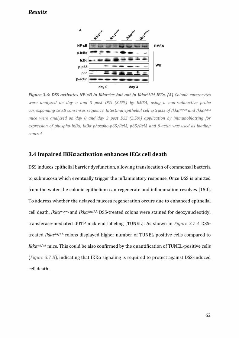
Results
62
Figure 3.6: DSS activates NF-κB in Ikkαwt/wt but not in IkkαAA/AA IECs. (A) Colonic enterocytes
were analyzed on day o and 3 post DSS (3.5%) by EMSA, using a non-radioactive probe
corresponding to κB consensus sequence. Intestinal epithelial cell extracts of Ikkαwt/wt and IkkαAA/A
mice were analyzed on day 0 and day 3 post DSS (3.5%) application by immunoblotting for
expression of phospho-IκBα, IκBα phospho-p65/RelA, p65/RelA and β-actin was used as loading
control.
3.4 Impaired IKKα activation enhances IECs cell death
DSS induces epithelial barrier dysfunction, allowing translocation of commensal bacteria
to submucosa which eventually trigger the inflammatory response. Once DSS is omitted
from the water the colonic epithelium can regenerate and inflammation resolves [150].
To address whether the delayed mucosa regeneration occurs due to enhanced epithelial
cell death, Ikkαwt/wt and IkkαAA/AA DSS-treated colons were stained for deoxynucleotidyl
transferase-mediated dUTP nick end labeling (TUNEL). As shown in Figure 3.7 A DSS-
treated IkkαAA/AA colons displayed higher number of TUNEL-positive cells compared to
Ikkαwt/wt mice. This could be also confirmed by the quantification of TUNEL-positive cells
(Figure 3.7 B), indicating that IKKα signaling is required to protect against DSS-induced
cell death.
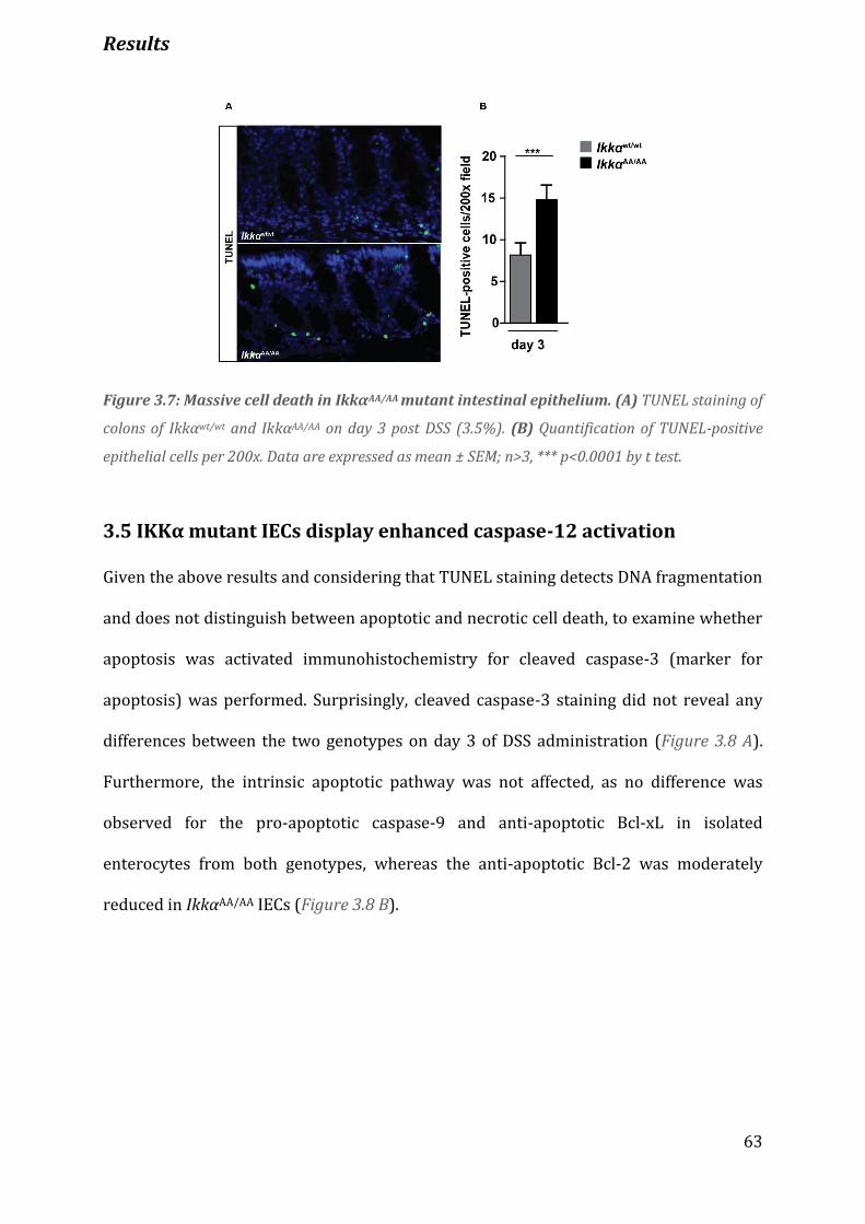
Results
63
Figure 3.7: Massive cell death in IkkαAA/AA mutant intestinal epithelium. (A) TUNEL staining of
colons of Ikkαwt/wt and IkkαAA/AA on day 3 post DSS (3.5%). (B) Quantification of TUNEL-positive
epithelial cells per 200x. Data are expressed as mean ± SEM; n>3, *** p<0.0001 by t test.
3.5 IKKα mutant IECs display enhanced caspase-12 activation
Given the above results and considering that TUNEL staining detects DNA fragmentation
and does not distinguish between apoptotic and necrotic cell death, to examine whether
apoptosis was activated immunohistochemistry for cleaved caspase-3 (marker for
apoptosis) was performed. Surprisingly, cleaved caspase-3 staining did not reveal any
differences between the two genotypes on day 3 of DSS administration (Figure 3.8 A).
Furthermore, the intrinsic apoptotic pathway was not affected, as no difference was
observed for the pro-apoptotic caspase-9 and anti-apoptotic Bcl-xL in isolated
enterocytes from both genotypes, whereas the anti-apoptotic Bcl-2 was moderately
reduced in IkkαAA/AA IECs (Figure 3.8 B).
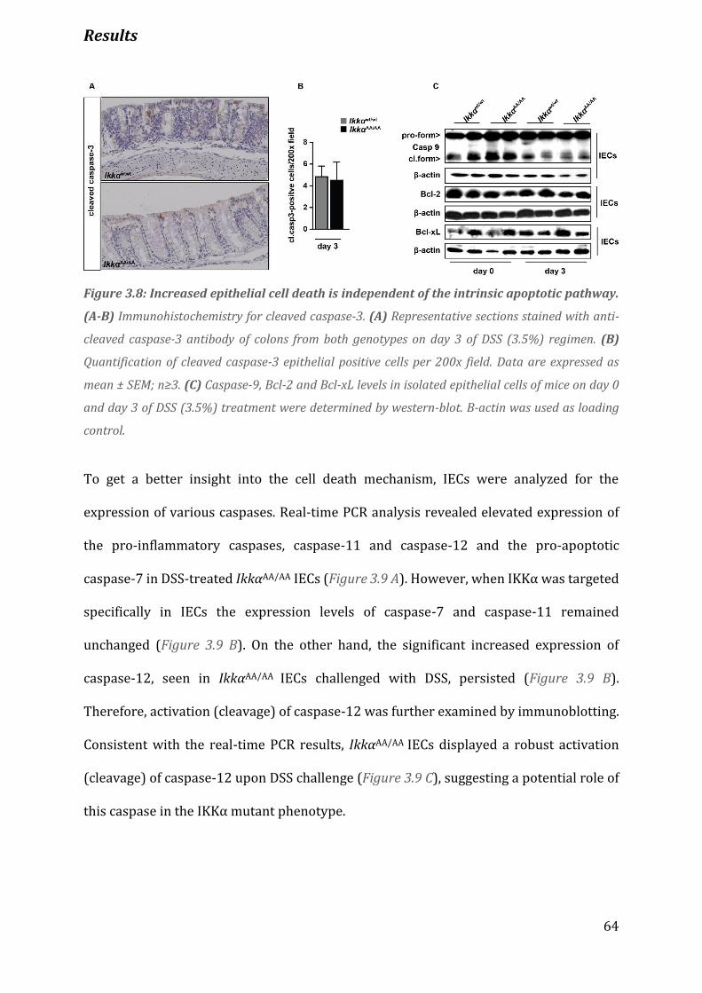
Results
64
Figure 3.8: Increased epithelial cell death is independent of the intrinsic apoptotic pathway.
(A-B) Immunohistochemistry for cleaved caspase-3. (A) Representative sections stained with anti-
cleaved caspase-3 antibody of colons from both genotypes on day 3 of DSS (3.5%) regimen. (B)
Quantification of cleaved caspase-3 epithelial positive cells per 200x field. Data are expressed as
mean ± SEM; n≥3. (C) Caspase-9, Bcl-2 and Bcl-xL levels in isolated epithelial cells of mice on day 0
and day 3 of DSS (3.5%) treatment were determined by western-blot. B-actin was used as loading
control.
To get a better insight into the cell death mechanism, IECs were analyzed for the
expression of various caspases. Real-time PCR analysis revealed elevated expression of
the pro-inflammatory caspases, caspase-11 and caspase-12 and the pro-apoptotic
caspase-7 in DSS-treated IkkαAA/AA IECs (Figure 3.9 A). However, when IKKα was targeted
specifically in IECs the expression levels of caspase-7 and caspase-11 remained
unchanged (Figure 3.9 B). On the other hand, the significant increased expression of
caspase-12, seen in IkkαAA/AA IECs challenged with DSS, persisted (Figure 3.9 B).
Therefore, activation (cleavage) of caspase-12 was further examined by immunoblotting.
Consistent with the real-time PCR results, IkkαAA/AA IECs displayed a robust activation
(cleavage) of caspase-12 upon DSS challenge (Figure 3.9 C), suggesting a potential role of
this caspase in the IKKα mutant phenotype.

Results
65
Figure 3.9: Caspase-12 is activated in IkkαΑΑ/AA IECs after DSS administration. (A-B) Expression
of caspase genes. (A) Relative mRNA expression levels were quantified in IECs of Ikkαwt/wt and
IkkαΑΑ/AA mice on day 0 and 3 of DSS regimen. (B) Relative mRNA expression levels were quantified
in IECs of Ikkαfl/fl and IkkαΔIEC mice on day 0 and 3 of DSS regimen. The mRNA levels were quantified
by real-time PCR and normalized to the level of cyclophilinB mRNA. Data are expressed as mean ±
SEM; n=3; * p<0.05, ** p<0.001, *** p<0.0001 by ANOVA followed by Bonferroni post hoc test for
multiple data sets. (C) Caspase-12 expression in IECs of Ikkαwt/wt and IkkαΑΑ/AA mice on day 0 and 3
of DSS (3.5%) regimen was determined by WB. B-actin was used as loading control. Intestinal
epithelial cells (IECs), western-blot (WB).
3.6 IkkαAA/AA mutants display decreased IL-18 serum levels
Caspase-12 was reported to exert a dominant-negative effect on caspase-1 [127] and
caspase-1 activation downstream of the NLRP3 and/or NLRP6 inflammasomes is
required for the maturation of the related cytokines IL-1β and IL-18 [107, 112, 121]. Thus,
in order to investigate the molecular basis of the impaired epithelial regeneration in
IkkαΑΑ/AA mutants and given the substantial induction of caspase-12 in intestinal epithelial
cells at both mRNA (Figure 3.9 A-B) and protein levels (Figure 9.C), serum levels of both
IL-1β and IL-18 in Ikkαwt/wt and IkkαΑΑ/AA mice were examined. Although there was no
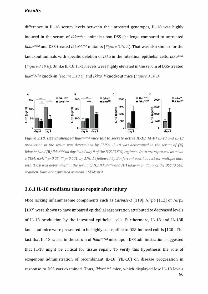
Results
66
difference in IL-18 serum levels between the untreated genotypes, IL-18 was highly
induced in the serum of Ikkαwt/wt animals upon DSS challenge compared to untreated
Ikkαwt/wt and DSS-treated IkkαΑΑ/AA mutants (Figure 3.10 A). That was also similar for the
knockout animals with specific deletion of Ikkα in the intestinal epithelial cells, IkkαΔIEC
(Figure 3.10 B). Unlike IL-18, IL-1β levels were highly elevated in the serum of DSS-treated
IkkαΑΑ/AA knock-in (Figure 3.10 C) and IkkαΔIECknockout mice (Figure 3.10 D).
Figure 3.10: DSS-challenged IkkαΑΑ/ΑΑ mice fail to secrete active IL-18. (A-D) IL-18 and IL-1β
production in the serum was determined by ELISA. IL-18 was determined in the serum of (A)
IkkαΑΑ/ΑΑ and (B) IkkαΔIEC on day 0 and day 9 of the DSS (3.5%) regimen. Data are expressed as mean
± SEM; n≥4; * p<0.05, ** p<0.001, by ANOVA followed by Bonferroni post hoc test for multiple data
sets. IL-1β was determined in the serum of (C) IkkαΑΑ/ΑΑ and (D) IkkαΔIEC on day 9 of the DSS (3.5%)
regimen. Data are expressed as mean ± SEM; n≥4.
3.6.1 IL-18 mediates tissue repair after injury
Mice lacking inflammasome components such as Caspase-1 [119], Nlrp6 [112] or Nlrp3
[107] were shown to have impaired epithelial regeneration attributed to decreased levels
of IL-18 production by the intestinal epithelial cells. Furthermore, IL-18 and IL-18R
knockout mice were presented to be highly susceptible to DSS-induced colitis [120]. The
fact that IL-18 raised in the serum of Ikkαwt/wt mice upon DSS administration, suggested
that IL-18 might be critical for tissue repair. To verify this hypothesis the role of
exogenous administration of recombinant IL-18 (rIL-18) on disease progression in
response to DSS was examined. Thus, IkkαΑΑ/AA mice, which displayed low IL-18 levels
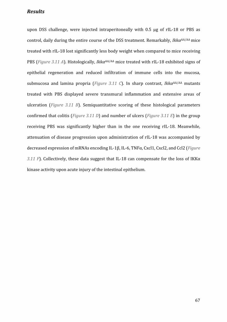
Results
67
upon DSS challenge, were injected intraperitoneally with 0.5 μg of rIL-18 or PBS as
control, daily during the entire course of the DSS treatment. Remarkably, IkkαΑΑ/AA mice
treated with rIL-18 lost significantly less body weight when compared to mice receiving
PBS (Figure 3.11 A). Histologically, IkkαΑΑ/AA mice treated with rIL-18 exhibited signs of
epithelial regeneration and reduced infiltration of immune cells into the mucosa,
submucosa and lamina propria (Figure 3.11 C). In sharp contrast, IkkαΑΑ/AA mutants
treated with PBS displayed severe transmural inflammation and extensive areas of
ulceration (Figure 3.11 B). Semiquantitative scoring of these histological parameters
confirmed that colitis (Figure 3.11 D) and number of ulcers (Figure 3.11 E) in the group
receiving PBS was significantly higher than in the one receiving rIL-18. Meanwhile,
attenuation of disease progression upon administration of rIL-18 was accompanied by
decreased expression of mRNAs encoding IL-1β, IL-6, TNFα, Cxcl1, Cxcl2, and Ccl2 (Figure
3.11 F). Collectively, these data suggest that IL-18 can compensate for the loss of IKKα
kinase activity upon acute injury of the intestinal epithelium.

Results
68
Figure 3.11: Administration of recombinant IL-18 rescues severity of colitis. (A) IkkαΑΑ/AA mice
were fed with 3.5% DSS for 5 days, followed by 4 days of regular drinking water. One group received
intraperitoneal injections with 0.5 μg recombinant IL-18 (rIL-18) daily until the end of the DSS
treatment, while the other control cohort received PBS instead. Data are expressed as mean ± SEM;
n≥5, ** p<0.001 by t test. Representative histologies from H&E-stained colons of IkkαΑΑ/AA receiving
PBS (B) and (C) rIL-18, on day 9 of the DSS treatment. (D) Histological scoring based on loss of crypts,
infiltration of immune cells into the mucosa, submucosa and muscle layer and (E) number of ulcers
on day 9 of DSS treatment. Data are expressed as mean ± SEM; n≥5; * p<0.05, by t test. (F) Expression
of inflammatory genes. Relative mRNA expression levels were quantified in whole colonic mucosa
tissue of mice on day 0 and 9 of DSS regimen. The mRNA levels were quantified by real-time PCR and
normalized to the level of cyclophilinB mRNA (n ≥ 4).
3.6.2 IECs comprise the main source of IL-18 production during DSS
colitis
Contrary to IL-1β, which is mainly produced by hematopoietic cells, IL-18 has been
identified in cells of both hematopoietic and non-hematopoietic origin and within the gut
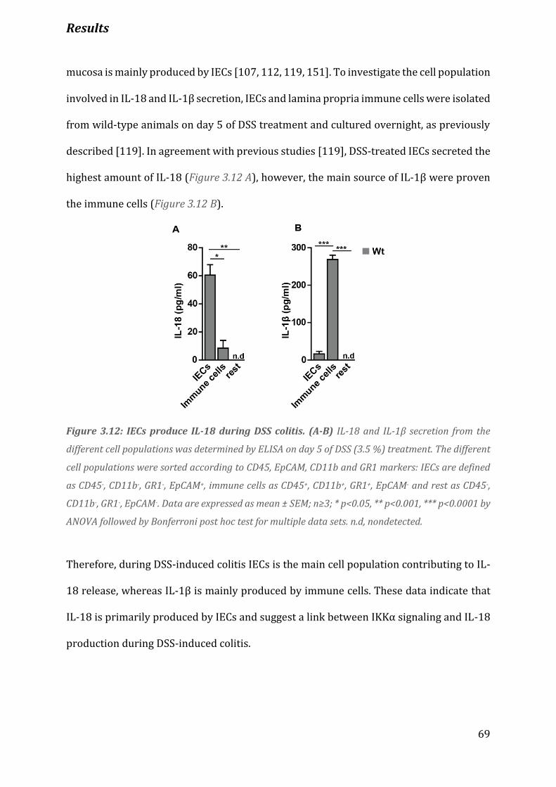
Results
69
mucosa is mainly produced by IECs [107, 112, 119, 151]. To investigate the cell population
involved in IL-18 and IL-1β secretion, IECs and lamina propria immune cells were isolated
from wild-type animals on day 5 of DSS treatment and cultured overnight, as previously
described [119]. In agreement with previous studies [119], DSS-treated IECs secreted the
highest amount of IL-18 (Figure 3.12 A), however, the main source of IL-1β were proven
the immune cells (Figure 3.12 B).
Figure 3.12: IECs produce IL-18 during DSS colitis. (A-B) IL-18 and IL-1β secretion from the
different cell populations was determined by ELISA on day 5 of DSS (3.5 %) treatment. The different
cell populations were sorted according to CD45, EpCAM, CD11b and GR1 markers: IECs are defined
as CD45-, CD11b-, GR1-, EpCAM+, immune cells as CD45+, CD11b+, GR1+, EpCAM- and rest as CD45-,
CD11b-, GR1-, EpCAM-. Data are expressed as mean ± SEM; n≥3; * p<0.05, ** p<0.001, *** p<0.0001 by
ANOVA followed by Bonferroni post hoc test for multiple data sets. n.d, nondetected.
Therefore, during DSS-induced colitis IECs is the main cell population contributing to IL-
18 release, whereas IL-1β is mainly produced by immune cells. These data indicate that
IL-18 is primarily produced by IECs and suggest a link between IKKα signaling and IL-18
production during DSS-induced colitis.
Results
70
3.7 Deficiency of caspase-12 rescues the phenotype of IkkαAA/AA mice
Caspase-12 deficiency has been associated with resistance to acute and chronic colitis
induced by DSS [119] and with efficient clearance of systemic and abdominal bacterial
infections [127, 138]. Based on the observation that IkkαAA/AA DSS-treated IECs showed
substantial activation of caspase-12 and reduced IL-18 production the role of caspase-12
during colitis was examined. Therefore, to investigate whether caspase-12 plays a role in
the severe phenotype of IkkαAA/AA mice, casp12-/-IkkαAA/AA double mutants were generated
by crossing caspase-12 knockout mice (casp12-/-) with IkkαAA/AA mice. Notably, challenge
of casp12-/-IkkαAA/AA mice with 2.5% DSS for 5 days and 4 additional days of regular water,
revealed that the double mutants lost less weight compared to IkkαAA/AA mutants reaching
that of casp12-/- mice (Figure 3.13 A). In sharp contrast to IkkαAA/AA (Figure 3.13 B), casp12-
/-IkkαAA/AA (Figure 3.13 C) and casp12-/- (Figure 3.13 D) mice showed significant
improvement at the end of the treatment and were capable of restricting mucosal damage
and immune cell infiltration by inducing crypt regeneration. Histological scoring revealed
significantly less tissue damage (Figure 3.13 E) and lower ulcer numbers (Figure 3.13 F)
in casp12-/-IkkαAA/AA related to IkkαAA/AA DSS-treated animals. No difference was observed
between casp12-/-IkkαAA/AA and casp12-/- DSS-treated mice (Figure 3.13 E-F). Thus, the data
indicate that deletion of caspase-12 ameliorates tissue damage and induces re-
epithelialazation in IkkαAA/AA mice.
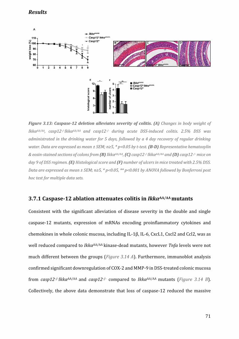
Results
71
Figure 3.13: Caspase-12 deletion alleviates severity of colitis. (A) Changes in body weight of
IkkαΑΑ/AA, casp12-/-IkkαΑΑ/AA and casp12-/- during acute DSS-induced colitis. 2.5% DSS was
administrated in the drinking water for 5 days, followed by a 4 day recovery of regular drinking
water. Data are expressed as mean ± SEM; n≥5, * p<0.05 by t-test. (B-D) Representative hematoxylin
& eosin-stained sections of colons from (B) IkkαΑΑ/AA, (C) casp12-/-IkkαΑΑ/AA and (D) casp12-/- mice on
day 9 of DSS regimen. (E) Histological score and (F) number of ulcers in mice treated with 2.5% DSS.
Data are expressed as mean ± SEM; n≥5, * p<0.05, ** p<0.001 by ANOVA followed by Bonferroni post
hoc test for multiple data sets.
3.7.1 Caspase-12 ablation attenuates colitis in IkkαAA/AA mutants
Consistent with the significant alleviation of disease severity in the double and single
caspase-12 mutants, expression of mRNAs encoding proinflammatory cytokines and
chemokines in whole colonic mucosa, including IL-1β, IL-6, CxcL1, Cxcl2 and Ccl2, was as
well reduced compared to IkkαAA/AA kinase-dead mutants, however Tnfα levels were not
much different between the groups (Figure 3.14 A). Furthermore, immunoblot analysis
confirmed significant downregulation of COX-2 and MMP-9 in DSS-treated colonic mucosa
from casp12-/-IkkαAA/AA and casp12-/- compared to IkkαAA/AA mutants (Figure 3.14 B).
Collectively, the above data demonstrate that loss of caspase-12 reduced the massive

Results
72
inflammatory response to an extent adequate to prevent DSS-induced mortality and
morbidity observed in IkkαAA/AA mice.
Figure 3.14: Deletion of caspase-12 reduces the expression of proinflammatory factors. (A)
Expression of inflammatory genes. Relative mRNA expression levels were quantified in whole colonic
mucosa tissue of mice on day 0 and 9 of DSS regimen. The mRNA levels were quantified by real-time
PCR and normalized to the level of cyclophilinB mRNA (n ≥ 4). (B) COX-2 and MMP-9 expression in
whole colonic extracts on day 0 and 9 of DSS regimen. COX-2 and MMP-9 expression were determined
by western blot (WB).
3.8 Impaired IKKα activation results in enhanced ER stress
Based on the finding that the severe phenotype in IkkαAA/AA mice was attributed to
caspase-12, the mechanism by which caspase-12 is activated was further explored.
Caspase-12 was found to localize at the cytoplasmic site of the endoplasmic reticulum
(ER) [128] and has been suggested to be activated upon ER stress [126]. In notion of that,
several ER stress markers, including IRE1α, XBP1, IRE1β, GRP78, GRP94, and CHOP were
examined. Real-time PCR analysis revealed that the expression of Ire1α was significantly
elevated in both untreated and DSS-treated IkkαAA/AA IECs, while the expression of Grp78
raised in IkkαAA/AA enterocytes only upon DSS challenge (Figure 3.15 A). Also, the
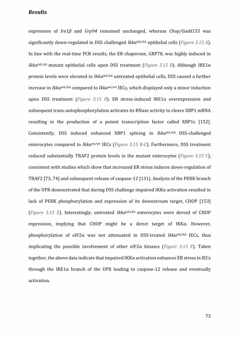
Results
73
expression of Ire1β and Grp94 remained unchanged, whereas Chop/Gadd153 was
significantly down-regulated in DSS challenged IkkαAA/AA epithelial cells (Figure 3.15 A).
In line with the real-time PCR results, the ER chaperone, GRP78, was highly induced in
IkkαAA/AA mutant epithelial cells upon DSS treatment (Figure 3.15 B). Although IRE1α
protein levels were elevated in IkkαAA/AA untreated epithelial cells, DSS caused a further
increase in IkkαAA/AA compared to Ikkαwt/wt IECs, which displayed only a minor induction
upon DSS treatment (Figure 3.15 B). ER stress-induced IRE1α overexpression and
subsequent trans-autophosphorylation activates its RNase activity to cleave XBP1 mRNA
resulting in the production of a potent transcription factor called XBP1s [152].
Consistently, DSS induced enhanced XBP1 splicing in IkkαAA/AA DSS-challenged
enterocytes compared to Ikkαwt/wt IECs (Figure 3.15 B-C). Furthermore, DSS treatment
reduced substantially TRAF2 protein levels in the mutant enterocytes (Figure 3.15 C),
consistent with studies which show that increased ER stress induces down-regulation of
TRAF2 [73, 74] and subsequent release of caspase-12 [131]. Analysis of the PERK branch
of the UPR demonstrated that during DSS challenge impaired IKKα activation resulted in
lack of PERK phosphorylation and expression of its downstream target, CHOP [153]
(Figure 3.15 E). Interestingly, untreated IkkαAA/AA enterocytes were devoid of CHOP
expression, implying that CHOP might be a direct target of IKKα. However,
phosphorylation of eIF2α was not attenuated in DSS-treated IkkαAA/AA IECs, thus
implicating the possible involvement of other eIF2α kinases (Figure 3.15 E). Taken
together, the above data indicate that impaired IKKα activation enhances ER stress in IECs
through the IRE1α branch of the UPR leading to caspase-12 release and eventually
activation.
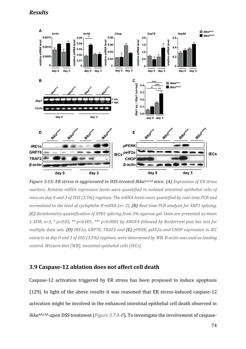
Results
74
Figure 3.15: ER stress is aggravated in DSS-treated IkkaAA/AA mice. (A) Expression of ER stress
markers. Relative mRNA expression levels were quantified in isolated intestinal epithelial cells of
mice on day 0 and 3 of DSS (3.5%) regimen. The mRNA levels were quantified by real-time PCR and
normalized to the level of cyclophilin B mRNA (n= 3). (B) Real time PCR analysis for XBP1 splicing.
(C) Densitometry quantification of XPB1 splicing from 3% agarose gel. Data are presented as mean
± SEM; n=3, * p<0.05, ** p<0.001, *** p<0.0001 by ANOVA followed by Bonferroni post hoc test for
multiple data sets. (D) IRE1α, GRP78, TRAF2 and (E) pPERK, peIF2α and CHOP expression in IEC
extracts at day 0 and 3 of DSS (3.5%) regimen, were determined by WB. Β-actin was used as loading
control. Western blot (WB), intestinal epithelial cells (IECs).
3.9 Caspase-12 ablation does not affect cell death
Caspase-12 activation triggered by ER stress has been proposed to induce apoptosis
[129]. In light of the above results it was reasoned that ER stress-induced caspase-12
activation might be involved in the enhanced intestinal epithelial cell death observed in
IkkαAA/AA upon DSS treatment (Figure 3.7 A-B). To investigate the involvement of caspase-
Results
75
12 activation in mediating cell death, casp12-/-IkkαAA/AA and IkkαAA/AA mice were subjected
to DSS treatment for 3 days and TUNEL staining was performed. TUNEL staining of DSS-
treated colons revealed no difference in epithelial cell death upon caspase-12 ablation
(Figure 3.16 A). This result is in agreement with recent studies which suggest that caspase-
12 deficient cells are as prone to apoptosis induced by various stimuli as caspase-12
proficient cells [127, 133].
Figure 3.16: Caspase-12 deletion does not affect cell death. (A) Quantification of TUNEL-positive
intestinal epithelial cells of colons of IkkαAA/AA and casp12-/-IkkαAA/AA mice on day 3 of DSS (2.5%)
regimen. Data are expressed as mean ± SEM; n≥3.
3.10 Defective IKKα activation impairs autophagy
Several lines of evidence highlight the close link between UPR and autophagy [75].
Furthermore, ER stress has been shown to be involved in autophagy induction [154]. In
order to investigate whether ER stress triggered activation of autophagy, IECs isolated
from Ikkαwt/wt and IkkαAA/AA mice that were left either untreated or were challenged with
DSS, were analysed for autophagic markers. LC3, the mammalian Atg8 homolog,
undergoes proteolysis to produce the cytosolic LC3-I form, which is then lipidated to
generate the LC3-II form. The LC3-II form is then attached to both faces of the
Results
76
autophogosomal membrane [155]. Both Ikkαwt/wt and IkkαAA/AA IECs exhibited obvious
accumulation of LC3-II, consistent with the fact that basal, constitutive level of autophagy
occurs many cells as a quality control mechanism [155] (Figure 3.17 A). However, in
contrast to Ikkαwt/wt, IkkαAA/AA IECs subjected to DSS showed only a modest increase in
LC3-I (Figure 3.17 A) indicating autophagosome formation. Quantification of the lipidated
LC3-II form revealed that autophagosome formation was significantly elevated in
IkkαAA/AA DSS-treated IECs (Figure 3.17 B).
P62 acts as a receptor that binds ubiquitinated proteins and organelles targeting them
into autophagosomes. Eventually, when autophagy completion occurs p62 is degraded
within the lysosomes [156]. p62 accumulation occurs when autophagic protein
degradation is blocked [85]. Interestingly, whereas p62 was decreased in Ikkαwt/wt treated
IECs, IkkαAA/AA DSS-treated IECs displayed accumulated p62 levels (Figure 3.17 C),
indicating that autophagy was impaired. Moreover, ATG16L1 is an important autophagy
protein in Paneth cells [14]. Given the recent association of, another ATG16L1 isoform,
ATG16L2 with IKKα in pancreas, the potential interaction between ATG16L1 and IKKα in
IECs was explored. Remarkably, mutant and wild-type IKKα interacted with ATG16L1 in
unchallenged IECs, indicating that IKKα might be involved in the regulation of this
autophagic protein in the colon. Although this interaction was maintained in DSS-treated
Ikkαwt/wt IECs it was abrogated in the mutant IECs after DSS administration (Figure 3.17
D). Furthermore, the total levels of ATG16L1 decreased after DSS challenge and were
further diminshed, in the mutant DSS-treated enterocytes (Figure 3.17 E), designating that
in response to stress ATG16L1 was severely affected by the mutant IKKα. However the
relevance of this interaction and the impact of IKKα on ATG16L1 remains still elusive.
Taken together, these data suggest that defective IKKα activation might interfere with
proper autophagic degradation thus leading to p62 accumulation.
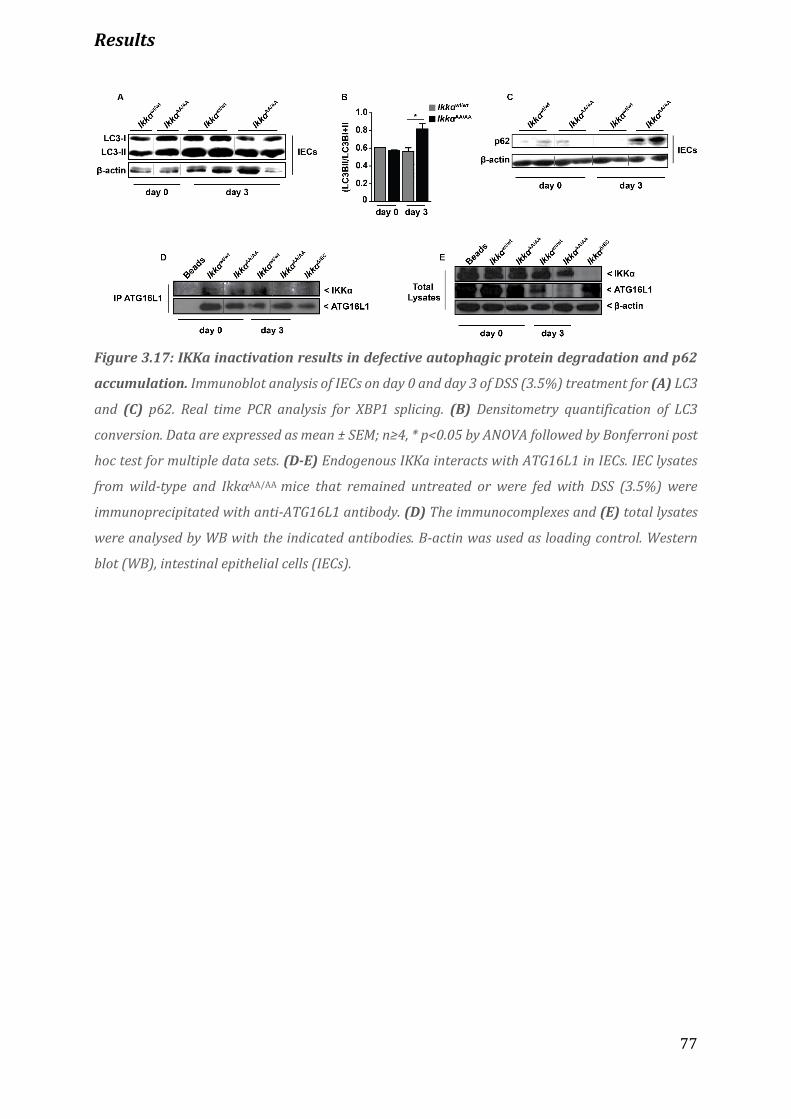
Results
77
Figure 3.17: IKKa inactivation results in defective autophagic protein degradation and p62
accumulation. Immunoblot analysis of IECs on day 0 and day 3 of DSS (3.5%) treatment for (A) LC3
and (C) p62. Real time PCR analysis for XBP1 splicing. (B) Densitometry quantification of LC3
conversion. Data are expressed as mean ± SEM; n≥4, * p<0.05 by ANOVA followed by Bonferroni post
hoc test for multiple data sets. (D-E) Endogenous IKKa interacts with ATG16L1 in IECs. IEC lysates
from wild-type and IkkαAA/AA mice that remained untreated or were fed with DSS (3.5%) were
immunoprecipitated with anti-ATG16L1 antibody. (D) The immunocomplexes and (E) total lysates
were analysed by WB with the indicated antibodies. B-actin was used as loading control. Western
blot (WB), intestinal epithelial cells (IECs).

Discussion
78
4. Discussion
IKKα, initially identified as a subunit of the IκB kinase complex that directly
phosphorylates IκB, is an important protein kinase involved in numerous biological
processes such as, cell survival, inflammation, development of secondary lymphoid
organs and transcriptional regulation [145]. The fact that mice lacking IKKα die soon after
birth [57] highlights its major significance during development and tissue homeostasis
that cannot be substituted by other members of the IKK complex. Although its biological
importance has been addressed in several human diseases the exact mechanisms remain
still ill defined. This report here, provides important information for IKKα mode of action
in maintaining intestinal homeostasis. Using IkkαAA/AA mutants in a model of acute colitis
this study provides strong evidence of a novel function of epithelial IKKα in suppressing
excessive inflammatory responses in the large intestine, thus maintaining a fine balance
between the intestinal epithelium and the host immune system.
4.1 Activation of IKKα suppresses inflammation in the large intestine
Given the fact that IBDs affect the quality of life of million people in the western world and
its incidence has sharply enhanced since the early 1950’s [157], identification of factors
and singnaling pathways implicated in disease pathogenesis comprise an urgent need.
One of the most commonly used model of IBD is by challenging mice with DSS salt. This
model, which closely mimics UC in humans, induces inflammation in rodents that starts
distally after about 5 days and is confined to the colonic mucosa [146]. The mechanism of
action is by affecting directly the inner mucus layer and inducing death of IECs. IECs death
disrupts the intestinal barrier and eventually allows bacterial translocation to the mucosa
that induce a strong local inflammatory response [158].
Discussion
79
In the present study IKKα is proven a critical regulator of intestinal homeostasis. Is
showed that mice engineered to express an inactive form of IKKα are unable to restrict
excessive inflammatory responses when colon injury is induced by DSS. The colon of those
mice is characterized by complete loss of crypts, massive infiltration of immune cells and
large areas of ulceration (Figure 3.1). The enhanced inflammation seen in IkkαAA/AA
mutants is attributed to defective IKKα signaling in the intestinal epithelium (Figure 3.3
and 3.4). Interestingly, several studies have demonstrated that IKKα of epithelial origin is
capable of suppressing enhanced inflammatory responses in various organs, including
skin, lungs, pancreas and intestine [51, 66, 159-162]. In the skin and lungs impaired IKKα
signaling enhances inflammation and macrophage recruitment and elevates the
expression of cytokines and chemokines inducing tissue pathology [139, 159, 160],
mirroring the events occurring in the intestine upon DSS-induced injury. The enhanced
inflammatory phenotype seen in the skin and lung epithelia of mice defective in IKKα
signaling is mainly attributed to elevated EGFR/ERK activities, which eventually lead to
enhanced production of cytokines and chemokines allowing macrophages infiltration
[139, 159, 160]. In the intestine, on the other hand, IKKα suppresses inflammation by
inhibiting caspase-12 over-activation, most likely indirectly, thereby promoting
inflammasome activation and downstream IL-18 production by the IECs. These findings
suggest that impaired IKKα signaling orchestrates an inflammatory microenvironment
which eventually triggers pathology in multiple epithelial organs, in both humans and
mice. However, IKKα activation in the intestine limits the recruitment of IFNγ-producing
M1-like polarized myeloid cells, thereby, promoting tumorigenesis in a nonautonomous
manner [162]. Therefore, under inflammatory conditions cell autonomous IKKα signaling
is essential for limiting exaggerated immune responses and to restore intestinal
homeostasis. However, in circumstances where environmental triggers cause cell
Discussion
80
mutagenesis, IKKα seems to act in a nonautonous manner to suppress recruitment of
myeloid cells and to promote tumorigenesis. It seems that IKKα can act in many different
ways, either in an autocrine or paracrine manner, depending on the external signals and
pathways activated. This in turn results in differential contribution of IKKα in the final
phenotype.
4.1.1 Pathology in IkkαAA/AA mice is independent of the alternative NF-
κB pathway
The canonical NF-κB pathway relies for its activation on the IκB kinase complex, which
phosphorylates and marks the IκB inhibitors for proteasomal degradation. However, the
alternative pathway is governed exclusively by IKKα and depends for its activation on the
phosphorylation and partial degradation of the NF-κB2/p100 IκB-like inhibitor [144].
The data obtained here argue for the fact that IKKα acts independently of the alternative
NF-κB activation, as deletion of the NF-κB2 precursor protein, p100, did not affect
pathology induced by DSS (Figure 3.5). Nonetheless, this is not surprising since IKKα has
been shown to have additional NF-κB-independent functions, including suppression of
skin inflammation [159] and promotion of tumor cell proliferation in the intestine [162].
The alternative signaling seems to be mainly important for developmental processes, such
as development of mammary gland [149] or secondary lymphoid organs [60, 67].
Defective IKKα activation results in diminished NF-κB activity (Figure 3.6), suggesting
that it is involved in the canonical pathway. It has been shown that Ikkβ deletion in IECs
renders mice susceptible to DSS-induced colitis [163], while simultaneous deletion of
both Ikkα and Ikkβ kinases induces spontaneous colitis in mice [51]. This indicates that
proper activation of NF-κB in the intestinal epithelium is required to maintain intestinal
homeostasis. Inactivation of NF-κB upon defective IKKα signaling has been documented
Discussion
81
in other studies as well [148, 149, 164]. On the contrary, IKKα has been as well reported
to limit NF-κB activation in response to specific signals [62, 64]. The reason why in some
tissues or cell types IKKα acts in different ways might depend on various factors such as,
its expression, subcellular localization or interaction with different partners and on the
specific signal.
4.2 IKKα activates inflammasome function and IL-18 production
Caspases are cysteine proteases that regulate tissue homeostasis, embryonic
development, cell differentiation and removal of injured or harmful cells from many
tissues in the body. Their activity is mainly regulated at the posttranslational level,
allowing their rapid activation and response to cellular stress and pathogenic stimuli
[165]. Some caspases, such as caspase-1, -5, -12 and -11 (-4 in humans), are activated
during innate immune responses and control the inflammatory reaction by processing the
pro-form of IL-1 family members (IL-18 and IL-1β) into their active forms, and therefore
are called inflammatory caspases [165].
In the present study caspase-12 was of particular interest due to the significant activation
(cleavage) found in IECs from DSS-treated IkkαAA/AA animals (Figure 3.9 C). Nevertheless,
the functional role of this activation remains still unidentified. The reason for that is the
highly restricted substrate specificity and low catalytic efficiency of this enzyme, as it
possesses proteolytic activity, which is, however, restricted to its self [133].
The importance of caspase-12 in the inflammatory phenotype of IkkαAA/AA mice was
supported by the rescue of IkkαAA/AA animals upon deletion of this caspase. Abrogation of
caspase-12 limited mucosal damage by promoting regeneration of crypts and surface
epithelia (Figure 3.13), and inhibited production of proinflammatory factors (Figure 3.14).
Discussion
82
Moreover, it is demonstrated that IL-18 levels are significantly downregulated in the
serum of IkkαAA/AA mice upon DSS treatment (Figure 3.10). The current data suggest that
the phenotype of IkkαAA/AA mice is ascribed to decreased IL-18 production, since
administration of this recombinant cytokine induced epithelial regeneration and reduced
inflammation in IkkαAA/AA mutants treated with DSS (Figure 3.11). In agreement with
former studies [107, 119], the data obtained here show that IEC comprise the major
source of IL-18 during DSS colitis (Figure 3.12).
IKKα-dependent activation of the inflammasome seems to be cell-type specific, since IKKα
was recently shown to limit inflammasome function in macrophages [166]. The authors
reported that macrophages from IkkαAA/AA and IkkαK44A (in which the ATP-binding domain
of IKKα is mutated resulting in loss of kinase activity) hyperproduce IL-1β in response to
stimuli that activate the inflammasomes [166]. In agreement with that the enhanced IL-
1β serum levels seen in DSS-treated IkkαAA/AA mice may have arrived from overactivation
of the inflammasome in immune cells, as it is shown that IL-1β is mainly produced by the
immune cells (Figure 3.12 B). Contrary to IkkαAA/AA mice, mice lacking Ikkα specifically in
the myeloid cells (IkkαΔmye) have less tissue damage than wild-type controls (Figure 3.4 F-
J) and low IL-1β serum levels (not shown here), suggesting that IL-1β levels correlate with
the extent of tissue damage. IL-1β has been shown to increase epithelial permeability
[167] and increasing levels of this cytokine correlate with severity of intestinal
inflammation in patients with CD and UC [168], which could explain how IL-1β
contributes to tissue damage. Nevertheless, since IL-1β signaling was not profoundly
explored in the present study it is still not well defined what the exact role of myeloid-
derived IL-1β is in the absence of proper IKKα signaling.
The role of IL-18 is complex and its possible contribution to the maintenance of chronic
inflammation in the intestine is unclear. Several studies have suggested that IL-18 could

Discussion
83
be an effector cytokine in IBD, as circulating and local IL-18 levels have been associated
with disease severity [116, 169, 170]. On the contrary, more recent investigations have
shown that mice deficient in IL-18 production are more susceptible rather than resistant
to DSS-induced colitis [107, 110, 112, 119, 171]. However, excessive production of IL-18
is pathogenic: inflammasome hyperactivation leads to increased IL-18 and IL-1β
secretion by macrophages, thereby, resulting in increased DSS-induced colitis [87]. It
appears that a dramatic shift in the cellular source of IL-18, from IECs to lamina propria
macrophages and dendritic cells, occurs as the severity of disease progresses in patients
with CD and UC [116, 151]. In the early phase of IBD injury of mucosa results in innate
immune responses to environmental factors, such as commensal or pathogenic bacteria,
that in turn may activate IECs to produce IL-18 [172]. However, in the later stages of
chronic and severe inflammation the mucosa is infiltrated by lymphocytes, macrophages
and other immune cells, which provide the cellular source of IL-18 and other cytokines
(e.g IL-12) to mount a prototypic Th1-mediated immune response [151, 172].
Caspase-1 and Asc (the inflammasome adaptor) knockout mice develop severe colitis
induced by DSS [107, 112, 119], while Caspase-12 deficient mice are protected [119].
Furthermore, caspase-12 was shown to interact with caspase-1 and to inhibit its
activation [127, 133]. The unique function of caspase-12 as a dominant-negative regulator
of the inflammatory caspase cascade has been reported in both humans [136] and mice
[119, 127]. Given the dominant negative effect of caspase-12 on caspase-1, the data
propose that IECs unable to activate IKKα upon DSS treatment, display increased caspase-
12 activation (Figure 3.9 C), which in turn acts as dominant negative regulator of caspase-
1 and therefore, limits the production of the cytoprotective IL-18. As both NLRP6 [112,
171] and NLPR3 [107, 110] inflammasomes were shown to be required for IL-18
production in vivo, it remains still unknown whether IKKα signals through the NLRP3 or
Discussion
84
NLRP6 inflammasome to induce IL-18 secretion. Likewise, the mechanism by which IL-18
production confers its protective effect on the colonic mucosa is not very clear yet and
whether it acts directly or indirectly on the IECs remains a subject of investigation
4.3 Defective IKKα activation results in ER stress
ER stress has been genetically associated with both forms of IBD, Crohn’s disease and
Ulcerative colitis, through a candidate gene study of XBP1 [16]. The intestinal epithelium
is highly sensitive, even to minor impairments in UPR signaling as can viewed by animal
models that either lack or have mutated certain proteins of the UPR signaling. For
instance, mice deficient in Atf6a and p58IPK [78], Ire1β [77], Arg2 [173], Xbp1[16] or
bearing hypomorphic allele of the Site 1 protease (S1P)-encoding gene, Mbtps1 [174] or
Xbp1[16] develop severe colitis induced by DSS or in the case of Arg2 and Xbp1
spontaneous inflammation of the small intestine. Chemical chaperones such as TUDCA
and PBA, which promote ER homeostasis and increase ER folding capacity, reduce ER
stress in IECs and alleviate acute and chronic colitis in mice, further demonstrating the
significance of proper UPR signaling in maintaining intestinal homeostasis [78].
It is demonstrated that activation of the UPR in the setting of impaired IKKα signaling,
results in increased Grp78, Ire1α and Xbp1 mRNA splicing (Figure 3.15 A-D). It has been
shown that hypomorphic XBP1 function in the epithelium results in ER stress, manifested
by an upregulation of GRP78 and profound induction of IRE1α [79]. Moreover, mice
deficient in Ire1β, which is specifically expressed in the IECs, are susceptible to DSS [77],
further emphasizes the importance of IRE1α-XBP1 axis in preserving homeostasis of the
intestinal epithelium.
Previous studies have shown that ER stress induces the formation of IRE1α and IKK
complex through TRAF2 [73, 175] and dissociation of TRAF2 from caspase-12 [131].
Discussion
85
TRAF2 is also considered a protein sensitive to degradation and was shown to be reduced
under ER stress conditions [73]. Moreover, Traf2 knockout mice develop spontaneous
colitis illustrating its crucial role in controlling homeostasis of the colon [176]. In this
study it is shown that DSS-treated IkkαAA/AA IECs display critically low levels of TRAF2
(Figure 3.15 D), which in agreement with these studies, underlie the importance of TRAF2
in maintaining colon physiology.
Examination of the PERK pathway revealed that it is significantly down-regulated (Figure
3.15 A and E), suggesting that IKKα loss induces enhanced ER stress that cannot be
compensated by the other UPR pathways. Previous studies reported that loss of
PERK/CHOP signaling cascade is linked to defects in cell death sensitivity and ROS-
mediated ER stress [153]. PERK signaling is also required for proper differentiation of
intestinal epithelial cells [79], delineating the importance of PERK pathway in the
intestine. Thus, it seems that elevated IRE1α and reduced PERK signaling leads to
increased sensitivity of the epithelium to environmental triggers, such as DSS, that further
challenge the UPR and its compensatory pathways.
ER stress can be both a primary reason (as seen in Xbp1 knockout mice [16]) and also a
consequence of intestinal inflammation (as observed in Il10 deficient mice). Several
studies have proposed evidence of ER stress, based on increased Grp78 and Xbp1 splicing,
in a significant proportion of UC and CD patients [27, 177], suggesting that ER stress is a
consequence of inflammation. The present report proposes that caspase-12 activation,
which occurs as a result of ER stress, is causing the massive inflammation seen in IkkαAA/AA
mice in response to DSS, suggesting ER stress as an upstream activator of inflammation
(chapters 3.7 and 3.7.1). Nevertheless, based on these findings, the reverse order of events
cannot be completely excluded and it should be further investigated.
Discussion
86
Given the substantial caspase-12 activation found in IkkαAA/AA IECs (Figure 3.9 C), it could
be therefore assumed that in IkkαAA/AA mutant IECs DSS triggers activation of the
IRE1α/XBP1 branch of the UPR, down-regulation of TRAF2 and thus, dissociation from
caspase-12. Caspase-12 in turn is activated causing an inflammatory environment in the
colon that results in the severe pathology of IkkαAA/AA mice (Figure 3.13 and 3.14).
4.4 IKKα controls autophagy
Perturbations in the function of autophagy-related proteins may contribute not only to
increased susceptibility to infection, but also to chronic inflammatory diseases and
autoimmune diseases. The only well-characterized link is between mutations in
autophagy proteins and IBD, especially Crohn’s disease [178]. The significance of
autophagy in intestinal physiology can be seen in animal models lacking certain
components of the autophagic machinery, such as Atg16l1, Atg7 and Atg5 [14, 86, 179-
181]. These mice develop severe spontaneous Crohn’s-disease-like transmural ileitis or
DSS-induced colitis, due to inefficient autophagic clearance of enteric pathogens and
aberrant immune priming that eventually act as a trigger for intestinal inflammation.
The IKK complex has been suggested a biological ‘switch’ for the rapid induction of
autophagy. It was reported that IKK activation is able to effectively trigger autophagy and
reversely, optimal induction of autophagy by several stimuli requires IKK activation [182,
183], supporting an intimate link between IKK complex and autophagy Here, it is shown
that IECs unable to activate IKKα in response to DSS are defective in autophagy
completion, which results in inefficient degradation and accumulation of p62 (Figure
3.17). LC3 conversion, which is used as a marker for autophagosome formation, is greatly
elevated in IkkαAA/AA IECs challenged with DSS, indicating that autophagy is activated.
However, p62 is significantly increased, suggesting that alterations occur in the late stage
Discussion
87
of the autophagic process. Loss of IKKα in pancreatic cells has been shown to impair
autophagy, by means of increased LC3 conversion and p62 accumulation, and eventually
to trigger inflammation [66]. It seems that IKKα, at least in some tissues, utilizes similar
mechanisms to maintain homeostasis.
P62 has been suggested to regulate formation of protein aggregates and to be cleared out
by autophagy [93]. Autophagy-deficient cells display elevated p62 levels and genetic
ablation of p62 suppresses the appearance of protein aggregates and rescues pathology
caused by autophagy deficiency [66, 93-95]. P62 as an adaptor protein has been linked to
IKK/NF-κB activation in response to diverse stimuli, such as IL-1 [96], RANK ligand [97]
or nerve growth factor (NGF)[98]. Another report suggesting the existence of a signalling
connection between IKK/NF-κB activation and p62 came from Duran et. al where they
showed that p62 is essential for Ras-induced transformation of lung epithelial cells and
IKK/NF-κB activation [184]. However, under conditions of stress, autophagy-defective
cells accumulate p62, which suppress canonical NF-κB activation [185]. Specifically,
under metabolic stress autophagy-deficient tumor cells preferentially accumulate p62
and fail to exert protein quality control, leading to enhanced ER stress and ROS
production. This failure of autophagy-defective cells to eliminate p62 is sufficient to
suppress NF-κB, impair survival and drive oncogenesis [185]. In a similar way is thought
that inadequate IKKα signaling in IECs results in defective autophagy that causes p62
accumulation (Figure 3.17 C) and ER stress (Figure 3.15 C). It seems that under conditions
where autophagy must be activated, IKK/NF-κB axis contributes to p62 degradation by
inducing autophagy. Nevertheless, to connect the striking phenotype in IkkαAA/AA mutants
and the relevance of p62 accumulation in disease outcome, it is essential to explore
whether loss of p62 will resolve the inflammatory phenotype observed in IkkαAA/AA mice.
The role of p62 ablation in IkkαAA/AA phenotype is currently under investigation.
Discussion
88
Atg16l1 is an essential autophagy gene [87] and a candidate gene responsible for
susceptibility to Crohn’s disease [81]. Mice deficient in Atg16l1 die within one day of
delivery, indicating that is required for survival during neonatal starvation [87].
Importantly, IKKα is shown to interact with ATG16L1 (Figure 3.17 D) in IECs indicating
that is somehow implicated in the regulation of this autophagy protein. This interaction,
however, is abrogated when IkkαAA/AA mutant IECs are challenged with DSS. Wild-type
DSS-treated IECs show reduced ATG16L1 protein levels and intact ATG16L1-IKKα
interaction (Figure 3.17 E), indicating that functional IKKα might be essential for inducing
autophagy completion together with ATG16L1. Recently, it was reported that the Crohn’s-
disease-associated Atg16l1 (T300A) risk variant is highly susceptible to proteolytic
degradation resulting in defective autophagy, which in turn establishes a chronic
inflammatory state [86, 181]. The critically low levels of ATG16L1 seen in the DSS-treated
IkkαAA/AA IECs and the lack of ATG16L1-IkkαAA/AA interaction upon DSS challenge (Figure
3.17 E) presumably designates that under conditions of stress, mutant IKKα sensitizes
ATG16L1 for degradation. As IKKα is a serine/threonine kinase it could be speculated that
phosphorylation of ATG16L1 might be required to maintain this interaction with IKKα
upon stress, as at resting state mutant IKKα is able to associate with ATG16L1 (Figure 3.17
D). It is possible that activation of IKKα is required to direct ATG16L1 to the autophagic
process. Since it is claimed here that autophagy is compromised at later stages, ATG16L1
might function as an adaptor protein involved in autophagy completion, as suggested
elsewhere [66]. Furthermore, ATG16L1 was required for Cullin-3 (an E3 ubiquitin ligase)
activation and thus, for Cul-3-dependent ubiquitination and degradation of p62 [95]. This
suggests a possible mechanism by which ATG16L1 regulates p62 degradation and
thereby, limits inflammatory conditions. Nonetheless, whether IKKα is a kinase for
Discussion
89
ATG16L1 and/or IKKα activation is essential to direct ATG16L1 to autophosomes and
through which mechanism requires further examination.
The UPR and autophagy are integrally linked pathways. Not only may ER stress induce
autophagy [83, 179, 186], but also vice versa, impaired autophagy can lead to ER stress
[179, 185, 187]. In light of these findings and given the close relationship of ineffective
autophagy with ER stress induction, it is likely that in the model of the present study
impairment of autophagy might have triggered ER stress. Nonetheless, to suggest that
further investigation is required and p62 ablation might shed light into this speculation.
Altogether, it is suggested that IKKα might control autophagy through the regulation of
ATG16L1, a highly implicated protein in IBD. IECs in which IKKα is mutated cannot
complete autophagy successfully, resulting in accumulation of p62. This in turn triggers
ER stress and subsequently caspase-12 release, which compromises mucosal immunity
and augments intestinal inflammation in the presence of DSS. Most likely this mucosal
inflammation depends on the microbiome and proinflammatory cytokines and derives
from increased IEC death, ER stress and caspase-12 activation, which are synergistically
increased when autophagy is impaired.
Conclusion
90
5. Conclusion
Physiologic levels of inflammation are protective, while excessive inflammation is
deleterious and is at the basis of IBD. One of the foremost inflammation-associated
pathways is the NF-κB pathway, with IKKα being one of the central regulators of this
pathway. IKKα is a protein kinase involved in inflammatory processes in various organs,
yet, its specific function during acute inflammation of the colon has not been investigated
to date.
The results presented in this study show that mice bearing an inactive form of IKKα are
significantly more susceptible to DSS-induced colitis. The exacerbated inflammatory
response is attributed to defective IKKα signaling in the IECs, suggesting that proper IKKα
activation in local cells of the colonic mucosa is critical for protection against DSS-induced
injury. The current model proposes that in response to environmental factors that can
damage the intestinal mucosa, IKKα activation is essential to stimulate autophagy via the
autophagy adaptor protein ATG16L1, resulting in efficient degradation of p62 and other
ubiquitinated proteins. This in turn prevents ER stress and caspase-12 activation, thus,
limiting the deregulated inflammatory responses in the colon (Figure 5. A).
On the other hand, absence of IKKα phospho-activation in the epithelial compartment,
compromises the autophagic process, presumably by regulating ATG16L1. Inactivation of
autophagy causes accumulation of p62 and other proteins and leads to ER stress, which
in turn activates and releases caspase-12. Caspase-12 suppresses caspase-1 activity and
downstream IL-18 production, which is required for epithelial regeneration and tissue
repair. The failure of the injured epithelium to regenerate allows eventually bacterial
products or commensals to penetrate into the mucosa and submucosa, where they
stimulate resident immune cells through pattern recognition receptors. Enhanced
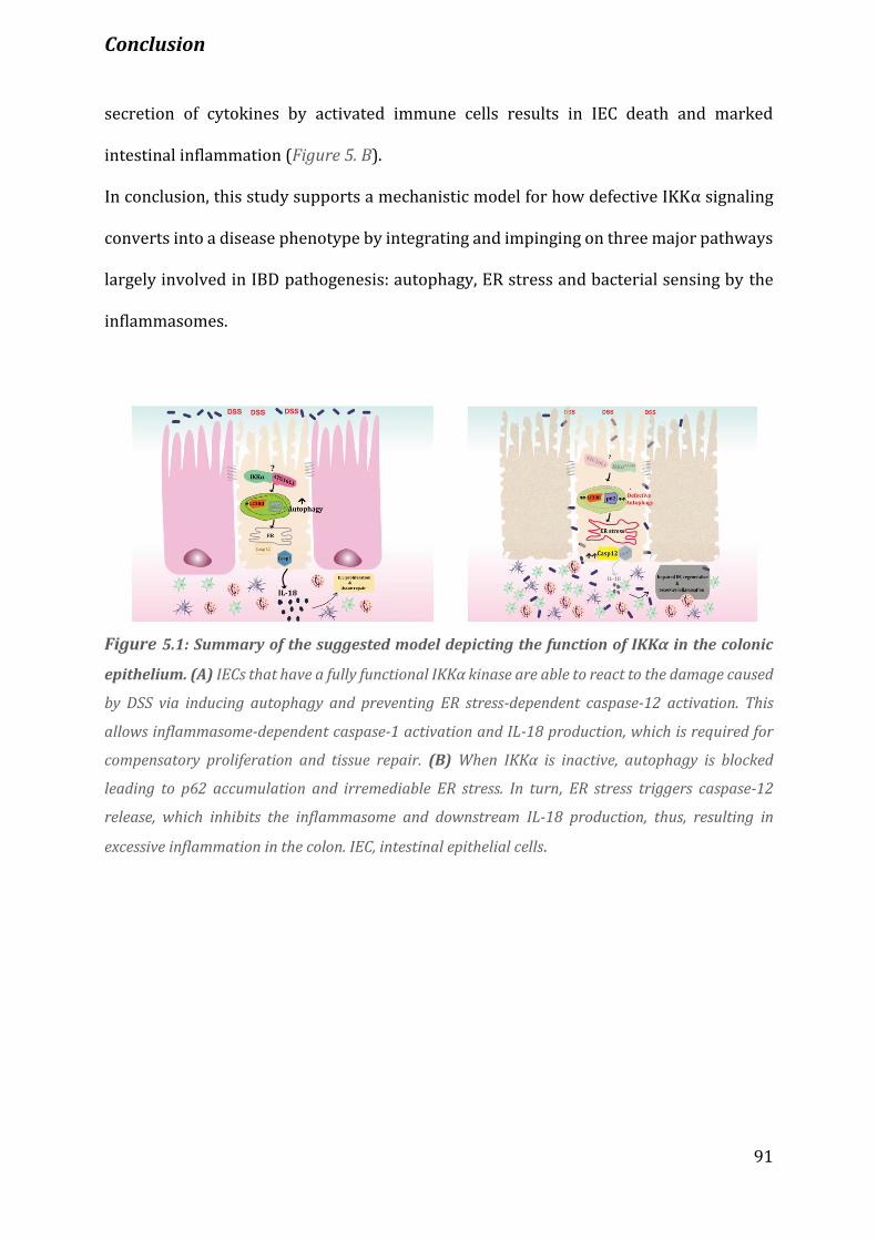
Conclusion
91
secretion of cytokines by activated immune cells results in IEC death and marked
intestinal inflammation (Figure 5. B).
In conclusion, this study supports a mechanistic model for how defective IKKα signaling
converts into a disease phenotype by integrating and impinging on three major pathways
largely involved in IBD pathogenesis: autophagy, ER stress and bacterial sensing by the
inflammasomes.
Figure 5.1: Summary of the suggested model depicting the function of IKKα in the colonic
epithelium. (A) IECs that have a fully functional IKKα kinase are able to react to the damage caused
by DSS via inducing autophagy and preventing ER stress-dependent caspase-12 activation. This
allows inflammasome-dependent caspase-1 activation and IL-18 production, which is required for
compensatory proliferation and tissue repair. (B) When IKKα is inactive, autophagy is blocked
leading to p62 accumulation and irremediable ER stress. In turn, ER stress triggers caspase-12
release, which inhibits the inflammasome and downstream IL-18 production, thus, resulting in
excessive inflammation in the colon. IEC, intestinal epithelial cells.
References
92
6. References
1. Liao, D.-H., Gastrointestinal tract modelling in health and disease. World Journal of Gastroenterology, 2009. 15(2): p. 169.
2. Clevers, L.G.v.d.F.a.H., Stem cells, self-renewal, and differentiation in the intestinal epithelium. Annual Review of Physiology, 2009. 71: p. 241-60.
3. Medema, J.P. and L. Vermeulen, Microenvironmental regulation of stem cells in intestinal homeostasis and cancer. Nature, 2011. 474(7351): p. 318-26.
4. Barker, N., M. van de Wetering, and H. Clevers, The intestinal stem cell. Genes Dev, 2008. 22(14): p. 1856-64.
5. Noah, T.K., B. Donahue, and N.F. Shroyer, Intestinal development and differentiation. Exp Cell Res, 2011. 317(19): p. 2702-10.
6. Barker, N., et al., Crypt stem cells as the cells-of-origin of intestinal cancer. Nature, 2009. 457(7229): p. 608-11.
7. Arthur Kaser, S.Z., and Richard S. Blumberg, Inflammatory bowel disease. Annual Review of Immunology, 2010. 28: p. 573-621.
8. Zhang, Y.Z. and Y.Y. Li, Inflammatory bowel disease: pathogenesis. World J Gastroenterol, 2014. 20(1): p. 91-9.
9. Wu, G.D., F.D. Bushmanc, and J.D. Lewis, Diet, the human gut microbiota, and IBD. Anaerobe, 2013. 24: p. 117-20.
10. Yu, L.C., et al., Host-microbial interactions and regulation of intestinal epithelial barrier function: From physiology to pathology. World J Gastrointest Pathophysiol, 2012. 3(1): p. 27-43.
11. Chassaing, B. and A. Darfeuille-Michaud, The commensal microbiota and enteropathogens in the pathogenesis of inflammatory bowel diseases. Gastroenterology, 2011. 140(6): p. 1720-28.
12. Rance K. Sellon , S.T., Michael Schultz, Levinus A. Dieleman, Wetonia Grenther, Ed Balish, Donna M. Rennick and R. Balfour Sartor, Resident Enteric Bacteria Are Necessary for Development of Spontaneous Colitis and Immune System Activation in Interleukin-10-Deficient Mice. Infection and immunity, 1998. 66(11): p. 5224–5231.
13. Gkouskou, K.K.D., C. Tsatsanis, C. Eliopoulos, A. G., The gut microbiota in mouse models of inflammatory bowel disease. Front Cell Infect Microbiol, 2014. 4: p. 28.
14. Cadwell, K., et al., A key role for autophagy and the autophagy gene Atg16l1 in mouse and human intestinal Paneth cells. Nature, 2008. 456(7219): p. 259-63.
15. Wehkamp, J., et al., NOD2 (CARD15) mutations in Crohn's disease are associated with diminished mucosal alpha-defensin expression. Gut, 2004. 53(11): p. 1658-64.
16. Kaser, A., et al., XBP1 links ER stress to intestinal inflammation and confers genetic risk for human inflammatory bowel disease. Cell, 2008. 134(5): p. 743-56.
17. Takeuchi, O. and S. Akira, Pattern recognition receptors and inflammation. Cell, 2010. 140(6): p. 805-20.
18. Matam Vijay-Kumar, C.J.S., Rebekah T. Taylor, Amrita Kumar, Jesse D. Aitken, Shanthi V. Sitaraman, Andrew S. Neish, Satoshi Uematsu, Shizuo Akira, Ifor R. Williams and Andrew T. Gewirtz, Deletion of TLR5 results in spontaneous colitis in mice. The Journal of Clinical Investigation, 2007. 117(12): p. 3909–3921.
19. Vijay-Kumar, M., et al., Metabolic syndrome and altered gut microbiota in mice lacking Toll-like receptor 5. Science, 2010. 328(5975): p. 228-31.
20. Slack, E., et al., Innate and adaptive immunity cooperate flexibly to maintain host-microbiota mutualism. Science, 2009. 325(5940): p. 617-20.
21. Shipra Vaishnava, C.L.B., Anisa S. Ismail, Lars Eckmann and Lora V. Hooper, Paneth cells directly sense gut commensals and maintain homeostasis at the intestinal host-microbial interface. PNAS, 2008. 105(52): p. 20858-63.
22. Jan Wehkamp, N.H.S., Edith Porter, Sabine Nuding, Michael Weichenthal, Robert E. Petras,Bo Shen, Elke Schaeffeler, Matthias Schwab, Rose Linzmeier, Ryan W. Feathers, Hiutung Chu, Heriberto Lima, Jr., Klaus Fellermann, Tomas Ganz, Eduard F. Stange and
References
93
Charles L. Bevins, Reduced Paneth cell alpha-defensins in ileal Crohn's disease. PNAS, 2005. 102(50): p. 18129-34.
23. Kuballa, P., et al., Impaired autophagy of an intracellular pathogen induced by a Crohn's disease associated ATG16L1 variant. PLoS One, 2008. 3(10): p. e3391.
24. Johansson, M.E., et al., The inner of the two Muc2 mucin-dependent mucus layers in colon is devoid of bacteria. Proc Natl Acad Sci U S A, 2008. 105(39): p. 15064-9.
25. Velcich, A., et al., Colorectal cancer in mice genetically deficient in the mucin Muc2. Science, 2002. 295(5560): p. 1726-9.
26. Van der Sluis, M., et al., Muc2-deficient mice spontaneously develop colitis, indicating that MUC2 is critical for colonic protection. Gastroenterology, 2006. 131(1): p. 117-29.
27. Chad K. Heazlewood, M.C.C., Rajaraman Eri, Gareth R. Price, Sharyn B. Tauro, Douglas Taupin,, C.W.P. David J. Thornton, Tanya L. Crockford, Richard J. Cornall, Rachel Adams, Masato Kato,, and N.A.H. Keats A. Nelms, Timothy H. J. Florin, Christopher C. Goodnow, Michael A. McGuckin, Aberrant Mucin Assembly in Mice Causes Endoplasmic Reticulum Stress and Spontaneous Inflammation Resembling Ulcerative Colitis. PLoS Medicine, 2008. 5(3).
28. Brown, E.M., M. Sadarangani, and B.B. Finlay, The role of the immune system in governing host-microbe interactions in the intestine. Nat Immunol, 2013. 14(7): p. 660-7.
29. Wehkamp, J., M. Schmid, and E.F. Stange, Defensins and other antimicrobial peptides in inflammatory bowel disease. Curr Opin Gastroenterol, 2007. 23(4): p. 370-8.
30. Vaishnava, S., et al., The antibacterial lectin RegIIIgamma promotes the spatial segregation of microbiota and host in the intestine. Science, 2011. 334(6053): p. 255-8.
31. Brandl, K., et al., MyD88-mediated signals induce the bactericidal lectin RegIII gamma and protect mice against intestinal Listeria monocytogenes infection. J Exp Med, 2007. 204(8): p. 1891-900.
32. Hooper, L.V. and A.J. Macpherson, Immune adaptations that maintain homeostasis with the intestinal microbiota. Nat Rev Immunol, 2010. 10(3): p. 159-69.
33. Suzuki, K.M., B. Doi, Y. Muramatsu, M. Chiba, T. Honjo, T. Fagarasan, S., Aberrant expansion of segmented filamentous bacteria in IgA-deficient gut. PNAS, 2004. 101(7): p. 1981-6.
34. Harrington, L.E., et al., Interleukin 17-producing CD4+ effector T cells develop via a lineage distinct from the T helper type 1 and 2 lineages. Nature Immunology, 2005. 6(11): p. 1123-32.
35. Bettelli, E., et al., Reciprocal developmental pathways for the generation of pathogenic effector TH17 and regulatory T cells. Nature, 2006. 441(7090): p. 235-8.
36. Mangan, P.R., et al., Transforming growth factor-beta induces development of the T(H)17 lineage. Nature, 2006. 441(7090): p. 231-4.
37. Takeda, H.K.K., Regulation of Intestinal Homeostasis by Innate and Adaptive Immunity. International Immunology, 2012. 24(11): p. 673-680.
38. Yen, D., et al., IL-23 is essential for T cell-mediated colitis and promotes inflammation via IL-17 and IL-6. J Clin Invest, 2006. 116(5): p. 1310-6.
39. Hue, S., et al., Interleukin-23 drives innate and T cell-mediated intestinal inflammation. J Exp Med, 2006. 203(11): p. 2473-83.
40. Peter J. Mannon, M.D., M.P.H., Ivan J. Fuss, M.D., Lloyd Mayer, M.D., et al., Anti–Interleukin-12 Antibody for Active Crohn’s Disease. The New England Journal of Medicine, 2004. 351(120).
41. Ralf Kühn, J.L., Donna Rennick, Klaus Rajewsky, and Werner Müller, Interleukin-10 Deficient Mice Develop Chronic Enterocolitis. Cell 1993. 75: p. 263-274.
42. M Shull, I.O., A Kier, S Pawlowski, R Diebold, M Yin, R Allen, C Siedman, G Proetzel, D Calvin, N Annunziata and T Doetschman, Targeted Disruption of the Mouse Transforming Growth Factor-β1 Gene Results in Multifocal Inflammatory Disease. Nature, 1992. 359.
43. Greten, M.K.a.F.R., NF-kappaB: linking inflammation and immunity to cancer development and progression. Nature Reviews Immunology, 2005. 5(10): p. 749-59.
44. Grivennikov, S.I., F.R. Greten, and M. Karin, Immunity, inflammation, and cancer. Cell, 2010. 140(6): p. 883-99.
References
94
45. Hayden, M.S. and S. Ghosh, NF-kappaB, the first quarter-century: remarkable progress and outstanding questions. Genes Dev, 2012. 26(3): p. 203-34.
46. Sun, S.C., Non-canonical NF-kappaB signaling pathway. Cell Research, 2011. 47. Bollrath, J. and F.R. Greten, IKK/NF-kappaB and STAT3 pathways: central signalling hubs in
inflammation-mediated tumour promotion and metastasis. EMBO Rep, 2009. 10(12): p. 1314-9.
48. Rogler, G., et al., Nuclear factor kappaB is activated in macrophages and epithelial cells of inflamed intestinal mucosa. Gastroenterology, 1998. 115(2): p. 357-69.
49. Neurath, M.F., et al., Local administration of antisense phosphorothioate oligonucleotides to the p65 subunit of NF-kappa B abrogates established experimental colitis in mice. Nat Med, 1996. 2(9): p. 998-1004.
50. Eckmann, L., et al., Opposing functions of IKKbeta during acute and chronic intestinal inflammation. Proc Natl Acad Sci U S A, 2008. 105(39): p. 15058-63.
51. Nenci, A., et al., Epithelial NEMO links innate immunity to chronic intestinal inflammation. Nature, 2007. 446(7135): p. 557-61.
52. Kris A. Steinbrecher, E.H.-L., Raquel Sitcheran, and Albert S. Baldwin, Loss of Epithelial RelA Results in Deregulated Intestinal Proliferative/Apoptotic Homeostasis and Susceptibility to Inflammation. The journal of Immunology, 2008.
53. Egan, L.J., et al., IkB-kinase -dependent NF-kB activation provides radioprotection to the intestinal epithelium. Proceedings of the National Academy of Sciences, 2004. 101(8): p. 2452-2457.
54. Greten, F.R., et al., IKKbeta links inflammation and tumorigenesis in a mouse model of colitis-associated cancer. Cell, 2004. 118(3): p. 285-96.
55. Zaph, C., et al., Epithelial-cell-intrinsic IKKbeta expression regulates intestinal immune homeostasis. Nature, 2007. 446(7135): p. 552-6.
56. Qiutang Li, D.V.A., Frank Mercurio, and I.M.V. Kuo-Fen Lee, Severe Liver Degeneration in Mice Lacking the IB Kinase 2 Gene. Science, 1999. 284(5412): p. 321-325.
57. Qiutang Li, Q.L., Jason Y. Hwang, Dirk Buscher, Kuo-Fen Lee, and a.I.M.V. Juan Carlos Izpisua-Belmonte, IKK1 deficient mice exhibit abnormal development of skin and skeleton. Genes and Development, 1999. 13: p. 1322–1328.
58. Takeda, K., Limb and Skin Abnormalities in Mice Lacking IKK. Science, 1999. 284(5412): p. 313-316.
59. Yinling Hu, V.B., Mireille Delhase, Peilin Zhang,, M.E. Thomas Deerinck, Randall Johnson,, and M. Karin, Abnormal Morphogenesis But Intact IKK Activation in Mice Lacking the IKK Subunit of IB Kinase. Science, 1999. 284(5412): p. 316-320.
60. Senftleben, U., et al., Activation by IKKalpha of a second, evolutionary conserved, NF-kappa B signaling pathway. Science, 2001. 293(5534): p. 1495-9.
61. Yixue Cao, G.B., Tiffany N. Seagroves, Florian R. Greten, Randall Johnson, Emmett V. Schmidt, and Michael Karin, IKKα Provides an Essential Link Between RANK Signaling and Cyclin D1 Expression During Mammary Gland Development Cell, 2002.
62. Toby Lawrence, M.B., George Y. Liu, Victor Nizet and M. Karin, IKKa limits macrophage NF-kB activation and contributes to the resolution of inflammation. Nature, 2005.
63. Li, Q., et al., Enhanced NF-kappaB activation and cellular function in macrophages lacking IkappaB kinase 1 (IKK1). Proc Natl Acad Sci U S A, 2005. 102(35): p. 12425-30.
64. Shembade, N., et al., The kinase IKKalpha inhibits activation of the transcription factor NF-kappaB by phosphorylating the regulatory molecule TAX1BP1. Nat Immunol, 2011. 12(9): p. 834-43.
65. Liu, B., et al., Proinflammatory stimuli induce IKKalpha-mediated phosphorylation of PIAS1 to restrict inflammation and immunity. Cell, 2007. 129(5): p. 903-14.
66. Li, N., et al., Loss of acinar cell IKKalpha triggers spontaneous pancreatitis in mice. J Clin Invest, 2013.
67. Giuseppina Bonizzi, M.B., Dennis C Otero, Kirsten E Johnson,Vroom, Yixue Cao, Don Vu, Anil G Jegga, Bruce J Aronow, Gourisankar Ghosh, Robert C Rickert and Michael Karin,
References
95
Activation of IKKa terget genes depends on recognition of specific kB binding sites by RelB-p52 dimers. The EMBO Journal, 2004. 23.
68. Mancino, A., et al., I kappa B kinase alpha (IKKalpha) activity is required for functional maturation of dendritic cells and acquired immunity to infection. EMBO J, 2013.
69. Malhotra, J.D. and R.J. Kaufman, The endoplasmic reticulum and the unfolded protein response. Semin Cell Dev Biol, 2007. 18(6): p. 716-31.
70. Lin, J.H., P. Walter, and T.S. Yen, Endoplasmic reticulum stress in disease pathogenesis. Annu Rev Pathol, 2008. 3: p. 399-425.
71. Fumihiko Urano, X.W., Anne Bertolotti, Yuhong Zhang, Peter Chung, Heather P. Harding, David Ron, Coupling of Stress in the ER to Activation of JNK Protein Kinases by Transmembrane Protein Kinase IRE1. Science, 2000. 287(5453): p. 664-666.
72. Masayuki Kaneko, Y.N., and Yasuyuki Nomura, Activation Signal of Nuclear Factor-k B in Response to Endoplasmic Reticulum Stress is Transduced via IRE1 and Tumor Necrosis Factor Receptor-Associated Factor 2. Biol. Pharm. Bull, 2003. 26(7): p. 931-935.
73. Hu, P., et al., Autocrine tumor necrosis factor alpha links endoplasmic reticulum stress to the membrane death receptor pathway through IRE1alpha-mediated NF-kappaB activation and down-regulation of TRAF2 expression. Mol Cell Biol, 2006. 26(8): p. 3071-84.
74. Mauro, C., et al., Central role of the scaffold protein tumor necrosis factor receptor-associated factor 2 in regulating endoplasmic reticulum stress-induced apoptosis. J Biol Chem, 2006. 281(5): p. 2631-8.
75. Kaser, A. and R.S. Blumberg, Autophagy, microbial sensing, endoplasmic reticulum stress, and epithelial function in inflammatory bowel disease. Gastroenterology, 2011. 140(6): p. 1738-47.
76. Zhang, K. and R.J. Kaufman, From endoplasmic-reticulum stress to the inflammatory response. Nature, 2008. 454(7203): p. 455-62.
77. Anne Bertolotti, X.W., Isabel Novoa, Rivka Jungreis, Karni Schlessinger, and A.B.W. Judy H. Cho, and David Ron, Increased sensitivity to dextran sodium sulfate colitis in IRE1b-deficient mice. J. Clin. Invest, 2001. 107: p. 585–593.
78. Cao, S.S., et al., The unfolded protein response and chemical chaperones reduce protein misfolding and colitis in mice. Gastroenterology, 2013. 144(5): p. 989-1000 e6.
79. Heijmans, J., et al., ER stress causes rapid loss of intestinal epithelial stemness through activation of the unfolded protein response. Cell Rep, 2013. 3(4): p. 1128-39.
80. Kaser, A. and R.S. Blumberg, Endoplasmic reticulum stress and intestinal inflammation. Mucosal Immunol, 2010. 3(1): p. 11-6.
81. Hampe, J., et al., A genome-wide association scan of nonsynonymous SNPs identifies a susceptibility variant for Crohn disease in ATG16L1. Nat Genet, 2007. 39(2): p. 207-11.
82. McCarroll, S.A., et al., Deletion polymorphism upstream of IRGM associated with altered IRGM expression and Crohn's disease. Nature Genetics, 2008. 40(9): p. 1107-12.
83. Fritz, T., et al., Crohn's disease: NOD2, autophagy and ER stress converge. Gut, 2011. 60(11): p. 1580-8.
84. Levine, B. and G. Kroemer, Autophagy in the pathogenesis of disease. Cell, 2008. 132(1): p. 27-42.
85. Johansen, T. and T. Lamark, Selective autophagy mediated by autophagic adapter proteins. Autophagy, 2011. 7(3): p. 279-296.
86. Murthy, A.L., Y. Peng, I. Reichelt, M. Katakam, A. K. Noubade, R. Roose-Girma, M. DeVoss, J. Diehl, L. Graham, R. R. van Lookeren Campagne, M., A Crohn's disease variant in Atg16l1 enhances its degradation by caspase 3. Nature, 2014. 506(7489): p. 456-62.
87. Saitoh, T., et al., Loss of the autophagy protein Atg16L1 enhances endotoxin-induced IL-1beta production. Nature, 2008. 456(7219): p. 264-8.
88. Jean-Pierre Hugot, M.C., Habib Zouali, Suzanne Lesage, Jean-Pierre Cezard, Jacques Belaiche, Sven Almerk, Curt Tysk, Colm A. O'Morain, Miquel Gassull, Vibeke Binder, Yigael Finkel, Antoine Cortot, Robert Modigliani, Pierre Laurent-Puig, Corine Gower-Rousseau, Jeanne Macrykk, Jean-Frederic Colombel, Mourad Sahbatou & Gilles Thomas, Association

References
96
of NOD2 leucine-rich repeat variants with susceptibility to Crohn's disease. Nature, 2001. 411.
89. Homer, C.R., et al., ATG16L1 and NOD2 interact in an autophagy-dependent antibacterial pathway implicated in Crohn's disease pathogenesis. Gastroenterology, 2010. 139(5): p. 1630-41, 1641 e1-2.
90. Travassos, L.H., et al., Nod1 and Nod2 direct autophagy by recruiting ATG16L1 to the plasma membrane at the site of bacterial entry. Nat Immunol, 2010. 11(1): p. 55-62.
91. Bitto, A., et al., p62/SQSTM1 at the interface of aging, autophagy, and disease. AGE, 2014. 92. Moscat, J. and M.T. Diaz-Meco, p62 at the crossroads of autophagy, apoptosis, and cancer.
Cell, 2009. 137(6): p. 1001-4. 93. Komatsu, M., et al., Homeostatic levels of p62 control cytoplasmic inclusion body formation
in autophagy-deficient mice. Cell, 2007. 131(6): p. 1149-63. 94. Korolchuk, V.I., et al., Autophagy inhibition compromises degradation of ubiquitin-
proteasome pathway substrates. Mol Cell, 2009. 33(4): p. 517-27. 95. Lee, J.K., H. R. Quinley, C. Kim, J. Gonzalez-Navajas, J. Xavier, R. Raz, E., Autophagy suppresses
interleukin-1beta (IL-1beta) signaling by activation of p62 degradation via lysosomal and proteasomal pathways. J Biol Chem, 2012. 287(6): p. 4033-40.
96. Laura Sanz, M.a.T.D.-M., Hiroyasu Nakano and Jorge Moscat, The atypical PKC-interacting protein p62 channels NF-κB activation by the IL-1-TRAF6 pathway. The EMBO Journal, 2000. 19(7): p. 1576–1586.
97. Angeles Dura´n, M.S., Michael Leitges, Juana M. Flores, Sylvain Picard, Jacques P. Brown, Jorge Moscat, and Maria T. Diaz-Meco, The Atypical PKC-Interacting Short Article Protein p62 Is an Important Mediator of RANK-Activated Osteoclastogenesis. Developmental Cell, 2004. 6: p. 303–309.
98. Marie W. Wooten, M.L.S., Vidya Mamidipudi, Maria T. Diaz-Meco, Philip A. Barker and Jorge Moscat, The atypical protein kinase C-interacting protein p62 is a scaffold for NF-kappaB activation by nerve growth factor. J Biol Chem, 2001. 276(11): p. 7709-12.
99. Wooten, M.W., et al., The p62 scaffold regulates nerve growth factor-induced NF-kappaB activation by influencing TRAF6 polyubiquitination. J Biol Chem, 2005. 280(42): p. 35625-9.
100. Jin, Z., et al., Cullin3-based polyubiquitination and p62-dependent aggregation of caspase-8 mediate extrinsic apoptosis signaling. Cell, 2009. 137(4): p. 721-35.
101. Lee, H.M., et al., Mycobacterium abscessus activates the NLRP3 inflammasome via Dectin-1-Syk and p62/SQSTM1. Immunol Cell Biol, 2012. 90(6): p. 601-10.
102. Choe, J.Y., et al., Enhanced p62 expression through impaired proteasomal degradation is involved in caspase-1 activation in monosodium urate crystal-induced interleukin-1beta expression. Rheumatology (Oxford), 2014.
103. Chen, G.Y. and G. Nunez, Inflammasomes in intestinal inflammation and cancer. Gastroenterology, 2011. 141(6): p. 1986-99.
104. Schroder, K. and J. Tschopp, The inflammasomes. Cell, 2010. 140(6): p. 821-32. 105. Elinav, E., J. Henao-Mejia, and R.A. Flavell, Integrative inflammasome activity in the
regulation of intestinal mucosal immune responses. Mucosal Immunol, 2013. 6(1): p. 4-13. 106. Villani, A.C., et al., Common variants in the NLRP3 region contribute to Crohn's disease
susceptibility. Nat Genet, 2009. 41(1): p. 71-6. 107. Zaki, M.H., et al., The NLRP3 inflammasome protects against loss of epithelial integrity and
mortality during experimental colitis. Immunity, 2010. 32(3): p. 379-91. 108. Zaki, M.H., et al., IL-18 production downstream of the Nlrp3 inflammasome confers
protection against colorectal tumor formation. J Immunol, 2010. 185(8): p. 4912-20. 109. Allen, I.C., et al., The NLRP3 inflammasome functions as a negative regulator of
tumorigenesis during colitis-associated cancer. J Exp Med, 2010. 207(5): p. 1045-56. 110. Hirota, S.A., et al., NLRP3 inflammasome plays a key role in the regulation of intestinal
homeostasis. Inflammatory Bowel Disease, 2011. 17(6): p. 1359-72. 111. Bauer, C., et al., Colitis induced in mice with dextran sulfate sodium (DSS) is mediated by the
NLRP3 inflammasome. Gut, 2010. 59(9): p. 1192-9.
References
97
112. Elinav, E., et al., NLRP6 inflammasome regulates colonic microbial ecology and risk for colitis. Cell, 2011. 145(5): p. 745-57.
113. Chen, G.Y., et al., A functional role for Nlrp6 in intestinal inflammation and tumorigenesis. Journal of Immunology, 2011. 186(12): p. 7187-94.
114. Wlodarska, M., et al., NLRP6 Inflammasome Orchestrates the Colonic Host-Microbial Interface by Regulating Goblet Cell Mucus Secretion. Cell, 2014. 156(5): p. 1045-1059.
115. Zhernakova, A., et al., Genetic analysis of innate immunity in Crohn's disease and ulcerative colitis identifies two susceptibility loci harboring CARD9 and IL18RAP. Am J Hum Genet, 2008. 82(5): p. 1202-10.
116. Theresa T. Pizarro, M.H.M., Marcia Bentz, Jan Cominelli, Christopher A. Moskaluk, Stephen J. Bickston and Fabio Cominelli, IL-18, a Novel Immunoregulatory Cytokine, Is Upregulated In Crohn's Disease: Expression and localization in Intestinal Mucosal Cells. Journal of Immunology, 1999. 162: p. 6829-6835.
117. Siegmund, B., et al., IL-1 beta-converting enzyme (caspase-1) in intestinal inflammation. Proc Natl Acad Sci U S A, 2001. 98(23): p. 13249-54.
118. Kanai, T., et al., Macrophage-derived IL-18-mediated intestinal inflammation in the murine model of Crohn's disease. Gastroenterology, 2001. 121(4): p. 875-888.
119. Dupaul-Chicoine, J., et al., Control of intestinal homeostasis, colitis, and colitis-associated colorectal cancer by the inflammatory caspases. Immunity, 2010. 32(3): p. 367-78.
120. Takagi, H., et al., Contrasting action of IL-12 and IL-18 in the development of dextran sodium sulphate colitis in mice. Scand J Gastroenterology, 2003. 38(8): p. 837-44.
121. Huber, S., et al., IL-22BP is regulated by the inflammasome and modulates tumorigenesis in the intestine. Nature, 2012. 491(7423): p. 259-63.
122. Saleh, M. and G. Trinchieri, Innate immune mechanisms of colitis and colitis-associated colorectal cancer. Nat Rev Immunology, 2011. 11(1): p. 9-20.
123. Martinon, F. and J. Tschopp, Inflammatory caspases and inflammasomes: master switches of inflammation. Cell Death Differ, 2007. 14(1): p. 10-22.
124. Amal Nadiri, M.K.W.a.M.S., The Inflammatory Caspases: Key Players in the Host Response to Pathogenic Invasion and Sepsis. Journal of Immnunology, 2006. 177: p. 4239-4245.
125. Heinz Fischer, U.K., Leopold Eckhart, and Erwin Tschachler, Human caspase-12 has acquired deleterious mutations. Biochemical and Biophysical Research Communications, 2002. 293: p. 722–726.
126. Kalai, M., et al., Regulation of the expression and processing of caspase-12. J Cell Biol, 2003. 162(3): p. 457-67.
127. Saleh, M., et al., Enhanced bacterial clearance and sepsis resistance in caspase-12-deficient mice. Nature, 2006. 440(7087): p. 1064-8.
128. Rao, R.V., et al., Coupling endoplasmic reticulum stress to the cell death program. Mechanism of caspase activation. J Biol Chem, 2001. 276(36): p. 33869-74.
129. Toshiyuki Nakagawa, H.Z., Nobuhiro Morishima, En Li, and B.A.Y.J.Y. Jin Xu, Caspase-12 mediates endoplasmic reticulum-specific apoptosis and cytotoxicity by amyloid-β. Letters to Nature, 2000. 6.
130. Yuan, T.N.a.J., Cross talk between two cystein protease families. Activation of caspase-12 by calpain in apoptosis. The Journal of cell biology, 2000. 150: p. 887–894.
131. Yoneda, T., et al., Activation of caspase-12, an endoplastic reticulum (ER) resident caspase, through tumor necrosis factor receptor-associated factor 2-dependent mechanism in response to the ER stress. J Biol Chem, 2001. 276(17): p. 13935-40.
132. E Fujita, Y.K., A Jimbo, A Isoai, K Maruyama and T Momoi, Caspase-12 processing and fragment translocation into the nuclei of tunicamycin treated-cells. Cell Death and Differentiation 2002. 9: p. 1108 ± 1114.
133. Roy, S., et al., Confinement of caspase-12 proteolytic activity to autoprocessing. Proc Natl Acad Sci U S A, 2008. 105(11): p. 4133-8.
134. Di Sano, F., et al., Endoplasmic reticulum stress induces apoptosis by an apoptosome-dependent but caspase 12-independent mechanism. J Biol Chem, 2006. 281(5): p. 2693-700.
References
98
135. Hiroshi Shiraishi, H.O., Akihiko Yoshimura and Hiroki Yoshida, ER stress-induced apoptosis and caspase-12 activation occurs downstream of mitochondrial apoptosis involving Apaf-1. Journal of Cell Science, 2006.
136. Maya Saleh, J.P.V., Rona K. Graham, Matthew Huyck,, et al., Differential modulation of endotoxin responsiveness by human caspase-12 polymorphisms. Nature, 2004. 429(6987): p. 72-5.
137. Labbe, K., et al., Caspase-12 dampens the immune response to malaria independently of the inflammasome by targeting NF-kappaB signaling. J Immunol, 2010. 185(9): p. 5495-502.
138. LeBlanc, P.M., et al., Caspase-12 modulates NOD signaling and regulates antimicrobial peptide production and mucosal immunity. Cell Host Microbe, 2008. 3(3): p. 146-57.
139. Liu, B., Xia, X., Zhu, F., Park, E., Carbajal, S., Kiguchi, K., DiGiovanni, J., Fischer, S. M., Hu, Y., IKKalpha is required to maintain skin homeostasis and prevent skin cancer. Cancer Cell, 2008. 14(3): p. 212-25.
140. Paxian, S., et al., Abnormal organogenesis of Peyer's patches in mice deficient for NF-κB1, NF-κB2, and Bcl-3. Gastroenterology, 2002. 122(7): p. 1853-1868.
141. Madison, B.B., et al., Cis elements of the villin gene control expression in restricted domains of the vertical (crypt) and horizontal (duodenum, cecum) axes of the intestine. J Biol Chem, 2002. 277(36): p. 33275-83.
142. Clausen, B.E., et al., Conditional gene targeting in macrophages and granulocytes using LysMcre mice. Transgenic Res, 1999. 8(4): p. 265-77.
143. Moolenbeek, C. and E.J. Ruitenberg, The "Swiss roll": a simple technique for histological studies of the rodent intestine. Lab Anim, 1981. 15(1): p. 57-9.
144. Hayden, M.S. and S. Ghosh, Signaling to NF-kappaB. Genes Dev, 2004. 18(18): p. 2195-224. 145. Sun, S.-C., The noncanonical NF-κB pathway. Immunological Reviews, 2012. 246: p. 125-
140. 146. Shuji Kitajima, S.T.a.M.M., Changes in colonic mucosal permeability in mouse colits induced
with Dextran Sulfate Sodium. Exp. Anim., 1999. 48: p. 137-143. 147. Ben-Neriah, M.K.a.Y., Phosphorylation Meets Ubiquitination: The Control of NF-kB Activity.
Annual Review of Immunology, 2000. 18: p. 621-663. 148. Adli, M., et al., IKKalpha and IKKbeta each function to regulate NF-kappaB activation in the
TNF-induced/canonical pathway. PLoS One, 2010. 5(2): p. e9428. 149. Yixue Cao, G.B., F.R.G. Tiffany N. Seagroves, and E.V.S.a.M.K. Randall Johnson, IKKα Provides
an Essential Link between RANK Signaling and Cyclin D1 Expression during Mammary Gland Development. Cell, 2001. 107: p. 763-775.
150. Yan, Y., et al., Temporal and spatial analysis of clinical and molecular parameters in dextran sodium sulfate induced colitis. PLoS One, 2009. 4(6): p. e6073.
151. Pizarro, B.K.R.a.T.T., The role of the IL-18 system and other members of the IL-1R/TLR superfamily in innate mucosal immunity and the pathogenesis of inflammatory bowel disease: friend or foe? Eur J Immunol, 2004. 34(9): p. 2356-64.
152. Marcella Calfon, H.Z., Fumihiko Urano, Jeffery H. Till, and H.P.H. Stevan R. Hubbard, Scott G. Clark & David Ron, IRE1 couples endoplasmic reticulum load to secretory capacity by processing the XBP-1 mRNA. Nature, 2002. 415.
153. Verfaillie, T., et al., PERK is required at the ER-mitochondrial contact sites to convey apoptosis after ROS-based ER stress. Cell Death Differ, 2012. 19(11): p. 1880-91.
154. Watanabe, T., et al., NOD2 is a negative regulator of Toll-like receptor 2-mediated T helper type 1 responses. Nat Immunol, 2004. 5(8): p. 800-8.
155. Yang, Z. and D.J. Klionsky, Mammalian autophagy: core molecular machinery and signaling regulation. Curr Opin Cell Biol, 2010. 22(2): p. 124-31.
156. Komatsu, M., S. Kageyama, and Y. Ichimura, p62/SQSTM1/A170: physiology and pathology. Pharmacol Res, 2012. 66(6): p. 457-62.
157. Lakatos, P.L., Recent trends in the epidemiology of inflammatory bowel diseases: Up or down? World Journal of Gastroenterology, 2006. 12(38): p. 6102-6108.

References
99
158. Malin E. V. Johansson, J.K.G., Karolina E. Sjöberg, Joel Petersson, Lena Holm, Henrik Sjovall, Gunnar C. Hansson, Bacteria Penetrate the Inner Mucus Layer before Inflammation in the Dextran Sulfate Colitis Model. Plos One, 2010. 5: p. e12238.
159. B Liu, J.W.-B., S Liu, X Chen, SM Fischer and Y Hu, IKKalpha represses a network of inflammation and proliferation pathways and elevates c-Myc antagonists and differentiation in a dose-dependent manner in the skin. Cell Death Differ, 2011. 18(12): p. 1854-64.
160. Xiao, Z., et al., The pivotal role of IKKalpha in the development of spontaneous lung squamous cell carcinomas. Cancer Cell, 2013. 23(4): p. 527-40.
161. Park, E., et al., Reduction in IkappaB kinase alpha expression promotes the development of skin papillomas and carcinomas. Cancer Research, 2007. 67(19): p. 9158-68.
162. Goktuna, S.I., et al., IKKalpha Promotes Intestinal Tumorigenesis by Limiting Recruitment of M1-like Polarized Myeloid Cells. Cell Reports, 2014.
163. Greten, F.R., Eckmann, L., Greten, T. F., Park, J. M., Li, Z. W., Egan, L. J., Kagnoff, M. F., Karin, M., IKKbeta links inflammation and tumorigenesis in a mouse model of colitis-associated cancer. Cell, 2004. 118(3): p. 285-96.
164. Yumi Yamamoto, U.N.V., Shashi Prajapati, Youn-Tae Kwak and R.B. Gaynor, Histone H3 phosphorylation by IKKa is critical for cytokine-induced gene expression. Nature Letters, 2003.
165. Sokolovska, A., et al., Activation of caspase-1 by the NLRP3 inflammasome regulates the NADPH oxidase NOX2 to control phagosome function. Nature Immunology, 2013. 14(6): p. 543-53.
166. Martin, B.N., et al., IKKalpha negatively regulates ASC-dependent inflammasome activation. Nature Communications, 2014. 5: p. 4977.
167. Rana Al-Sadi, D.Y., Karol Dokladny, and Thomas Y. Ma, Mechanism of IL-1β-Induced Increase in Intestinal Epithelial tight junction permeability J Immunol, 2008. 180(8): p. 5653–5661.
168. H.C. Reinecker, M.S., T. Witthoeft, I. Pflueger, S. Schreiber, and R.P.M.A. Raedler, Enhanced secretion of tumour necrosis factor-alpha, IL-6, and IL-1 beta by isolated lamina propria mononuclear cells from patients with ulcerative colitis and Crohn's disease. Clin Exp Immunol, 1993. 94: p. 174-181.
169. Dinarello, C.A., Interleukin-18 and the pathogenesis of inflammatory diseases. Semin Nephrol, 2007. 27(1): p. 98-114.
170. P V Sivakumar, G.M.W., S Kanaly, K Garka, T L Born, J M J Derry, J L Viney, Interleukin 18 is a primary mediator of the inflammation associated with dextran sulphate sodium induced colitis: blocking interleukin 18 attenuates intestinal damage. Gut, 2002. 50: p. 812–820.
171. Normand, S., et al., Nod-like receptor pyrin domain-containing protein 6 (NLRP6) controls epithelial self-renewal and colorectal carcinogenesis upon injury. Proc Natl Acad Sci U S A, 2011. 108(23): p. 9601-6.
172. Siegmund, B., Interleukin-18 in intestinal inflammation: friend and foe? Immunity, 2010. 32(3): p. 300-2.
173. Zhao, F., et al., Disruption of Paneth and goblet cell homeostasis and increased endoplasmic reticulum stress in Agr2-/- mice. Dev Biol, 2010. 338(2): p. 270-9.
174. Brandl, K., et al., Enhanced sensitivity to DSS colitis caused by a hypomorphic Mbtps1 mutation disrupting the ATF6-driven unfolded protein response. Proc Natl Acad Sci U S A, 2009. 106(9): p. 3300-5.
175. Masayuki Kaneko, Y.N., and Yasuyuki Nomura, Activation Signal of Nuclear Factor-k B in Response to Endoplasmic Reticulum Stress is Transduced via IRE1 and Tumor Necrosis Factor Receptor-Associated Factor 2. Biol. Pharm. Bull., 2003. 26(7): p. 931-935.
176. Piao, J.H., et al., Tumor necrosis factor receptor-associated factor (TRAF) 2 controls homeostasis of the colon to prevent spontaneous development of murine inflammatory bowel disease. J Biol Chem, 2011. 286(20): p. 17879-88.
177. Shkoda, A., et al., Interleukin-10 blocked endoplasmic reticulum stress in intestinal epithelial cells: impact on chronic inflammation. Gastroenterology, 2007. 132(1): p. 190-207.
References
100
178. Levine, B., N. Mizushima, and H.W. Virgin, Autophagy in immunity and inflammation. Nature, 2011. 469(7330): p. 323-35.
179. Adolph, T.E., et al., Paneth cells as a site of origin for intestinal inflammation. Nature, 2013. 503(7475): p. 272-6.
180. Cadwell, K., et al., Virus-plus-susceptibility gene interaction determines Crohn's disease gene Atg16L1 phenotypes in intestine. Cell, 2010. 141(7): p. 1135-45.
181. Lassen, K.G., et al., Atg16L1 T300A variant decreases selective autophagy resulting in altered cytokine signaling and decreased antibacterial defense. Proc Natl Acad Sci U S A, 2014.
182. Criollo, A., et al., The IKK complex contributes to the induction of autophagy. EMBO J, 2010. 29(3): p. 619-31.
183. Criollo, A., et al., Autophagy is required for the activation of NFkappaB. Cell Cycle, 2012. 11(1): p. 194-9.
184. Duran, A., et al., The signaling adaptor p62 is an important NF-kappaB mediator in tumorigenesis. Cancer Cell, 2008. 13(4): p. 343-54.
185. Mathew, R., et al., Autophagy suppresses tumorigenesis through elimination of p62. Cell, 2009. 137(6): p. 1062-75.
186. Fujita, E., et al., Two endoplasmic reticulum-associated degradation (ERAD) systems for the novel variant of the mutant dysferlin: ubiquitin/proteasome ERAD(I) and autophagy/lysosome ERAD(II). Hum Mol Genet, 2007. 16(6): p. 618-29.
187. Yang, L., et al., Defective hepatic autophagy in obesity promotes ER stress and causes insulin resistance. Cell Metab, 2010. 11(6): p. 467-78.
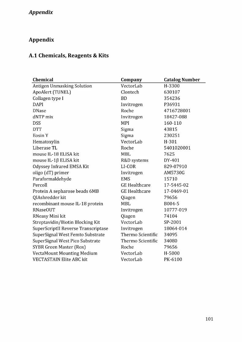
Appendix
101
Appendix
A.1 Chemicals, Reagents & Kits
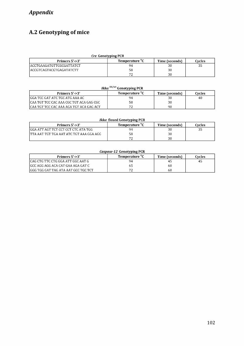
Appendix
102
A.2 Genotyping of mice
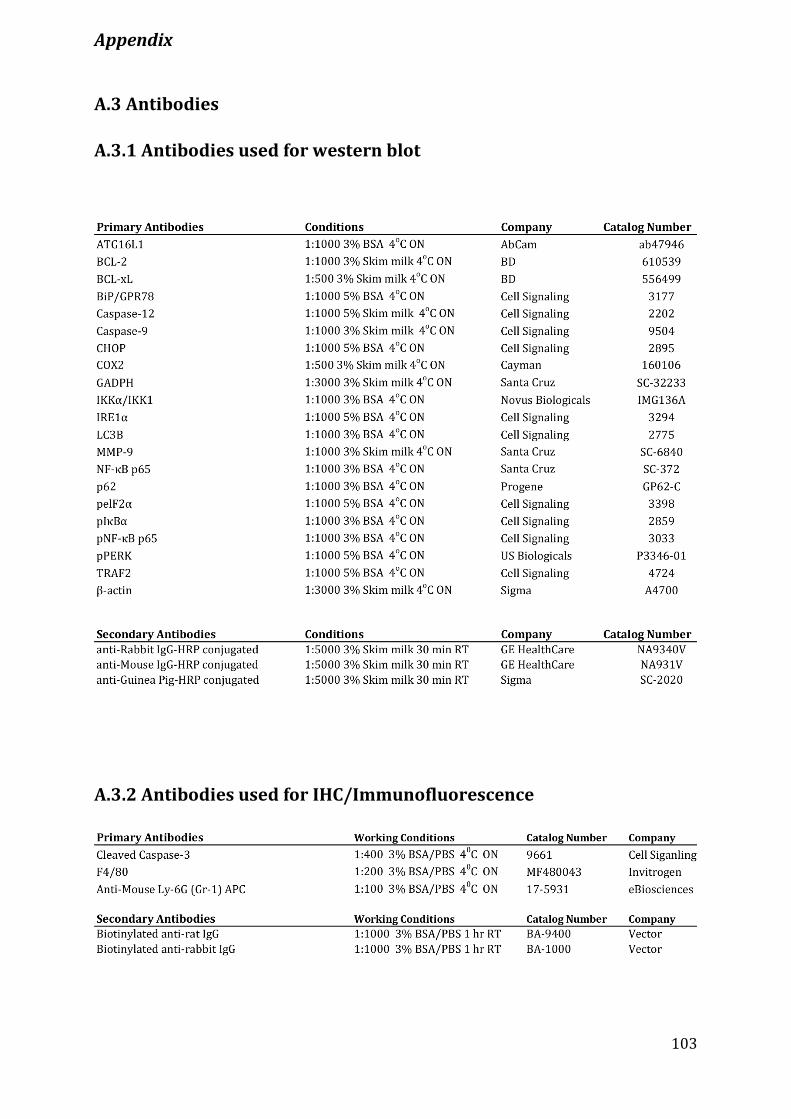
Appendix
103
A.3 Antibodies
A.3.1 Antibodies used for western blot
A.3.2 Antibodies used for IHC/Immunofluorescence
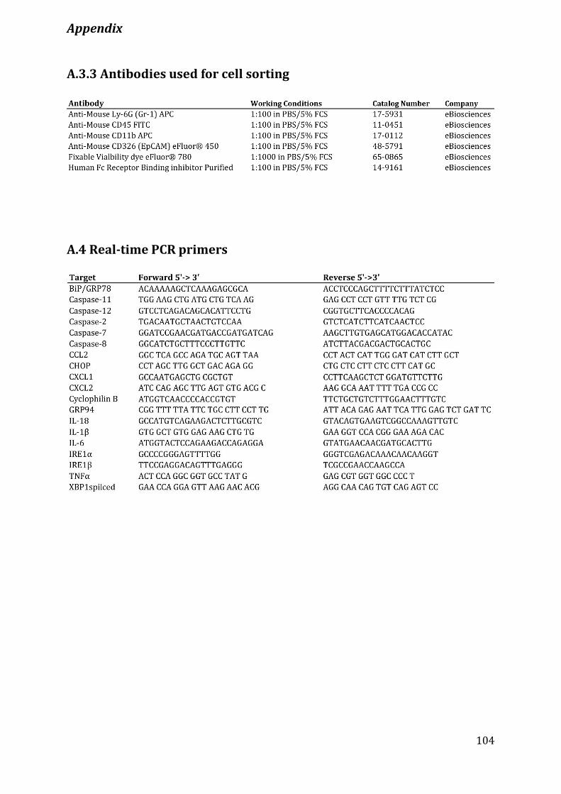
Appendix
104
A.3.3 Antibodies used for cell sorting
A.4 Real-time PCR primers
Publications
105
1) Serkan I. Göktuna, Özge Canli, Julia Bollrath, Alexander A. Fingerle, David Horst,
Michaela A. Diamanti, Charles Pallangyo, Moritz Bennecke, Tim Nebelsiek, Arun K.
Mankan, Roland Lang, David Artis, Yinling Hu, Thomas Patzelt, Jürgen Ruland, Thomas
Kirchner, M. Mark Taketo, Alain Chariot, Melek C. Arkan and Florian R. Greten. ‘IKKα
Promotes Intestinal Tumorigenesis by Limiting Recruitment of M1-like Polarized Myeloid
Cells. Cell Reports 2014; 7.
2) Martina E. Spehlmann, Carolin F. Manthey, Sara M. Dann, Elaine Hanson, Sukhman S.
Sandhu, Linus Y. Liu, Farid K. Abdelmalak, Michaela A. Diamanti, Kristin Retzlaff,
JürgenScheller, Stefan Rose-John, Florian R. Greten, Jean Y. J. Wang and Lars Eckmann.
‘Trp53 Deficiency Protects against Acute Intestinal Inflammation’. Journal of Immunology
2013; 191:837-847.


![Review Role of tumor microenvironment in tumorigenesis · Review Role of tumor microenvironment in tumorigenesis Maonan Wang1,2, ... (Figure 1) [1]. Although researchers now have](https://img.pdfslide.tips/doc/110x75/5f1d575396da9a7fe415bbde/review-role-of-tumor-microenvironment-in-tumorigenesis-review-role-of-tumor-microenvironment.jpg)