Embed Size (px)
Citation preview

Micro-Robots in Medical
Applications
헤난도 레온 로드리게스Hernando Leon-Rodriguez

Contents
Introduction
Principals of electromagnetics actuators Circular coils
Saddle coils
Six-mag coils
Octomag coils
Medical therapies micro-robot
Polymer material for micro-robot
Self-folding polymers in micro-robot
Printing micro-robots

History of Micro-robots
3
1600’s: Optical microscope
1850: Micro-assembly in watch industry
1930, 1952, 1973: Micro-organisms
1900’s: Manipulation of small objects (magnetic, electrical, optical) 1950’s: Integrated
circuits
1998: Manipulation of single cells
2003: Biomedical micro-robots 1995:
Micromanipulation/ Micro-assembly
1980-90’s: MEMS research
1982: Silicon as mechanical material

Introduction – Micro-robot technology
Electromagnetic actuator - EMA Micro robot - magnetic
Region of interest - ROI

- Wireless resonant magnetic micro-actuator
- Microrobot configuration : Nickel attractor, Swing mass (MEMS Technique)
- 2 pair of Helmholtz coil Moving the two-dimensional plane
- Drive mechanism: Resonant impact force using linear motion
SOA of Cell Manipulation using Electromagnetic Field
B. J. Nelson (Applied Physics Letters, 2008)
Resonant impact force http://www.iris.ethz.ch/

- Driven by an external magnetic field Untethered microrobot
- Micro-robot configuration : Permanent magnets (laser cutting)
- 3 pair Helmholtz coil Moving the two-dimensional plane
- The drive mechanism: Stick–slip motion using linear motion
SOA of Cell Manipulation using Electromagnetic Field
M. Sitti (The International Journal of Robotics Research, 2009)
Gradient magnetic field http://pi.is.mpg.de/

SOA of Cell Manipulation using Electromagnetic Field
- Artificial bacterial flagella
- Micro-robot configuration: Helical tail, soft magnetic head (MEMS Technique)
- 3pair of Helmholtz coil are using to moving in the three-dimensional space
- Drive mechanism: Rotational magnetic field uses linear motion
B. J. Nelson (Applied Physics Letters, 2009)
Rotational magnetic field http://www.iris.ethz.ch/

- Microrobot Configuration: Magnetic composite body Micro-molding technique
- 2 pair of Helmholtz coil and Maxwell coil uses to move 2 dimensional surface
- Drive mechanism: Magnetic torque & force using linear motion
SOA of Cell Manipulation using Electromagnetic Field
S. Park (Mechatronics, 2013)
Magnetic composite body

Manipulation of Heterogeneous Micro-particles
Multiple micro-particles manipulation
Manipulation using a multiple micro-robot system
Experimental methods
Two optional powered micro- robots capable Micro-robot: tip : U, V shape
Block : 8 blue square, 8 red disk
- Lithography uses process
A number of the independent operation of the micro-robot Through micro- particles 2-D assembly
Thermally Responsive Micro-clamper
S. Park (BioRob, IEEE, 2016)

Medical therapies by micro-robot

Light-actuated microrobots for biomedical science
1µm-diameter polystyrene beads are loaded inside a micro-robot.
Light-driven micro-robots
Experimental methods
Optical forces are non-invasive and can operate through sealed and sterile biological chambers
Metal-vapor deposition of titanium adhesion and a gold layers (of 1 and 5nm thickness) as a circular disk inside the body of each light robot.
these light robots is practically transparent to the trapping beam wavelength and thus generates very little heat.
Glückstad, Lasers & Sources, 2017)
Mask is fabricated on top of the structure to secure exposure of only certain regions by
metal-vapor deposition.
Applications in the Eye
Model eye
23G Needle with robot
Experiments in the laboratory

“Bacteria swim by rotating their flagella filaments” (1973, Berg) A
rtif
icia
l B
acte
rial
Fla
gell
a
Red cell: 8µ
Micro-robot
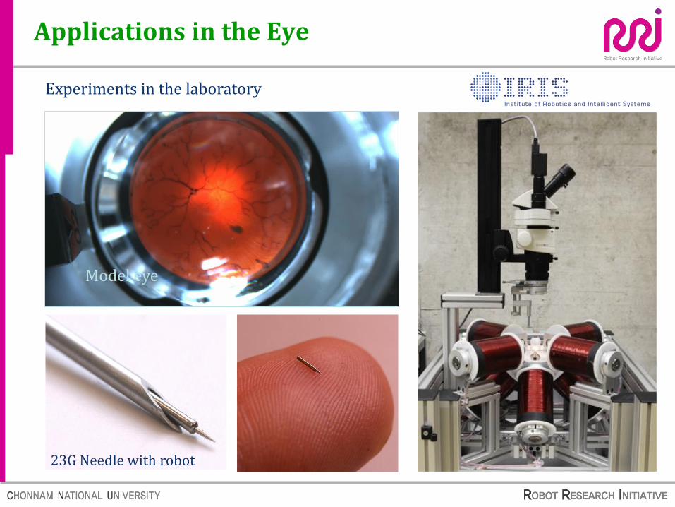
μm Robot Liposomal Bacteria-based Micro-robots
Concept of bacteria-based microrobot
Liposome
nonpathogenic/attenuated bacterial strain
Active targeting therapeutic bacteria-based micro-robot Using liposomes

mm Robot Capsule Endoscope Robot (active)
Active capsule endoscope
Active capsule endoscope
Passive capsule endoscope
Endoscope
• Pain and infection
• Small intestine diagnosis only• Time required 24 hours
• Pre-digestive target• Diagnosis and treatment function• Time required 20 minutes
Prototype II
Prototype I

Magnetic Capsule Endoscope
S. Yim and M. Sitti, “Design and rolling locomotion of a magnetically actuated soft capsule endoscope,” IEEE Trans. Robotics, vol. 28, no. 1, pp. 183–194, 2012.

Poly( N -isopropylacrylamide)-clay (PNIPAM-clay) nanocomposite (NC) hydrogels with both excellent responsive bending and elastic properties are developed as
temperature-controlled manipulators. University Chengdu , Sichuan 610065 , P. R. China Adv. Funct. Mater. 2015, 25, 2980–2991
Bending and elastic properties

J.C. Kuo, S.W. Tung, and Y.J. Yang, “A hydrogel-based intravascular micro-gripper manipulated using magnetic field,” The International Conference on Solid-State Sensors, Actuators and Microsystems, 2013, National Taiwan University, TAIWAN.
Temperature controller: Alternating magnetic field (AMF)
3D Locomotion with polymer material

S. Fusco, M. S. Sakar, S. Kennedy, C. Peters, R. Bottani, F. Starsich, A. Mao, G. A. Sotiriou, S. Pané, S. E. Pratsinis, D. Mooney, B. J. Nelson, "An Integrated Microrobotic Platform for On-Demand, Targeted Therapeutic Interventions," Advanced Material, 2014, ETH Zurich , Zurich, Switzerland.
Temperature controller: Near-infrared light (NFR)
3D Locomotion and folding

SELF-FOLDING THERMO-MAGNETICALLY RESPONSIVE SOFT MICROGRIPPERS
The Johns Hopkins University in Baltimore, MD, a team working in a new area called soft robotics is developing tiny,
self-folding devices that could one day be used to perform biopsies or precisely deliver drugs inside living tissue.
Source: http://www.medicalnewstoday.com/articles/289078.php
ACS Appl Mater Interfaces 2015 Feb 28
Self-folding devices with biopsy capability

Drug-load Bead Trap with Electromagnetically actuated ability
Self-folding Micro-robot
Bilayer (NIPPAM, PEG-DA)
Shape change with temperature
Application
Tissue engineering
Micro-particle assembly
Drug & cell delivery
Reference temperature = 33℃
Programmable Self-folding Micro-robot with Electromagnetically Actuated Ability

3D EMA system with 2-pair Helmholtz and Maxwell coils on each axis. Composition: 3D Fixed HC, MC. Characteristic: 3D driving and alignment direction Advantage: HC and MC are suitable for 6DOF for rotation and pulling.
Local EMA manufactured system
3D Locomotion System with HC and MC
Nueva Granada Military University of Colombia

3D EMA system with 2-pair Helmholtz and Maxwell coils on each axis.
Autonomous control of Micro-robot
2D Autonomous motion with HC and MC N
uev
a G
ran
ada
Mil
itar
y U
niv
ersi
ty o
f C
olo
mb
ia

Macro-Surgical Robots and Devices

Tendency of Robotic Systems
Industrial
Robot
Structured
Environment
Unstructured
Environment
Medical
Robot
Life
Supporting
Robot
Entertainment Robot
Battle/
Rescue
Robot

Open Surgery MIS SILS NOTES
Minimally Invasive Surgery Natural Orifice Transluminal Endoscopic
Surgery
Single Incision Laparoscopic Surgery
Development of Surgical Robotics and Devices

1987 16 century
Dead or Alive
1886
Large damage
mple complex pprreecciissee
2001
Small damage
No damage?
Ancient
surgery
Open
surgery
MIS (minimally
invasive surgery)
MIS
with robot Future
rough si
Development of Surgical Robotics and Devices

Neuro surgery
Covering almost all surgeries
Development of Surgical Robotics and Devices

The Most Successful Surgical Robot
Da Vinci System – Laparoscopic Surgery Robot
-6-
Surgical Arm Cart
Surgeon Console
InSite High Resolution
3D Endoscope
EndoWrist Instruments
Master Console
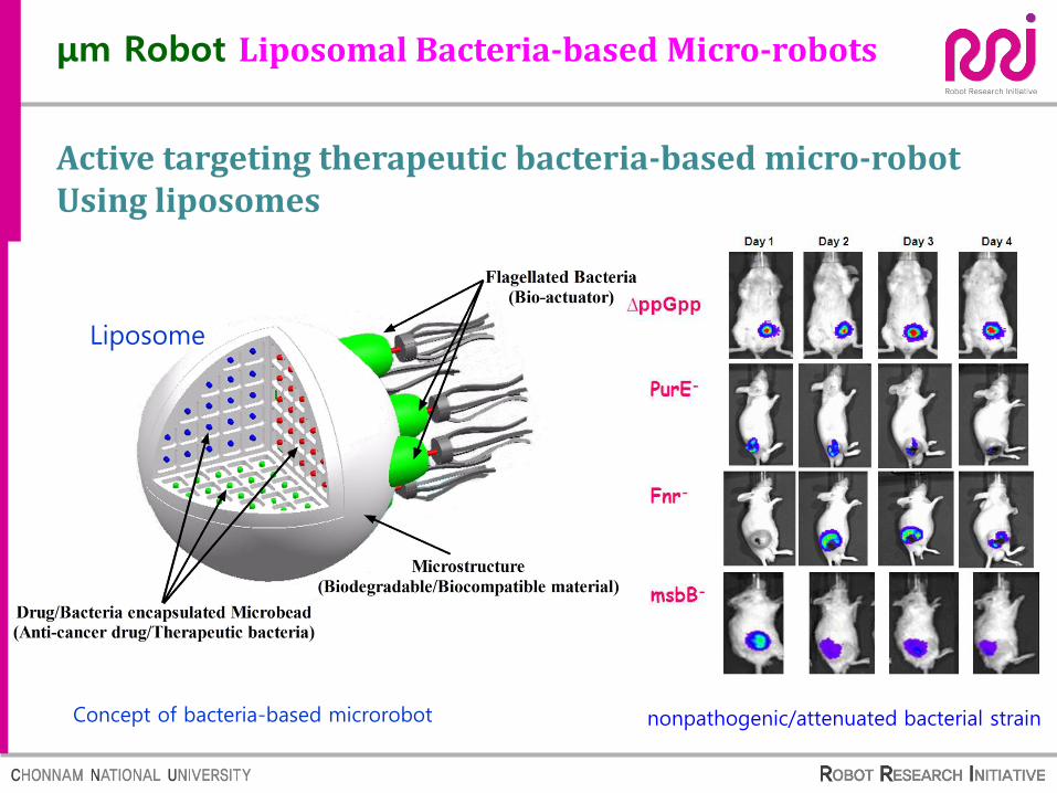
Minimally Invasive Surgery (MIS) :
Single Port Laparoscopic Robot
-7-
ARAKNES (Scuola Superiore Sant'Anna)
FDA: Da Vinci
(Intuitive Surgical),
2011
SPIDER
(TransEnterix)

-8-
(Hitachi) 2000
(Nagoya Univ) 2004
Tyson, M. D. & Humphreys, M. R. (2014) Urological applications of natural orifice transluminal endoscopic surgery (NOTES); Nat. Rev. Urol. doi:10.1038/nrurol.2014.96
Minimally Invasive Surgery (MIS) :
NOTES? (Natural Orifice Transluminal Endoscopic Surgery)

MM-1 Tokyo University, Japan
Diameter:5 mm
DOF:6
Cardioarm Cardiorobotics and Carnegie Mellon University, USA
Diameter:10mm
DOF:snake-like
ARTEMIS Karlsruhe Research Centre, Germany
DOF:6
Diameter:10mm
Development of Surgical Robotics and Devices

Operation:Manual
Size:110cmx18mm
Channel:4 (7mm,6mm,4mm,4mm)
NOTES/R-scope
Penn State Hershey Medical Center,
Hershey, Pennsylvania, USA.
Operation:Manual
Size: 130cmx11.7mm
TransPortTM
USGI Medical, San Capistrano, CA
Development of Surgical Robotics and Devices

Channel:3个(6mm,
4mm,4mm) Size:16
x22mm; Length:55cm
Cardioarm
Cardiorobotics and Carne
gie Mellon University
Diameter: 10mm
DOF: Snake-like
DDES
Boston Scientific Inc. USA
Development of Surgical Robotics and Devices

MicroHand System for MIS
Development of Surgical Robotics and Devices

Tele-surgery

Tele-surgery

Interactive mode
Remote mode
Interactive mode
Core haptic technology and remote control technology
Master-slave remote control technology
Force echo technique of two-way remote control
F/T Sensor based force echo technology and safety control
Direct intervention of physician
Restriction of robot movement
Joint Surgery Robot Intelligence Control – RRI-Korea

Stiff-Flop ANUBIS
Jamming gripper
Tissue
Device
Future of Surgical Robotics and Devices

Diameter:22mm
Force:2N
Layer Jamming Mechanism
the Samsung Advanced Institute of
Technology,Korea
FlexHand Tianjin University
Future of Surgical Robotics and Devices

Flexible Sensors
Haptic Sensors (Liu 2012)
Sensors in surgical devices
Future of Surgical Robotics and Devices

Sources:
Shuxin Wang Tianjin
University
Robot Research Initiative, Chonnam National University Jong-Oh Park, Sukho Park, Seong Young Ko, Ph.D Professors
http://internetmedicine.com/robotics-lecture/